Open Access Journals on Biomedical Research

Frequency of Blood Glucose Monitoring in Relation to Glycemic Control in Patients with Type-2 Diabetes

Introduction

Diabetes is among the principal sources of morbidity and mortality around the World. Diabetes prevalence has been increased from 108 million cases in 1980 to 422 million cases of diabetes in 2014, and this trend of increasing burden has been speculated to raise from 425 to 629 million diabetic people from 2017 to 2045 [1]. Diabetes is among the major health problems facing the human population around the world today. It also poses economic problem because it is estimated that 10% of National Health Service (NHS) expenditure is spent on diabetes which is equal to £1 million per hour. Presently, 2.3 million people have been reported with diabetes and above 500,000 people with type-2 diabetes are not aware of their diabetic status. It is expected that more than 4 million people will have diabetic till 2025 and probably a large number those will have type-2 diabetes, leading to an increase in aged, overweight and obese individuals. It is alarming that growing unhealthy lifestyle has been a key reason of type-2 diabetes, once observed only in the over-40s, being diagnosed in a increasing number of younger people and even children [2]. Diabetes is manifested when blood glucose concentration reaches the higher level than normal blood sugar level. Blood sugar is a main source of energy found in foods which we eat. Insulin is hormone which is made by the pancreas which facilitates glucose from food get into cells for its use as energy. At times body doesn’t make sufficient or any insulin or doesn’t use insulin properly, then glucose remains unused in blood and doesn’t enter cells.

Three types of diabetes are commonly found as type-1, type-2 and gestational diabetes. In type-1 diabetes the body doesn’t make insulin and the patient immune system attacks and damages those cells in pancreas which are responsible for making insulin. Type-1 diabetes is typically detected in children and young adults, though it can appear in every age. Individuals in type-1 diabetes need to take insulin every day for his/her survival. In type-2 diabetes, which is most common type of diabetes the body doesn’t produce or use insulin properly. Type-2 diabetes can start at any age, even during childhood. However, this type of diabetes takes place mostly in middle-aged and older people. Gestational diabetes is caused in certain women when they are pregnant. Most of the times, this form of diabetes goes away after the birth of baby. There are some other less common types of diabetes including monogenic diabetes, which is an inherited form of diabetes, and type of diabetes associated with cystic fibrosis [3]. Type-2 diabetes has a number of causes but most important are known as genetics and lifestyle, but the combination of both factors can create insulin resistance, when body doesn’t use insulin as it should. Insulin resistance mostly causes type-2 diabetes. Genes do play important role in type-2 diabetes, but lifestyle also play important role. Lifestyle choices that affect the development of type-2 diabetes includes lack of exercise, unhealthy meal planning choices, overweight or obesity [4]. Some risk factors of diabetes include being overweight or obese, hypertension (high blood pressure), and low level of “good” cholesterol (HDL), elevated level of triglycerides in blood, sedentary lifestyle and family history of diabetes. Symptoms of diabetes include increase urination, excessive thirst, weight loss or gain, hunger, fatigue, skin problem, blurred vision, nausea, vomiting etc. Diabetes can cause microvascular (damaged to small vessels) and macrovascular (damaged to large vessels) diseases [5].

Self-monitoring of blood glucose (SMBG) refers to check blood glucose of diabetic patient at home. SMBG is an important modern therapy of diabetic patients. It is used to attain a specific level of glycemic control and to avoid hypoglycemia. The aim of SMBG is to gather detailed information about blood glucose concentration at various times. SMBG can be used to help in the fixing of a therapeutic regimen in response to blood glucose values and to assist individuals in adjusting their dietary intake, physical activity, and insulin dosages to improve glycemic control on a daily basis [6]. SMBG can be measured by strips or by glucometer before meal (fasting blood sugar, FBS) or anytime (random plasma sugar, RBS). Hba1c refers to glycated hemoglobin is actually the protein in the RBCs carrying oxygen all over the body joins with glucose and become glycated. Through measuring hba1c we are able to get the whole status of average blood glucose level over a period of weeks/ months. The normal Hba1c is below 42mmol/mol or below 6.0%. Prediabetics have 42-47mmol/mol or 6.0%-6.4% and diabetic have 48mmol/mol or over and/or 6.5% or over. There is a difference between hba1c and blood glucose level. In hba1c we know about the how high sugar level has been over a period of time, it provides a longer-term trend. While blood glucose, is the concentration of glucose in blood at a single point in time. It is measured as FBS and RBS [7]. Study reported that that SMBG concentration is linked with improved glycaemic control in patients and rise in rate of SMBG with growing HbA1c value was proportional to the higher ratio of insulin- treated patients in higher HbA1c categories [8,9]. Also, the better quality of metabolic control demonstrating self-monitoring of blood glucose improved glycemic control in the majority of non– insulin-treated and also insulin treated type-2 diabetic patients [10-12]. Studies have reported that substantial numbers of diabetic patients have poor glycaemic control and older age, duration of diabetes, poor dietary habits, rural lifestyle, poor medication and low education are the eliciting elements of poor glycaemic control [13-15]. Inadequate glycaemic control prevailed in the majority of aged Pakistani diabetic subjects. SMBG levels have been reported to be associated with clinically and statistically improved glycaemic control irrespective of diabetes types or therapies. Eliciting factors of poor glycaemic control and increase awareness on the significance of SMBG and strongly promote this practice among diabetic patients should be taken seriously by the healthcare authorities in targeting multidimensional interventions to accomplish good glycaemic control [8,9]. In the year 2017, about 6.9% (7,474,000 individuals) of the Pakistani population were suffering from diabetes and the country was on 10th position (projected to be on 8th position in 2045) among high burden diabetes countries all over the world [1]. Numerous studies on SMBG have been done in different countries and also in Pakistan but there is no such study conducted in Mardan district of Khyber Pakhtunkhwa province (KP) in Pakistan [8,9]. Therefore, the present study is aimed at investigating the association between blood glucose, measured as Hba1c and frequency of SMBG in diabetic patients from Mardan Pakistan [16].

Materials and Methods

Study area: This study was conducted in District Mardan of Khyber Pakhtunkhwa. Data were obtained from patients with diabetes from District Mardan. The Mardan city is on 23rd position in the list of big cities of Pakistan and the second largest city of Khyber Pakhtunkhwa with human population size of 331,837. Mardan is located in the southwest of the district at 34°12’0N 72°1’60E and at altitude of 283 metres (928 ft). An economic zone is planned as a part of the multi-billion-dollar China-Pakistan Economic Corridor (CPEC) near Rashakai. Mardan features a hot semi-arid climate. The average temperature in Mardan is 22.2°C, while the annual rainfall averages 559 mm. (https://en.wikipedia.org/wiki/Mardan).

Study Population

The data were collected from 100 diabetic patients of Mardan including male, female and children related to different age groups. These patients were suffering from type-2 diabetes. Data collection: Data were collected from the patients of diabetes diagnosed in Mardan Medical Complex, private clinics and private hospitals in District Mardan. during January 2019 to March 2019 through convenience survey. Information was obtained by structured questionnaires on diabetes therapy and blood glucose self-monitoring. All the participants were briefed about the study and then their written consent was obtained. All the subjects were subjected to interview for filling the questionnaire proforma regarding the study. Data regarding glycemic values, socio demographic and clinical characteristics of the patients were recorded. All those subjects with severe illness, having accidental physical disabilities, unable to comprehend this study, those having life threatening diseases and those suffering from cholera, dengue or malaria.

The Questionnaire Proforma Encompassed the Following Parameters

General Information: Patient name, age, gender, weight, ethnicity, type of diabetes, duration of diabetes, diabetes therapy, fasting plasma glucose, random plasma glucose, Hba1c.

Additional Information: Family history, additional disease with diabetes, allergies, hypoglycemic episodes, education, income and employment.

Study Variables: These variables included glycemic control parameters including FPG, RPG and HbA1c. Socio-demographic and clinical features, depression, cognitive status, physical status, weakness, nutritional status, pain and level of self-care were independent variables. The target value for HbA1c was <7%, FPG was 80–130 mg/dL and RPG was <180 mg/dL [17]. Subjects having HbA1c, FPG, and RPG levels above the upper threshold of the target levels were declared as having poor glycemic control.

Statistical Analysis: SPSS version 21.0 was used for statistical analyses of collected data. Descriptive statistics and bivariate correlation were applied. Mean and standard deviation were computed for quantitative variables. Qualitative variables were subjected to frequencies and percentage analyses.

Results and Discussion

Descriptive analysis showed that age of subjects was between 15-81 years with mean ± SD as 47.2±15.7. almost equal number of subjects belonged to urban (52%) and rural (48%) areas. Most were females (60%) subjects, and 82% subjects were suffering from years. Overall, 44% of the patients used to check their diabetes less than twice a week and 22% check their diabetes once daily. Stratification for frequency and percent of patients who test their FBS and RBS was 82% and 89% among which some people couldn’t test either FBS or RBS. The recorded FBS value was 53-345mg/dl with mean ± SD of 164.6±58.4 and of RBS was 50-560mg/dl with mean ± SD of 263.7±97.2. The frequency of hba1c was 80 with recorded value of 4.20-16.90% and mean ± SD was 10.9±23.8. Most of the people (67%) used medications along with insulin from years. Maximum of the (52%) people were not following diet to control it. Majority of the patients (78%) had high blood sugar. Weight of the patients was between 30-115kg with average between 60-70kg. Most (67%) subjects were not doing regular exercise. Maximum of the patients (73%) were consuming tea 42% showed no family history of diabetes while 40% subjects had one parent affected and 18% had both parents affected. Most of the patients (98%) had other problems like vision problem, nausea, numbness etc. Sixty percent subjects were literate with majority of unemployed, and 55% were middle class (Table 1). By using bivariate correlation, we performed Pearson correlation coefficient. The values p˃0.05 and p˃0.01 show significant linear relationship. From this correlation analysis a linear relationship between age and weight was determined as well as between gender and SMBG, insulin, diet and weight. There was no correlation between SMBG and Hba1c (Table 2).

biomedres-openaccess-journal-bjstr

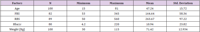
Table 1: Descriptive Statistics of factors associated with diabetes.

biomedres-openaccess-journal-bjstr

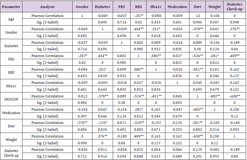
Table 2: Bivariate correlation among the study variables.

In past few decades, huge rises in diabetes prevalence have been confirmed in almost all areas of the world, with 415 million people worldwide now living with diabetes [17]. Diabetes is among the metabolic disorders characterized by elevated blood sugar levels for a long time period. Diabetes afflicts various complications like diabetic ketoacidosis, hyperosmolar hyperglycemic state, or death. Other severe complications can be cardiovascular disease, stroke, chronic kidney disease, foot ulcers, and eyes damage. (https:// en.wikipedia.org/wiki/Diabete). In 2000 the global prevalence of diabetes was estimated to be 151 million by International Diabetes Federation (IDF). In 2003 the number reached to 194 million, in 2006 the number increase to 246 million, in 2009 it reached to 285 million, in 2011 it increased to 366 million, in 2013 it reached to 382 million, in 2015 it reached to 415 million. In recent study it was estimated that in 2017 about 425 million cases of Diabetes in age group 20-79 years throughout the globe, If the age range is increased to 18-99 the estimated case of diabetes rises to 451 million, the number of diabetes cases are increased day by day and reached to 629 million people for age range 20-79 years [17].

According to a recent study in Pakistan, 11.77% prevalence of type-2 diabetes mellitus has been recorded. Males were more affected (11.20%) than females (9.19%). Likewise, this trend was reported in 3 provinces of Pakistan as well reporting 16.2%, 12.14% and 13.3% prevalence in males as compared to females as 11.70 %, 9.83% and 8.9% respectively in Sindh, Punjab and Baluchistan province respectively. While in Khyber Pakhtunkhwa (KP) Province, females (11.60%) were more affected than males (9.2%). This prevalence was higher in urban areas (14.81%) as compared to that reported from rural areas of Pakistan (10.34%) [18]. In our study we focused on the frequency of blood glucose monitoring in relation to glycemic control in patients with type- 2 Diabetes. This kind of study was first reported from Scotland by Evans et al. [10] to investigate patterns of self-monitoring of blood glucose concentration in diabetic patients and to determine whether frequency of self-monitoring is related to glycaemic control. Further studies are conducted in different countries. In Pakistan similar study has been reported from Agha Khan University Karachi by Khowaja, et al. [8], to explore the association between selfmonitoring of blood glucose (SMBG) levels and improved glycaemic control (HbA1c level) among type-2 diabetic patients.

Our study revealed that 89% subjects were suffering from type- 2 diabetes, 5% from type 1 diabetes, 1% from gestational and 5 from other types of diabetes. Out of 100 cases 40% were male and 60% were female. Stratification for frequency of normal blood glucose in patients suffering with diabetes mellitus with regards to duration of diabetes mellitus was recorded which shows that out of 100 cases 82 subjects have diabetes from years, 15 have from months and 3 subjects have diabetes from days. Overall, 52% belonged to urban areas and 48% from rural. Frequency of Hba1c was 80%. Seventy nine percent did not develop any types of allergies. The normal blood glucose in patients suffering with diabetes with regards to financial status shows that out of 100 cases 10% were of lower class (10,000-20,000), 60% were of middle class (20,000- 40,000) and 30% were from upper class (> 40,000). Frequency of control of blood glucose in patients suffering with diabetes mellitus with regards to educational status showed that 60% patients were literate, and 43% were employed. By using bivariate correlation, we performed Pearson correlation coefficient. Basically, this was performed the extent to which two variables are linearly related. The value p˃0.05 and p˃0.01 show significance. From this correlation we found that there is significance association between age and weight. There is also significance link between gender and SMBG, insulin, diet and weight. According to frequency of and time of doing SMBG, the results of the participants who did SMBG were above or below the target for glycemic control. This means that doing SMBG is not currently associated with better glycemic control. There is no liner relationship between self-monitoring blood glucose and Hba1c.

Evans et al. [10], have reported a direct relationship of haemoglobin A1c level with the number of strips collected during previous 6 months in diabetic patients. They have not found any association of HbA1c level and SMBG in diabetic patients using insulin. It may be due to the fact that SMBG is more effective in true insulin deficiency unlike the insulin resistant state. Or diabetic patients might have no aware of insulin use, more worried about the risks of hypoglycaemia, and hence less likely to act on the results of tests. Our study is similar to Harris [11] who evaluated the association of SMBG and HbA1c. They reported no such association between the frequency of SMBG and HbA1c level of those diabetic patients treated with insulin, oral agents, or diet alone. More educated, having a diabetes patient education class, and frequent visitor to physician were found frequently practicing self-monitoring. However, SMBG was not associated with higher economic status or to having health insurance. Our study was quite contradictory to the study of Khowaja and Waheed [9] because they examined that self-monitoring of blood glucose concentration is associated with improved glycaemic control, which prevents complications resulting from diabetes. Their study indicated that there was a significant association of self-monitoring of blood with glycaemic control. It was contradictory may be because their sample size was 5 times higher in number than our study samples. Davis, et al. [15] also determined, like our study that both cross-sectional and longitudinal Fremantle Diabetes Study (FDS) data revealed that Hba1C was not statistically different between SMBG users and nonusers. This study is also similar to Alzahrani, et al. [18] establishing that the results of the patients who followed SMBG were beyond the target for glycemic control which reveals that SMBG is not linked with improved glycemic control. In our study we do not support the association of SMBG to glycemic control. The results of our study reveal that SMBG is important, but it also indicates that majority of subjects were checking their blood sugar occasionally. It was also observed that participants doing SMBG, were managing their diabetes well and self-monitoring may be recommended particularly in those patients who are the most difficult to control.

It is established that education and economic status are also the possible effect modifiers for controlling the blood glucose levels. Besides regular checking of blood glucose, lifestyle reform also needed to be done in diabetic patients including diet and regular exercise. The results of this study may be handy for creating awareness of blood glucose control during diabetes ultimately reducing the morbidity level in due to diabetes [19].

Conclusion

According to the result of our study, regular self-monitoring in patients with diabetes is uncommon and the self-monitoring of blood glucose is not associated to glycemic control among type- 2 diabetes. There is a lack of knowledge about SMBG and Hba1c testing and control in public.

For More Articles: Biomedical Journal Impact Factor: https://biomedres.us

Open Access Journals on Medical drug and theraputics

Premature Mortality due to Breast Cancer in a Mountainous Province of Vietnam from 2005 to 2018

Abstract

Objectives: To describe the time trend of esophageal cancer (EC) mortality that occurred in Nghe An province during 2005-2014.

Methods: Mortality due to EC and all causes were annually collected from the A6 death register system using the “Validated mortality registration forms” from 2005 to 2014. There was a mandatory monthly active mortality registration by 480 state health stations following the A6 death register system. Age-standardized rates (ASR) of mortality and mortality rate ratio and 95% confidence interval (MRR (95%CI)) by time-period were estimated.

Results: Among 140,670 reported deaths, 639 were due to EC (Men 527, women 112). The mortality rate (WHO-ASR) per 100,000 was 4.70 in men and 0.77 in women, giving the men to women ratio was 6.1. Mortality from EC was significantly increased during the 10 years, 2013-2014 vs. 2005-2006, MRR (95%CI): 2.79 (1.95, 3.99), p for trend < 0.05.

Conclusion: We observed an increased time trend of mortality due to EC; men are responsible for over 80% of this fatal disease, therefore, a program of primary and secondary prevention against EC focused on men is highly needed in the Nghe An province.

Keywords: Esophagus; Cancer; Time-Trend; Mortality

Abbreviations: EC: Esophageal Cancer; HDI: Human Development Index; SHS: State Health Stations; ALDH2: Aldehyde Dehydrogenase 2; MRR: Mortality Rates Ratio; CI: Confidence Interval

Introduction

Breast cancer has been worldwide recognized to have a high mortality rate among women despite wide variations in ethnicity, culture, and economics. It was reported that breast cancer is the most commonly diagnosed cancer among women in 140 of 184 countries worldwide [1]. More than one million new cases of female breast cancer are diagnosed each year. Approximately one in 4 of all new cancer cases diagnosed in women worldwide are breast cancer, followed by 6.6% of cancer death in 2018 [2]. The breast cancer mortality rate has been substantially increasing in the world during the past 25 years [3]. According to the American Cancer Society, the overall breast cancer incidence rates increased among Asian/ Pacific Islander (1.7% per year), non-Hispanic back (0.4% per year), and Hispanic (0.3% per year) [4]. In China, mortality from breast cancer rose progressively during the past three decades in both rural and urban areas [5]. An estimated 268 600 women were diagnosed and 69 500 died of breast cancer in 2015, accounting for 15.1% of all new cancer cases and 6.9% of all cancer deaths in Chinese women, respectively [6].

Vietnam, situated in Southeast Asia, is a developing country with a GNP per capita of USD 2,540 in 2019. Social health insurance is voluntary of approximately US$20.00 per annum, which is paid by individuals and their households. In Vietnam, breast cancer is reported to be the most common cancer in Vietnamese women [7]. There were 15,229 cases of breast cancer were diagnosed in 2018, accounting for 5.3% of all cancer deaths [2]. In response to this situation, the Vietnamese government has introduced nationwide breast self–examination education as the method for early detection [8]. Moreover, the usage of mammography and hormone therapy for patients with estrogen-receptor-positive breast cancers has been improved the life expectancy of patients with breast cancer [9]. However, lacking population-based studies about this fatal disease hindered the establishment of focused primary prevention strategies. The mortality rate is a basic and critical indicator for the development of appropriate and effective intervention programs and monitoring the health of patients with breast cancer. Lang Son is a mountainous province in the Northeast region with a natural area of 8,187.25 km2, bordering Guangxi province in China.

The population of the province in 2019 is 782,811 people, mainly living in rural areas (accounting for 80.7%). Lang has a high proportion of ethnic minorities (84.74% of the province’s population). Currently, there are 30,583 poor households, accounting for 15.83%; 21,267 near-poor households, accounting for 11.01% [10]. Having these geographic economic conditions, Lang Son province is considered as the representative of mountainous provinces in Vietnam where the citizens have a limited chance to approach high-quality health care services. In this context, this study was conducted to clarify the status of breast cancer mortality in Lang Son province from January 2005 to December 2018 to provide an updated and comprehensive understanding of recent trends of breast cancer mortality in this province.

Material and Methods

Study Design and Data Source

This is a population-based study of the time series of deaths from breast cancer of women living in Lang Son province. The data was collected in two steps. At first, the mortality information was recorded in an official book referred to by The Ministry of Health’s death recording systems (A6) managed by the Lang Son CDC. The A6 mortality systems were validated and presented to be a reliable and feasible system for mortality recording [11]. These unique systems were introduced to be used nationwide in Vietnam in 1992. All deaths occurring in the communities were registered at the state commune health stations. The data from the A6 was collated and determined monthly by the head of the state commune health stations who in turn forward the information every year to the Lang Son CDC from 2005 to date to develop a database of mortality there. Five indicators included the case’s ID, age, sex, date – place – cause of death, and ICD-10 coding. To prevent duplicate records, the head of the commune health station, trained medical workers, followed carefully the medical care for each morbidity case for each resident for at least 6 months until the outcome was identified. By this follow–up process, all deaths in the list have described the cause of death based on medical records. Besides, data on population was collected with careful cross-check with several independent information sources such as the departments for provincial statistics, the department of Population – Family – Children, the Committee of Family and Planning. We included all cases that were dead of breast cancer from January 2005 to December 2018, ICD-10: C50 for the present study. The Lang Son population-based mortality registration covered over 226 state commune health stations of 11 cities/districts of the province. The average resident number of each state commune health station was annually collected by the Lang Son CDC.

Data Analysis

Data was reviewed and cross-checked between information sources, cleaned, encoded, and reported by Excel software, analyzed by STATA 15.0. For the calculation of the mortality rates of breast cancer, the ratio was determined between the number of deaths from breast cancer in women and the female population in that year, multiplied by 100,000. The world population structure and the Segi’s world population standard were used to estimate agestandardized mortality rates per 100,000 (ASR). Mortality rates ratio and 95% confidence interval adjusted for age groups (0-9, 10- 19, 20-29, 30-39, 40-49, 50-59, 60-69, 70-79, 80+) was estimated to observe time trend from 2005 to 2018.

Ethical Approval

This is a population-based study about mortality rate with no interfere with no physical intervention. This information to identify patients including name, personal address, date of birth, would be not published. Moreover, due to the database purpose, which is to provide accessible data to help improve the mortality of breast cancer in the community, the public interest is considered to outweigh personal interests in privacy or autonomy that would otherwise be protected by consent mechanisms. The research protocol was approved by the Hanoi Medical University Review Board in Bio-Medical Research # 61/HMURB, dated 25 November 2008 and by the Board of Ethics in Bio-Medical Research at University of Medicine and Pharmacy at Ho Chi Minh City #106/ UMP-BOARD, dated 20 March 2019.

Results

From January 2005 to December 2018 (missing data for 2009- 2010), there were 210 deaths out of 17,990 women diagnosed with breast cancer in Lang Son province. Table 1 showed the crude mortality rate and the age-standardized mortality rates from breast cancer at all ages between 2005 and 2018. The crude mortality rate of breast cancer was 4.7. After adjusting using the World Health Organization standard population for 2000 – 2025 and the Segi World standard population (in the 1960s), the ASR was 5.2 and 4.7, respectively. Over four-fifths of death cases died under the age of 70 (81.5%). In Table 2, the crude rate and the ASR rate were annually calculated. The lowest ASR per 100,000 person-year was found in 2006 (3.2). The value of 7.0 was the highest ASR that occurred in 2012. When the first two-year 2005-2006 was the reference group, the mortality rate ratio for 2018 was increased, but not statistically significant, age-adjusted MRR, 95%CI: 1.36, 0.78, 2.35, p for trend=0.209, (Table 3). Figure 1 presented the age-specific mortality rate per 100,000 person-years during the study period. The mortality rate was found to be at a low and stable level from the age group 0-9 to the age group 20-29. There was, on the other hand, a rapid rate of increase at the reproductive age, especially at the age group 40 – 59, and slow down after that. The highest specific cancer mortality rates per 100,000 were seen for the age group 60-69. The curve thereafter was seen to be declined (Figure 2).

biomedres-openaccess-journal-bjstr

Figure 1: Location of the study population, Lang Son province in Viet Nam.
(Source: https://commons.wikimedia.org/wiki/File:Lang_Son_in_Vietnam.svg)

biomedres-openaccess-journal-bjstr

Figure 2: Age-specific mortality rate per 100,000 person-years in women during 2005-2018 (missing data for 2009-2010) due to breast cancer

biomedres-openaccess-journal-bjstr

Table 1: Mortality due to breast cancer in women during 2005-2018 in Lang Son province & Crude rate per 100,000 person-years; @ Age-standardized rate per 100,000 person-years using the World Health Organization standard population for 2000-2025; # Proportion of death cases aged under 70 year-olds. @ Age-standardized rate per 100,000 person-years using SEGI World standard population (in the 1960s). Missing data for 2009-2010.

biomedres-openaccess-journal-bjstr

Table 2: Mortality due to breast cancer in women by year from 2005 to 2018 in Lang Son province. The estimated proportion of deaths due to breast cancer was 1.17% (210 cases of breast cancer vs. 17,990 total cases) in women. & Crude rate per 100,000 personyears; $ Age-standardized rate per 100,000 person-years using the World Health Organization standard population for 2000-2025; # Proportion of death cases aged under 70 year-olds. Missing data for 2009-2010.

biomedres-openaccess-journal-bjstr

Table 3: Mortality rates ratio and 95% confidence interval by years from 2005 to 2018 in Lang Son province.

Note: ## adjusted for age groups (0-9, 10-19, 20-29, 30-39, 40-49, 50-59, 60-69, 70-79, 80+). Missing data for 2009-2010.

Discussion

We observed a serious prelature breast cancer mortality in the Lang Son province, remote mountainous areas. Female breast cancer has presented at young age women and the proportion of premature death was very high, over four-fifth of total death cases. The result has highlighted the importance of public health policies for better prevention and management of breast cancer screening to reduce premature death in the study population. Moreover, financial support policies should be implemented to promote access to appropriate diagnosis and treatment for women living in disadvantaged areas. After adjusting with the World Health Organization standard population, 2000-2025, the ASR of breast cancer in Lang Son province is 5.2/100,000 during study time from 2005 to 2018. In consistence, the studies in Chinese women living in rural areas, the ASR in 2013 and 2014 of breast cancer were 5.59/100 000 and 5.79/100 000, respectively [6,12]. The ASR of breast cancer of our study is lower compared to one among women living in Ha Noi – the capital of Vietnam in the period from 1996- 2005 and the ASR of breast cancer in Vietnam in 2012, which were as high as 13 and 9.9, respectively [13,14]. Hanoi City is the highest urbanization that can be explained why the breast cancer in this city was higher than in the Lang Son province.

To reduce young age death from breast cancer, the National Cancer Control Programs increasing the awareness of early detection of breast cancer and providing free screening for breast cancer and cervical cancer has been conducted in Vietnam since 2008. There were other several programs such as the “Early detection of breast cancer and cervical cancer in women” program implemented from 2012, the “We care for her” happening in 2013–2014. The Vietnam Women’s Union has representatives at every commune has been also running programs to educate how to do breast self–examination [8]. Moreover, treatment for breast cancer in Vietnam has been remarkably improved recently. Bettertolerated therapies have been replacing ablative surgery and aggressive chemotherapy. Tamoxifen or other hormonal therapies, cytotoxic, and targeted therapies, shown to significantly reduce breast cancer recurrence and mortality in breast cancer patients [15,16], are all available in Vietnam [8]. In addition, the population variation of our study comparing the previous studies might be likely the inducement. Nationwide, Vietnam has many cities in the progress of “modernization”, where women are at increasingly high risk of breast cancer such as decreased parity, delayed childbirth, less breastfeeding which was demonstrated to be risk factors of breast cancer in low–income countries as well as in Vietnam [17,18]. However, these programs have been piloted at the areas of urbanization of the Hanoi and Ho Chi Minh Cities, but that might not be ready in the Lang Son province.

In terms of the trend mortality rates during the study period, there was a non-increasing trend during 2005-2018 but it is suggested to be increasing soon due to many environmental factors and lifestyles of an unhealthy diet, tobacco smoking, harmful usage of alcohol, and lack of physical activities. This result is consistent with previous studies showing the rise of breast cancer mortality in other territories. For instance, according to a global analysis, there was a significant increase in breast cancer mortality rates in all super regions. For total world countries, the mean breast cancer mortality rate was 13.77 per 100,000 in 1990 and the overall increasing slope of the mortality rate was 0.7 per 100,000 from 1990 to 2015 [3]. Another study presented a tendency of increased deaths from breast cancer in Brazilian women, particularly in young women from 1996 to 2013 [19]. In China, the standardized mortality rate of breast cancer was similarly shown to have an upward trend [20]. To improve cancer prevention for Vietnamese women, it is important to establish better public health policies and management of breast cancer, especially in remotes areas. It was reported that the majority of breast cancer patients in Vietnam are detected at the advanced stages [21], which was revealed to be the result of poor knowledge and awareness among the general public [22].

In a study conducted in the mountainous area in Northern Vietnam, the level of knowledge and practice about breast selfexamination, clinical breast examination, breast ultrasound, and mammography were still adverse. Approximately 17% of women mentioned clinical breast examination, and only 13.8% reported practicing breast self-examination [23]. Among women living in one rural district, more than half of all the participants, including both younger (69.5%) and older (53.3%) women, believed that they would not get breast cancer if they took good care of themselves [24]. In central cities such as Hanoi and Ho Chi Minh City, the prevalence of sufficient knowledge and practice breast self–examination among female textile workers were only 22.7% and 15.8% [25]. In this context, increasing awareness about the importance of breast cancer screening and developing early detection strategies for breast cancer is essential. Community education programs and low-cost screening approaches such as clinical breast examination should be conducted widely to ensure the accessibility of all Vietnamese women, especially ones residing in remotes areas. The mammography screening should be suggested in high-risk women according to the age-specific mortality rate results.

Furthermore, the long treatment course of breast cancer has been causing a significant financial burden to the patients, especially the patients without health insurance. A recent study conducted in South East Asia, including Vietnam, showed that 48% of cancer patients incurred financial catastrophe within one year after the diagnosis [26]. It was estimated that the annual medical expenses for breast cancer treatment amounted to 18% of gross national income (GNI) per capita in Vietnam in 2010 ($195 vs. $1,100) [27]. These facts again emphasize the role of breast cancer screening in Vietnamese women since the diagnosis at the early stage can reduce significantly the cost of initial treatment. Besides, the financial burden of the treatment course could be a barrier to seeking care and to appropriate treatment compliance, which may contribute to the higher mortality of breast cancer. Therefore, establishing a policy of universal health insurance coverage along with other financial supports would improve access to medical care and the prognosis of breast cancer patients in Vietnam. The government should give financial support to breast cancer patients whose cost of illness exceeds their ability to pay. Furthermore, a network of primary health care such as home care and community care should be promoted to provide health care services to breast cancer women throughout the country. These settings may improve compliance with treatment and reduce costs for patients in Vietnam, where access to health facilities for cancer treatment has been limited. This combination would encourage the patients to comply with their long-term treatment and thus contribute to reducing deaths from cancers, including breast cancer.

Moreover, the current vertical and central organization of health care services in Vietnam may limit the accessibility of medical treatment for breast cancer patients living in mountainous areas. The lack of surgical oncologists, medical and radiation oncologists, anesthetists, and pathologists at the low level of the health system also has a significant effect on providing comprehensive treatment to breast cancer patients. Authorizing and supporting district and commune health stations with the appropriate health infrastructure and trained healthcare workforce could have a positive impact in terms of reducing in-direct costs for women with low – income to encourage them not to bypass them in favor of seeking suitable treatment as well as in reducing overcrowding in central and provincial hospitals. Breast cancer deaths in our study mostly occurred in reproductive-aged women, increasing rapidly from 40 years old, reaching a peak at the ages of 60-69, and then falling. This result is logical according to one study showing that the breast cancer incidence in Vietnamese women was highest at the age of 50 [21]. Meanwhile, the overall survival rate for breast cancer patients was 85.01 ± 1.61 months [28]. Similarly, according to a study investigating the breast cancer mortality rate in China during 1991 – 2011 showing that breast cancer usually occurs post-puberty, and its incidence increases slowly from 30 years old, reaching a peak at the ages of 40-60, and the first death peak occurring at 55 to 65 years [20]. In this sense, the clinical protocols and public policies that encourage early detection for breast cancer, and the key population should be among women between ages 40 to 65. Breast cancer screening using clinical breast examination for women aged 40 to 55 years was identified to be very costeffective in Vietnam according to the World Health Organization criteria [29]. Another study demonstrated offering the first round of mammography screening to Vietnamese women aged 50-59 years was cost-effective, with the given threshold of three times the Vietnamese GDP per capita [30]. Therefore, breast cancer screening programs need to be provided to women aged 40-65 in the combination of clinical breast cancer and mammography to improve the life expectancy for breast cancer patients as well as reduce the public economic burden.

Several limitations should be noted in this study. Firstly, it was done in only one mountainous province in the North of Vietnam, so the findings may not be generalized to all Vietnamese women. Second, there was a deficit of the information system concerning factors associated with mortality from breast cancer that is restricted in the death record. This study, however, is the first investigation of the mortality rate of breast cancer in the province at a continuous long-term period. The findings presented here were similar to and different from those reported elsewhere. Overall, the results of this study will increase our understanding and guide interventions to improve early screening for breast cancer strategies specifically for Vietnamese women, especially in remote areas, with adverse socioeconomic status. Cancer mortality rates estimated in this study are reliable because there were no duplicate records. At each commune, the head of the commune Health station followed up carefully all fatal cases occurring at his commune while giving medical care and household visiting until the result as neighborhood relationship as well as a duty of the appointed medical worker position. Despite these limitations, the present findings had highlighted the public health problem of premature breast cancer in the low-medium economic countries that warrant global action plans against this preventable disease.

For More Articles: Biomedical Journal Impact Factor: https://biomedres.us

Open Access Journal on Microbiology Research

Time Trend of Mortality from Esophagus Cancer During 2005-2014 in the Nghe An Province, Viet Nam

Introduction

Esophageal cancer (EC) is the seventh most common malignancy with 572,034 (3.2%) new cases in 2018. EC ranked sixth in mortality with 505,585 cases in 2018, accounting for 5.3% of total cancer deaths. According to GLOBOCAN 2018, more than 75% of death from EC occurred in Asia, mainly in developing countries [1,2]. The prognosis of EC is poor with the 5-year relative survival rate ranging from 4-40% depending on the stages of cancer. The five-year survival rate was less than 20% [3]. Although the incidence and mortality rate of EC did not increase significantly in the past, EC is expected to be a global burden in the future. The main reasons are the transition of age structure and the increasing proportion of people with harmful alcohol consumption, smoking, obesity, and low fruit and vegetable intake [4,5]. Viet Nam has an average human development index (HDI) and is facing a double burden of both communicable and non-communicable diseases. In Viet Nam, non-communicable diseases are responsible for about 68% of the disease burden and are the leading cause of death (accounting for 77% of total deaths) [6,7]. According to GLOBOCAN 2018, Viet Nam reported 2,411 new cases and 2,222 deaths from EC (accounting for 1.9% of all cancer deaths) [8]. Data in Viet Nam on mortality from all cancers combined and from EC were limited. We conducted this study to describe the time trend of EC mortality in Nghe An province from 2005 to 2014.

Methods

Nghe An province has a total area of 16,493.7 km² and is located in the North Central Region. Nghe An is bordered to the East by the sea, to the West by Laos, to the North by Thanh Hoa province, and to the South by Ha Tinh province (Figure 1). Nghe An has 21 districts, cities, towns, and 480 state health stations with a population of 3,215,179 in 2009. The descriptive epidemiological method was used to conduct the study. Mortality due to EC and all causes were annually collected from the A6 death register system using the “Validated mortality registration forms” from 2005 to 2014. There was a mandatory monthly active mortality registration by 480 state health stations (SHS) following the A6 death register system. Deaths recorded in the family register management system of Nghe An (regardless of location) from January 1, 2005, to December 31, 2014, were indexed in the A6 records of 480 SHS in 21 districts/town/cities. We excluded deaths that occurred in the study area but were not recorded in the family registry of Nghe An. For each reported death, the investigators annually collected the patient’s information (name, age, gender, date of death, and cause of death assigned by ICD-10 code) and filled in the printed form of “Validated mortality registration forms”. During the 10 years, our study collected 140,670 deaths, of which 639 were deaths from EC. In 2014, the list of deaths from the A6 death register system was validated by the Verbal Autopsy instrument. The quality of mortality from cancer in general and EC, in particular, was very good regarding accuracy and completeness. The data were checked, cleaned, encrypted, and then entered using Excel software. Deaths from EC (C15) were selected. We used STATA 10 analysis software to calculate the age-standardized mortality rate per 100,000 persons by using 3 standard populations: Segi World Standard; European standard; WHO World Standard (2000-2025).

biomedres-openaccess-journal-bjstr

Figure 1: Location of Nghe An province in Viet Nam.

We used the following equation to calculate the agestandardized mortality rate:

di: number of deaths in the ith age group, wi: population in the ith age group of the standard population, Yi: person-years in the ith age group [9,10].

Mortality rates ratio and 95% confidence interval (MRR, 95% CI) was estimated by performing logistic regression analysis to examine time trends and risk of deaths due to EC, adjusted for sex, age, the proportion of unknown underlying cause of death, and allcause mortality rates by time. For time trends during 2005-2014, we divided into five periods of 2005-2006 (the reference group), 2007-2008, 2009-2010, 2011-2012, and 2013-2014. The research protocol was approved by the Ethics Committee of Hanoi Medical University on Nov. 25, 2008. The study was conducted under the approval of local authorities and health authorities in the study area. The collected information was completely confidential and only used for scientific purposes.

Results

The total number of deaths from EC in Nghe An from 2005 to 2014 was 639 out of 140,670 reported deaths; 527 were men (82.47%) and 112 were women (17.53%). The rates of death from EC in men increased steadily over the years (Table 1). The standardized mortality rates that used WHO and EU standard populations were higher than the Segi standard. The age-standardized mortality rate (WHO World Standard) in 2013-2014 was 6.32/100,000 which is 2 times higher than in 2005-2006 (3.29/100,000). From 2005 to 2014, the mortality rates in women with EC were always less than 1.00/100,000 (Segi and WHO standards) and increased slowly, regardless of standard populations. (Figure 2) showed that the mortality rates of people with EC had a steady increase. The mortality rates in men were always higher than in women. In 2013-2014, the mortality rate in women was 0.89/100,000; the rate in men was 6.32/100,000, 7.1 times higher than in women. Compared to the period of 2005-2006, the risk of death from EC was significantly increased in 2013-2014, mortality rate ratio – MRR (95% CI) was 2.79 (1.95, 3.99), p for trend 0.001, (Table 2). The age-specific rates of EC had an upward trend. The rates were low in age groups under 40 (<1.00) and increased considerably from 40 years old in all periods. Patients aged 70 or older had the highest mortality rate, increasing from 11.33 in 2005-2006 to 15.37 in 2013-2014; the highest was in 2011-2013 with a rate of 18.01 (Figure 3).

biomedres-openaccess-journal-bjstr

Figure 2: Age-standardized mortality rate per 100,000 by sex and time (WHO world standard).

biomedres-openaccess-journal-bjstr

Figure 3: Age-specific mortality rate per 100,000 from the esophagus by time.

biomedres-openaccess-journal-bjstr

Table 1: Mortality from esophagus during 2005-2014 by sex.

Note: # Percent against total deaths; ASR: Segi (“world”) standard; EU: Scandinavian (“European”) standard; WHO: WHO World Standard.

Discussion

We conducted this study in Nghe An province for 10 years to describe the trend of deaths from EC. The results showed that from 2005 to 2014, Nghe An had a total of 639 deaths from EC. With this relatively long time and a large study population, we found a significant increasing trend of EC mortality during the 10 years and over 80% of EC occurred in men. This study used three standard populations including Segi, WHO, and the EU to calculate the age-standardized mortality rates of EC for further comparison of the present study findings with other populations worldwide. The results showed that the mortality rates calculated by using 3 standard populations were different. In all periods, agespecific rates calculated by using the EU standard were the highest compared to Segi and WHO standards. [10]. The mortality rate in 2011-2012 (WHO standard) of Nghe An (3.1/100,000) was higher than the estimated mortality rate of Viet Nam by GLOBOCAN-2012 in the same period (2012) which was 2.9/100,000; however, the rate of Nghe An in 10 years (2.31/100,000) was lower [11]. From 2005-2014, the age-standardized mortality rate of EC in Nghe An province increased from 1.75/100,000 (2005-2006) to 3.34/100,000 (2013-2014).

Data from GLOBOCAN 2008 and 2012 showed that the mortality rate of EC in Asia declined from 5.8/100,000 to 5.00/100,000. However, more than 75% of deaths from EC were Asian and occurred mostly in developing countries [1,12,13]. This may be due to some recognized risk factors that induce EC: smoking, alcohol consumption, obesity, low intake of vegetables and fruits, and inefficient facilities to implement cancer control programs by annual screening for EC [4,5,11,14]. Furthermore, 36% of Asians also had an alcohol flushing reaction, which is caused by a deficiency of aldehyde dehydrogenase 2 (ALDH2). People who had ALDH2 heterozygotes and were heavy drinkers had an increased risk of EC [15]. The 5-year relative survival rate of EC is low, ranging from 4 to 40% depending on the stages of cancer at the time of diagnosis. The 5-year survival rate was below 20%, suggesting the importance of cancer prevention. However, in Viet Nam, the national screening programs for EC in particular and gastrointestinal cancers, in general, were rarely conducted [3]. In addition, the Vietnamese population is shifting from a young population structure to an aging population and the reporting system of the preventive healthcare system is improving. All of these reasons could partially explain the increased mortality rate of EC in Nghe An province during the study time. Mortality from EC was related to age with the highest rate being in the elderly group. In Nghe An, deaths from EC started to increase rapidly in people aged 40 and higher.

The age group over-70 had the highest mortality rate (Figure 2). This result is similar to other studies in the world. Mortality rates of EC in the UK started to increase rapidly in the age group 45-49 and the highest mortality rate was reported in the age group 89-90 [16]; in the United States, the results were 50-55 years old and 70-80 years old, respectively [17,18]. In this study, 82.47% of EC deaths were men. This result is similar to WHO estimation of Viet Nam’s mortality from EC in 2012: 88% were men and 12% were women [11]. In Nghe An, the age-standardized mortality rates in men were higher than in women. The results were similar to data from GLOBOCAN 2008 and 2012. Rates of death from EC were higher in men than in women and increased in both sexes in four years: men increased from 8.6/100,000 to 9.9/100,000, women increased from 3.4/100,000 to 3.8/100,000 [12,13]. Our results also showed that in women, during the 10 years, the rate increased slightly and was less than 1/100,000 (Table 2). In men, the rate doubled from 2005 to 2014 (Table 1). This result is similar to the estimated age-standardized mortality rate of Viet Nam in 2012 which reported the rate in men was 5.5 and in women was 0.6 [11]. In 2012, the mortality rate of men with EC in Nghe An was lower than the data of Asia (9.9/100,000) but higher than Southeast Asia (3.3/100,000) [1,11]. The main reason could be the higher rates of alcohol consumption and smoking among men compared to women [19,20].

biomedres-openaccess-journal-bjstr

Table 2: Mortality from esophagus during 2005-2014.

Note: $ Adjusted for sex, age, the proportion of unknown underlying cause of death and all-cause mortality rates.

Alcohol and tobacco were shown to be risk factors of EC [21,22]. In Viet Nam, the alcohol consumption per capita per year was estimated to increase from 4.7 liters in 2010 to 8.3 liters in 2016. Alcohol consumption in men was 7 times higher than in women [19,23]. The rate of people with harmful alcohol consumption in men (44.2%) was 40 times higher than in women (1.2%) [19]. According to GATS 2015, the proportion of men smokers in Viet Nam was 45.3% but only 1.1% in women [20]. The higher risk of death from EC in men than in women could be explained partially due to a high prevalence of men smokers in Viet Nam. From January 1, 2020, the Government’s Decree No. 100/2019/ND-CP on administrative penalties for road traffic and rail transport violations, and the Law on Prevention and Control of the harms of alcohol and beer abuse took effect which may contribute to reducing alcohol and beer consumption of Vietnamese people in the future, therefore, EC mortality in men will be decreased. Longitudinal studies are needed to evaluate the impacts on alcohol-related cancer cases including EC cases in Viet Nam [24,25].

Conclusion

Mortality from EC in Nghe An increased significantly over the years with MRR (95% CI) being 2.79 (1.95, 3.99). In 2013-2014, the rate was 6.32/100,000 which was doubled from 2005-2006 (3.29/100,000). The mortality rate began to rise rapidly at the age of 40 with the highest in the age group over 70. Men had higher mortality rates than women. An annual screening program for EC in men from 40 years old for the high-risk group to detect early EC for treatment at the early stage is highly recommended.

For More Articles: Biomedical Journal Impact Factor: https://biomedres.us

Open Access Journals on Microbiology Research

Recent Trends in Analytical Techniques for Impurity Profiling

Quality and purity parameters remain under the spotlight while focusing on the safety of the drug product. The imp urities have been defined by the International Conference on Harmonization (ICH) guidelines to be the component of a new drug product, which is not the drug substance, nor the excipient added in the formulation. However, no drug substance and product is 100% pure, if one looks into depth for the analysis of impurities in the product. Considering this, guidelines have established the limits of impurity identification according to the daily dose of the drug products [1]. Three types of impurities are known and are classified by ICH as; organic, inorganic, and residual solvent impurity based on the nature and source of origin. Toxicity of impurities is the reason behind the continuing approach to detect and control them in pharmaceutical drug products. Interestingly, a smattering of impurities does not pose health risks while some have the potential to cause significant damage to human health including physiological damage, organ- and genotoxicity [2]. Consequential toxicities of impurities have been continuously investigated and reported in the active pharmaceutical ingredient (API) and synthesis materials of numerous drugs including pantoprazole [3], ceritinib [4], ranitidine [5], metformin [6], atorvastatin [7] and many more. Impurity profiling is the principal step towards controlling impurities in pharmaceuticals. The process of identification refers to) and qualification (acquiring and evaluating biosafety data) of impurities are the two main components ascertained during impurity profiling.

Outstanding advances have been observed in the development of analytical instruments for impurity profiling of pharmaceuticals. A brief overview of recently emerged and increasingly used techniques are covered in this editorial. Mass spectrometry (MS) has found significantly increasing applications in the analytical field including analysis of impurities, proteomics, pollutants, and polymers. The different forms of MS including inductively-coupled plasma MS (ICP-MS), ultra-performance liquid chromatography – MS (UPLC-MS), liquid chromatography-quadrupole time-offlight high-resolution MS (LC-Q-TOF-HRMS), vacuum outlet gas chromatography MS (GC-MS), Fourier transform ion cyclotron resonance MS (FT-ICR-MS), and other sophisticated techniques were used in impurity profiling purposes. Drugs including alfentanil hydrochloride [8], arginine vasopressin [9], difluprednate [10], cefteram pivoxil [11], alalevonadifloxacin [12] and many other have been recently profiled for impurities by MS techniques. On the other hand, electrophoretic and spectrometric techniques have also been brought into use for impurity analysis. [13] has employed nuclear magnetic resonance (NMR) for the impurity analysis in rat urine and feces. Interestingly, analytical methods for impurity profiling have also been developed using capillary electrophoresis. Impurity profiling of drug products containing biomolecules, drugs with stereochemical centers, and biopharmaceuticals can be performed using capillary electrophoresis [14].

The technologies employed in impurity profiling are depicted in (Figure 1). Besides, maturing updates in chromatographic separation have been developed to efficiently execute a broad range of functions. Chromatographic techniques including hightemperature liquid chromatography (HTLC), hydrophilic interaction liquid chromatography (HILIC), supercritical fluid chromatography (SFC), UPLC, GC, and size exclusion chromatography (SEC) have established their scope of utilization in impurity profiling of drug products. Moreover, the hyphenation of chromatography with spectrometric techniques is also practiced. The equipment update in chromatographies for column and detector systems has also been observed. The control of impurities in drug products can be done if analysed. The methods of analysis have got significant advancements in recent years. The chromatographic and spectrometric systems are continuously getting new updates and being merged to expand their scope of applications. Observing at a large scale, the hyphenation of chromatographic and spectrometric methods is mostly used for impurity profiling. However, specific analytical methods for impurity profiling of drugs need to be developed.

biomedres-openaccess-journal-bjstr

Figure 1: Overview of techniques employed in impurity profiling.

For More Articles: Biomedical Journal Impact Factor: https://biomedres.us

Open Access Journals on Medical Research

A Case of Portal System Formation by Direct Joining of the Inferior Mesenteric Vein with the Superior Mesenteric Vein Observed in Anatomy Practice

Introduction

The inferior mesenteric vein (IMV) in humans flows into the portal vein (PV). In addition to the IMV, major veins constituting the portal system flowing into the PV include the superior mesenteric vein (SMV) and splenic vein (SV), and anomalies are frequently observed upon the joining of each vein. Anomalies have been also reported in gross anatomical studies [1,2,3] and on computed tomography (CT) [4-8]. Normally, the IMV ascends on the dorsal surface of the parietal peritoneum, distributes in the dorsal surface of the transverse colic attachment site, and enters the inferior margin of the pancreas, from where it flows into the SV, connecting to the PV. Regarding anomaly of this inflow region, there are 3 types: Type A directly flowing into the SV, Type B flowing into the SV/SMV junction, and Type C directly flowing into the SMV (Figure 1).

biomedres-openaccess-journal-bjstr

Figure 1: Inferior mesenteric vein 3 variations

• Type A) Variation of the venous drainage pattern of the inferior mesenteric vein into the splenic vein

• Type B) Variation of the venous drainage pattern of the inferior mesenteric vein into the junction between the splenic vein and the superior mesenteric vein

• Type C) Variation of the venous drainage pattern of the inferior mesenteric vein into the supeiror mesenteric vein.

a. Portal vein

b. Superior mesenteric vein,

c. Splenic vein

d. Inferior mesenteric vein

e. Sp: Spleen

In gross anatomical reports, Types A, B, and C accounted for 37, 21, and 42% of 112 autopsied bodies, respectively, in a report from Weinhaus [1], 65, 18, and 12% of 85 autopsied bodies, respectively, reported by Zilaie [2], and 73, 20, and 6% of 11 autopsied bodies reported by Kaur [3]. In CT reports, Types A, B, and C accounted for 54, 17, and 27% of 300 cases, respectively, in a report from Papavasiliou [4], 56, 18, and 26% of 54 cases, respectively, reported by Graf [5], 68.5, 7.60, and 18.50% of 102 cases, respectively, reported by Sakaguchi [6], 48.5, 10.6, and 40.9% of 66 cases, respectively, reported by Arimoto [7], and 40, 30, and 20% of 916 cases, respectively, reported by Krumm [8], demonstrating a slight difference in the frequency of each type (Table 1).

biomedres-openaccess-journal-bjstr

Table 1: Drainage site of IMV and frequency of occurrence. IMV= inferior mesnteric vein, SV= splenic vein, SMV= superior mesenteric vein, GA= gross anatomy, CT= computed tomography.

We report a case of direct joining of the SMV constituting the portal system in a corpse observed during anatomy practice. Embryologically, partial atrophy and disappearance of the venous system occur from the bilateral vitelline veins and their anastomotic branches as the intestine rotates at approximately 6 weeks of embryogenic age [9-12]. At this time point, a certain abnormality may have occurred when the distribution of bilateral vitelline veins started to become that observed in adults around the intestine through their development and regression, forming an anomaly in the IMV inflow region.

Case Report, Observed Body and Methods

The anomaly of the IMV noted in an 89-year-old female (autopsy number 1989: senility) donated to Kanagawa Dental University for anatomy practice in the fiscal year of 2019 was excised using gross anatomical techniques, and the SMV, IMV, SV, and spleen were excised while connected to the PV (Figure 2). This report was prepared based on the ethical codes of the Japanese Association of Anatomists after approval (approval number: 557) by the Kanagawa Dental University Research Ethics Committee. There was no COI-related organization or institution.

biomedres-openaccess-journal-bjstr

Figure 2: Anatomic variants of the inferior mesentric vein (IMV): reaely drainage of the IMV into the superior mesentric vein (SMV) can be found. SP= Spleen, DC= Descending colon, SC= Sigmoid colon, PV= Portal vein, SV= Splenic vein, SMV= Superior mesenteric vein, IMV= Inferior mesenteric vein, MCV= Middle colic vein, RCV= Right colonic vein, LCV= Left colonic vein, SmV= Sigmoidl vein, *= Location of the IMV drained into SMV.

Results

The SV from the splenic hilum ran on the top surface of the pancreas on the posterior surface of the gastric corpus and flowed into the PV. The right and middle colic veins joined the SMV and flowed into the portal vein. In addition, the IMV joined by the jejunoileal vein joined at a site approximately 2 cm distal to the SPV from the region of the SMV and SV flowing into the PV (Figure 2).

Discussion

The portal vein is a functional blood vessel related to functions, such as detoxification and metabolism in the liver and bile production. The main veins constituting the portal system are the SMV, which transports nutrients absorbed in the jejunum and ileum, and water absorbed in a part of the ascending and transverse colon to the portal vein, the IMV, which transports water absorbed in the rest of the transverse, descending, and sigmoid colon and upper rectum, and the SV, which transports a component of red blood cells destroyed in the spleen, indirect bilirubin. Nutrients ingested by humans start from the oral cavity, are absorbed in the small and large intestine, and stored in the liver through the portal system. The portal system plays an important clinical role in absorption, metabolism, and storage of nutrients [13,14]. In addition, an increase in venous blood flow of the portal system was suggested to alter intrahepatic blood flow components of the portal vein, reducing the hepatic functional reserve. An anomaly was noted in this gross anatomical observation, in which the IMV joined the SMV and flowed into the portal vein. Anomalies of blood vessels constituting the portal system slightly differ among reports [1-8] but as shown in Table 1, the mean frequency of Type C in which the IMV directly joins the SMV was 24.1%.

Many veins constituting the digestive system gather in the portal vein and congenital abnormality in the distribution of the portal vein is considered markedly rare, even though the embryological timing is the same as that of the bile duct and celiac artery system. During development of the portal vein, 2 vitelline veins that develop from the yolk sac at 4 weeks of embryogenic age distribute to be positioned on the bilateral sides of the archenteron, which becomes the future duodenum, and then join the main vein and umbilical vein, and flow into the venous sinus. At 5 weeks of embryogenic age, 3 anastomotic branches of the bilateral vitelline veins on the cranial side, middle anastomotic branches, and anastomotic branches on the caudal side are formed on the ventral or dorsal side of the archenteron. At 6 weeks of embryogenic age, the venous system partially starts to atrophy, and disappears from the bilateral vitelline veins and their anastomotic branches as the intestine rotates [9-12]. At this time point, a certain abnormality may have occurred when the distribution of the bilateral vitelline veins around the intestine started to become that observed in adults through development and regression and formed an anomaly in the IMV inflow region. It has recently become possible to acquire detailed information before surgery due to the development of imaging diagnostic methods, including angiography, in all fields, thus increasing the frequency of surgical approach to the portal system in the digestive field. By identifying abnormalities in the distribution of the portal system before surgery, decisions regarding the surgical approach to the portal system can be easily made and its limitations are known.

For digestive surgery, CT is essential for treatment, and abnormality of the portal system distribution can be accurately diagnosed by ultrasonography and angiography in addition to CT, being a promising auxiliary diagnosis. Moreover, there are many case reports of abnormality of the portal system distribution based on imaging diagnosis [4-8]. It is necessary to identify the venous system from venules originating in the jejunoileum and colon that join the main veins of the portal system, i.e., the SMV, IMV, and SV, to investigate not only approaches in digestive surgery, but also the states of liver function and nutrition in patients in all fields. For anomalies of the portal system, confirmation on imaging, and gross and clinical anatomical information are desired.

For More Articles: Biomedical Journal Impact Factor: https://biomedres.us

Open Access Journals on Microbiology Research

Successful Mobile ECMO In COVID-19 and Varicella Patient: Case Report

Up to this date the World Health Organization had recorded more than 270 million confirmed cases of SARS CoV-2 infection, with over than 5 million deaths. Many hospitalized patients developed critical illness, requiring extracorporeal membrane support. In Serbia, there is only one center with experience in this technique. Some patients develop severe form of acute respiratory distress syndrome and cannot be transferred to reference hospital just by using conventional mechanical ventilation. This case reported successful treatment and first interhospital transport in Serbia of severe SARS CoV-2 and primary varicella co-infection using extracorporeal membrane oxygenation.

Keywords: Extracorporeal Membrane Oxygenation; Mobile ECMO; Covid-19; Acute Respiratory Distress Syndrome; Varicella

Background

Last year declaration of the coronavirus outbreak as pandemic by World Health Organization was followed by rising number of patients infected with SARS-CoV-2 requiring hospitalization and intensive care unit (ICU) admission [1]. Although mechanically ventilated patients fulfilled criteria for acute respiratory distress syndrome (ARDS) by Berlin definition of ARDS [2], some distinctive features of SARS-CoV-2 infection made this ARDS more difficult to treat [3]. The Surviving Sepsis Campaign Guidelines released in January this year suggested to use venovenous ECMO in mechanically ventilated adults with COVID-19 and refractory hypoxemia despite optimized ventilation, use of rescue therapies and proning [4].

Case Presentation

We present the case of thirty-nine-year-old male patient, without other pre-existing conditions, who was admitted to remote University Hospital due to the bilateral covid pneumonia, proven by PCR analysis of nasopharyngeal swab. During the sixteendays hospitalization period he was treated with oxygen therapy, corticosteroids, tocilizumab, and other supportive therapy. On the day of the planned hospital discharge, fever appeared as well as maculopapular rash on the skin of the face and in the oral cavity. Additional anamnestic data revealed that a few days prior to the hospital admission the patient’s children suffered from chickenpox. Serological enzyme-linked immunosorbent assay confirmed high levels of Varicella Zoster IgM antibodies and acyclovir treatment was initiated. The patient’s condition rapidly deteriorated, respiratory failure required invasive mechanical ventilation, including the trial of prone position, with PaO2/FiO2 ratio of 85. Hypotension was bridged with the use of vasopressors. Severe refractory hypoxemia with Murray Score for Acute Lung Injury of 3.5 was the indication for VV-ECMO.

ECMO circuit was set by ultrasound guided placing of stiff wires in the right jugular vein and right femoral vein, followed by radiographic confirmation of adequate wire positions. Next, the cannulas were placed and the ECMO procedure with ultraprotective mechanical ventilation (Volume Control Ventilation, Tidal Volume 280ml, PEEP 12cmH2O, RR 12, FiO2 0.6, plateau pressure 22cmH2O) was started. On Day 3, after the hemodynamic stabilization was achieved, the patient was transferred to our ICU. Although ECMO transports are considered as high-risk and complex, this was the first interhospital transport on ECMO in Serbia and it went neatly. Team members included an anesthesiologist accompanied by ICU physician and ICU nurse. No staff has been proven infected during transport. The duration of the transport, defined by the time of leaving the hospital until arriving to our ICU, was not greater than 15 minutes. Potential ECMO transport complications were reduced by using our previously made ECMO checklists for interhospital transport.

For the time while VV-ECMO was performed the patient tailored anticoagulation was done with use of heparin, with targeted APPT-R of 1.5-2.0. Functional antithrombin III level was always above 80%. We did not observe any thrombotic circuit complication, nor bleeding. Platelet count were at the bottom level of normal range. Native lung shunt was 37% at the beginning, while membrane lung shunt was 23% and there were no major deviations in shunt percentages during the procedure. On the eleventh day of ECMO procedure, ECMO weaning was successfully done. Specimens submitted for microbiological testing at admission were negative. However, ICU stay was accompanied by Acinetobacter cloaceticus ventilator associated pneumonia treated with combined intravenous and nebulized colistin. Direct therapy led to significant clinical improvement within 24 hours. The day after ECMO weaning the patient was extubated. Nevertheless, severe ICU delirium along with urosepsis appeared so the patient was reintubated.

Bacteriological analysis of urine confirmed the presence of Enterococcus faecalis, wherefore carbapenem was added to the therapy. After three days the patient was successfully weaned from mechanical ventilation again. Three weeks after ICU admission he was transferred to the step-down unit. Further hospital stay was accompanied with intensified respiratory rehabilitation, cough, and expectoration stimulation. With the help of a physiotherapist the patient managed to start walking and preform active exercises a week after ECMO weaning. A color duplex scan of veins and arteries of the lower extremities was performed and was described as normal, while a color duplex scan of the neck and arms showed partial thrombosis in the jugular vein where ECMO cannula was placed. After thirty days of hospital treatment the patient was discharged home. He was recommended to use a home oxygen concentrator until the scheduled check-up as well as apixaban.

Discussion

In this paper we reported a case of SARS-CoV-2 and primary varicella co-infection resulting in bilateral pneumonia which progressed to acute respiratory distress syndrome requiring mechanical ventilation and ECMO. To our knowledge, this is the first reported case of the kind in adults with such devastating consequences. We searched PubMed, Cochrane database, Toxnet, Cinahl. Key words were varicella, COVID-19, coinfection, ARDS, ECMO. Coronaviruses belongs to a family of enveloped positivesense single-stranded RNA viruses [5]. A novel coronavirus named Severe Acute Respiratory Syndrome Coronavirus 2 causes COVID19 [6]. As of January 2020, more than 270 million people were tested positive for SARS CoV-2, with more than 4 million deaths worldwide [7]. It was first described in Wuhan, China, and soon it led to a global health crisis. There is a long list of symptoms and signs associated with COVID-19 such as fever, dry cough, aches and pains, diarrhea, headache, loss of taste and smell, skin rash, etc. Varicella-zoster virus is highly contagious a -stranded DNA virus that belongs to Herpesviridae family. It causes varicella (chickenpox) as a primary infection, which usually affects children under age of ten in parts of the world where vaccine against varicella is not available. Reactivation of the virus causes zoster (shingles) [8,9].

Interaction between these viruses is unknown. The immunological features of COVID19 and varicella separately are complex enough and adding tocilizumab in that equation makes pathophysiological mechanism of this case even more difficult to understand and explain. Cell-mediated immunity is necessary for fighting against viruses and bacteria. However, SARS-CoV-2 infection affects T lymphocytes, leading to immunosuppressed state [10]. Data from other study described functional exhaustion of NK and CD8+ T cells with the increased expression of inhibitory receptor NKG2A [11]. In addition to the above, humoral immune response have important role in COVID-19 infections [12].

VZV sets off robust innate and acquired immune responses [13]. While it causes mild disease in most children and healthy adults, immunocompromised patients are in risk of developing complications like pneumonia, secondary bacterial infections [14,15]. Latest published data confirmed that T-Cell mediated immune response is essential for preventing life-threatening VZV infections [16]. Other mechanisms causing immunosuppression include use of corticosteroids and IL-6-receptor-blocker (Tocilizumab) for COVID19 treatment which occurred in early phase of hospitalization. Tocilizumab is a humanized, monoclonal, antihuman interleukin-6 (IL-6) receptor antibody. It is approved for treatment of rheumatoid arthritis, giant-cell arteritis, cytokine releasing syndrome [17-21]. Based on preliminary non-peer reviewed report from Recovery trial group Tocilizumab may improve the course of COVID-19 [22]. However, in our case, this therapy probably additionally altered immune response and enabled VZV to cause severe pneumonia. Another risk factor for severe form of disease in our patient is cigarette smoking.

Our patient did not have history of chickenpox, and he was not vaccinated against varicella. Latency time between last contact with his children and development of skin lesions was almost three weeks. Alternative diagnosis of insect bite was ruled out because he was hospitalized at the moment of appearance of skin lesions, and continuously monitored during treatment for COVID-19. Also, patient did not report any insect bites. Type and distribution of skin and mucosa lesions was typical for varicella. In adult patients admitted in ICU requiring mechanical ventilation due to respiratory failure caused by varicella mortality rate is up to 50% [23]. Treatment options are antiviral therapy (acyclovir, valaciclovir, famciclovir, brivudine, foscarnet), corticosteroids, and respiratory support. Antiviral agents have been associated with reduction of severity of the disease, but there are no large, randomized control trials to confirm this. Benefit is greater in patients who receive antiviral drugs in first 24h of skin rash appearance.

Role of corticosteroids is controversial. In some studies utilization of steroids was not associated with mortality reduction but was associated with increased risk of superinfection [24,25]. Alternative treatment option in patients who develop severe ARDS refractory to optimized conventional care is ECMO. Based on what we know today ECMO is worth of considering in patients ARDS associated with COVID-19 [23]. Also, there are several case reports on ECMO procedure in patients with severe ARDS caused by varicella. They showed that ECMO was safe and effective [26,27]. Still there is not enough data on this topic to conclude whether ECMO should be used in these patients with more confident if needed. Interhospital transport of patients on ECMO is complex and associated with great risks. Therefore, it should be done by specialized teams with most experience to avoid complications. There are different models of organizing transportation of ECMO patients which can be considered [28]. In Serbia there are no specialized teams for interhospital transport patients on ECMO. In our case transport was organized by team of medical experts who is responsible for treatment ECMO patients in our hospital with technical help of colleagues from Institute for emergency medical aid Novi Sad. This case demonstrates that ECMO should be considered as rescue therapy in patients with profound respiratory failure caused by varicella refractory to standard care. Despite being complex and risky, interhospital transport patients on VVECMO is feasible if necessary.

For More Articles: Biomedical Journal Impact Factor: https://biomedres.us

Open Access Journals on Internal Medicine

Cancer CGH+SNP Unmasked Multiple Noncontiguous Deletions on Chromosome 7q and Cryptic Genomic Imbalances in a CMML Patient with an Apparently Balanced t(4;12) Translocation. A Case Report and Literature Re-View

Chronic myelomonocytic leukemia (CMML) is a clonal hematopoietic stem cell disorder with overlapping features between myelodysplastic syndromes (MDS) and myeloproliferative neoplasms and an inherent leukemic risk of ~15% over 3-5 years [1,2]. The 2017 WHO classification has recommended its partitioning into three categories based on peripheral blood and bone marrow (BM) blasts percentage [2]. In addition, the previously used 1994 FAB Cooperative Leukemia Group subdivision into a “dysplastic” (MD) and a “proliferative” CMML variant has been revived. Median age at diagnosis is 70 years, with a male preponderance. In many cases the diagnosis is occasional, with a median survival of 24-36 months [3]. Over the years several studies aimed to identify clinical and biological features associated with CMML survival outcomes, leading to the development of different prognostic models for individual patients’ treatment decision-making [4]. Like acute myeloid leukemia, CMML patients demonstrate ~10-15 mutations per kilobase of coding DNA regions, [5] while clonal cytogenetic abnormalities are observed in 20-30% of cases, including +8, -Y, chromosome 7 abnormalities, +21, and complex karyotypes [1]. In 2014 an international collaborative study between Mayo clinic and French consortium stratified CMML patients into three cytogenetic risk groups: high: complex karyotype, chromosome 7 abnormalities, monosomal karyotype; intermediate: +8, +21, others; and low: normal karyotype, -Y, der(3q) [3].

Here, we describe the case of a 76-year-old patient who was admitted to our hospital because of suspected CMML and for whom an array CGH was performed to better define the genomic imbalances at submicroscopic level and identify involved genes. In November 2018, a 76-year-old woman was referred to our hospital because of persistent monocytosis. A BM biopsy was then performed, showing increased age-adjusted cellularity and granulocytic proliferation associated with dyserithropoiesis and dysmegakaryopoiesis. A diagnosis of CMML-1, MD-subtype, was made according to the 2017 WHO classification. BM cytogenetic analysis revealed a karyotype characterized by the presence of two different cell lines, the largest one [18/20 metaphases] with an interstitial deletion of chromosome 7q at the bands q21-q36 and an apparently balanced translocation between chromosomes 4q24 and 12q15. Altogether, the karyotype was 46,XX,del(7)(q21q36),t(4;12) (q24;q15)[18]/46,XX[2] (Figure 1). A Cancer CGH+SNP array was then performed to define the real nature of the translocation. Array CGH analysis unveiled the t(4;12) unbalanced nature with three cryptic genomic imbalances: two deletions on chromosome 4 (one of 4.7Mb at band q24 spanning the bases 101944715-106679408, and one deletion of 10Mb at bands q13.1-q13.3, spanning the bases 64116915-74323464) and one deletion of 6Mb on chromosome 12 at bands q21.33-q23.1, spanning the bases 90077323-96215823 (Figure 2).

biomedres-openaccess-journal-bjstr

Figure 1: QFQ-banding abnormal karyotype of patient: white arrow showing the t(4;12) and red arrow the interstitial deletion of chromosome 7.

biomedres-openaccess-journal-bjstr

Figure 2: Cancer CGH+SNP array results of the patient: three noncontiguous deletions on chromosome 7q at bands q21.11-q22.1, q22.1-q32.2 and q34-q36.1; two deletions on chromosome 4 at bands q13.1-q13.3 and q24; one deletion on chromosome 12 at bands q21.33-q23.1. The breakpoints are according to the 37 build (March 2009) of the Human Genome Reference Consortium (GRch37/hg19).

Furthermore, the 7q deletion was composed of three noncontiguous deletions: a 15Mb loss at bands q21.11-q22.1, spanning the bases 82769585-98521920, a 30Mb loss at bands q22.1-q32.2, spanning the bases 100139536-130148949, and a 11Mb loss at bands q34-q36.1, spanning the bases 140529849- 151559567. Finally, the analysis did not detect any copy number neutral loss of heterozygosity. Based on these results, NGS analysis was then performed, showing the presence of TET2 c1870 (VAF 25.4%) and c3344 mutations (VAF 38.9%). These results are consistent with the presence of a normal cell line together with an abnormal one. As already reported in the literature, chromosome 7 aberrations are found in about 20% of CMML patients harboring cytogenetic abnormalities, classifying these cases as at high cytogenetic risk. On the long arm of chromosome 7 map several tumor suppressor genes and their loss of function via monoallelic deletion may play a role in CMML pathogenesis and progression. At present, tumor suppressor genes in 7q are believed to operate in a haplo insufficient manner, and new powerful technologies such as microarray comparative genomic hybridization allows to overcome this limit and new genes located in bands 7q22 and 7q34-36 have been discovered [6,7]. While chromosome 7q cytogenetic analysis could not detect the precise intervals and the genes involved in the deletion, with array CGH we identified five genes already known to have a potential role in tumorigenesis.

In details, EZH2 is a component of the polycomb repressive complex-2 and encodes for a methyltransferase, initiating epigenetic silencing of many genes involved in different cell pathways. CUX1 encodes for a homeobox transcription factor involving in tumorigenesis, with a possible role as a tumor suppressor gene. SAMD9 and SAMD9L compound heterozygous deletions with high frequency in adult and childhood myeloid leukemia. In contrast with previous reports, KMT2C/MLL3, despite being an epigenetic regulator acting as a gene silencer, is not involved in our deletion. In our patient, together with a del7q, we found an apparently balanced t(4;12) translocation, which was proved to be unbalanced by array CGH. The three deletions found on chromosome 4 involve many OMIM genes, with TET2 and NFKB1 playing an important role in disease progression. Somatic TET2 mutations occur in ~60% of CMML, even if they are not specific for the disease and can also be detected as a part of age-related clonal hematopoiesis. Moreover, they have not proven to negatively impact either on overall (OS) or leukemia-free survival [8,9]. On the contrary, in the absence of clonal ASXL1 involvement, TET2 mutations were shown to favorably impact on OS [10]. Interestingly, we found the coexistent loss of EZH2 due to the 11Mb deletion at bands q34-q36.1 of chromosome 7. Indeed, its deletion is known to contribute to myeloid tumorigenesis in association with TET2 variations. The 6Mb deletion of chromosome 12q involving 25 OMIM genes was not commonly described in association with hematological malignancies, so that its biological significance remains unclear. At the same time, we cannot exclude that some of the involved genes could play a minor role in disease onset or progression.

In conclusion, this case shows both common recurrent rearrangements and rare copy number alterations. Clarifying the role of these alterations could contribute to elucidate the mechanisms involved in CMML leukemogenic network, possibly contributing to define a more accurate prognosis. This case also underlines the importance of including different molecular cytogenetic tests in CMML diagnostic workup, so providing prognostic information and a strategy to develop personalized therapies, especially considering that NGS analysis is not always available.

For More Articles: Biomedical Journal Impact Factor: https://biomedres.us

Open Access Journals on Medical Research

Perception of the Quality of Life of People with Kidney Transplants and Transplant Candidates in Mérida, Yucatán, México

Introduction

Chronic kidney disease (CKD) affects around 11% of the population over 20 years of age worldwide, with an increase in incidence in recent years [1]. Peritoneal dialysis, hemodialysis, and kidney transplantation are treatments that have been effective in increasing the life expectancy of people with CKD [1,2]. In the last three decades, the analysis of quality of life has been integrated as an indicator of the evolution of the state of health in patients with CKD to see beyond the number of years of survival. The quality of life is, according to the WHO, “the perception that an individual has of his place in existence, in the context of the culture and value system in which he lives and in relation to his objectives, his expectations, his standards, your concerns. It is a concept that is influenced by the physical health of the subject, their psychological state, their level of independence, their social relationships, as well as their relationship with the environment ”. This concept encompasses both objective and subjective aspects that reflect the degree of physical, emotional, social and economic well-being of each individual. The analysis of the quality of life in people with CKD allows us to understand the impact of the disease and its treatment, to know more about the patients, how they evolve and how they adapt to the organic alteration [3,4].

At present, the analysis of the quality of life in people with CKD seeks to generate evidence, qualitative and quantitative, to facilitate: the process of assessing human needs and the implementation of quality interventions in healthcare sectors [5]. In health sciences, phenomenological research, and those with a qualitative approach in general, generate evidence that serves as a guide to practice that is sensitive to the realities of the people to whom care is directed, their cultural diversity and the contexts in which their lives unfold [6,7]. In studies related to quality of life in transplanted people and candidates for kidney transplantation, the participants manifest as the main human responses: recurrent hospitalizations, uncertainty about the work situation, deterioration of body image, deterioration of sexual functionality, dependence on third parties, stress and guilt [2,8-12]. Specifically, people who are candidates for kidney transplantation show anxiety and depression as the main human responses. Transplants report acute rejections, side effects of medication, and emotional instability; [12-14] immediately, after transplantation, they can perceive liberation with respect to dependence on renal replacement therapy, but as time passes they have to face various adaptation problems: side effects of medications, medical and social complications, among the latter the reincorporation of work, social and family life [12,13,15].

The analysis of quality of life, with its respective components and human responses in patients with a history of CKD is recent. Therefore, the inherent needs of the nursing care process may go unnoticed when directing care for people with these characteristics. Although there are numerous studies that quantitatively address health-related quality of life, [4,16,17] qualitative studies such as this one provide particular evidence to integrate it into the holistic process of the nurse-patient relationship at different levels of care [18,19]. Therefore, the objective of this study is to analyze the perception of quality of life of people with kidney transplants and candidates for kidney transplants treated at the High Specialty Medical Unit of Mérida, to identify related human responses through a phenomenological approach. interpretative.

Methodology

Design

A qualitative study with an interpretive phenomenological approach was carried out. From this design it is possible to understand the experiences and the articulation of similarities and differences in the meanings and human experiences of people with kidney transplants and kidney transplant candidates. Although it is not possible to make generalizations from the results of this study, particular data are achieved with transferability to other populations with similar characteristics [6,7,20]. This article followed the COREQ criteria (Consolidated criteria for reporting qualitative research) to enhance its quality and clarity [21].

Study and Sampling Population

An intentional sampling was carried out, obtaining a final sample was made up of 11 people with a history of CRI: 7 candidates for kidney transplantation and 4 transplants, who received health services at the High Specialty Medical Unit of Mérida (UMAE) of the Mexican Institute of Social Security (IMSS) during the period from November 2019 to February 2020.

Data Collection

The data were collected through semi-structured interviews conducted during their follow-up consultations. Interviews lasted 30 to 40 minutes, were recorded in audio format and field notes were taken. Table 1 presents the questions asked during the semistructured interviews.

biomedres-openaccess-journal-bjstr

Table 1: Questions from the semi-structured interviews.

Ethical Considerations

The study respects the ethical principles: beneficence, nonmaleficence, justice and autonomy. The study research protocol, with folio R-2018-785-129, was approved by the ethics committee of the High Specialty Medical Unit of the Mexican Institute of Social Security. The testimonies presented herein are referenced with codes to safeguard the identity of the participants.

Information Processing

The semi-structured interviews were transcribed verbatim and then analyzed through content analysis. This analysis process consisted of:

1) Coding the data and establishing a data index;

2) Categorize data content into meaningful categories; and

3) Determine the issues related, in this case human responses, with the previously defined categories. [7,22]. In the results section, tables are presented that allow the visualization of the analysis categories delimited in table 2 based on Urzúa and Caqueo [23], the human responses within the categories and, finally, the testimonies of the participants; all of the above accompanied by an interpretive narrative.

biomedres-openaccess-journal-bjstr

Table 2: Categories for grouping and analysis of qualitative data.

Note: *Categories of the concept of quality of life from Urzúa and Caqueo

Quality Criteria

Once the transcription of the interviews was completed, the 11 participants were asked to verify that the interpreted information was correct. Also the protocol related to the organization of the data, the detailed and meticulous description of the selection of the sample and the context in which the study is carried out, facilitate the possibility of transfer and reproducibility of the same under similar conditions, providing this otherwise qualitative quality criterion.

Results

Participant Characteristics

The years of age resulted with a median of 37 (mean 39) and SD = 13 in the 11 participants. In people who were candidates to receive KT, the median was 37 (mean 41) and in those with KT it was 35.7 years (mean 41), respectively. In this last group, two people were 6 months or less after having received RT, one was 1 year old and one person was 10 years after receiving this treatment. Table 3 shows that the majority of the total sample was made up of men who worked as employees.

biomedres-openaccess-journal-bjstr

Table 3: Sociodemographic characteristics of the 11 participants included in the study.

Quality

Once the transcription of the interviews was completed, the 11 participants were asked to verify that the interpreted information was correct. Also the protocol related to the organization of the data, the detailed and meticulous description of the selection of the sample and the context in which the study is carried out, facilitate the possibility of transfer and reproducibility of the same under similar conditions, providing this otherwise qualitative quality criterion.

Quality of Life: Perception in People who are Candidates for Kidney Transplantation

Table 3 shows the interpretations related to the categories: concept of quality of life with their respective domains: physical, economic, family and social, then the identified human responses are presented. Most of the participants stated that quality of life is to be well physically, mentally and emotionally, as well as having all the basic services and not depending on kidney replacement treatments: dialysis or hemodialysis. In the physical domain, people highlight discomforts, pain and discomfort related to the procedures of renal replacement therapies or of the body itself: chronic or bone pain, for example. These human responses largely condition the inability to enter the labor field. In the economic domain, the participants report that they are unable to carry out the activities of any job due to physical disability, and therefore, they consider that their monetary income from a trade or job is limited, scarce or nil. In addition, they highlighted that the economic resources are focused on financing the management of one’s own health: laboratory tests, transportation, extraordinary treatments, medical appointments and consultations, among others; These efforts are complicated precisely by the lack of monetary inputs. In the family domain, people identify the importance of the support, attention and understanding that they receive, received and expect to receive from their family in the ups and downs related to their state of health and well-being. In this regard, some express feelings of feeling a burden for their relatives due to the extra activities that the latter carry out in health care, which generates tension and uncertainty. However, the interviewees expressed the motivation generated by their family environment: mothers, children and grandchildren, among other ties, drive the desire to want to get out of their problem and be patients while waiting for the transplant.

In the emotional domain, each of the people interviewed expressed their affectation at different points that leads them to present low self-esteem: fear, frustration, depression, sadness and uncertainty are some of the emotions they expressed in their testimonies. Participants follow a continuous coping process, because not every day they feel with all the energy and motivation to continue with daily life. The emotional perception of the interviewees was reflected in their features during the interviews, points were touched that led them to tears, they expressed how difficult it is to live with a dysfunctional organ, the uncertainty before latent complications that can even lead them to lose life (Table 4).

biomedres-openaccess-journal-bjstr

Table 4: Quality of life: perception of kidney transplant candidates.

Note: *Categories of the concept of quality of life from Urzúa and Caqueo.

Quality of Life: Perception in People with Kidney Transplantation

Table 5 shows that most of the participants consider that quality of life involves physical, environmental and personal well-being as components. For one of the interviewees it means no longer depending on external factors to maintain life; another considered that the longer he can extend his life the better for its quality, he considered that discomforts are companions of life. In the physical domain, the interviewees expressed the freedom to carry out various activities and eat food without affecting their quality of life. They expressed that they can move and travel without thinking about the need to carry too many supplies related to their treatment. They also stated that they can eat food without causing discomfort or altering their clinical parameters, especially water, which was previously restricted. In the economic domain, the participants report that they have time and autonomy to build opportunities for insertion to trades, jobs and professional or educational training. One case mentioned that the ability to acquire economic resources improves their quality of life, another participant refers that they can work freely without thinking about the times of any kidney therapy, finally, one case reports that they returned to normal by taking fully these opportunities than before approached discreetly.

biomedres-openaccess-journal-bjstr

Table 5: Quality of life: perception of kidney transplants.

Note: *Categories of the concept of quality of life from Urzúa and Caqueo

In the family domain, the perception and feelings of being considered a burden for their families has decreased along with the amount of care related to kidney replacement therapies from which transplant participants are already exempt; People mentioned that despite the constant support of their relatives there was a physical distancing seeking to reduce the cross-infection of infections, a situation that has recently ended and they can share more time and experiences together. In the emotional domain, confidence and emotional balance were interpreted in the participants. Two people mentioned that they feel they have a new opportunity in life, to restart it and have new experiences that they previously did not consider possible. Two people mentioned the need to have confidence and know how to take the advice of health personnel: doctors and nurses. Finally, a participant described that he was overwhelmed by living a few days in isolation after his transplant, necessary to prevent infections, but at the same time accepting that it is necessary to improve his quality of life.

Discussion

The quality of life of people with a history of kidney disease is affected from the first clinical manifestations, QoL in this sector has shown deficiencies, low levels or areas of opportunity compared to the rest of the population [24]. Physical, environmental and personal well-being are part of the conception of quality of life in people with kidney disease, whether they have been transplanted or not. In the early stages of the disease, a series of negative perceptions of the disease and its immediate and intermediate quality of life are experienced that, ultimately, can influence their coping actions, these perceptions can trigger anxiety, depression, coping, autonomy, self-esteem and accelerated progression of the disease [25]. In the identification of human responses in patients with chronic kidney disease, the main physiological risks related to this pathology have been highlighted. Farias et. to the. point out the overestimation of human biological responses and those related to complications by the nursing staff who provide care to patients with nephropathies in a renal center. Among 24 diagnostic labels identified, the most frequent were “risk of infection”, “excess fluid volume”, “hypothermia”, among others whose main domains were located in Safety / Protection and Activity / Rest, on the other hand, “ low situational self-esteem ”was ranked 16th in frequency [26] corresponding to the Self-perception domain in the NANDA-I [18]. The above shows what Spilogon et. to the. (2018) points out as an area of opportunity in the nursing process since it has the flexibility and openness to consider the perceptions and preferences of the user, in this case of the patient with nephropathies [27].

In the emotional category, low self-esteem was detected in the participants with CKD without transplantation, and it is that a patient with CKD has recognition and esteem needs, therefore the people in charge of their care should promote favorable behaviors in coping with the pathology and adherence to treatment, avoiding judging and repressing the failures of our human condition [28]. In contrast, the participants who had received a kidney transplant showed confidence and emotional balance, something that could be considered normal after receiving the expected transplant according to Tucker et. to the [29]. From a quantitative approach, Rocha et. to the. point out that the higher the quality of life, the better the self-esteem assessment of people with chronic kidney disease after transplantation [30]. In the economic category, while people who had not received a kidney transplant conceived the inability to enter the job market among their perception of quality of life, those who had received a kidney transplant indicated more time and autonomy to build job and academic opportunities. Reports indicate that chronic kidney disease patients face many barriers to staying or joining the workforce after starting dialysis: few opportunities, lack of financial resources to invest, fatigue and other symptoms of kidney failure, potential loss of disability benefits or medical follow-up, dialysis programming and employer biases. The social perception that CKD patients cannot work completes a vicious cycle of low job expectations [25,31].

In the family category, the perception of “being a burden” for family members influences is an important component in the perception of the quality of life of people with and without kidney transplantation. The evidence indicates that family members of patients with a history of kidney disease manifest sleep interruptions, depression, anxiety, among other disorders associated with unforeseen responsibilities related to the treatment and logistics of their relatives; they must also deal with insufficient information, medication regimen and accompany periodic hospitalizations [32]. The NANDA International classifies problems in plausible diagnostic labels of interventions focused on promoting the health of individuals, the family. and community, we can cite: Risk of fatigue of the caregiver role, Tiredness of the caregiver role, Dysfunctional family processes, Willingness to improve family processes, among others [18]. In the physical category, participants without kidney transplantation identified pain and discomfort as a condition for quality of life, a common and often severe manifestation in various populations with CKD; with prevalence’s of 40% to 60%, it constitutes a strong imperative to establish the management of chronic pain as a clinical and research priority [33]. In this regard, the labels acute and chronic pain are available in the NANDA-I [18]. Although pain and physical limitation decrease after a kidney transplant, it is important to mention that the physical and nutritional autonomy indicated by the present participants can generate an excess of confidence and the acquisition of unhealthy practices. Regulated physical training by physiotherapy specialists appears to be safe in kidney transplant recipients and is associated with better quality of life and exercise capacity [34]. With regard to diet, the Mediterranean and DASH (Dietary Approaches to Stop Hypertension) diets have been shown to be the most beneficial dietary patterns for the population after kidney transplantation by focusing on less meat and food while increasing the intake of fresh foods and plant-based options [35]. Knowledge and awareness in the kidney transplant population should be a cornerstone of therapy and an integral part of nursing responsibilities.

Therefore, nurses must educate patients about self-care behaviors and remind them of the dangerous complications of abandoning them [28]. In the participants who had not received a kidney transplant, there was an expectation of receiving a kidney transplant to improve their quality of life and, from there, improve their quality of life. In this regard, we can mention the benefits in anticipation of receiving a kidney transplant mentioned by Santos et. to the. who in a group of people with Brazilian kidney disease detected that patients who were not waiting for a transplant had a risk of poor quality of life, mainly in the emotional and physical aspects; those who were not awaiting transplantation died more frequently in the following 12 months [36]. However, betting on kidney transplantation to improve the quality of life in patients with kidney disease is not entirely recommended, in this regard we can cite the studies by Schulz et. to the. and Smith et. to the. published in 2014 and 2019, [29,37] who reported that before transplantation, patients can overestimate the gains in quality of life without finding significant improvements in it after being transplanted. Kidney transplantation is not a guarantee of improvement in quality of life in all patients with kidney disease. In the present study, those people who had received kidney transplantation did not consider an absolute improvement in their quality of life. The literature indicates that kidney transplants can provide dramatic improvements in quality of life and health status, however, the effects on the improvement are not universal and patients live in constant uncertainty as they are aware of the probability of kidney dysfunction Graft [29]. There are samples that have indicated that the expectation about the functionality or rejection of the graft generates greater fear and uncertainty than death itself [38]. The results on the perception of quality of life in people receiving renal replacement therapy support the trend of the last decade focused on the analysis of this category beyond just assessing life expectancy [39]. Among the limitations of the present, the risk of bias due to the same interpretive approach and the inability to generalize the results to the study population stands out. To compensate for the above, criteria of methodological rigor were followed and from a particular context a search for generalities was made, reinforcing the results with respect to other studies (twenty-one).

Conclusion

In transplant patients, a perception of absolute quality of life or free from discomfort is not reached and human responses are still manifested that require care and interventions to achieve the maximum level of well-being. The construction of the concept of quality of life includes physical, mental, personal and social elements that are feasible to document and in which to carry out interventions for the benefit of trafficked persons and their families, it is evident that human responses not only obey physiological needs.

For More Articles: Biomedical Journal Impact Factor: https://biomedres.us

Open Access Journals on Cancer Research

B Vitamin Intake and the Risk of Colorectal Cancer Development: A Systematic Review and Meta-Analysis of Observational Studies

Introduction

Recent cancer research has increased interest in lifestyle factors like diet, physical activity, stress level or habits which are influenced by socio-economical state and socio-behavioral factors as well. They affect the human physiology and have significant impact on the development of cancer and other diseases [1-3]. According to GLOBOCAN 2020, colorectal cancer (CRC) is the third most frequent cancer type and the second most common cause of cancer death worldwide, although around 40% of the cases would be preventable [4]. Countries with better and careful cancer prevention programs have more chance to fight against CRC [5]. Dietary intake of methyl donors (such as folate, choline, betaine, methionine and vitamin B2, B6 and B12) could have important role in cancer prevention by reducing the risk of cancer and could contribute to the success of cancer therapies and to reach better quality of life (QoL) of the patients [6-8]. Dietary methyl donors are food components, which provide methyl groups for the one-carbon metabolism, which consists of two main metabolic cycles: the folate cycle and the methionine cycle [9]. Methionine has a universal methyl group and can be added to several molecules; thus, its sufficient amount supports the normal DNA methylation [10]. It is also well known that inadequate DNA methylation may lead to development of cancer [6].

The optimal function of one-carbon metabolism requires specific vitamins as well as minerals. B vitamins are catalytic co-enzymes in these processes; therefore, they can influence the availability of methyl groups [10]. Moreover, B vitamins are important in energy-yielding metabolism, oxygen transport and neuronal functions. They play essential roles in basic metabolic pathways and fundamental cellular functions consequently have an impact on cognitive and psychological processes, including mental and physical fatigue [7,11]. Besides nutritional and other lifestyle factors, genetically determined components influence the development of CRC as well. One of these is the single nucleotide polymorphism (SNP) of the methylenetetrahydrofolate reductase (MTHFR) gene. MTHFR is involved in the one-carbon metabolism, where this enzyme activates folic acid. It has a common SNP at the position of 677 (MTHFR C677T). The heterozygous mutation (CT) results in a reduced enzyme activity around 65% of the normal level, while the homozygous (TT) mutation causes only 30% enzyme activity, and both reduce the level of DNA methylation [12-14].In this meta-analysis our aim was to systematically collect publicly available data, and summarize and update the scientific knowledge about the associations between dietary B2, B6 and B12 vitamin intake and the risk of CRC in adult patients, which has already published until 15th March 2021. Moreover, we aimed to highlight the importance of the need for standardization of the way how to explain the result of a meta-analysis as well.

Materials and Methods

Study Characteristics

Our systematic review and meta-analysis based on Preferred Reporting Items for Systematic Reviews and Meta-Analyses (PRISMA) statements [15] (Table S1) focused on vitamin B2, B6 and B12 intake and the polymorphisms of MTHFR (where data were collected from cohort and case-control studies, respectively), and their effects on colorectal cancer risk in adults.

Literature Search

We carried out a systematic scientific literature search in PubMed, Ovid-Medline, Web of Science (WOS) and ProQuest electronic databases to identify observational studies presenting results on the relationship between B vitamin intake and colorectal cancer risk. Searches were accomplished in all available years until 15th March 2021. We collected publications based on combinations of the following searching terms: B vitamins, vitamin B2, vitamin B6, vitamin B12, colorectal cancer and dietary intake (i.e. PubMed: B vitamins AND colorectal cancer, vitamin B2 AND colorectal cancer, vitamin B6 AND colorectal cancer, vitamin B12 AND colorectal cancer; Ovid-Medline: vitamin B and colorectal cancer and dietary intake; Web of Science/ProQuest: vitamin B2 and colorectal cancer, vitamin B6 and colorectal cancer, vitamin B12 and colorectal cancer). We used advanced search in case of Ovid-Medline, Web of Science, and ProQuest. Electronic search, study selection and review of selected papers were undertaken by two independent authors.

Study Selection and Quality Assessment

Identified records were screened by titles and abstracts and after removal of duplicated studies, publications were reviewed based on inclusion and exclusion criteria. Inclusion criteria were:
1. Publications had to be written in English.
2. Papers had to be original articles.
3. Patients had to be adults.
4. The exposure of interest was vitamin B2, B6 and B12.
5. The outcome of interest was the diagnosis of colorectal cancer. All studies with only animal or in vitro experiments were excluded. After screening process, the remained 35 studies were assessed by eligibility criteria, which were: 1. odds ratio (OR), relative risk (RR) or hazard ratio (HR) with 95% confidence interval (CI) had to be calculated in the article; 2. the studies had to be cohort or case-control studies (these only were accepted if they discussed the association between B2, B6 and B12 vitamin intake and MTHFR polymorphism in CRC. Articles, which met all the criteria were reviewed again and these publications formed the basis of our quantitative analysis. We applied the Newcastle-Ottawa Scale (NOS) for assessing the quality of included publications in our meta-analysis [16].

Statistical Analysis

We summarized the observed treatment effect sizes including odd ratios (ORs), confidence intervals (CIs) and weights of the studies using random effects model [17-19]. Overall ORs (combined effect size, CES) and the corresponding 95% CIs and 95% prediction intervals (PIs) were calculated. The studies were tested using I2 statistic and Cochran’s Q test. In order to identify possible sources of heterogeneity, we explored studies with outlier effect sizes using funnel plot and Galbraith plot [20]. We also used the “Trim and fill” method within funnel plot to estimate true effect size and the dispersion of the combined effect size (heterogeneity) [19]. In this process both observed and adjusted combined effects size (CES) were calculated with related CI and PI, respectively [21,22]. We carried out Egger’s regression test [23] and Begg & Mazumdar’s rank correlation test to inspect possible publication bias [24]. Publication and other biases of the individual studies were evaluated according to the information found in the original articles. All statistical analysis were implemented by the tools of Meta-Essentials [25].

Results

Literature Search

A total of 1021 articles (199 from PubMed, 178 from Ovid- Medline, 624 from WoS and 20 from ProQuest) were identified through the electronic search. After screening titles and abstracts and excluding duplicates, 84 items were reviewed according to inclusion and exclusion criteria. 35 articles went through full-text review of which 9 cohort studies focused on the effects of B vitamin intake on CRC risk and further 5 eligible items (case-control studies) discussed the connection between MTHFR polymorphism, CRC risk and B vitamin intake. Finally, 14 eligible studies were included in the quantitative analysis. The selection procedure is presented on the detailed PRISMA flow diagram (Figure 1).

biomedres-openaccess-journal-bjstr

Figure 1: PRISMA flow diagram of study selection for meta-analysis.

Study Characteristics

In the first analysis consisting of 9 selected articles, we calculated overall ORs for vitamin B2, B6, B12 intake and CRC risk without consideration of MTHFR polymorphism. These studies were cohort studies, 5 from America, 1 from Sweden, China, Japan and Australia. The overall sample size was 777 117 and number of cases was 8146 (Table 1). We stratified the analysis according to the type of B vitamin and individual forest plots were generated for vitamin B2, vitamin B6 and vitamin B12 with 5 [26-30], 7 [27- 29,31-34] and 4 [27-29,34] cohort studies, respectively (Figure 2). In the second analysis we evaluated the 5 eligible articles [13,14,35-37] (Figure 3A). Calculated overall OR represented the association between MTHFR C667T homozygous polymorphism and B vitamins, influencing the risk of CRC development caused by this gene variant. Regarding study design, these were case-control studies conducted mainly in Europe and the US. The 5 studies had a total of 7790 participants with 2230 cases (Table 2). The daily intake of B vitamins was categorized into low or high groups, using tertiles, quartiles or quintiles. Dose of intake varied between studies (Tables 1 & 2); therefore, we compared the highest versus lowest intake and related ORs in all cases.

biomedres-openaccess-journal-bjstr

Table 1: List and characteristics of publications, discussing the intake of vitamin B2, B6, B12 and the risk of colorectal cancer, included in the meta-analysis.

biomedres-openaccess-journal-bjstr

Table 2: List and characteristics of publications discussing the association of vitamin B2 and B6 intake, influenced by MTHFR C667T polymorphism, with the risk of colorectal cancer development included in the meta-analysis.

Association between Intake of B Vitamins and the Risk of CRC

We applied combined effect size (CES) to demonstrate the association between the highest versus lowest intakes of vitamin B2, B6, B12 and the risk of CRC.

Vitamin B2: The combined effect size for the risk of CRC for highest versus lowest categories of vitamin B2 intake was 0.90 with CI95% 0.83 – 0.97, indicating higher intake of vitamin B2 had inverse association with risk of CRC. There was not difference between CI95% and PI95% values. Heterogeneity among studies was not observed (I2 = 0.00%; p = 0.910, PI95% = 0.83 – 0.97) (Figure 2A). According to the “Trim and fill” method there was also no evidence for heterogeneity in case of vitamin B2, and funnel and Galbraith plots did not show any outliers among effect sizes as well. Egger’s regression test (p = 0.202) and Begg & Mazumdar’s rank correlation test (p = 0.094) showed no possible evidence of publication bias.

Vitamin B6: The results of the meta-analysis showed a reduced risk of CRC development by higher dietary intake of vitamin B6 (CES = 0.80; CI95% 0.68 – 0.92). PI95% value (0.64 – 0.96) was similar to CI95%. A low statistical heterogeneity was detected (I2 = 9.17%; p = 0.359; PI95% 0.64 – 0.96) (Figure 2B). According to the “Trim and fill” method there was no evidence for heterogeneity in case of vitamin B6, and funnel and Galbraith plots did not show any outliers among effect sizes as well. Publication bias was not indicated according to Egger’s (p = 0.880) and Begg & Mazumdar’s (p = 0.174) tests.

biomedres-openaccess-journal-bjstr

Figure 2: Meta-analysis for the association of vitamin B2 (A), vitamin B6 (B), vitamin B12 (C) intake and colorectal cancer risk. Effect sizes of selected studies, discussing the association of vitamin B intake (highest versus lowest categories) and colorectal cancer, were included. The size of each dot is proportional to the weight of the study.

Vitamin B12: Based on combined effect size calculated from ORs of the 5 selected cohort studies, we observed that higher dietary intake of vitamin B12 could increase the risk of CRC (CES = 1.10; CI95% 0.80 – 1.39; PI95% 0.50 – 1.69) in some populations. A significant substantial heterogeneity was presented with I2 = 64.01%; p = 0.011; PI95% = 0.50 – 1.69. The “Trim and fill” method also showed significant heterogeneity (p = 0.002) as well. We visualized effect sizes of vitamin B12 intake to select outliers but neither funnel plot nor Galbraith plot (Figure 3B) suggested outliers, despite the study of Ishihara et al. was more likely to be a possible one. Excluding the results published by Ishihara et al., the meta-analysis on vitamin B12 intake changed significantly. Based on 4 studies, the recalculated CES fell below 1, changed to 0.98 (CI95% 0.74 – 1.21; PI95% 0.60 – 1.36). Heterogeneity became moderate and non-significant (I2 = 36.69%; p = 0.177) based on the regularly used calculations (Figure 2C), but not on the one proposed by Borenstein. There was no potential publication bias anymore after exclusion (p = 0.975 and p = 0.500).

Association between B Vitamin Intake and MTHFR Polymorphism

According to random effects model we found that higher dietary intake of vitamin B2 and B6 could decrease the risk of CRC in patients with MTHFR C667T polymorphism. The calculated CES was 0.81 with CI95% 0.64 – 0.98 (PI95% value was the same). Heterogeneity was not detected among the included studies (I2 = 0.00%; p = 0.515) (Figure 3A). There was also no evidence for heterogeneity by “Trim and fill” method as well. We assessed publication bias in which Egger’s regression test and Begg & Mazumdar’s rank correlation test did not show publication bias with levels of significance 0.759 and 0.340, respectively.

biomedres-openaccess-journal-bjstr

Figure 3: (A) Meta-analysis for the influence of MTHFR C667T polymorphism on the association of B vitamin intake and the risk of CRC. Effect sizes of selected studies, discussing colorectal cancer risk and vitamin B2 and B6 intake, were included. The size of each dot is proportional to the weight of the study. (B) Identification of outliers among studies addressing vitamin B12. Studies outside the skew boundary line of funnel or Galbraith plots are possible outliers.

Discussion

The importance of nutritional vitamin and mineral intake has increased over the last three decades parallel with the negative environmental factors affecting the human body. Lifestyle factors such as diet, physical activity, stress level and habits, which influenced by social and economic state can increase the risk of cancers. Nutrition of cancer patients requires more attention because their nutritional status is determinative not only for successful cancer treatment but to maintain their physical strength, general well-being or to reduce side effects of their therapies. Therefore, there is an expectation and necessity to measure and evaluate the effects of these vitamins, compounds and products [6,7,10,38]. Several studies suggested that dietary methyl-donors and related vitamins can contribute to cancer prevention [8,39-41]. Dietary methyl-donors, such as folate, betaine, choline,methionine and B vitamins provide methyl groups for the one-carbon metabolism of which vitamin B2, B6 and B12 can influence the availability of methyl groups [38,7,10]. B vitamins, additionally, take part in energy-yielding metabolism, oxygen transport and neuronal functions thus they affect the cognitive and psychological processes, including mental and physical fatigue [7,11].

We performed a systematic review and meta-analysis to collect recently available scientific data about the effect of dietary intake of vitamin B2 (riboflavin), B6 (pyridoxine) and B12 (cobalamin) on the risk of CRC development as well as their importance in counteracting MTHFR C677T polymorphism and consequently decrease the risk of CRC development [13,14,35-37]. Although there are well known protocols how to prepare a systematic review or meta-analysis, the interpretation of the results is varied by papers and by selected research area. Most analysis use Cochrane Q, p value and I2 statistics, applying subgroup analysis and calculate heterogeneity as well as publication bias. Heterogeneity regularly interpreted as low, moderate, substantial as follows: 30-60%, 50- 90% and 75-100%, respectively. However, we used additional measurements, the PI95% as well, to interpret our findings according to Michael Borenstein’s recently published book entitled “Common mistakes in Meta-analysis and how to avoid them” [19].

Our meta-analysis suggests a decreased risk of CRC for the highest versus the lowest intake of vitamin B2 and B6. Overall effect was determined as combined effect sizes (CES) with the related CI95% values. In general, if overall effect size is above 1, it means the risk increases, when it is placed below 1 that means the risk of CRC decreases. Our results showed that the values of CES are 0.90 for vitamin B2 and 0.80 for B6, thus these vitamins could decrease the risk of CRC. However, there are two additional, regularly used metrics in a meta-analysis, the I2 and the p value. In a regular basis these are used to evaluate the heterogeneity reflecting on how much the effect sizes varies. However, Borenstein explains that I2 is a ratio and describes us “what proportion of the variance in observed effects reflects variation in true effects, rather than sampling error”, and does not say anything about the heterogeneity. In case of heterogeneity, it is more important to answer the question: “how much the true effect size varies across the studies”, and the measurement called prediction interval (PI) are able to depict it. In our cases, the PI 95% values are 0.83 – 0.97 for vitamin B2 and 0.64 – 0.96 for vitamin B6. PI95% does not crossing 1 that means the true effect sizes are below 1, and as the interval is quite small, it means there is no heterogeneity in these studies.

With regard to the association between vitamin B12 and the risk of CRC, the analysis of the 5 included cohort studies showed that CES is 1.10 with CI95% 0.80 – 1.39 and PI95% 0.50 – 1.69. The range of PI95% crossing 1, which suggests that dietary intake of vitamin B12 could increase the risk of CRC in some populations. I2 was 64.01% (p = 0.011), which is considered as a high variance between effect sizes. As a result of the identification of outliers in ORs, we excluded the effect sizes published by Ishihara et al. Even though ORs of this study were inside the skew boundary line of funnel and Galbraith plots, our calculation suggested it is a possible outlier because the I2 reduced to 36.69% (p = 0.177) after exclusion. Although CES changed to 0.98, CI95% and PI95% still passed through 1. This suggested that we still could claim that vitamin B12 could has a negative effect on the risk of CRC in some populations because the range of PI95% suggested high heterogeneity.

Some publication has already been written that patients in the higher quartile of vitamin B12 intake had more chance to smoking and drinking alcohol, and because of this the utilization of vitamin B12 is decreased in their case [42,43]. As stated by Ishihara et al., there is possibility for positive association between vitamin B12 intake and the risk of CRC, written in their study, which remained after the adjustment of smoking habits and alcohol intake. Therefore, their result represents more likely the effect of smoking and alcohol consumption on the risk of CRC, which is a well-known positive association, rather than the dietary intake of vitamin B12 [33]. All the smoking habits, alcohol consumption and gastrointestinal disorders should be considered if we examine the effect of vitamin B12 intake on the risk of CRC as these factors make it difficult to involve patients properly into any study group based only on their known vitamin B12 intake [33,44]. This information led us to exclude vitamin B12 intake from the further analysis. After the exclusion of the study of Ishihara et al. the group of the studies became homogeneous, which is essential criterion for calculating publication bias.

Genetic polymorphisms also can influence the risk of CRC. The most well-known is the single nucleotide polymorphism of MTHFR gene at the position in C677T. This substitution is resulted in decreased enzyme activity in homozygous TT mutation with lower DNA methylation level, thereby increased risk of CRC, however it highly depends on nutritional status [12-14]. Vitamin B2 is the cofactor of MTHFR, which catalyses the formation of 5,10-methyltetrahydrofolate (5,10-THF), and through S-adenosylmethionine (SAM) influences DNA methylation. Depletion of vitamin B2 or folate causes inadequate formation of 5,10-THF and leads to increased homocysteine / S-adenosylhomocysteine (SAH) level and insufficient methylation of DNA, which increases the possibility of development of cancer [6,45,46]. Vitamin B6 is a cofactor of cystathionine-β-synthase which converts homocysteine to cysteine in the liver. Low vitamin B6 level can result in an increased homocysteine and SAM levels, which then similarly can arrest DNA methylation [6,38].

In the second part of our analysis, we investigated the association between MTHFR C667T polymorphism and intake of vitamin B2 and B6. We could confirm that appropriate intake of vitamin B2 and B6 could be possibly protective in diminishing or even eliminating the negative effect of the reduced enzyme activity in the folate cycle in case of homozygote TT patients. Additionally, vitamin B2 intake have already been reported as a protective factor for breast and cervical cancer as well, highlighting its potential protective role in cancer prevention [47-49]. There was no evidence for publication bias, indicating that the pooled results may be unbiased. We excluded the group of vitamins B12 from this analysis as well, because the effect of vitamin B12 is influenced by numerous factors as we have already described above. In contrast to the first analysis, in the second, the effect of B vitamins was handled altogether as both vitamin B2 and B6 play role in the onecarbon cycle, which is regulated by MTHFR.

The limitations of our study are similar to other meta-analysis, where several confounding factors (e.g., inadequate controls, misclassification of exposure when using FFQ, dietary intake obtained at baseline may have changed over the long follow-up period, high intake of vitamins may have been at lower risk due to other healthy habits and behaviors, adjusted variables differed in the studies) could affect the pooled result. Additionally, nutrients which was not measured in the studies could influence the risk of CRC even after an adjustment process. Details of other possible biases were described in the original papers. We used more searching engines to increase the chance for achieve the highest amount of searching terms related to our analysis as possible. We used additional metrics from Borenstein, which gives additional, valid and meaningful interpretation of the results.

In conclusion, we found that vitamin B2 and B6 may be an effective dietary component to decrease CRC risk, and they can be an important part of a dietary intervention, or a special diet during/ after cancer treatment. We found that an adequate intake of vitamin B2 and B6 – and probably B12 – could compensate the consequence of the reduced enzyme activity of MTHFR in CRC development. Therefore, it may give the opportunity to incorporate a genetic test of the MTHFR polymorphism into the screening process of CRC with recommendations for specific diet for those in need.

For More Articles: Biomedical Journal Impact Factor: https://biomedres.us