Journals on Medical Research

Successful Treatment of a Chronically Infected and Occluded Aorto-Bifemoral Dacron® Bypass with Bacteriophages

In vascular surgery, infections of the vascular grafts are considered to be severe complications [1]. Especially infections of aortic grafts are associated with a high morbidity and mortality of up to 75 % [2]. Since these procedures are often performed in patients with multiple comorbidities, the required explantation of the infected graft and the extensive struggle with the related abdominal infection is related with an early postoperative morbidity and mortality of even over 20 % [3]. Despite the initial achievement of a successful treatment, the general rate of reinfection can be up to 20 % of cases [4]. This is mainly due to bacterial colonies embedded in the peri-prosthetic tissue, which then form a surface-adherent biofilm and hence have an up to 1000-fold greater resistance to antibiotic administration [5]. Even a targeted antibiosis appropriate to antimicrobial susceptibility testing can only suppress a graft infection but does not constitute a curative treatment option [6]. The most common pathogenic bacteria associated with graft inflammation are Staphylococcus aureus, Staphylococcus epidermidis and other coagulase-negative staphylococci, Enterobacterales, Pseudomonas aeruginosa and corynebacteria [7]. These bacteria regularly enhance their specific virulence by attaching to the prosthetic material, and hence averting the local immune response by forming biofilms, that hinder phagocytosis. Furthermore, systemic antibiotic therapy is often inadequate due to the lack of effective saturation concentrations within the inflammatory periprosthetic tissue. In order to reduce the morbidity and mortality associated with the often inevitable surgical treatment, less invasive approaches to adequately treat the infection of the surrounding tissue are urgently needed. In this context, bacteriophages and their bacteriolytic activity are a promising therapeutic option.

Case

In November 2020, a 66 year-old male patient was referred to the emergency ward by his general practitioner with the clinical symptom of an acute abdomen. The examination revealed ubiquitous tenderness on all quadrants with peritonism in the lower abdomen. An infection with SARS-CoV-2 was ruled out. Further examination showed an elevated body temperature of 39.2 °C, and blood testing revealed a leukocyte count of 9.4 x 10⁹/l, as well as an elevated serum C-reactive protein of 90.2 mg/l. The chest X-ray depicted no evidence of pneumonia. An endocarditis was ruled out. Calculated antibiotic therapy with ampicillin/sulbactam was started in the usual dosage intravenously. Blood cultures were positive for Methicillin-susceptible Staphylococcus aureus. Secondary findings included the status of ubiquitous arterial occlusive disease. Due to the necessity of numerous vascular operations on both legs, the patient had eventually undergone a thigh amputation on the right side 12 months earlier; the left side revealed a chronically occluded polytetrafluorethylen (PTFE) Stockmann bypass still in situ.

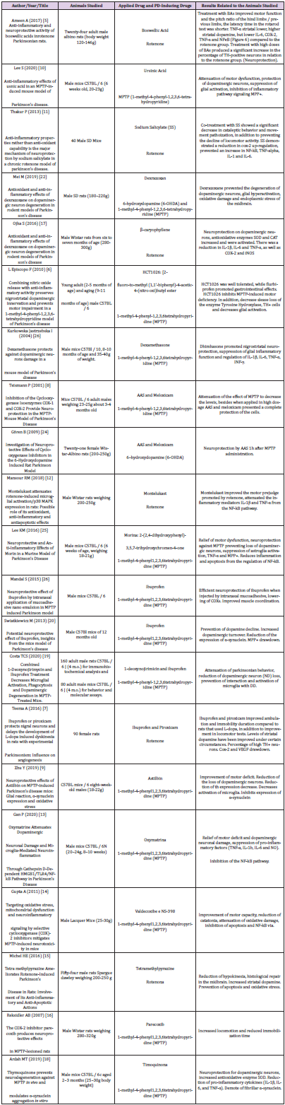
After various transfemoral surgical recanalization attempts in the anamnesis, there were hostile tissue conditions bifemoral with a chronic wound infection leading to exposed graft material. Wound swabs exposed the presence of Staphylococcus aureus and Escherichia coli, indicating a polymicrobial infection. The peripheral blood flow of the lower limbs was compensated. Initially, a CT scan of the abdomen was performed, whereupon an occluded and infected aorto-bifemoral graft was assumed. The subsequently performed PET- CT scan displayed a visibly increased metabolic activity in the area of the graft, so that we diagnosed a chronically occluded and infected aorto-bifemoral prosthetic bypass with subsequential bifemoral infections, leading to the cutaneous wound healing disorder (Figure 1). Due to the patient’s comorbidities, we generally intended an operation and anesthesia time as short as possible with an efficacious treatment by explanting the prosthetic bypass. Further we planned to forego a lavage program for the septic abdomen and intended a primary closure of the abdomen.

In order to treat the local inflammation in the abdominal and femoral areas in the long term intra- and postoperatively, the use of bacteriophages was considered to be plausible alternative therapy option in this case. The patient himself favored an alternative solution compared to an indefinite lasting systemical antibacterial treatment. Therefore, an experimental approach using local bacteriophage application was intended as a last resort treatment in line with Article 37 of the Declaration of Helsinki and in unity with the local ethics committee (A 2021-0208).

biomedres-openaccess-journal-bjstr

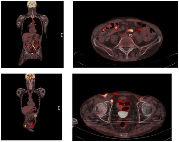
Figure 1: Preoperative PET-CT.
Preoperative PET-CT imaging with increased metabolic activity in the area of the aortobifemoral Dacron® bypass as well as the enhancement surrounding the femoral chronic wound infection.

Bacteriophage Treatment

As a curative therapeutic strategy an intra and extra abdominal application of SniPha 360 (Phage24.com, Austria) was executed. SniPha 360 is a commercially available bacteriophage cocktail of lytic bacteriophages against Escherichia coli, Staphylococcus aureus, Pseudomonas aeruginosa, Streptococcus pyogenes, Proteus vulgaris and Proteus mirabilis. After outlining the potential risks but also benefits of the experimental procedure, the patient consented to the therapy. When performing the relaparotomy, cloudy fluid appeared within the abdomen. After an initial lavage, the retroperitoneum was opened, and the proximal aorta was prepared for clamping. The aorto- bifemoral Dacron® prosthesis presented a shell of a biofilm and was embedded in putrid fluid. The infected aortic prosthesis was extirpated, and the aorta was then sutured over. The prosthesis was retrieved femorally after mobilization of the legs of the prosthesis. The bacteriophage suspension was instilled on Tabotamb-Snow®, which was placed retroperitoneally around the infection.

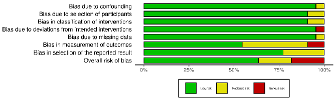
The retroperitoneum and abdomen were primarily closed without further drainage. After removing the femoral anastomoses, the wound conditions were debrided, mobilized and lavaged with a sharp spoon. A bacteriophage-soaked fleece was then placed bilaterally on the femoral side by the same principle, and the wounds were closed again without further drainage (Figure 2). The operation time was 52 minutes, without significant blood loss. Subsequently the patient could be taken to the intensive care unit and extubated without the need for catecholamines. After 10 days of hospitalization, the patient could be discharged with subjective well-being, irritation-free wound conditions and normal findings for inflammatory values in the blood. PET-CT imaging at three months post intervention did not show signs of infection enclosing the aorta or both femoral regions (Figure 3).

biomedres-openaccess-journal-bjstr

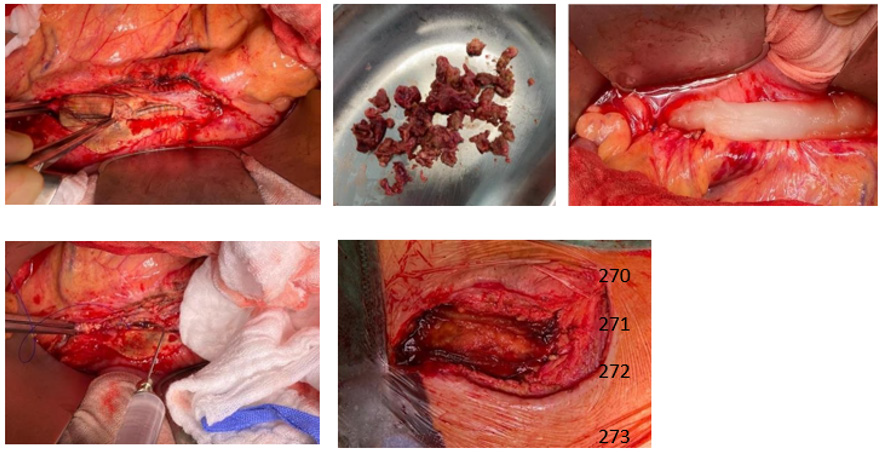
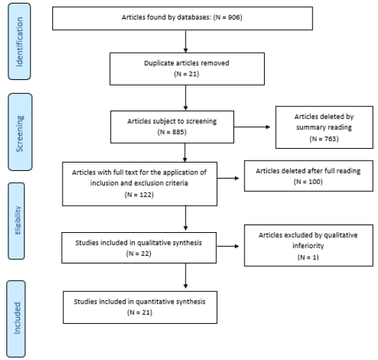
Figure 2: Intraoperative images
Intraoperative pictures showing infected aorto-bifemoral Dacron® bypass. Bacteriophage suspension application on a Tabotamb-Snow®, which was placed retroperitoneally.

biomedres-openaccess-journal-bjstr

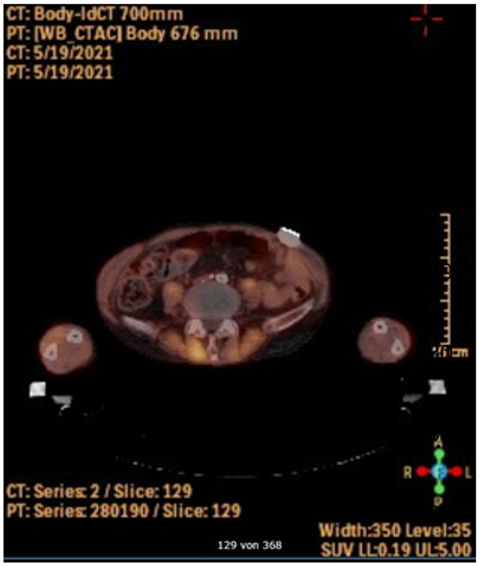
Figure 3: Postoperative PET-CT
Postoperative PET-CT imaging showing no increased metabolic activity surrounding the bypass.

Discussion

This case demonstrates a successful treatment of a chronically infected occluded aorto-bifemoral Dacron® bypass by a local bacteriophage application. It is assumed that around 50-65% of prosthesis infections are a result of bacterial contamination during surgery [7-10]. A general distinction is made between early (up to 30 days postoperatively) and late infections, although the classification is arbitrary [7,10,11]. Early prosthesis infections are often assumed to be a consequence of intraoperative contamination and late infections to be a result of hematogenous bacterial spread, but profound evidence for this is limited. Late infections are usually caused by insufficient tissue integration of the prosthesis into the graft bed. Common pathogenic agents are staphylococci, enterobacteria and corynebacteria [7,10]. Bacteriophages (or simply ‘phages’; Greek: “bacteria eater”) are viruses that selectively infect bacterial cells and were first described in 1917 by the Canadian Félix Hubert d’Hérelle [12].

Currently Bacteriophages are known as a potent anti-bacterial treatment due to their lytic activity [13]. They are considerably stable when exposed to the inflammatory environment and contribute significantly to the regulation of global bacterial mass. A bacteriophage can only multiply where its host is. They are highly specific and therefore predominantly affect strains within one bacterial species, rarely crossing species boundaries [14]. In the first (lytic) cycle of viral reproduction, phages kill their corresponding bacteria through lysis: once infected, the bacterium host cell then starts the process of reproduction, the destruction of the bacterium, and the release of new phage particles; this process is controlled by enzymes and an interaction of bacterial and phage genes. In the second (lysogenic) cycle, the bacteriophage nucleic acid is integrated into the host bacterium’s genome or forms a circular replicon in the bacterial cytoplasm. Compared to other antibacterial therapeutic strategies like local Rifampine treatment [6], no cytotoxic effects on vascular cells could be found for bacteriophages [15].

In addition, they are effective on multi-drug resistant bacteria as well as biofilm-organized bacteria. Recently, in a case series of eight patients with infections of vascular grafts, surgical wounds or implanted medical devices further demonstrated the feasibility of using different bacteriophages with lytic activity for successful treatment of bacterial infections [16]. Although bacteriophages were used for successful treatment of infections of vascular implants, bacteriophage treatment is still not common and not an officially recommended option for infections in the westernized hemisphere [17]. The retro- and intraabdominal application of phages directly to the infection site ensured a maximum concentration, contact time and invasion of the bacteriophages into the infected peri graft tissue. We were able to perform a short operation time, a definite treatment in respect to complete skin/wound closure and the forego of any drainages. No bacteriophage related clinical adverse events had been detected in our case. A three-month follow-up PETCT scan revealed no signs of infections. It could be assumed that the bacteriophage treatment was successful.

In order to treat the local inflammation in the abdominal and femoral areas in the long term intra- and postoperatively, we perceived the use of bacteriophages as an alternative therapy option in antibacterial local therapy. However, there is an ongoing follow-up for the patient to assure a lasting treatment success. In summary, this case report demonstrates that bacteriophage treatment could be a curative treatment option for patients with bacterial graft- and peri graft infections that are not suitable for extensive surgical approaches.

For More Articles: Biomedical Journal Impact Factor: https://biomedres.us

Open Access Journals on Medical Research

Diagnostic Approach in X-Linked Adrenoleukodystrophy in the Pediatric Patient. A Case Report

Introduction

X-linked adrenoleukodystrophy (X-ALD) is the most common peroxisomal disorder, caused by mutations in the ABCD1 locus Xq28 gene due to deficiency of the ALDP protein of the peroxisomal membrane resulting in accumulation of long-chain fatty acids (VLCFA) mainly in the adrenal cortex and central nervous system [1]. It has an incidence of 1 in 21,000 hemizygotes and 1 in 16,800 in heterozygotes. Cerebral infantile X-ALD is the most devastating and progressive phenotype, occurs between three and ten years (peak seven years) is characterized by developmental regression, severe sensory and neurological deficits, in addition to clinical data characteristic of adrenal insufficiency (Addison) [2].

Case Presentation

A 7-year-old boy with no major medical or perinatal history. He started his current condition 6 months ago with asthenia and adynamia, loss of previously learned fine motor skills (buttoning pants, tying shoes), decreased interaction with people and impaired academic performance. Likewise, areas of hyperpigmentation were observed in folds (neck, armpits, elbows, English), so he was requested levels of Adrenocorticotropa hormone ACTH levels of 5455pg/ml (normal: 12 – 76pg/ml) and serum cortisol within normal limits for age. Prednisolone was started at physiological doses. He presented an episode of adrenal crisis secondary to community-acquired pneumonia, so the dose was adjusted to stress doses in the previous 5 months. In this hospitalization, contrasted brain Magnetic Resonance Imaging (MRI) was performed, where hyperintensity was found in the T2 sequence at the level of the white matter in the posterior region of the parietal and occipital lobes, as well as at the level of the midbrain and bridge (Figure 1). He was send to the Genetics department with suspicion of X-linked adrenoleukodystrophy. During his follow-up by Pediatric Endocrinology 2 months after diagnosis, he presented progression of neurological deterioration with ataxic gait, impaired verbal communication and urinary and fecal incontinence. The Genetics department who started management with Lorenzo’s oil (40ml/ day) and lovastatin, as well as a diet low in fatty acids, and requested serum determination of very long chain fatty acids (VLCFA) evaluated him. A follow-up was carried out for 3 months, finding poor response to treatment with persistent cognitive impairment, spasticity and rigidity.

biomedres-openaccess-journal-bjstr

Figure 1: Contrast Brain MRI in T2 sequence.
A. Hyper uptake in the region of the internal capsules and parietal lobes;
B. Hyper uptake is observed in the thalamus, as well as in the white matter of the parietal lobes;
C. Hyper uptake is observed in the corpus callosum, in the bridge and mesencephalon.

Discussion

More than 800 variants in the ABCD1 gene mutation are described, which cause defects in ALDP (adrenoleukodystrophy protein) resulting in the elevation of VLCFA mainly hexadecanoic acids (C26:0) and lignoceric acids (C24:0), causing the accumulation of these in the central nervous system among other tissues [1,2]. VLCFA modify phosphatidylcholine in the myelin membrane can cause instability and inflammation due to altered β-oxidation peroxisomal and increased fatty acid elongation, elevated levels of VLCFA in membranes alter myelin structure and function, this mechanism is described in cerebral adrenoleukodystrophy (CALD) [1]. This causes damage to the endothelium of the cerebral microvasculature critical for the initiation and progression of inflammatory demyelination, tight junction proteins have been shown in autopsies to be displaced, along with massive bloodbrain barrier disruption [1,2]. X-ALD is sensitive to oxidative stress that arises from accumulation of VLCFA and when an excess of free radicals occur they cause the opening of the transition pore of mitochondrial permeability resulting in cell death [1]. Clinical phenotypes of ALD have been described: cerebral infantile, adrenomyeloneuropathy (AMN) and primary adrenal insufficiency [2]. Due to the natural evolution of the disease the phenotypes actually represent a spectrum of the condition, practically all males with ALD will develop adrenal insufficiency and progressive myelopathy in adulthood, and may also develop rapidly progressive cerebral demyelination, which can occur during childhood, but also in adulthood [2].

ALD initially presents as adrenal insufficiency between the ages of 3-10 years, of these 35-40% may progress to a rapidly progressive form of inflammatory demyelination (CALD), leading to progressive neurological deterioration with a peak incidence observed between the ages of 3-8 years causing a vegetative state until death within 2-3 years [2,3]. It is clear that other factors modulate this phenotypic conversion in addition that there is an average delay in the diagnosis of adrenal insufficiency of 3.5 years, which is considered a predetermining factor for clinical progression since an asymptomatic period may be followed [2,3]. The presentation of the clinical case had a delay in the diagnosis of more than 6 months, combining the progression of the disease with clinical data such as asthenia and adynamia in addition to hyperpigmentation at the same time demonstrating manifestations of adrenal insufficiency, it is necessary to start the approach by assessing adrenal function [3]. Adding affectation of fine motor skills involvement suggests brain magnetic resonance imaging (MRI) to rule out brain tumor, neuro infection or autoimmune encephalitis [4]. Brain magnetic resonance imaging (MRI) demonstrates demyelination of the brain white matter, which usually precedes clinical symptoms, ALD brain lesions are initially observed in the splenium of the corpus callosum and the parieto occipital white matter [4,5].
Given the age of the patient, the insidious and progressive onset together with the episode of adrenal crisis provoked by an infection in addition to the data provided by the MRI with which a Loes score of 6 was obtained, it is necessary to approach the patient as a childhood cerebral phenotype of ALD, requesting VLCFA levels [4,5]. Plasma VLCFA analysis is the most commonly used diagnostic test for ALD in men [3]. Given the prevalence of 1 in 21,000 newborns in addition to the difficulty in detecting ADL-X in female patients due to negative family history with onset between the ages of 30- 40 years plus few clinical data (progressive spastic paraparesis more common presentation after age 60) and the need for a panel of laboratory tests [2,3]. In February 2016, newborn screening for X-ALD was added to the recommended uniform screening panel in the United States since ALD has been identified as a newborn screening condition (Figure 2) [2,3]. It was recommended that asymptomatic males identified by newborn screening in New York should have their adrenal function quantitatively assessed every 6 months and the first brain MRI performed until 12 months, then annually until age 3 years, then every 6 months, until age 10 years, and annually thereafter [3]. Once identified, it is suggested to refer the family for counseling, as well as confirmatory tests in the members that require them, in addition, the adrenal function is assessed, since it has been demonstrated that in newborn patients there is biochemical evidence of adrenal insufficiency from 5 weeks and at 4.5 months [2,3].

biomedres-openaccess-journal-bjstr

Figure 2: Proposed algorithm for the diagnosis of ALD-X. It is suggested to investigate clinical history in women with clinical data and perform newborn screening [3]. Normal values VLCFA C24:0/C22:0 0.84μg/mL C26:0/C22:0 0.01μg/mL.

Follow-up with brain MRI is suggested to assess disease progression [3,5]. Abnormalities in MRI are evaluated using the Loes scale, points are awarded based on location, extent of brain parenchymal signal changes, ALD patterns, and the presence of focal and global atrophy [5]. Hyperintensity in T1 or T2 may be seen in areas including white matter: supratentorial, corpus callosum, visual pathway, frontopontine or corticospinal tract and major projection fibers [4,5]. The score is rated from 0 (no evidence of disease) to 34 (severe involvement). 5 In conjunction, the neurological function scale that evaluates the clinical progression of the disease in the patient has been used, in our case resulting in a score of 11 (Table 1) [4]. Both scales should be used to assess progression at a minimum of 1 follow-up evaluation [4,5]. CALD is defined as arrested CALD ≥ 2 consecutive MRI scans within a minimum of 6 months with no increase in Loes Score, no contrast enhancement and no progression of brain symptoms (neurological function scale) [4,5]. There is a limitation in the treatment therapies used in patients with ALD, once the diagnosis is made therapeutic interventions are indicated when the Loes score is between 0.5- 9.2.6 Allogeneic hematopoietic stem cell transplantation is effective for cerebral ALD in the early stages of the disease (Loes 0. 5-9 and neurological function score ≤ 1) allowing to stop the disease progression at the brain level, for symptomatic patients with a Loes score ≥ 10 transplantation produces unfavorable results [4,6]. Transplantation does not alter the progression of adrenal insufficiency so corticosteroid replacement should be initiated, hydrocortisone is the glucocorticoid of choice in children and should be started at stress doses as soon as adrenal insufficiency is diagnosed [3].

biomedres-openaccess-journal-bjstr

Table 1: Assessment of brain involvement. Neurologic Function Scale is used to evaluate the general clinical neurologic status in patients with CALD to determine the degree of risk prior to stem cell transplantation and post-transplant for follow-up [4]. A score of 0 represents no signs of brain disease but more severe signs (*) represent 12 points that determine a high-risk progression domain [4]. Loes < 10 are considered seriously for allogeneic hematopoietic stem cell transplantation at standard risk. Loes > 10 have a more complicated.

Supportive therapies such as Lorenzo’s oil (GTO/GTE) (mixture of glycerol trioleate [GTO] and glycerol trierucate [GTE] in a ratio 4: 1, at doses 2-3ml/kg/day, this reduces the synthesis of very long chain fatty acids (VLCFA) by competitive inhibition of the enzyme responsible for the elongation of saturated fatty acids, but is ineffective in halting disease progression (CALD). Metabolic modulators such as bezafibrate demonstrated a reduction of VLCFA in ALD fibroblasts, but not in plasma, recently a combination of multiple antioxidants at high doses normalizes biomarkers of oxidative damage and inflammation [2,6].

Conclusion

In this case, the importance of making a diagnosis when clinical suspicion persists, recognizing disease patterns with mild to severe patient-dependent manifestations, which are used to assess disease prognosis, as well as the importance of VLCFA analysis to prompt diagnosis of ALD early to allow hormone replacement and prevent disease progression to a devastating phenotype. MRI images of the brain obtained as part of the patient evaluation are used to determine the use of a specific therapy by assessing the risk-benefit ratio, as well as to evaluate progression and determine if the disease is arrested.

For More Articles: Biomedical Journal Impact Factor: https://biomedres.us

Journals on Medical Science Research

The Cultivation of Creativity in the Classroom

Introduction

Conceptually, creativity is defined as the ability to produce a new work or idea based on imagination. Younger psychologists argue that creativity is not a special skill or ability of a few people, but rather is the result of special training and learning through specific processes, which enable each individual to activate inexhaustible forces of his mind [1]. Many view creativity as a tendency to activate or recognize alternative ideas or possibilities, which can prove useful in problem solving, in communicating with others, or even in the field of entertainment. Creativity according to others is to think outside the blueprints or frameworks, approaching new areas and achieving results, which are able to provide answers to problems that concern them [2]. In this process, a clear distinction is made between predetermined abilities. Of course, the first concerns creativity, learning ability and communication. The latter are directly related to production, economy, marketing, etc. Creativity therefore moves within an indefinite pattern and can be evaluated through different processes. Many also argue that creativity can be seen as a process by which new original and useful ideas are activated, which help to deal with everyday problems and challenges. But it is important to see human creativity as the art of the different in terms of thinking. But in order to formulate a definition that will be based on scientific data, it is important to do a historical review and to trace the way in which science used to deal with, and continues to deal with, the concept of creativity. But in order not to get lost in the vastness of the various sciences, we will approach it both as an object of psychological research and as an object of pedagogical practice [3].

It is a fact that creativity has preoccupied researchers and around the subject has developed a large bibliography that gives new dimensions to creative thinking and opens new perspectives. As a starting point for creativity, we refer to the discomfort Guilford posed to the American Psychological Society in 1950 over the way the scientific community approached creativity, which was listed in the international literature as an “American challenge.” This discomfort and Guilford’s concerns in general were the trigger for the development of a number of approaches to creative thinking [4]. The interest of scientists and researchers to deal with the creative thinking of man and the abandonment of the concept of the “divine gift”, gave a series of definitions which we present below in order to arrive at a more widely accepted definition, but also through management of the concept of creativity to be able to understand the nature of creative thinking and the structural principles that govern it. Guilford made a first attempt to define the concept, according to which “creativity covers the most characteristic abilities of creative individuals, which determine the probability for an individual to express a creative behavior, which is manifested by ingenuity, composition and design.” of course, this definition is simplistic, but some facts emerge that are then confirmed by researchers. This ability seems to be linked to certain personality traits.

These characteristics speculate if and how creativity will manifest. Creativity affects all individuals and is not a rare phenomenon only of gifted individuals [5]. “Differentiation between individuals is quantitative, a matter of classification, not qualitative.” Getzels and Jackson (1962) define creativity as the combination of those elements that are considered original and different [5]. They point out that creativity is one of the most valuable human possibilities, but it is difficult to examine it systematically. Lowenfeld and Brittain (1975) argue that creativity is directly related to who gives the definition. Thus, some psychologists distinguish as qualitative elements of creativity:
a. Flexibility of thought
b. The originality of the idea
c. The ability to think differently
d. How to solve problems.

Of course, here we have to contrast Einstein’s view, which argues that formulating a problem is more important than solving it. Piaget (1960) defines creativity as a process of problem solving, problem finding, exploration, experimentation, an intellectual energy that implies respect and thoughtful decision making. Torrance (1966) identifies creativity with the ability of the individual to face various problems, with sensitivity, originality but also with method and calm. Creativity according to Lee, Webberlen and Litt (1987) is a multifaceted phenomenon and every issue that arises is addressed through different processes. We must also cite the view of Bruner (1962) who defines creativity as an energy from which arises a special and effective surprise [3]. Freud (1972) defines creativity as an instinctive impulse that aims at creation but also correlates it with the impulse of destruction. Creativity can include shaping new systems, transferring familiar relationships to a different field, and shaping new correlations. Through the conceptual approach it follows that it is difficult to integrate creativity into a definition. We adopt what Davis (1992) states: “There are as many infinite definitions and ideas of creativity as there are people who have written their ideas on a piece of paper.” Of course, if we want to categorize the prevailing positions on creativity, we could mention:
a. The traditional view, which claims that there are a number of “intelligent”, “gifted” people, this category includes people with exceptional talent or some special skills that stand out from the rest and cite as examples personalities such as Mozart and Einstein and according to this creativity is not the same in all people, so it is not cultivated.
b. The modern view, which argues that talent is mainly the result of practice and hard work, and all individuals have the opportunity to reach a degree of creativity and the cognitive processes followed in the emergence of ideas are no different from every day and therefore creativity can be cultivated.

The nature of Creativity

Creativity is about observing known things, it is based on previous ideas – experiences and the search extends to something new or a different approach. Among the main reasons that drive creativity, we distinguish:
a) The need for an innate impulse inherent in the human mind for something new.
b) The communicative need for exchange of ideas.
c) The human need to solve problems and create new ideas.

The human brain is the one that plays an important role in every creation, whether it is associated with human survival and the construction of the first tools, with mental functions, with artistic creations or even with the confrontation of everyday human problems. Creativity does not start with zero states [6]. It can be built on pre-existing knowledge or experiences. In the nature of creativity, we must mention an important element that runs through the whole process. It is the element of imagination that enables people and much more children to successfully process everyday situations and develop their creative abilities [6]. Imagination and creativity could be said to move in parallel and are interconnected. Of course, we must emphasize that creativity does not start from scratch, like the imagination, in the sense that the pre-existing elements that are inscribed in the consciousness help to create new representations in the form of images or ideas. This begs the question, are the representations of memory the same as the representations that exist and are recorded in the imagination? Their difference is more in the form and not in the content. It is important to point out that there is a danger in childhood that the imaginary will become an extension of reality.

The Evaluation of Creativity

Evaluation is the stage at which an account of what has been produced is made. It is an important stage in the whole process through which the ideas produced are evaluated [7]. Without it, the process would be “incomplete”. Furthermore, every evaluation of ideas has basic principles, such as: “it is a continuous process, it must be done for all ideas, it must have the meaning of collectivity, it must be objective, and it must be a guide for further paths” [8]. It is suggested that the convergent thinking be evaluated separately from the divergent one in order to understand the differences. Evaluating creativity is important for the following reasons:
a) It contributes decisively for the younger generations to show their abilities and to take advantage of their inclinations and interests.
b) It is a determining factor and a steady step towards selfknowledge.
c) It prepares future generations to adapt to the rapid changes that are taking place” [8].

Creativity is not an objective feature, because we have the ability to use indicators to evaluate the creative possibilities through which objective determination is achieved. We should mention that there are no surefire ways to guarantee the reproduction of innovative ideas. Source inspiration does not fall into measurement scales. Of course, there have been references to specific brainstorming processes, but the issue remains open, as discussions persist in the qualitative dimension, which is not measurable [9].

Evaluation Methods

From the search in the foreign language literature regarding the methods of evaluation of the creative inclination and ability, we have distinguished numerous and flexible methods that enable the evaluation. Of course, we should point out the research controversy in the scientific community regarding the evaluation of creativity [10]. Hocevar in a thorough review of creativity presented key points – axes used in creativity studies:
a. Convergent thinking exercises.
b. Divergent thinking exercises.
c. Recording the behavior and interests of individuals.
d. Recording of special personality elements.
There are other ways to measure creativity, such as:
1. Plot titles: here the participants are given the plot of a story and asked to come up with original titles.
2. Quick reactions to word associations: this is where unusual answers are scored.
3. Conception of shapes and forms: here are presented simple drawings of people and objects and they are asked to find common properties and characteristics in two or more paintings. Scoring is again based on unusual answers.
4. Unusual uses: here are given everyday objects, e.g., a toothpick and unusual applications are required.
5. Remote correlations: here participants are asked to create a new word from two other simple ones.
6. Distant effects: this calls for the activation of a list of consequences of unexpected events.
7. Creativity can also be calculated based on the response to a variety of test scenarios, such as:
8. The expression of ideas: the ability to easily develop a variety of reasoning and correlations, when presented with a simple word or image.
9. The combination of ideas in a new way: the development of a wide range of innovative approaches and solutions, when we are asked to explore new possibilities for an ordinary simple object of our daily life (eg a brick).
The emergence of new benefits for existing ideas: the activation of original ideas or solutions based on pre-existing ideas. Investigation: the ability to process an idea in order to make it practically functional. Focus and distinction: identifying the most important elements of an idea and then approaching them in an effort to solve a problem while simultaneously evaluating the difficulties. Perspective exchange: the ability to suggest ways to view and solve a problem in the light of different perspectives.

Children and Creativity the Cultivation of Creativity in the Classroom

Teachers should review the teaching practices they apply in order to be able to judge the extent to which they have been able to instill in students a creative way of thinking. Some ways to boost creativity by teachers are:
Enhancing divergent thinking:
a) Allow the teacher to ask questions of the student.
b) Be educationally receptive and sensitive to the problems faced by students.
c) To make the children realize the maximum importance of the questions, but also not to be afraid to trust their senses.
d) Problems should not be presented simply but discovered.
e) Attempt to try a second Tuesday etc. to find a solution to each problem.
f) The taught subjects of the courses to be examined from different angles.
g) To convey to the students the message that they should not rest on the first correct answer they will give.
h) In general, is there anything going on in the school that could be part of the concept of divergent thinking?
i) In any case, Learning should not be a mechanical storage of knowledge from textbooks and teachers.
j) Existence of motivation and encouragement:
k) Students’ questions should be accepted by teachers so that they can then develop.
l) Children’s curiosity to be supported and additionally provoked.
m) Opportunities for self-directed learning should be provided.
n) The teacher appreciates and supports the personal interests of the student.
o) Unnecessary repetition of a theme should be avoided.
p) Receptivity to the new
q) The school should be not only a place of traditional teaching, but also of enjoyment, fun and challenge for spiritual adventures.
r) Teaching should convey the real world within the school.
s) As a place the school should be offered for the cultivation of imagination.
t) The school must be able to dispel stress and provide a sense of comfort and relaxation to the student.
u) The school must provide opportunities and opportunities for a subject to be examined in an experimental and at the same time pleasant way.
v) Finally, the uniqueness and individual personality of each student should be assessed, and a conservative attitude should not be imposed.
Cultivation of creativity must be an integral part of the educational process (Jullien, 2004). We emphasize that the cultivation of creative tendencies should not be pursued only within the framework of some ‘special hourly support programs that will be repeated at sparse intervals or at a predetermined time and place. The desire to cultivate such an important element must overcome all limitations and be systematically systematized, in every activity that accompanies school and extracurricular life.

Conclusions

Creativity is a multifaceted concept. Its special nature leads to the non-existence of a unified psychological theory that will explain and include all its dimensions. Many people still associate it with the arts and avoid associating it with other cognitive objects such as the sciences which are considered uncreative. They believe that creativity is a special feature of some people who are involved in the arts. The importance of creativity for art is very important but it is equally important in the sciences and in other cognitive areas that result from the composition of two areas. For example, the use of art in the natural sciences and vice versa. Today we have come to the conclusion that creativity is characteristic of every human being. All people can be creative as long as they are given the opportunity and find themselves in an environment where the conditions are right for them to develop and cultivate their creative skills. The development of creativity in the school context is important and there are specific ways to enhance and promote it that must be taken care of by teachers. The evaluation of creativity also plays an important role, which is a key factor in the course of children’s education at all levels of education.

For More Articles: Biomedical Journal Impact Factor: https://biomedres.us

Open Access Journals on Medical Research

Rapid and Practical Screening Method for the Detection of Colistin-Resistant Bacteria in Food

Introduction

Colistin is recognized as one of the few remaining available antibiotics for the treatment of intractable infections caused by multidrug-resistant Gram-negative bacteria [1]. Recent studies have shown that bacteria carrying the mcr gene, which confers colistin resistance to most members of the Enterobacteriaceae, are widely disseminated, particularly in Asia [2,3]. Since colistin is widely used in animal husbandry [4], the spread of colistin-resistant (CR) bacteria in communities via livestock food is a potential risk factor. Moreover, CR bacteria are often found in animals and animalfood [5-7]; thus, monitoring CR bacteria in animal-food is essential. However, the conventional culture method [8] for detecting CR bacteria in food is laborious and time-consuming. Rapid detection of colistin resistance genes at the research level is now possible using the SYBR green method [9], but its widespread practicality is limited due to the need for complex steps and equipment involved in DNA extraction from samples and determination of result specificity. To overcome this limitation, we here report a simple, rapid, and practical detection method of Escherichia coli harboring mcr-1, as a representative CR bacterium, using a highspeed real-time polymerase chain reaction (PCR) kit. We further verified the utility of this method for detecting CR bacteria in retail meat samples. Although a real-time PCR assay for the detection of mcr genes from bacterial isolates has already been established, this newly proposed detection method holds practical relevance for widespread use, as the entire procedure, from food sample processing to the final result, can be completed within only 1 h.

Materials and Methods

A total of 27 retail meat samples, including pork and chicken, were collected from 10 markets (two supermarkets and eight local traditional markets) in Vietnam and five supermarkets in Japan during November and December 2019. None of the eight traditional markets in Vietnam maintained a refrigerator for meat preservation. In contrast, the two supermarkets in Vietnam and all five supermarkets in Japan had refrigerators for food storage. Each sample was collected from one meat type per market. Bacterial cultures and DNA extraction were performed on the collection day. Ten grams of each meat sample were placed in a stomacher bag (AS ONE, Osaka, Japan) containing 90 mL buffered peptone water. The samples were hand-homogenized for 2 min. The resulting homogenate was inoculated on CHROMagar COL-APSE (CHROMagar, Paris, France), a selective medium for CR Gram-negative bacteria, and cultured at 37 °C for 24 h. CR E. coli-like colonies were distinguished based on colony color (dark pink to reddish) after cultivation [8,10]. A representative colony was isolated by sub-culturing on MacConkey agar, and bacterial identification was performed. The colistin minimum inhibitory concentration (MIC) was estimated, and colistin resistance genes (mcr-1 to -5) were detected by multiplex PCR as described previously [6,11].
In parallel, DNA was extracted from 1 mL of the homogenate using the Kaneka Easy DNA Extraction Kit version 2 (Kaneka, Tokyo, Japan). The presence of E. coli and the colistin resistance gene mcr-1 in the DNA extracts was determined by real-time PCR using a mobile PCR device, PicoGene PCR1100 (Nippon Sheet Glass, Tokyo, Japan). PCR primers and TaqMan probes for realtime PCR detection of E. coli 16S rRNA and mcr-1 were prepared as described previously (Table 1) [12]. Details regarding the realtime PCR, including PCR mixtures and thermal cycling conditions, are provided in Tables 2 & 3, respectively. The DNA extract of the CR E. coli strain (E362) [6] carrying mcr-1 was utilized as a positive control in PCR. The entire 50 PCR cycles were completed within only 21 min. Moreover, the real-time PCR device could simultaneously measure fluorescence at three different wavelengths for the same sample. Two fluorescent dye-labeled TaqMan probes (Integrated DNA Technologies, Singapore), Cy5 for E. coli 16S rRNA and FAM for mcr-1, were used for each sample. The entire protocol is outlined in Figure 1. Figure 2 shows representative real-time PCR profiles of the samples.

biomedres-openaccess-journal-bjstr

Figure 1: Outline of the screening protocol using mobile real-time PCR PicoGene® PCR1100. BPW, buffered peptone water.

biomedres-openaccess-journal-bjstr

Figure 2: Representative plots obtained from real-time PCR amplification of mcr-1 and E. coli 16S rRNA genes in meat samples.
a) Positive control, mcr-1 E. coli.
b) mcr-1–negative pork sample, H-A market pork.
c) mcr-1–positive chicken sample, H-E market chicken.

Results and Discussion

The detection sensitivity of the method was assessed using pork meat samples spiked with an mcr-1-positive E. coli strain culture. The lower limit of mcr-1-E. coli detection for the entire method, from DNA extraction to detection by real-time PCR, was 7 × 10² CFU/g; however, a minimum of 7 × 10³ CFU/g was required for quantification using a linear correlation. In the validation study using retail meat samples, CR E. coli-like bacteria were detected using the culture-based method in eight out of ten chicken and in three out of seven pork samples purchased in Vietnam (Table 4). The semi-quantitative levels of CR bacteria in these samples were in the range 103‒108 CFU/g (Table 4). All representative CR E. coli isolates from each sample were confirmed to be resistant to colistin (MIC ≥ 4 μg/mL) and possessed mcr-1 but not mcr-2 to -5, except for the H-E market pork sample, which harbored mcr-3 in addition to mcr-1, as determined by multiplex PCR. No samples from the Japanese supermarkets were contaminated with CR bacteria. All samples, except for the H-E market pork sample, that were positive via the culture-based method were also positive by real-time PCR (Table 4). Some culture-negative samples such as H-B market pork, T-B market chicken, T-B market pork, and T-E market chicken were PCR-positive. Such contradictory results may be attributed to the features of the real-time PCR method and its ability to detect mcr-1 even in dead cells and/or non-E. coli cells. In contrast, a pork sample from the H-E market showed CR E. coli colonies after culturing but tested negative for mcr-1 by real-time PCR. Such discrepant cases could be due to a low level of mcr-1–positive bacteria below the detection limit of the real-time PCR method or the presence of bacteria expressing non-mcr CR determinants [13].

The new method presented herein detects the target gene and facilitates quantitative analysis. In addition, the method using TaqMan probes has high detection specificity, and is simple because it does not require specificity verification by melting curve analysis, even for one-step extracted DNA from food. The results output the ratio of bacteria carrying mcr-1 to the total number of E. coli cells, which may be mcr-1–positive or –negative bacteria (Figure 2). The detected quantitative mcr-1 levels were higher than the CR E. coli-like bacterial levels determined via the culture-based method because the real-time PCR method detects all mcr-1 regardless of bacterial species. The quantitative linear range detected via realtime PCR was between 103 and 106 CFU/g. Although the detected signal was below the quantitative linear range limit in some samples, they were still considered to have positive results via realtime PCR. The approach described in this study provides limited information regarding the degree of contamination; nevertheless, the developed method is reliable and practical owing to a short processing time, enabling the rapid screening of contaminating bacteria with mcr-1 in food.

Conclusion

A new rapid and practical screening method was developed for detecting CR E. coli in food samples. The developed method is advantageous because it is easy to perform, has a short processing time, and provides reliable results that are consistent with those obtained by traditional methods.

For More Articles: Biomedical Journal Impact Factor: https://biomedres.us

Open Access Journals on Medical Research

Remote Monitoring of the Health Status of Pregnant Women in the COVID-19 Pandemic

The Role of Remote Technologies in the Quality Management System and Safety of Medical Care

On April 26, 2021, Deputy Chairman of the State Duma Irina Yarovaya at a meeting of the Presidium of the Council of Legislators of the Russian Federation under the Federal Assembly of the Russian Federation called for simplifying the exchange of data between medical institutions and patients. In the Sverdlovsk region, an automated information system of mobile notifications «AIST_SMART» for pregnant patients and doctors began to operate. Using a smartphone or, say, a tablet, pregnant patients in their personal account get the opportunity to keep an electronic diary of self-control of their health. The diary has the functions of automatic interpretation of the results and the formation of signal information for the obstetrician-gynecologist. Now pregnant women do not need to fill out paper diaries of self-control, call their doctor or the reception of the antenatal clinic or wait for a doctor’s call in order to report the results – the process is fully automated. The women’s consultation received an IT tool for remote interaction with pregnant women and women in child child. The introduction of «AIST_SMART» technologies made it possible to replace paper diaries with electronic ones. Medical data of the patient are collected in a single database and allow you to track the dynamics of the patient’s health around the clock. The results of electronic diaries are automatically processed by the system and if no abnormalities are detected, the data is simply recorded in the system and does not disturb the doctor (Figure 1).

In case of detection of deviations in the patient’s state of health, the system marks the identified deviations and sends a notification to the doctor about the current state (Figure 2). Mobile notifications instantly convey accurate and detailed information about the patient’s state of health and thus contribute to the timely decision on hospitalization in case of detection of criteria for weighting the course of NCVI. All notifications in case of deviations are automatically sent to the attending physician and the doctor in the Obstetric Remote Consultation Center (hereinafter referred to as the ADCC) for the routing of the patient 24/7. Remote health monitoring functions as follows.

biomedres-openaccess-journal-bjstr

Figure 1: The data of the expanded diary of self-control at the NKVI, all indicators are normal.

biomedres-openaccess-journal-bjstr

Figure 2: The data of the expanded diary of self-control at COVID-19 with deviations in the state of health.

Registration in the System «AIST_SMART»

To register the patient in the personal account at the initial appointment of a pregnant patient, a consent-instruction [1] is issued to connect to the mobile service «AIST_SMART» with an individual QR code. At home, the patient reads the QR code using the camera of her smartphone or tablet and, according to the instructions, undergoes the registration procedure, forming a digital four-digit PIN-code. From now on, it is guaranteed 24/7 technical support. The QR code serves as the patient’s identifier and the link between her electronic medical record (EHR) in the AIST «RAM» and the personal account in the «AIST_SMART» system. To register a doctor in your personal account, you must log in to the medical information system – AIST «RAM», in which all medical personnel of the obstetric service in the region work. Open the «Personal Account» tab and register by scanning an individual QR code. So, in order to access electronic self-control diaries, the doctor and the patient connect to the AIST_SMART service, and after registering in the system, notifications about the results of remote health monitoring will be received on their mobile device. The doctor does not need to call on the phone to find out how she feels, what her temperature is, the symptoms of SARS, etc.

How the Mobile Alert System Works

Formation of Notification of the Result of Self-Control Diaries

This process is fully automated. AIST_SMART performs the role of an intellectual assistant to the obstetrician-gynecologist/ midwife. The patient fills in the diary data, and the doctor receives ready-made results with automatic interpretation. Now the patient will not forget to call the antenatal clinic, and the doctor will be able to make decisions on the tactics of conducting comprehensively, taking into account the results of the patient’s home self-control and his obstetric status.

Patients with COVID-19 are Asymptomatic/Mild and Receiving Care on an Outpatient Basis (at home)

Upon receipt of the results of testing in a pregnant woman / maternity for COVID-19, the data are entered by medical personnel in the AIST «RAM». Notifications about the results are automatically generated in the personal account «AIST_SMART» (Figures 3 & 4). These notifications are automatically sent to both the patient and the doctors. With what there is control that the patient is also informed about the result (Figure 5). If a positive result is detected on the COVID-19, the patient receives notifications 2 times a day about the need to fill out a self-control diary, which is also informed by the doctor – full feedback (Figure 6). The doctor of the ADC, based on the results of the self-control diary (Figure 7) and obstetric status according to the data in the electronic medical record (hereinafter referred to as the EHR) in the AIST «RAM», where there is information about all the results of the examination, the course of pregnancy and diagnoses, decides on further management tactics: to continue outpatient treatment or hospitalization in a covid hospital. The ADCC doctor fixes his decision in the EHR, making out a remote consultation for the attending physician of the antenatal clinic or obstetric hospital (if the patient is in the hospital at the time of detection of the COVID-19).

biomedres-openaccess-journal-bjstr

Figure 3: Mobile NOTIFICATION of PCR result for COVID-19: not detected.

biomedres-openaccess-journal-bjstr

Figure 4: Mobile NOTIFICATION of PCR result for COVID-19: DETECTED.

biomedres-openaccess-journal-bjstr

Figure 5: Marking about the patient’s reading of the results of the examination.

biomedres-openaccess-journal-bjstr

Figure 6: Mobile notification that a reminder has been sent to the patient to complete a self-monitoring diary.

If a decision is made on the need for hospitalization, the doctor of the ADC through a confidential «working» chat in AIST_SMART can contact the patient and clarify her consent to hospitalization and the possibility of transportation by personal transport. If consent is obtained (Figure 8), the ADC doctor makes an additional referral for (re-) hospitalization to a particular covid hospital for pregnant women and women in childcare, taking into account available places. The patient receives a notification about the referred referral indicating the covid hospital, the date and time of hospitalization (Figure 9). If it is necessary to organize transportation, the doctor of the ADC has resources through communication with the medical organization where the patient is on the dispensary register and agreeing on the method and time of transportation by the NSR team in compliance with epidemiological rules (Figure 10).

biomedres-openaccess-journal-bjstr

Figure 7: Dynamics of the state of health according to the electronic diary of self-control.

biomedres-openaccess-journal-bjstr

Figure 8: Communication with the patient through confidential “working” chat in AIST_SMART.

You do not Need to Receive a Paper Direction

If necessary, you can print the direction at the place of treatment of the patient, using a single information space of the regional obstetric monitoring of AIST «RAM». All the directions that a woman received during pregnancy are reflected in her personal account in the «My directions» section. The patient can open any document, even if the connection with the internet has disappeared.

biomedres-openaccess-journal-bjstr

Figure 9: Notification of referral to a Covid hospital and labeling of reading by a patient.

Advantages of Remote Monitoring of Health

The transition to electronic diaries of self-control allows you to identify the weighting of the course of ARVI / ARI in the case of outpatient treatment (at home) with COVID-19, and timely send the patient to hospitalization to prevent adverse events, which is from the main directions of the quality management system and safety of medical care. AIST_SMART allows you to create constant feedback [2] with the patient and thereby form a patient-centric model of care as one of the priority areas for the development of modern medicine and healthcare in general. All of the above increases the compliance of doctor-patient interaction and directly affects the quality and safety of medical care in the difficult conditions of the NCVI pandemic, which meets the modern needs of society and solves the tasks set by the Government of the Russian Federation in the field of digitalization of healthcare [3-8].

For More Articles: Biomedical Journal Impact Factor: https://biomedres.us

Open Access Journals in COVID-19 

Experience of Indian National Biobank in COVID-19 Pandemic and Future Directions

Introduction

The Coronavirus disease 2019 (COVID-19) is an infectious disease caused by the Severe Acute Respiratory Syndrome Coronavirus-2 (SARS-CoV-2) [1]. SARS-CoV-2 was first reported in China and till date accounted for 169 597 415 confirmed global cases of COVID-19, including 3 530 582 deaths. Worldwide [2]. On March 11th 2020, WHO declared public health emergency [3]. High transmissibility of this virus rapidly surged number of cases and many countries around the globe announced regionalnational lockdowns [4]. The lockdown situations adversely affected businesses, slow down of scientific activities and operational activities of several sectors including biobanks [5]. The National Liver Disease Biobank (NLDB), India is an advanced open resource sharing liver disease biobank for liver and associated disease research established with the joint efforts of the Department of Biotechnology (DBT) and Institute of Liver & Biliary Sciences (ILBS), New Delhi, Government of India [6]. NDLB is India’s first liver disease biobank with a storage capacity of more than 5.4 million biosamples and certified by Tissue Repository Network (CTR. Net) in 2020 [7]. NLDB has been set up in an institute dedicated to patient care and research in liver diseases.
The biobank collects high quality biosamples across the country with clinical data. A total of 73,831 aliquots of serum, plasma, PBMC, urine, tissue, stool, and whole blood from 12,607 patients have been collected and stored at NLDB as of Dec 31st 2020. Biosample and access to the advance analytical facility openly available under one roof for all researchers. In order to deliver cutting edge services for collaborative liver disease research NLDB acquired a non-profitable business and financial model, charging only the cost for utilization of services, NLDB engaged trained and highly competent staff with world class storage and advanced analytical infrastructure, aiming to become a nodal centre for providing the clinical and basic researchers to reliably store biosamples and carry out their research at one platform. The national sudden lockdown was placed on 24 March 2020 in India for 68 days in different phases when the number of confirmed coronavirus cases were approximately 500 [8]. The lockdown restricted people to stay in their homes [9] and all transport services were suspended with exceptions for essential emergency services [10].

Impacts

The sudden lockdown brought both the opportunities and challenges to the biobank. Although, the National Liver Disease Biobank (NLDB) is a liver and related diseases biobank, the government of India designated it as an add-on COVID biobank permitting for collection and storage of COVID-19 biosamples for research, developing diagnostics and vaccines. NLDB faced tri-directional challenges based on financial, operational and sustainability, but were accepted positively with changing in the processes and management.

Crisis Management

The storage facility and associated equipment are one of the key elements in operations of biobank. As per best practices published by International Society for Biological and Environmental Repositories [11], telephone numbers for professional assistance should be clearly posted in the repository and accompanying administrative areas (e.g., engineering or facilities personnel, power companies, fuel supply companies, transportation services). The emergency planning was focused to maintain cryopreservation of biosamples from various possible events that may breakdown the freezers. NLDB has 10 % of the total storage capacity as backup, maintained at operating temperature at all times. Safe guarded by 24×7 CCTV surveillance and a security personal and all mechanical freezers connected with datalogger equipped with SMS alert system. Three biobank personnel are trained and even prepared for 24×7 shifts in case of emergency. Contact numbers of emergency response team (engineering, electricity and security office) are posted on all storage units. Earlier the emergency plan was only focused for natural calamities. Learning from the current situation, an upgraded emergency plan based on management and transportation of sample at satellite center, business strategy, financial planning and operations of biobank is under review. Moreover, NLDB also started to develop contingency plan to keep operating in pandemic positions. There were difficulties in taking consent with COVID infected patients. Leftover diagnostic samples stored at biobank without consent will be utilised for research after approval from ethics board.

Sample Collection

The NLDB follows the “decentralized collection, centralized storage, distribution and informatics” model. (Figure 1). It has collaboration with 18 hospitals for collection of biosamples and supports many research projects by providing biosamples along with associated data. Biosamples are collected with necessary precautions, however, in this pandemic, the need of PPE kit, sanitizer, and establishment of BSL2/BSL3 facility was critical, considering all samples as highly infectious.

biomedres-openaccess-journal-bjstr

Figure 1: NLDB Model for biosample collection, transportation and distribution.

The challenges confronted while functioning in this pandemic:
1. There has been a significant decline in the number of samples collected from both host institute and the satellite centres because Outpatient Department and surgeries are only limited for emergency cases (Figure 2). 2. Co-ordination with satellite centers and maintenance of samples became difficult because of limited staff.
2. At initial period, hospitals were not prepared to screen for COVID-19 for all patients, leading to high chances of collecting COVID-19 contaminated samples from asymptomatic patients. Sample processing protocols were revised and precautions were made even for handling samples apparently COVID negative.
3. Biobank was instructed to collect COVID-19 biosamples but processing and storage area was not designed to handle highly infectious samples. To avoid cross contamination, urgent requirements for separate space for processing and storage of COVID and non-COVID samples was flagged.
4. Dedicated routes to transport cryoshippers containing aliquoted COVID biosamples were made from patient ward to BSL2+ facility and then to the storage area.

biomedres-openaccess-journal-bjstr

Figure 2: Effect of biosample collection during lockdown.

Logistics and Supply of LN2 Gas

The surging Covid-19 in March, 2020 and the Indian government’s decision to contain the disease outbreak through lockdown adversely affected the domestic logistics sector, especially road transportation, production and supply of essential goods [12]. With increasing number of active cases of Covid-19, the consumption and demand of oxygen was increased throughout the country [13]. Some LN2 industries directed to produce more oxygen in comparison to LN2 gas. The resource management for consumables, refilling of LN2 in cryoshippers, transient storage and transportation of biosamples are managed from main centre established at New Delhi, India. The sudden nationwide lockdown almost got NLDB in a standstill affecting the operational chain such as managing the collection, storage, transportation of biosamples from satellite centres. Biobank has consumption of 100 litres/day to maintain temperature of two LN2 tank. NLDB does not have LN2 plant and dependents only on LN2 supply from outside. Closedown of LN2 factories due to movement of labours, local shortage/limited access to liquid nitrogen, shortage of drivers, made it difficult to get the LN2 tanks refilled. Moreover, the market price of LN2 was hiked up to three times in comparison to the previous routine rate. The pandemic taught that biobank should have inhouse plant for LN2 supply. To avoid such problem in future, NLDB processed to establish an LN2 plant in ILBS premises with capacity to produce approx. 250 litres of LN2/ day (Figure 3).

biomedres-openaccess-journal-bjstr

Figure 3: Elements which affected the LN2 supply in the pandemic.

Operations

The ban and restrictions on public transport effected the employees resulting in only 40% attendance of the staff. Biobank staff were seconded in COVID-19 testing lab and the sudden focus and orders to quickly set up procedures to test covid-19 samples, and two-technicians infected with COVID-19 at different time periods and others quarantined for coming in close contact, were big challenges faced. IT experts were not able to resolve the technical issues in the biobank software from home due to nonavailability of remote access for the software. There was temporary interruption of collection and distribution activities as hospitals redirected to treat critical cases and COVID -19 patients only. During lockdown, one of the -80⁰C freezers stopped working, consequently the samples were shifted to the backup freezer. Repair was delayed due to restricted movement and limited supply of spare parts and backup LN2 freezer was being utilised for storage of COVID-19 samples. Biobank samples were on complete risk in case of any failure in storage system as the backup freezers were already in use. Emergency purchase of two -80 freezers was done to accommodate more COVID samples as left over covid-19 samples from hospital diagnostic centers were directed to store in biobank for future research. SOPs were revised as per the knowledge gained in the pandemic. A separate SOP is developed as per guidelines of Indian Council for Medical Research/ Government of India for collection, storage and distribution of COVID-19 samples for research.

Personnel Wellbeing

Commuting for the personnel was big issue in lockdown. However, staff working in COVID lab were provided accommodation in hospital. The safety guidelines issued by Ministry of Health and Family Welfare Government of India to maintain social distancing at work place and transport were followed with necessary compliance [14,15]. Routine test, thermal scanning, sanitizing machine, touch free mechanism installed at all entry and exit points and common areas. Complete ban on non-necessary visit and emergency visits were allowed only after negative rapid antigen test. Two biobank technical staff resigned from their job because their family not allowed them to work on COVID-19 samples.

Management Related Issues

a. Finance: A project for add-on COIVD biobank facility was submitted to the Government of India which was approved and funds released on priority basis in December, 2020.
b. Biobank Information Management System (BIMS): IT related issues were impediment for biobank due to no remote access of clinical databases, biobank systems, slow adaptation and update of software. BIMS was updated with annotation for COVID-19 as per recommendations of ICMR, GOI.
c. HAZARD Management: The primary and basic requirement of biobank is safety of its staff and of the environment against biological and chemical hazards. There were no specific guidelines available for storage, collection, distribution and QMS of highly infectious samples in ISO20387, NCI and ISBER best practices. Sharing of COVID biosamples are not as easy as non-COVID samples thus National Oversight Committee was constituted by ICMR to review the same. NLDB has provision to share the sample after approval of Biosample release committee (BRC). Sample are released after signing MTA and undertaking by recipient to handle COVID-19 and it is informed that any violation or misuse would be dealt with strict action as per laws of Government of India.

Work Culture & Infection

Work culture of biobank has been totally changed due to COVID fright and implementation of new rule and SOPs. Handling the informed consent, annotation forms duly signed by COVID patients was a big issue. WHO and ICMR guidelines are being followed by NLDB to prevention from any infection, Intensive communication and training on good hygiene practices, PPE kit donning and doffing has been provided to biobank personnel. Technicians are equally divided for COVID and non-COVID related work. It is compulsory to wear N95-type masks, use of hand sanitizer, disinfect all documents coming through patents in Ultraviolet (UV) light, and to sanitize work area daily and disinfect the storage area twice a week.

Research Support

The government of India has released huge funds for research focused on Diagnostics, Vaccines, Novel Therapeutics, Repurposing of Drugs or any other intervention for control of COVID-19 as most of the research institutes were closed or had limited access to maintain necessary equipment during lockdown.

Discussion

The sudden lockdown consequent to the COVID-19 pandemic brought both the opportunities and challenges to the biobank. NLDB handled the tri-directional challenges that were operational, financial and sustainability. Sudden changes in operations, supply chain disruptions, manpower presence and remote access of software were major difficulties along with the Handling of Covid-19 biosamples, inaccessibility of donors and challenges in obtaining informed consent. Although, there was neither biobank practices and standards included any plan to run a biobank in a pandemic, NLDB followed the available national [15] and international standards [16] and guidelines [11,17,18] to handle the infectious samples. Though, biobank had an emergency plan for backup storage though there were no thoughts to have an emergency plan for LN2 supply and to work with limited man power. Flexibility in purchase rules, monitoring of efficient utilization, stock management for every one month can be a great help to run biobank in emergency. Biobank must have inhouse LN2 plant along with a rate contract with suppliers to supply LN2 in emergency at equivalent prices. All SOPs revised to treat all sample as infectious Remote monitoring and access of software during emergencies is a must. However, development of remote monitoring software is only possible after the contribution of key stakeholders, such as hospital administration, IT team, privacy legal expert and biobank operations team. In conclusion, NLDB used this pandemic as a learning experience and modifying its operational, emergency and business plans for future crisis and pandemics.

For More Articles: Biomedical Journal Impact Factor: https://biomedres.us

Journals on Hypertension

Tai Chi, Qigong, and the Treatment of Hypertension

Introduction

Tai chi, also referred to as taiji or taijiquan, is considered both a martial art and a kind of low-impact exercise. Its origins are unclear, but it apparently dates back at least to the thirteenth century. The oldest style is the Chen style, which originated in the Chen village in China [1,2]. The second oldest style, and also the most popular style, practiced by more people than any other style, is the Yang style [3]. The other main styles are the Wu and Wu Hao styles [4], and the Sun style [5], which is the youngest of the five main styles. The various styles of tai chi have much in common, although there are some differences, which we need not discuss in this article. One of the main common features of all styles of tai chi is that they generate healing life energy (qi, pronounced chee), which serves to boost the body’s immune system and prevent the onset of illness and disease. Qi energy also has a beneficial effect on treating existing illness. Many articles and books have been written about the health benefits of tai chi [6-7], so we need not go into the details here. Suffice it to say that many medical studies have found that the regular practice of tai chi can lead to many health benefits, including the treatment of existing diseases and illnesses.
Qigong has been around a lot longer than tai chi, perhaps thousands of years [8]. Many books and articles have been written about this traditional Chinese medicine tool as well [9-74]. It is also a set of gentle exercises that generate qi, which has beneficial healing effects for a wide variety of ailments, including, but not limited to ankylosing spondylitis [75-76], anxiety and stress reduction [77-82], arthritis [83-89], autism [90], back pain [91-92], cancer [93-115], cognitive impairment [116-119], COPD [120-121], COVID-19 [122-123], depression [124-134], elder care [135-138], fibromyalgia [139-141], longevity [142-144], Parkinson’s Disease [145-146], and traumatic brain injury [147], to name a few. The present article focuses on the beneficial effects of tai chi and qigong exercises on hypertension and blood pressure. It reviews a few studies that have found beneficial effects and cites a number of other studies for further reading and research.

Methodology

The PubMed.gov database [148] was searched to find studies that had been done to determine the effectiveness of tai chi and qigong exercises on blood pressure and hypertension.

Findings

The findings reported upon in this article are representative of the numerous studies that have been done examining the effects of tai chi and qigong on blood pressure and hypertension. Additional studies on this topic are cited in the reference section below Liu et al. [149] conducted a meta-analysis to determine the effectiveness of tai chi and qigong exercises in the treatment of essential hypertension (EH). Specifically, they looked at blood pressure (BP), levels of nitric oxide (NO), and endothelin-1 (ET-1). Exercises were performed from 1.5 to 6 months. Nine randomized controlled tests (RCTs) of 516 EH patients in China found that those who did the exercises were able to reduce both systolic and diastolic blood pressure. The exercises also contributed to higher NO blood levels and lower ET-1 blood levels. Although the difference in treatment outcomes using tai chi and qigong exercises versus antihypertensive drugs was statistically insignificant, combining the two therapies resulted in significantly better outcomes than what would occur using only tai chi and qigong or drug therapy. Thus, tai chi and qigong exercises were equally effective as drug therapy in the treatment of hypertension, only without the side-effects that may be present with drug therapy. Liu et al. concluded that tai chi and qigong exercises could be an effective complementary and alternative therapy for EH patients.
The tai chi exercises varied by study, and included the Yang- 24 form, Yang-8, and Chen-style tai chi. The qigong exercises also varied by study, and included Mawangdui Daoyinshu and Baduanjin, among others. Subgroup analyses were performed for the different types of tai chi and qigong, and some were found to be more effective than others. One subgroup analysis of changes in systolic blood pressure ranked the effectiveness of the various exercises as follows, from most to least effective:
a) Chen-style tai chi
b) Mawangdui Daoyinshu Qigong
c) Self-compiled qigong
d) Yang-style tai chi
An examination of different subgroups found that some tai chi and qigong exercises were more effective than others in lowering diastolic blood pressure. The ranking, from most to least effective, was:
a. Chen-style tai chi
b. Self-compiled qigong
c. Mawangdui Daoyinshu Qigong
d. Yang-style tai chi
Liu et al. concluded that Chen-style tai chi might be most effective in reducing blood pressure, while Yang-style tai chi might be the least effective. The authors also compared the effectiveness of the various tai chi and qigong exercises on improving NO levels. The ranking from most to least effective was:
a. Yang-style tai chi
b. Baduanjin Qigong
c. Mawangdui Daoyinshu Qigong
Chen-style tai chi and self-compiled qigong were not statistically significant in improving NO levels. The authors also analyzed subgroup data on the effectiveness of tai chi and qigong in reducing ET-1. The ranking from most to least effective was:
a. Baduanjin Qigong
b. Yang-style tai chi
c. Mawangdui Daoyinshu Qigong
Self-compiled qigong was found not to be statistically significant in lowering ET-1 levels. Thus, it appears that Baduanjin and Yangstyle tai chi may be more effective than other exercises in improving NO and ET-1 scores.
If one were to interpret the findings of this study, one might conclude that choosing qigong and or tai chi therapy might be superior to drug therapy for the treatment of EH for two reasons. Although the study found that qigong/tai chi therapy and drug therapy are equally effective in treating EH, qigong/tai chi therapy has two distinct advantages over drug therapy: qigong/tai chi therapy has no adverse side-effects, and it does not cost anything. Drug therapy, on the other hand, sometimes has adverse sideeffects, and it is not free. The study also found that combining qigong/tai chi therapy with drugs might be superior to choosing just one of the two options.
Pan et al. [150] conducted a systematic review of randomized controlled trials on the effects of tai chi on blood pressure, body mass index (BMI), and quality of life (QOL) on patients suffering from hypertension. Their meta-analysis of 24 studies containing 2,095 patients (1,074 in the treatment group and 1,021 in the control group) found that the intervention group had significantly better outcomes for systolic blood pressure (SBP) [p ≤ 0.001], diastolic blood pressure (DBP) [p ≤ 0.001], physical functioning [ p ≤ 0.001], role-physical [p ≤ 0.001], general health [p = 0.001], bodily pain [p ≤ 0.001], vitality [p ≤ 0.001], social functioning [p = 0.027], role-emotional [p = 0.003], and mental health [p = 0.001] compared to the control group. However, the differences in BMI between the groups were insignificant. Pan et al. concluded that tai chi is an effective therapy to improve SBP and DBP for patients suffering from essential hypertension. Zou et al. [151] found that the practice of baduanjin was beneficial for quality of life (p = 0.004), sleep quality (p = 0.001), balance (p = 0.004), handgrip strength (p = 0.007), trunk flexibility (p = 0.006), systolic (p = 0.0004) and diastolic (p = 0.005) blood pressure, and resting heart rate (p = 0.0005). They examined the results of various studies on each of these topics. In the case of the effect of baduanjin on blood pressure, they examined 9 studies having a total of 743 participants.
Ladawan et al. [152] investigated the effects of qigong exercise on cognitive function, blood pressure and cardiorespiratory fitness in 12 healthy middle-aged subjects who performed qigong exercises in 60-minute sessions, three times a week for eight weeks. They found that the exercises resulted in significant improvements in Trail Making Tests Part A (p = 0.04), systolic blood pressure (p = 0.0001), diastolic blood pressure (p = 0.005), mean arterial pressure (p < 0.001) and maximal workload (p = 0.032). Twelve weeks after cessation of the exercises, they had all returned to the baseline. The authors concluded that it is necessary to perform qigong regularly to maintain the improved health effects.
Ching et al. [153] examined data on 370 subjects from seven randomized controlled trials (RCTs). The following six types of qigong exercises were used:
a) Conventional Qigong
b) Guolin Qigong
c) Shuxinpingxue Gong
d) Dongeui Qigong
e) Ba Duan Jin Qigong
f) Mawangdui Daoyinshu Qigong
They found that the practice of qigong exercises had a significant effect on reducing systolic (p < 0.001) and diastolic (p < 0.001) blood pressure. The above studies are representative of the studies that have been done in recent years on the effectiveness of tai chi and qigong on reducing high blood pressure. Some other recent studies are listed in the reference section at the end of this article [154-188].

For More Articles: Biomedical Journal Impact Factor: https://biomedres.us

Open Access Journals on Drug

Use of Anti-Inflammatory Drugs in the Treatment of Parkinson’s Disease: A Systematic Review of Perimental Studies

Introduction

Parkinson’s disease (PD) is a progressive neurodegenerative disease characterized by the loss of dopamine neurons (AD) in the substance nigra pars compacta (CNS) and accumulation of insoluble cytoplasmic protein inclusions called Lewy and Lewy neurites bodies [1]. The precise mechanism underlying the pathogenesis of PD is not yet fully understood. The accumulation of evidence suggests that soluble α-synuclein aggregates, known as oligomers, play a significant role in PD where the neurodegenerative process culminates in impairing several subcellular functions [1]. Thus, clinically, PD presents as muscle stiffness, tremor at rest, bradykinesia (abnormal slowness of voluntary movements), postural instability; some patients also have symptoms related to psychiatric and cognitive disorders. In this context, intraneuronal accumulation and aggregation of alpha-synuclein can start from several sites such as the intestinal tract, where this altered protein (alpha-synuclein) can be transported through the enteric route to the CNS through the parasympathetic pathway [2]. In addition to this hypothesis, there is genetic influence in the functional roles of genes identified as monogenic forms of PD. Mutations in SNCA, LRRK2 and VPS35 genes have been highly penetrating and cause autosomal dominant forms of PD [1]. Thus, showing the existence of multifactorial processes to support the underlying cause of this aberrant protein accumulation. Therefore, what most of these studies show is that when alpha-synuclein is lodged in the CNS itself, it is directly linked to damage triggered by the activation of microglia, which, by releasing inflammatory factors, causes an oxidative burst affecting neuronal cells leading to death [3].
Thus, since there is a pattern of inflammatory characteristics after the beginning of the accumulation of these proteins, this tangle of interleukins, TNF-α, TNF-γ, CCL2, ROS and NO may increase such accumulation and aggregation already in force, thus determining an even more cumulative and oxidative neurodegenerative picture, exponentially affecting the patient’s condition, becoming a real “Parkinson’s snowball”. Thus, this hypothesis suggests a clinical applicability of treatment with anti-parkinsonian drugs of antiinflammatory nature and drugs properly anti-inflammatory drugs (IANES and corticosteroids), where the anti-inflammatory action may provide a therapeutic resource for patients with the purpose of promoting a decrease in levels of dopaminergic cell lesions and lowering of alpha-synuclein accumulation. This study, therefore, aims to correlate the use of these two types of drugs with antiinflammatory attributes to the treatment of PD, observing whether there is an anti-inflammatory or neuroprotective response (via dopaminergic markers) and which group of drugs is better than the other.

Methodology

This study consisted of a systematic review prepared according to the Preferred reporting items for systematic review and metaanalysis protocols (PRISMA-P). The eligibility criteria defined for the inclusion of an article in this review were human and animal studies, contain relevant information regarding the neuroprotective action of the drug in PD, applicability of anti-inflammatory drugs, csf analysis, use of in-silico computational method and clinical results and be indexed in the electronic databases MEDLINE/ Pubmed, LILACS, EMBASE, Scopus and Web of Science. Using the PECOS strategy, the descriptors used in the searches were chosen based on the technical-scientific terms MeSH (Medical Subjective Heading) and DeCS (Descriptors in Health Sciences), combined by the Boolean operator “AND” or “OR” (Table 1). MEDLINE/ PubMed research strategy: “Idiopathic Parkinson’s Disease” OR “Lewy Body Parkinson’s Disease” OR “Parkinson’s Disease, Idiopathic” OR “Parkinson Disease, Idiopathic “ OR “Parkinson’s Disease, Lewy Body” OR “Parkinson’s Disease” OR “Idiopathic Parkinson Disease” OR “Lewy Body Parkinson Disease” OR “Primary Parkinsonism” OR “Parkinsonism, Primary” OR “Paralysis Agitans” AND “Neuroinflammation” OR “Inflammations” OR “Innate Inflammatory Response” OR “Inflammatory Response, Innate” OR “Innate Inflammatory Responses” AND “Anti Inflammatory Agents” OR “Agents, Anti-inflammatory” OR “Anti-inflammatories” OR “Anti-inflammatory Agents” OR “Agents, Anti-Inflammatory” OR “Agents, Anti Inflammatory” OR “Anti-Inflammatories” OR “Anti Inflammatories” OR “Anti-inflammatory Agents, Non-Steroidal” OR “NSAIDs” OR “Non-Steroidal Anti-Inflammatory Agents” OR “Non-Steroidal Anti Inflammatory Agents” OR “Nonsteroidal Anti-Inflammatory Agents” OR “Nonsteroidal Anti Inflammatory Agents” OR “Anti Inflammatory Agents, Nonsteroidal” OR “Antiinflammatory Agents, Nonsteroidal” OR “Nonsteroidal Antiinflammatory Agents” OR “Corticosteroids” OR “Corticoids” OR “Inhibitors, Cyclo-Oxygenase” OR “Inhibitors, Cyclo Oxygenase” OR “Inhibitors, Cyclooxygenase” OR “Prostaglandin Synthesis Antagonists” OR “Antagonists, Prostaglandin Synthesis” OR “Inhibitors, Prostaglandin-Endoperoxide Synthase” OR “Inhibitors, Prostaglandin Endoperoxide Synthase” OR “Prostaglandin Endoperoxide Synthase Inhibitors” OR “Prostaglandin Synthase Inhibitors” OR “Cyclo-Oxygenase Inhibitors” OR “Cyclo Oxygenase Inhibitors” OR “Inhibitors, Prostaglandin Synthase” OR “Inhibitors, Cyclooxygenase 2” OR “Cyclooxygenase-2 Inhibitors” OR “Inhibitors, Cyclooxygenase-2” OR “Coxibs” OR “COX-2 Inhibitors” OR “COX 2 Inhibitors” OR “Inhibitors, COX-2” OR “COX2 Inhibitors” OR “Inhibitors, COX2”.

biomedres-openaccess-journal-bjstr

Table 1: PECOS Strategy.

EMBASE research strategy: (‘parkinson disease’/exp/mj OR ‘parkinson disease’/mj OR ‘parkinson`s disease’/mj OR ‘parkinsons disease’/mj OR ‘paralysis agitans’/mj OR ‘parkinson disease, symptomatic’/mj) AND (‘anti-inflammatory agent’/exp/mj OR ‘antiinflammatory agent’/mj OR ‘anti-inflammatory agents’/mj OR ‘antiinflammatory agents, steroidal’/mj OR ‘anti-inflammatory agents, topical’/mj OR ‘anti-inflammatory drug’/mj OR ‘anti-inflammatory agent’/mj OR ‘anti-inflammatory agents’/mj OR ‘anti-inflammatory agents, steroidal’/mj OR ‘anti-inflammatory agents, topical’/mj OR ‘antiflogistic agent’/mj OR ‘antiinflammation agent’/mj OR ‘anti inflammatory agent’/mj OR ‘anti-inflammatory drug’/mj OR ‘antiinflammatory steroid’/mj OR ‘anti-inflammatory activity’/exp/mj OR ‘anti-inflammatory action’/mj OR ‘anti-inflammatory activity’/ mj OR ‘anti-inflammatory effect’/mj OR ‘anti-inflammatory action’/ mj OR ‘anti-inflammatory activity’/mj OR ‘anti-inflammatory effect’/mj OR ‘antiphlogistic action’/mj OR ‘antiphlogistic activity’/ mj OR ‘antiphlogistic effect’/mj OR ‘nonsteroid anti-inflammatory agent’/exp/mj OR ‘nsaid’/mj OR ‘anti-inflammatory agents, nonsteroidal’/ mj OR ‘anti-inflammatory agents, non-steroidal’/mj OR ‘anti-inflammatory agent, nonsteroid’/mj OR ‘non steroid antiinflammatory agent’/mj OR ‘non steroid anti-inflammatory drug’/ mj OR ‘non-steroidal anti-inflammatory agent’/mj OR ‘non-steroidal anti-inflammatory drug’/mj OR ‘non-steroidal anti-inflammatory agent’/mj OR ‘non-steroidal anti-inflammatory drug’/mj OR ‘nonsteroid anti-inflammatory agent’/mj OR ‘nonsteroid antiinflammatory drug’/mj OR ‘nonsteroid antirheumatic agent’/mj OR ‘nonsteroidal anti-inflammatory drug’/mj OR ‘nonsteroidal anti-inflammatory drugs’/mj OR ‘nonsteroidal anti-inflammatory drugs’/mj OR ‘nonsteroidal anti-inflammatory agent’/mj OR ‘nonsteroidal anti-inflammatory drug’/mj OR ‘prostaglandin synthase inhibitor’/exp/mj OR ‘cyclooxygenase inhibitor’/mj OR ‘cyclooxygenase inhibitors’/mj OR ‘prostaglandin synthase inhibitor’/mj OR ‘prostaglandin synthetase inhibitor’/mj OR ‘cyclooxygenase 2 inhibitor’/exp/mj OR ‘cox 2 inhibitor’/mj OR ‘cox 2 specific inhibitor’/mj OR ‘cox 2 specific inhibitors’/mj OR ‘cox- 2 inhibitor’/mj OR ‘cox-2 specific inhibitor’/mj OR ‘cox-2 specific inhibitors’/mj OR ‘cox2 inhibitor’/mj OR ‘cox2 specific inhibitor’/ mj OR ‘coxib’/mj OR ‘coxibs’/mj OR ‘cyclooxygenase 2 inhibitor’/ mj OR ‘cyclooxygenase 2 inhibitors’/mj) AND (‘modulation’/exp/ mj OR ‘modulation’/mj OR ‘protection’/exp/mj OR ‘protection’/ mj OR ‘protective factors’/mj OR ‘treatment outcome’/exp/mj OR ‘medical futility’/mj OR ‘outcome and process assessment (health care)’/mj OR ‘outcome and process assessment, health care’/ mj OR ‘outcome management’/mj OR ‘patient outcome’/mj OR ‘therapeutic outcome’/mj OR ‘therapy outcome’/mj OR ‘treatment outcome’/mj OR ‘disease management’/exp/mj)
LILACS Research Strategy: “Idiopathic Parkinson’s Disease” OR “Lewy Body Parkinson’s Disease” OR “Parkinson’s Disease, Idiopathic” OR “Parkinson Disease, Idiopathic “ OR “Parkinson’s Disease, Lewy Body” OR “Parkinson’s Disease” OR “Idiopathic Parkinson Disease” OR “Lewy Body Parkinson Disease” OR “Primary Parkinsonism” OR “Parkinsonism, Primary” OR “Paralysis Agitans” AND “Neuroinflammation” OR “Inflammations” OR “Innate Inflammatory Response” OR “Inflammatory Response, Innate” OR “Innate Inflammatory Responses” AND “Anti Inflammatory Agents” OR “Agents, Anti-inflammatory” OR “Anti-inflammatories” OR “Anti-inflammatory Agents” OR “Agents, Anti-Inflammatory” OR “Agents, Anti Inflammatory” OR “Anti-Inflammatories” OR “Anti Inflammatories” OR “Anti-inflammatory Agents, Non-Steroidal” OR “NSAIDs” OR “Non-Steroidal Anti-Inflammatory Agents” OR “Non-Steroidal Anti Inflammatory Agents” OR “Nonsteroidal Anti-Inflammatory Agents” OR “Nonsteroidal Anti Inflammatory Agents” OR “Anti Inflammatory Agents, Nonsteroidal” OR “Antiinflammatory Agents, Nonsteroidal” OR “Nonsteroidal Antiinflammatory Agents” OR “Corticosteroids” OR “Corticoids” OR “Inhibitors, Cyclo-Oxygenase” OR “Inhibitors, Cyclo Oxygenase” OR “Inhibitors, Cyclooxygenase” OR “Prostaglandin Synthesis Antagonists” OR “Antagonists, Prostaglandin Synthesis” OR “Inhibitors, Prostaglandin-Endoperoxide Synthase” OR “Inhibitors, Prostaglandin Endoperoxide Synthase” OR “Prostaglandin Endoperoxide Synthase Inhibitors” OR “Prostaglandin Synthase Inhibitors” OR “Cyclo-Oxygenase Inhibitors” OR “Cyclo Oxygenase Inhibitors” OR “Inhibitors, Prostaglandin Synthase” OR “Inhibitors, Cyclooxygenase 2” OR “Cyclooxygenase-2 Inhibitors” OR “Inhibitors, Cyclooxygenase-2” OR “Coxibs” OR “COX-2 Inhibitors” OR “COX 2 Inhibitors” OR “Inhibitors, COX-2” OR “COX2 Inhibitors” OR “Inhibitors, COX2” .

Web of Science Search Strategy

TÓPICO (Parkinson disease*) AND TÓPICO (inflammation*) AND TÓPICO (anti-inflammatory*).

Scopus Search Strategy

(TITLE-ABS-KEY (Parkinson AND disease) AND TITLE-ABSKEY ( inflammation ) AND TITLE ( anti-inflammatory ) ) .
The selection of articles was performed by two researchers blindly and independently through reading the titles, reading the abstracts and, finally, full reading of the articles. Any disagreement in the selection was resolved in consensus meetings. Articles that fully met the eligibility criteria were included in this study. The selection process is described in Flowchart 1 adapted from PRISMA (Figure 1). In order to analyze the methodological quality of the included studies, each article was evaluated by a researcher based on the items of the ACROBAT-NRSI (A Cochrane Risk of Bias Assessment Tool for Non-Randomized Studies) [4]. Acrobat-NRSI scores were used to exclude articles that did not present hardhitting information to the research, besides serving as a basis for discussing the methodological quality of the articles and the possible viruses in the generalization of their results (Figures 2 & 3). From each article included, data related to the objectives of this review were extracted, such as author, title, type of study, population, PD induction drug, drugs used applied, positive results. These data were computed and compared using the t-Student test for independent samples, with the purpose of comparing the percentage s percentages of the and effects on PD between NCAs and other anti-inflammatory drugs (Table 2).

biomedres-openaccess-journal-bjstr

Table 2: Characteristic of selected experimental clinical trials.

biomedres-openaccess-journal-bjstr

Figure 1: Adapted from PRISMA.

biomedres-openaccess-journal-bjstr

Figure 2.

biomedres-openaccess-journal-bjstr

Figure 3.

Findings

Twenty-one articles were analyzed, separated between two groups according to the drug used for pre-clinical study, antiparkinsonian drugs of anti-inflammatory nature and drugs properly anti-inflammatory drugs (IINES and corticosteroids). Improvement in motor function, decreased movement patriotization, increased levels of striatal dopamine, decreased interleukins and blockage of inflammatory pathways, such as those participating in MPP+ and COX-2, as well as increased and/or decreased loss of neurons armed with tyrosine hydroxylase (TH) enzyme, an important marker of neuroprotection, were identified.

Discussion

In view of these findings, this systematic review demonstrated that there is an effective therapeutic relationship in the use of anti-inflammatory drugs in PD through findings such as, mainly, quantitative increase or decrease in the loss of tyrosine hydroxylase enzyme [5-9]and improvement of motor function or prevention of motor decline [5,10-16]. However, since these are experimental studies in animals where clinical failures are commonly recorded in this methodology, caution should be exercised in the face of these findings, even if it shows clinical relevance. In addition, the importance of the therapeutic look is emphasized, especially in pathophysiological terms elapsed by the articles, observing in most of them that this disease, which affects the nicrostriatal region harboring the substantia nigra and quite rich in microglia, has the cumulative character of alpha synuclein in its altered form, which leads to the formation of a highly fibrillar aggregate by very little known pathways, thus, there is the beginning of a cascade of events that lead to the release of inflammatory toxic factors and a progressive dopaminergic neurodegeneration [17,18]. It is identified, therefore, that within this pathophysiological mechanism there is linked an inflammatory response, so there is a target to be investigated and possibly treated, demonstrating possible therapeutic purposes against PD.
In parallel, this review was able to investigate some other parameters found in experimental animal studies. Some motor tests showed improvement in the face of performance tests, applicability of previous training or open field observation, in addition, motor improvement of the forelimbs and later [5], significant decrease in cataleptic behavior [10], improvement of ambulation and immobilization time [7]and reduction of hypokinesia [15]. These results reinforce the hypothesis of a neuroinflammatory cause of Parkinson’s and once again the application of anti-inflammatory drugs for a possible therapy. It can be observed that characteristics that are found in patients such as muscle stiffness, tremor at rest, bradykinesia and postural instability could be solved or attenuated by a drug with function, absorption and mechanisms similar to what were found in this review. Therefore, there is a vast ness of possibilities for anti-inflammatory pharmacological use, in which, however, there is still a need to weigh the pros and cons, the latter being something of changeable capacity within the pharmaceutical industry, in which with investments in research and advanced technology can be achieved a less deleterious profile to the body, such as raising blood pressure, interaction with anti-hypertensive drugs, reduction of renal perfusion and gastrointestinal symptoms [16].
Within this context, it was also possible to identify an increase, then neuroprotection from levels of dopamine, TH enzyme and dopaminergic neurons in some animals. These results can be explained by the fact that the neuroinflammatory process, in its characteristic of exponential cascading lesion of dopaminergic neurons [8,19], was blocked and there was no more decrease in degenerative character. All this was observed from immunohistochemical analyses of TH (Tyrosine Hydroxylase) levels, an enzyme involved in dopamine synthesis through a series of biochemical reactions that has the amino acid tyrosine as a precursor and a molecular marker of dopaminergic neurons, along with dopamine dosage [5-9,18,19]. Thus, it was demonstrated what can occur in a neural system previously healthy, but with microglia activated by the pathophysiology of PD, in this case by mimetic drugs of PD such as rotenone and 1-methyl-4-phenyl-1,2,3,6 tetrahydropyridine (MPTP). Thus, it is envisaged, once again, the use of these drugs or something more advanced both in patients already diagnosed and living with the disease chronically, as well as in patients at the beginning of diagnosis and mild clinical picture, promoting neuroprotection and, consequently, a greater defense and increased quality of life.
Some drugs in the studies acted directly on microglia and other inflammatory foci, some of them are very common, such as ibuprofen, meloxicam, piroxicam, AAS, Valdecoxib and Parecoxib (NHEMS, which act by inhibiting COX-2, prostaglandin and ultimately reducing cytokines), dimethazone (Corticosteroid that reduces the gene expression of pro-inflammatory cytokines). All of them obtained good results regarding the lowering of glial hyperactivation and intracellular inflammatory, in addition to stimulating the recovery and regeneration phase, avoiding in some cases the toxicity of MPTP [20], which shows that even having extensive knowledge and applicability of these drugs, they can still be key parts for the advancement of neural therapy in PD. Similarly, oxymatrine, an alkaloid compound found at the root of a Chinese herb (Sophora flavescent), promoted relief of motor deficits induced by MPTP and conferred significant neuroprotection, in addition to inhibiting the activation of microglia and exacerbated release of pro-inflammatory as cytokines [13]. This shows that within the vastness of drugs known and disseminated by the pharmaceutical industry, there are still a gigantic number of other substances that can be used in the treatment of this disease [20-27].

Conclusion

Our study has concluded that there is a need for investment in quality, more robust, broad-spectrum preclinical studies, with minimal view to achieve the ideal pharmacological therapeutic for this target. Thus, it is necessary more clinic trials to confirm this relationship between an inflammatory profile and use of antiinflammatory drugs which possible therapeutic agents to treatment of PD.

For More Articles: Biomedical Journal Impact Factor: https://biomedres.us

Journals on COVID-19

Type 2 Diabetes Mellitus and COVID-19 in Mexico. A comprehensive Assessment

Introduction

On February 11, 2020, the International Committee for Taxonomy in Viruses named SARS-CoV-2. Composed of a genome of 30,000 base pairs, belonging to the Coronaviridae family of the order Nidovirales. Phylogenetically coronaviruses are classified into alpha, beta, gamma and delta. Coronaviruses were identified 50 years ago as pathogens responsible for the common cold, mainly HCoV-OC43, HCoV-229E among other variants. At the beginning of 2002, coronaviruses were considered exclusively veterinary pathogens, however, by 2019 they were identified in biological samples from patients diagnosed with pneumonia [1-4]. Showing an age trend initially with geriatric patients, it has been shown that the risk of mortality increases after 75 years [5]. However, today age is no longer a dependent factor for infection. It is important to mention this since it may be due to multiple etiologies in addition to infection, such as: comorbidities, lack of metabolic control, suspension of work in the outpatient clinic due to hospital oversaturation derived from the pandemic, sedentary lifestyle, among others.
Hence it is important to emphasize the lack of metabolic control derived from all those cardiometabolic diseases, such as: obesity, hypertension, dyslipidemias and mainly diabetes mellitus, which turns out to be the first pandemic that has not been adequately controlled since ancient times [6]. All these factors are directly and proportionally related to the risk of severe progression and poor prognosis due to the chronic inflammatory state that generate more the acute systemic inflammatory response derived from COVID-19. In the case of obesity, another factor shared by both pathologies increased even more derived from confinement due to the forced closure of sports centers, favoring a sedentary lifestyle. The anxiety derived from the pandemic favors a greater consumption of foods with low nutritional power, again favoring obesity and lack of metabolic control. Therefore, in the context of a controlled diabetic patient, the measures that had to be implemented as a strategy to reduce the rate of infections are one of the factors to generate lack of control. The percentage of uncontrolled diabetics since the beginning of the pandemic is more and more common and continues to rise, which entails greater spending on health, greater generation of medical supplies and resources. There is an excess of mortality in the Mexican Republic derived from the pandemic, not only due to COVID-19, but also due to other causes [7,8] without forgetting to mention the possibility of under- registration that exists, for example, in marginalized areas or those who could not have hospital access derived from the same scenario. That is why the relevance of this article where a comprehensive scenario is proposed for the knowledge and management of COVID-19 in those patients who already have a chronic damage such as Diabetes Mellitus.

Pathophysiology

The incubation period for SARS-CoV-2 is 5 days with a range of 2 to 14 days [9]. The spectrum of diseases generated by coronavirus infection is mainly acute respiratory, chronic, enteric, hematological, endothelial and of the central nervous system. The mechanism of transmission of the disease by SARS-CoV-2 is from person to person through the airway by the drops of Flügge that are exhaled when coughing, sneezing or speaking and are inhaled or deposited in the mouth and ocular conjunctiva, as well as surfaces, which can function as fomites [10]. The main structural proteins found on the membrane surface of the SARS-CoV-2 viral particles participate within the pathophysiology, which are: Spike (S), membrane (M) and envelope (E). Among other, these are responsible for the anchorage and entry of these microorganisms to the host’s cells. It should be noted the type 2 angiotensin converting enzyme (ACE 2) which is a type I membrane protein that contains receptors in the lung, heart, kidney and intestine, endothelium, nervous system, mainly. The ACE 2 receptors that are located in the lower respiratory tract of humans are the cellular receptors for SARS CoV-2. Since the virion has the S-glycoprotein or Spike protein, which projects through the viral envelope and forms the spicules of the crown, this is glycosylated and is responsible for mediating the binding of the receptor (protein S + ACE 2), as well as its fusion with the host cell [11,12].
This strong bond unites the entire SARS-CoV-2 membrane with the host cell membrane, entering it through endocytosis. Viral particles release their RNA that binds to viral DNA, initiating the viral replication cycle, which leave the host cell through exocytosis. Once the RNA of the SARS-CoV-2 particles begins its translation and transcription, two processes are generated: the first related to the high demand for manufacturing viral proteins causing cellular stress that ends in apoptosis of the target cells; while in the second, the viral RNA acts in a molecular pattern associated with pathogens, which leads it to be recognized by the cells of the immune system, initiating the activation of the cytokine cascade and the migration of neutrophils. Hypercoagulability, venous stasis and endothelial damage is another of the main characteristics mediated by the ACE 2 receptors that SARS-CoV-2 particles possess, being observed in the endothelium of the veins, arteries and arterial smooth muscle cells of the brain; This produces dysfunction and inflammation of the microvasculature that alters vascular flow and initiates platelet activation, increasing risk for macrovascular and microvascular thrombosis, pulmonary thromboembolism, deep vein thrombosis, catheter-related thrombosis, ischemic cerebrovascular disease, acrosyndromes, and capillary leak syndrome. in organs such as lungs, kidneys and heart, increasing mortality, one of the main complications [13] (Figure 1).

biomedres-openaccess-journal-bjstr

Image 1: Physcopathogenesis of COVID-19.

SARS-COV2 as a Diabetogenic Agent

Diabetes is associated with a chronic low-grade inflammatory state that favors the development of an exaggerated and constant inflammatory response. At the molecular level, there is an increase in the levels of IL-6 and C-reactive protein (CRP), so the proinflammatory state typical of diabetes can favor the cytokine storm and the systemic inflammatory response that accompanies the acute respiratory distress syndrome (ARDS) in patients with COVID 19 [14]. This is why diabetics infected with SARS-CoV-2 have a higher rate of hospital admission, severe pneumonia, and higher mortality compared to non-diabetic subjects [15]. SARS-CoV-2 is considered diabetogenic since it is also capable of causing direct damage to the pancreas, due to the expression of ACE 2 (mainly in islet cells) even in a higher proportion than at the lung level, which could worsen hyperglycemia and even induce the onset of diabetes in previously non- diabetic subjects [16]. It should be noted that only 1-2% of patients with mild COVID-19 infection present pancreatic lesions, while 17% of patients with severe cases present with lesions of the pancreas, which can accentuate the systemic inflammatory response and, therefore, Therefore, accelerate the appearance of ARDS [17]. On the other hand, the current scenario of the pandemic even in uninfected subjects may favor the deterioration of metabolic control due to difficulties in accessing the health system, lack of physical activity and increased stress associated with confinement.
Therapeutic strategies should be aimed at facilitating access to the health system through telemedicine to advise the patient on the adaptation of treatment or any other remotely manageable medical situation and guide patients and caregivers in the control of diabetes in order to prevent hospitalization [18]. Clinical symptoms. Different stages of SARS-CoV-2 disease have been described in humans depending on the clinical severity, which can range from mild symptoms such as: fever, myalgia, headache, cough, anosmia. Up to severe symptoms characteristic of pneumonia with severe respiratory impairment [19,20-25]. Table 1 Mild and moderate infections comprise 80.9% of the registered cases; the severe ones, 13.8% and the critical ones, 4.7%. In the adult population it is 1.2%; while in pediatric population it is 15.8% [26]. The prevalence of asymptomatic patients differs according to the age group and can be reported by up to 40% [27]. Due to the high percentage of asymptomatic patients not only in Mexico, but also worldwide, it is vitally important to continue using a facial mask in our daily lives in order to reduce the risk of contagion. Even people with a full vaccination schedule are not exempt from COVID-19 infection.

biomedres-openaccess-journal-bjstr

Table 1: Clinical symptoms of COVID-19 severity.

Prognostic factors for serious and severe disease are considered: cardiovascular disease, diabetes mellitus, hypertension, chronic lung disease, cerebrovascular disease, cancer, chronic kidney disease, obesity and smoking [28,29]. Some alterations in laboratory parameters associated with a pro-inflammatory and procoagulant state are indicative of a poor prognosis, such as multiorgan failure [30]:
• Lymphopenia.
• Elevated liver enzymes.
• Elevated LDH.
• Elevation of acute inflammation markers (CRP, ferritin, procalcitonin).
• D-dimer elevation.
• Prothrombin time lengthening.
• Elevation of troponins.
• CPK elevation.
• Markers of kidney damage (elevated creatinine, anuria). Diagnosis. There are different detection techniques for SARSCoV- 2, each with different sensitivity and specificity. We currently have three types of diagnostic tests [17,18]:
a) Nucleic acid detection tests (PCR). In the case of the gold standard. Being its high cost the main limitation for its application.
b) Antigen (Ag) detection tests.
c) Antibody detection tests (Ab): IgM / A and IgG.

We must emphasize that a negative result does not exclude infection, therefore, if the clinical suspicion is high (clinical data, epidemiological context, radiological findings, sometimes earlier in computed tomography than the positivity of the PCR and analytical studies), it is recommends repeating the same sample in 48-72 hours or trying to obtain it from the lower respiratory tract, especially in severe or progressive disease [16]. Throughout the pandemic, a high percentage of false negatives has been observed in the practice of antigenic tests, the most used in Mexico due to the difference in cost between PCR, which has perpetuated in the patient the uncertainty of being or not with the infection, which means that they do not follow the medical indications and finally contribute to continue perpetuating the contagion. Educating the patient about what a negative result implies despite high clinical suspicion is part of our work in this pandemic and therefore, as health professionals, we should not base our treatment on a laboratory test and the recommended measures should be initiated in the context of isolation, symptomatic treatment and continuous monitoring of associated comorbidities in order to avoid complications as explained in detail.
Treatment of diabetes mellitus in patients with COVID-19. Treatment depends on the clinical characteristics of each patient, risk of complications, age, ease of access to the health area, socioeconomic status, risk of drug interactions especially in patients with polypharmacy, etc. Treatment for COVID-19 infection should be symptomatic, that is, based on the clinical picture presented by each patient, which can be: antihistamines, cough suppressants, thromboprophylaxis, analgesics and anti-inflammatories, educate for self-monitoring of vital signs and provide all the necessary alarm data. As outpatient management in non-serious patients and mild symptoms, the following should be taken into account: prevention of infection, healthy lifestyle, general measures to improve diabetes control, treatment of hyperglycemia, treatment of comorbidities and support doctor (Figure 2). For the treatment of asymptomatic or non-severe patients, the following is recommended: home management, follow usual treatment for diabetes control, goal of fasting glucose 70-130 mg / dL, HbA1c <6.5%, use of telemedicine to clarify doubts and education, indicate alarm and isolation measures, adjust the medication only if there is lack of control. Speaking of telemedicine, Mexico is not fully prepared, since it has a technological development of around 25%, however, thanks to portable technology such as a cell phone that facilitates the use of telemedicine, it can favor the medical attachment of chronic degenerative diseases and likewise surveillance of the clinical evolution of COVID-19 in those patients with a high risk of complications. Up to 70% of the population could benefit from these programs [22,24,30].

biomedres-openaccess-journal-bjstr

Image 2: Measures to be implemented in diabetic patients with COVID-19 taken with modified from M.M. Lima-Martínez et al.

In the case of patients with mild-moderate infection: home management with close monitoring, assess risk of progression and assess the need for in-hospital management, medication adjustments according to glycemic control, fasting blood glucose target of 72-144 mg / dL, HbA1c <7%, close medical contact. For those with severecritical infection: use insulin in continuous intravenous infusion or basal-bolus-correction regimen, fasting glycemic goal of 72-180 mg / dL, HbA1c <8%, strict monitoring of plasma glucose, electrolytes, ketone bodies, renal and cardiovascular function, procoagulant markers among others. Always in-hospital (22,30). (Figure 3). With the above mentioned, the need for extra medication should be taken into account depending on the symptoms of COVID-19 according to the evidence reported so far. It is intended to exemplify the treatment of these two entities together, since if we only dedicate ourselves to treating the patient based exclusively on the diagnosis of COVID-19, forgetting about their underlying pathology, in this case diabetes mellitus, we increase the risk of complications and mortality. Special considerations for drugs for diabetes mellitus in COVID-19 should be taken into account, such as: Metformin, SGLT2-i, GLP-1 analogs, DPP-4 inhibitors, sulfonylureas, and insulin. Each one with specific indications, making the appropriate dose adjustments according to the patient’s needs, to optimize therapeutic goals, but it is important to emphasize that for those who require hospitalization derived from COVID-19, the drug of choice for glycemic control will be insulin [22].

biomedres-openaccess-journal-bjstr

Image 3: Indication in the management of covid 19 according to the clinical severity of diabetic patients. Takane and modified from M.M. Lime Martinez, et al. & Medina – Chavez JH, et al.

In diabetics hospitalized for COVID-19, the use of prophylactic doses of low molecular weight heparin, such as Enoxaparin, is suggested in the absence of contraindications (active bleeding or platelet count <25 × 109 / l, and others), with dose adjustment for patients with frank elevation of D-dimer and those that present severity criteria [15]. It is important to individualize the prothrombotic risk according to the age and associated comorbidities of each patient, even in patients with mild symptoms thromboprophylaxis is indicated, the duration of this measure will also depend on how many associated risk factors present and the clinical severity, which requires a minimum of 2 weeks in those asymptomatic or mild symptoms and up to 6 weeks in severe conditions. Even with the resolution of the symptoms and / or the hospital discharged, this measure must continue for a minimum of 7 days [30].

Conclusions

The union of protein S with ACE 2 is the most important point within the pathophysiology since it culminates in a systemic inflammatory response and endothelial damage, which opens the door for a wide panorama of complications in the organism, even that a patient debut as diabetic from infection. At the beginning of 2020, when the first case of COVID-19 was registered, to date, the Mexican population presents data of exhaustion derived from isolation. Despite this, the vaccination program that was established in Mexico has not been fast enough, placing itself practically in the last place in Latin America for complete coverage of vaccines and reducing the rate of infections to be able to restore daily activities in a greater proportion and better still reduce morbidity and mortality in vulnerable groups. In addition to this, the lack of supplies and medical personnel in the health sector remains constant, which does not favor the scenario of both pandemics since it also worsens the medical adherence required by patients with chronic degenerative diseases, leading to a greater risk of complications, greater risk of contagion and finally higher mortality; thus, generating a vicious circle. Offering a broad panorama as a comprehensive evaluation of what COVID-19 implies in a patient with Diabetes Mellitus offers us new opportunities to reduce complications and serious progression of the disease, emphasizing the need to establish strategies such as telemedicine if necessary for better medical surveillance, promote pharmacological adherence and provide timely help in case of seriousness, always treating together.
We are in a century where two pandemics converge with each other, increasingly diabetic patients with lack of metabolic control, generating catastrophic damage to health, psychosocial and the economy. It is necessary to control both, starting with preventive measures to be able to modify the impact that has been generated so far. The points to follow in the context of DM2 and COVID-19 will be prevention measures where isolation is the most important, educating the patient, surveillance of comorbidities and glucose self-monitoring to be able to adjust the dose or change the medication in case of lack of control, monitor alarm signs and offer symptomatic treatment according to the needs of the patient, without forgetting the necessary use of telemedicine as a support tool.

For More Articles: Biomedical Journal Impact Factor: https://biomedres.us

Journals on Medical Research

Dental and Oral Health Care Coverage for Seniors in the United States

Introduction

Oral health is a key component of general health. Estimated prevalence of oral health problems is a staggering 50% worldwide [1]. In addition, diseases of the mouth have been associated with serious chronic diseases, especially among elderly adults [2]. In the US, federal legislators are currently debating proposals to expand Medicare, the public insurance for adults over age 65, to provide dental, vision and hearing benefits. However, these proposals raise both cost and feasibility concerns. Interim steps can be undertaken now to facilitate planning for providing dental benefits to seniors in public insurance schemes.

Health Impact of Oral Disease

Chronic diseases correlated with poor oral health range from diabetes and heart disease to arthritis, and mouth pain interferes with eating which, in turn, causes nutritional deficits that impact overall health [2]. Also, tooth loss is disfiguring, with mental health sequelae, such as shame, isolation and loss of self-esteem. All these problems are more common and more severe among older individuals, especially those with disabilities and among racial/ ethnic minorities or low socioeconomic groups. Assessing the true extent of the problem is hampered by a lack of outcome measure standardization and reliability [3]. This knowledge gap creates an evidence vacuum, likely to be filled by political agendas and shortterm cost considerations.

Current Policy Debate

The Build Back Better Act of 2021 includes vision, hearing and dental benefits for seniors as part of a $3.5 Trillion spending bill for health and other topics. By September 16, the proposal had passed in two committees of the House of Representatives that are on the pathway to a full House vote. Unresolved issues include the fact that many low-income seniors are covered by Medicaid, instead of Medicare, and some states have not extended Medicaid dental coverage to all eligible residents. In addition, the Congressional Budget Office estimated that the cost of providing dental benefits would be higher than the costs for vision and hearing services ($238 Billion over 10 years for oral health for seniors, versus $30 billion for vision care and $89 Billion for hearing benefits). This led to provisions that phase-in coverage for dental treatment beginning in 2028. Additionally, debate between public health advocates for seniors and representatives of private practice dentistry center on whether patients and providers would actually participate in a public system, and about the feasibility of new government regulations [4,5]. One example of a regulatory barrier is that medical practice is reimbursed via diagnostic codes, but dental practices are typically reimbursed via treatment codes.

Interim Policy Options

If it is not possible to provide oral health benefits for all seniors now, then demonstration projects could focus on what works for seniors and private practice dentists. This applied research could be overseen collaboratively by health agencies and the US Small Business Administration. The projects should research the impact of various payment models (e.g., fee-for-service vs. Valuebased care) among small dental businesses in major regions of the country. Primary outcome measures would be cost efficiency, cost effectiveness and participation rates of both seniors and dental providers. Secondary study aims might be reliability of treatment outcome measures for dental function, esthetics, disease, and comfort, especially in high-risk seniors and those with disabilities.

Conclusion

US seniors have an urgent need for dental and oral health care. The minimum policy response would be research conducted now to pave the way for a workable system of dental coverage by 2028. Given the increasingly clear connection between oral health and overall health, some of these projects should be cost-effectiveness studies with both oral and general health outcomes. Investments in oral health today may not only save money on overall health costs in the long run, but improve the quality of life, and may even save the lives of seniors.

For More Articles: Biomedical Journal Impact Factor: https://biomedres.us