Use of Anti-Inflammatory Drugs in the Treatment of Parkinson’s Disease: A Systematic Review of Perimental Studies
Introduction
Parkinson’s disease (PD) is a progressive neurodegenerative disease characterized by the loss of dopamine neurons (AD) in the substance nigra pars compacta (CNS) and accumulation of insoluble cytoplasmic protein inclusions called Lewy and Lewy neurites bodies [1]. The precise mechanism underlying the pathogenesis of PD is not yet fully understood. The accumulation of evidence suggests that soluble α-synuclein aggregates, known as oligomers, play a significant role in PD where the neurodegenerative process culminates in impairing several subcellular functions [1]. Thus, clinically, PD presents as muscle stiffness, tremor at rest, bradykinesia (abnormal slowness of voluntary movements), postural instability; some patients also have symptoms related to psychiatric and cognitive disorders. In this context, intraneuronal accumulation and aggregation of alpha-synuclein can start from several sites such as the intestinal tract, where this altered protein (alpha-synuclein) can be transported through the enteric route to the CNS through the parasympathetic pathway [2]. In addition to this hypothesis, there is genetic influence in the functional roles of genes identified as monogenic forms of PD. Mutations in SNCA, LRRK2 and VPS35 genes have been highly penetrating and cause autosomal dominant forms of PD [1]. Thus, showing the existence of multifactorial processes to support the underlying cause of this aberrant protein accumulation. Therefore, what most of these studies show is that when alpha-synuclein is lodged in the CNS itself, it is directly linked to damage triggered by the activation of microglia, which, by releasing inflammatory factors, causes an oxidative burst affecting neuronal cells leading to death [3].
Thus, since there is a pattern of inflammatory characteristics after the beginning of the accumulation of these proteins, this tangle of interleukins, TNF-α, TNF-γ, CCL2, ROS and NO may increase such accumulation and aggregation already in force, thus determining an even more cumulative and oxidative neurodegenerative picture, exponentially affecting the patient’s condition, becoming a real “Parkinson’s snowball”. Thus, this hypothesis suggests a clinical applicability of treatment with anti-parkinsonian drugs of antiinflammatory nature and drugs properly anti-inflammatory drugs (IANES and corticosteroids), where the anti-inflammatory action may provide a therapeutic resource for patients with the purpose of promoting a decrease in levels of dopaminergic cell lesions and lowering of alpha-synuclein accumulation. This study, therefore, aims to correlate the use of these two types of drugs with antiinflammatory attributes to the treatment of PD, observing whether there is an anti-inflammatory or neuroprotective response (via dopaminergic markers) and which group of drugs is better than the other.
Methodology
This study consisted of a systematic review prepared according to the Preferred reporting items for systematic review and metaanalysis protocols (PRISMA-P). The eligibility criteria defined for the inclusion of an article in this review were human and animal studies, contain relevant information regarding the neuroprotective action of the drug in PD, applicability of anti-inflammatory drugs, csf analysis, use of in-silico computational method and clinical results and be indexed in the electronic databases MEDLINE/ Pubmed, LILACS, EMBASE, Scopus and Web of Science. Using the PECOS strategy, the descriptors used in the searches were chosen based on the technical-scientific terms MeSH (Medical Subjective Heading) and DeCS (Descriptors in Health Sciences), combined by the Boolean operator “AND” or “OR” (Table 1). MEDLINE/ PubMed research strategy: “Idiopathic Parkinson’s Disease” OR “Lewy Body Parkinson’s Disease” OR “Parkinson’s Disease, Idiopathic” OR “Parkinson Disease, Idiopathic “ OR “Parkinson’s Disease, Lewy Body” OR “Parkinson’s Disease” OR “Idiopathic Parkinson Disease” OR “Lewy Body Parkinson Disease” OR “Primary Parkinsonism” OR “Parkinsonism, Primary” OR “Paralysis Agitans” AND “Neuroinflammation” OR “Inflammations” OR “Innate Inflammatory Response” OR “Inflammatory Response, Innate” OR “Innate Inflammatory Responses” AND “Anti Inflammatory Agents” OR “Agents, Anti-inflammatory” OR “Anti-inflammatories” OR “Anti-inflammatory Agents” OR “Agents, Anti-Inflammatory” OR “Agents, Anti Inflammatory” OR “Anti-Inflammatories” OR “Anti Inflammatories” OR “Anti-inflammatory Agents, Non-Steroidal” OR “NSAIDs” OR “Non-Steroidal Anti-Inflammatory Agents” OR “Non-Steroidal Anti Inflammatory Agents” OR “Nonsteroidal Anti-Inflammatory Agents” OR “Nonsteroidal Anti Inflammatory Agents” OR “Anti Inflammatory Agents, Nonsteroidal” OR “Antiinflammatory Agents, Nonsteroidal” OR “Nonsteroidal Antiinflammatory Agents” OR “Corticosteroids” OR “Corticoids” OR “Inhibitors, Cyclo-Oxygenase” OR “Inhibitors, Cyclo Oxygenase” OR “Inhibitors, Cyclooxygenase” OR “Prostaglandin Synthesis Antagonists” OR “Antagonists, Prostaglandin Synthesis” OR “Inhibitors, Prostaglandin-Endoperoxide Synthase” OR “Inhibitors, Prostaglandin Endoperoxide Synthase” OR “Prostaglandin Endoperoxide Synthase Inhibitors” OR “Prostaglandin Synthase Inhibitors” OR “Cyclo-Oxygenase Inhibitors” OR “Cyclo Oxygenase Inhibitors” OR “Inhibitors, Prostaglandin Synthase” OR “Inhibitors, Cyclooxygenase 2” OR “Cyclooxygenase-2 Inhibitors” OR “Inhibitors, Cyclooxygenase-2” OR “Coxibs” OR “COX-2 Inhibitors” OR “COX 2 Inhibitors” OR “Inhibitors, COX-2” OR “COX2 Inhibitors” OR “Inhibitors, COX2”.
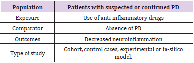
Table 1: PECOS Strategy.
EMBASE research strategy: (‘parkinson disease’/exp/mj OR ‘parkinson disease’/mj OR ‘parkinson`s disease’/mj OR ‘parkinsons disease’/mj OR ‘paralysis agitans’/mj OR ‘parkinson disease, symptomatic’/mj) AND (‘anti-inflammatory agent’/exp/mj OR ‘antiinflammatory agent’/mj OR ‘anti-inflammatory agents’/mj OR ‘antiinflammatory agents, steroidal’/mj OR ‘anti-inflammatory agents, topical’/mj OR ‘anti-inflammatory drug’/mj OR ‘anti-inflammatory agent’/mj OR ‘anti-inflammatory agents’/mj OR ‘anti-inflammatory agents, steroidal’/mj OR ‘anti-inflammatory agents, topical’/mj OR ‘antiflogistic agent’/mj OR ‘antiinflammation agent’/mj OR ‘anti inflammatory agent’/mj OR ‘anti-inflammatory drug’/mj OR ‘antiinflammatory steroid’/mj OR ‘anti-inflammatory activity’/exp/mj OR ‘anti-inflammatory action’/mj OR ‘anti-inflammatory activity’/ mj OR ‘anti-inflammatory effect’/mj OR ‘anti-inflammatory action’/ mj OR ‘anti-inflammatory activity’/mj OR ‘anti-inflammatory effect’/mj OR ‘antiphlogistic action’/mj OR ‘antiphlogistic activity’/ mj OR ‘antiphlogistic effect’/mj OR ‘nonsteroid anti-inflammatory agent’/exp/mj OR ‘nsaid’/mj OR ‘anti-inflammatory agents, nonsteroidal’/ mj OR ‘anti-inflammatory agents, non-steroidal’/mj OR ‘anti-inflammatory agent, nonsteroid’/mj OR ‘non steroid antiinflammatory agent’/mj OR ‘non steroid anti-inflammatory drug’/ mj OR ‘non-steroidal anti-inflammatory agent’/mj OR ‘non-steroidal anti-inflammatory drug’/mj OR ‘non-steroidal anti-inflammatory agent’/mj OR ‘non-steroidal anti-inflammatory drug’/mj OR ‘nonsteroid anti-inflammatory agent’/mj OR ‘nonsteroid antiinflammatory drug’/mj OR ‘nonsteroid antirheumatic agent’/mj OR ‘nonsteroidal anti-inflammatory drug’/mj OR ‘nonsteroidal anti-inflammatory drugs’/mj OR ‘nonsteroidal anti-inflammatory drugs’/mj OR ‘nonsteroidal anti-inflammatory agent’/mj OR ‘nonsteroidal anti-inflammatory drug’/mj OR ‘prostaglandin synthase inhibitor’/exp/mj OR ‘cyclooxygenase inhibitor’/mj OR ‘cyclooxygenase inhibitors’/mj OR ‘prostaglandin synthase inhibitor’/mj OR ‘prostaglandin synthetase inhibitor’/mj OR ‘cyclooxygenase 2 inhibitor’/exp/mj OR ‘cox 2 inhibitor’/mj OR ‘cox 2 specific inhibitor’/mj OR ‘cox 2 specific inhibitors’/mj OR ‘cox- 2 inhibitor’/mj OR ‘cox-2 specific inhibitor’/mj OR ‘cox-2 specific inhibitors’/mj OR ‘cox2 inhibitor’/mj OR ‘cox2 specific inhibitor’/ mj OR ‘coxib’/mj OR ‘coxibs’/mj OR ‘cyclooxygenase 2 inhibitor’/ mj OR ‘cyclooxygenase 2 inhibitors’/mj) AND (‘modulation’/exp/ mj OR ‘modulation’/mj OR ‘protection’/exp/mj OR ‘protection’/ mj OR ‘protective factors’/mj OR ‘treatment outcome’/exp/mj OR ‘medical futility’/mj OR ‘outcome and process assessment (health care)’/mj OR ‘outcome and process assessment, health care’/ mj OR ‘outcome management’/mj OR ‘patient outcome’/mj OR ‘therapeutic outcome’/mj OR ‘therapy outcome’/mj OR ‘treatment outcome’/mj OR ‘disease management’/exp/mj)
LILACS Research Strategy: “Idiopathic Parkinson’s Disease” OR “Lewy Body Parkinson’s Disease” OR “Parkinson’s Disease, Idiopathic” OR “Parkinson Disease, Idiopathic “ OR “Parkinson’s Disease, Lewy Body” OR “Parkinson’s Disease” OR “Idiopathic Parkinson Disease” OR “Lewy Body Parkinson Disease” OR “Primary Parkinsonism” OR “Parkinsonism, Primary” OR “Paralysis Agitans” AND “Neuroinflammation” OR “Inflammations” OR “Innate Inflammatory Response” OR “Inflammatory Response, Innate” OR “Innate Inflammatory Responses” AND “Anti Inflammatory Agents” OR “Agents, Anti-inflammatory” OR “Anti-inflammatories” OR “Anti-inflammatory Agents” OR “Agents, Anti-Inflammatory” OR “Agents, Anti Inflammatory” OR “Anti-Inflammatories” OR “Anti Inflammatories” OR “Anti-inflammatory Agents, Non-Steroidal” OR “NSAIDs” OR “Non-Steroidal Anti-Inflammatory Agents” OR “Non-Steroidal Anti Inflammatory Agents” OR “Nonsteroidal Anti-Inflammatory Agents” OR “Nonsteroidal Anti Inflammatory Agents” OR “Anti Inflammatory Agents, Nonsteroidal” OR “Antiinflammatory Agents, Nonsteroidal” OR “Nonsteroidal Antiinflammatory Agents” OR “Corticosteroids” OR “Corticoids” OR “Inhibitors, Cyclo-Oxygenase” OR “Inhibitors, Cyclo Oxygenase” OR “Inhibitors, Cyclooxygenase” OR “Prostaglandin Synthesis Antagonists” OR “Antagonists, Prostaglandin Synthesis” OR “Inhibitors, Prostaglandin-Endoperoxide Synthase” OR “Inhibitors, Prostaglandin Endoperoxide Synthase” OR “Prostaglandin Endoperoxide Synthase Inhibitors” OR “Prostaglandin Synthase Inhibitors” OR “Cyclo-Oxygenase Inhibitors” OR “Cyclo Oxygenase Inhibitors” OR “Inhibitors, Prostaglandin Synthase” OR “Inhibitors, Cyclooxygenase 2” OR “Cyclooxygenase-2 Inhibitors” OR “Inhibitors, Cyclooxygenase-2” OR “Coxibs” OR “COX-2 Inhibitors” OR “COX 2 Inhibitors” OR “Inhibitors, COX-2” OR “COX2 Inhibitors” OR “Inhibitors, COX2” .
Web of Science Search Strategy
TÓPICO (Parkinson disease*) AND TÓPICO (inflammation*) AND TÓPICO (anti-inflammatory*).
Scopus Search Strategy
(TITLE-ABS-KEY (Parkinson AND disease) AND TITLE-ABSKEY ( inflammation ) AND TITLE ( anti-inflammatory ) ) .
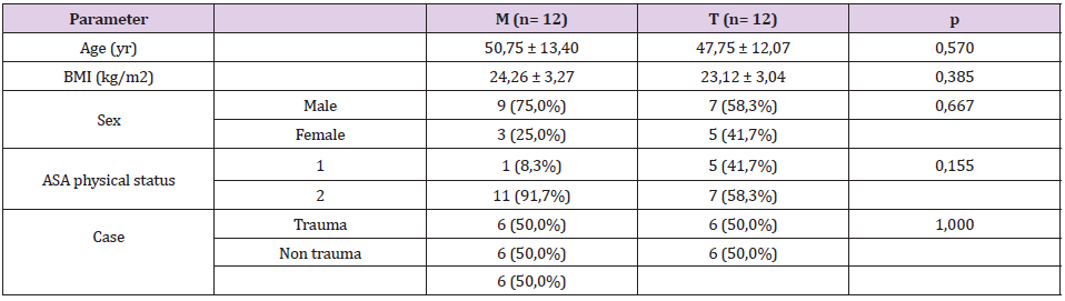
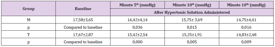
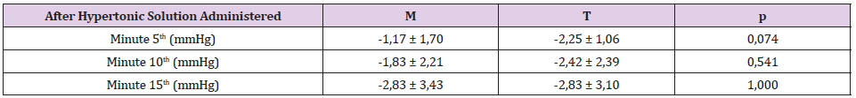
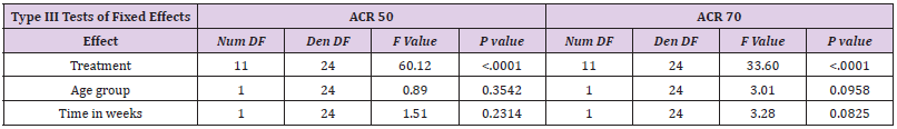
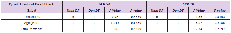
The selection of articles was performed by two researchers blindly and independently through reading the titles, reading the abstracts and, finally, full reading of the articles. Any disagreement in the selection was resolved in consensus meetings. Articles that fully met the eligibility criteria were included in this study. The selection process is described in Flowchart 1 adapted from PRISMA (Figure 1). In order to analyze the methodological quality of the included studies, each article was evaluated by a researcher based on the items of the ACROBAT-NRSI (A Cochrane Risk of Bias Assessment Tool for Non-Randomized Studies) [4]. Acrobat-NRSI scores were used to exclude articles that did not present hardhitting information to the research, besides serving as a basis for discussing the methodological quality of the articles and the possible viruses in the generalization of their results (Figures 2 & 3). From each article included, data related to the objectives of this review were extracted, such as author, title, type of study, population, PD induction drug, drugs used applied, positive results. These data were computed and compared using the t-Student test for independent samples, with the purpose of comparing the percentage s percentages of the and effects on PD between NCAs and other anti-inflammatory drugs (Table 2).
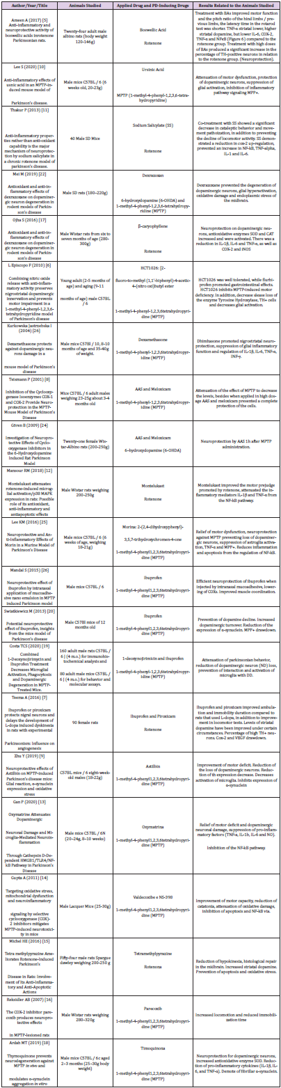
Table 2: Characteristic of selected experimental clinical trials.
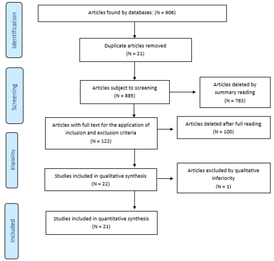
Figure 1: Adapted from PRISMA.
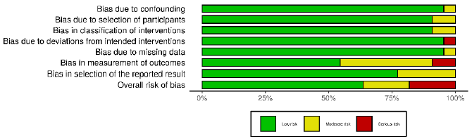
Figure 2.
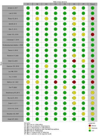
Figure 3.
Findings
Twenty-one articles were analyzed, separated between two groups according to the drug used for pre-clinical study, antiparkinsonian drugs of anti-inflammatory nature and drugs properly anti-inflammatory drugs (IINES and corticosteroids). Improvement in motor function, decreased movement patriotization, increased levels of striatal dopamine, decreased interleukins and blockage of inflammatory pathways, such as those participating in MPP+ and COX-2, as well as increased and/or decreased loss of neurons armed with tyrosine hydroxylase (TH) enzyme, an important marker of neuroprotection, were identified.
Discussion
In view of these findings, this systematic review demonstrated that there is an effective therapeutic relationship in the use of anti-inflammatory drugs in PD through findings such as, mainly, quantitative increase or decrease in the loss of tyrosine hydroxylase enzyme [5-9]and improvement of motor function or prevention of motor decline [5,10-16]. However, since these are experimental studies in animals where clinical failures are commonly recorded in this methodology, caution should be exercised in the face of these findings, even if it shows clinical relevance. In addition, the importance of the therapeutic look is emphasized, especially in pathophysiological terms elapsed by the articles, observing in most of them that this disease, which affects the nicrostriatal region harboring the substantia nigra and quite rich in microglia, has the cumulative character of alpha synuclein in its altered form, which leads to the formation of a highly fibrillar aggregate by very little known pathways, thus, there is the beginning of a cascade of events that lead to the release of inflammatory toxic factors and a progressive dopaminergic neurodegeneration [17,18]. It is identified, therefore, that within this pathophysiological mechanism there is linked an inflammatory response, so there is a target to be investigated and possibly treated, demonstrating possible therapeutic purposes against PD.
In parallel, this review was able to investigate some other parameters found in experimental animal studies. Some motor tests showed improvement in the face of performance tests, applicability of previous training or open field observation, in addition, motor improvement of the forelimbs and later [5], significant decrease in cataleptic behavior [10], improvement of ambulation and immobilization time [7]and reduction of hypokinesia [15]. These results reinforce the hypothesis of a neuroinflammatory cause of Parkinson’s and once again the application of anti-inflammatory drugs for a possible therapy. It can be observed that characteristics that are found in patients such as muscle stiffness, tremor at rest, bradykinesia and postural instability could be solved or attenuated by a drug with function, absorption and mechanisms similar to what were found in this review. Therefore, there is a vast ness of possibilities for anti-inflammatory pharmacological use, in which, however, there is still a need to weigh the pros and cons, the latter being something of changeable capacity within the pharmaceutical industry, in which with investments in research and advanced technology can be achieved a less deleterious profile to the body, such as raising blood pressure, interaction with anti-hypertensive drugs, reduction of renal perfusion and gastrointestinal symptoms [16].
Within this context, it was also possible to identify an increase, then neuroprotection from levels of dopamine, TH enzyme and dopaminergic neurons in some animals. These results can be explained by the fact that the neuroinflammatory process, in its characteristic of exponential cascading lesion of dopaminergic neurons [8,19], was blocked and there was no more decrease in degenerative character. All this was observed from immunohistochemical analyses of TH (Tyrosine Hydroxylase) levels, an enzyme involved in dopamine synthesis through a series of biochemical reactions that has the amino acid tyrosine as a precursor and a molecular marker of dopaminergic neurons, along with dopamine dosage [5-9,18,19]. Thus, it was demonstrated what can occur in a neural system previously healthy, but with microglia activated by the pathophysiology of PD, in this case by mimetic drugs of PD such as rotenone and 1-methyl-4-phenyl-1,2,3,6 tetrahydropyridine (MPTP). Thus, it is envisaged, once again, the use of these drugs or something more advanced both in patients already diagnosed and living with the disease chronically, as well as in patients at the beginning of diagnosis and mild clinical picture, promoting neuroprotection and, consequently, a greater defense and increased quality of life.
Some drugs in the studies acted directly on microglia and other inflammatory foci, some of them are very common, such as ibuprofen, meloxicam, piroxicam, AAS, Valdecoxib and Parecoxib (NHEMS, which act by inhibiting COX-2, prostaglandin and ultimately reducing cytokines), dimethazone (Corticosteroid that reduces the gene expression of pro-inflammatory cytokines). All of them obtained good results regarding the lowering of glial hyperactivation and intracellular inflammatory, in addition to stimulating the recovery and regeneration phase, avoiding in some cases the toxicity of MPTP [20], which shows that even having extensive knowledge and applicability of these drugs, they can still be key parts for the advancement of neural therapy in PD. Similarly, oxymatrine, an alkaloid compound found at the root of a Chinese herb (Sophora flavescent), promoted relief of motor deficits induced by MPTP and conferred significant neuroprotection, in addition to inhibiting the activation of microglia and exacerbated release of pro-inflammatory as cytokines [13]. This shows that within the vastness of drugs known and disseminated by the pharmaceutical industry, there are still a gigantic number of other substances that can be used in the treatment of this disease [20-27].
Conclusion
Our study has concluded that there is a need for investment in quality, more robust, broad-spectrum preclinical studies, with minimal view to achieve the ideal pharmacological therapeutic for this target. Thus, it is necessary more clinic trials to confirm this relationship between an inflammatory profile and use of antiinflammatory drugs which possible therapeutic agents to treatment of PD.
For More Articles: Biomedical Journal Impact Factor: https://biomedres.us