Open Access Journals on Medical Research

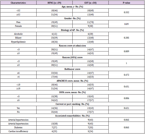
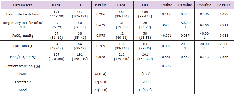
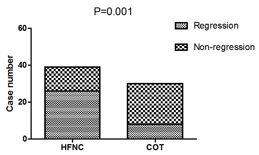
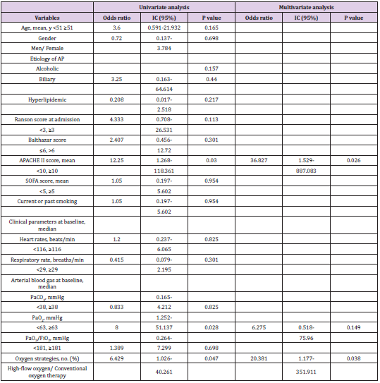
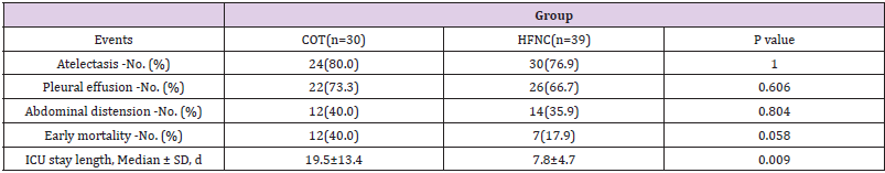
Insight into Secondary Metabolites of Circinella, Mucor and Rhizopus the Three Musketeers of Order Mucorales

Introduction

Fungi are rich sources of biologically active natural compounds, which are used in the manufacturing of wide range of clinically important drugs. Fungi produce important antibiotics such as the beta-lactam antibiotics members, penicillin and cephalosporin, which and their derivatives are dominating the most important antibiotic market until now [1-5]. Fungi generally and endophytic ones specifically represent future factories and potent biotechnological tools for production of bioactive natural substances which could extend healthy life span of humanity, as done by penicillin from centuries, and considered promising alternatives for some high costly produced chemicals and drugs [6- 12]. The present review highlights some bioactivity of secondary metabolites, produced by Circinella, Mucor, and Rhizopus, involved in medical, pharmaceutical, agricultural, and industrial applications. Zygomycetes constitute a remarkable group of microscopic fungi. These fungi are mainly soil inhabitants living as saprobes and decomposers of organic matter and herbivorous feces. Zygomycota, represent the most basal terrestrial phylum of the kingdom of Fungi [13].

Zygomycetes have an integral role in the development of microbial ecosystems, a property which has the potential to be converted for biotechnological and industrial applications ranging from food technology to drug development [14]. The Mucorales, which is classified into the subphylum Mucormycotina [15], is the largest order of fungi. Members of this group (Circinella, Mucor and Rhizopus) are abundant saprophytes in nature. They are commonly found in soil and decaying vegetation, and can also be found in grains [16,17]. Mucorales members (Example; Circinella, Mucor and Rhizopus) grow and invade quickly on easily digestible substrates, such as those containing starches, sugars, and hemicelluloses. Future studies should investigate Circinella, Mucor and Rhizopus their ability to produce extracellular enzymes and potential applications in biotechnology.

Circinella, Mucor and Rhizopus Description and Ecology

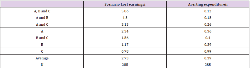
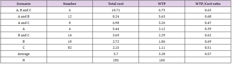
The Genus Circinella belonging to Phylum: Mucoromycota; Class: Zygomycetes; Order: Mucorales; Family: Syncephalastraceae. Colonies mucraceous, usually up to few mm high; sporangiophores erect, branching sympodially and each branch recurved (Circinate) terminated with a sporangium; sporangia globose, columellate, many-spored, covered with non-diffluent wall, incrusted with calcium oxalate, breaking into pieces; columellae globose, subglobose or cylindro-conic; sporangiospores globose or oval, smooth; heterothallic; zygospores when produced, born on erect hyphae, with more or less similar gametangia and suspensors. Colonies fast growing, attaining 7.5 cm after 7 days on malt extract agar at 25C. The most common Circinella species is Circinella muscae. Circinella colonies color is brownish and common present in soil (Figure 1) [11,13]. The Genus Mucor belonging to Phylum: Mucoromycota; Class: Zygomycetes; Order: Mucorales; Family: Mucoraceae. Colonies fast growing, often up to several cm in height, white to yellow becoming dark grey with age; mycelium non-septate, occasionally septa produced in old cultures; sporangiophores erect arising singly from the mycelium without rhizoids, forming a dense mat, unbranched or branched monopodially or sympodially, sometimes the branches recurved, bearing terminal sporangia; sporangia globose, many-spored, columellate, without apophysis; sporangial wall deliquescent, incrusted with needles of calcium oxalate; columellae of various shapes, colourless or coloured; sporangiophores colourless or colored, greyish or brownish, globose or elliptical, wall thin smooth; chlamydospores produced in some species , terminal, hyaline, smooth walled; zygospores produced in the aerial mycelium and arising from copulation of two straight gametangia.

biomedres-openaccess-journal-bjstr

Figure 1: Circinella spp., different species.

biomedres-openaccess-journal-bjstr

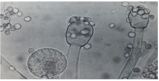
Figure 2: Mucor spp., different species.

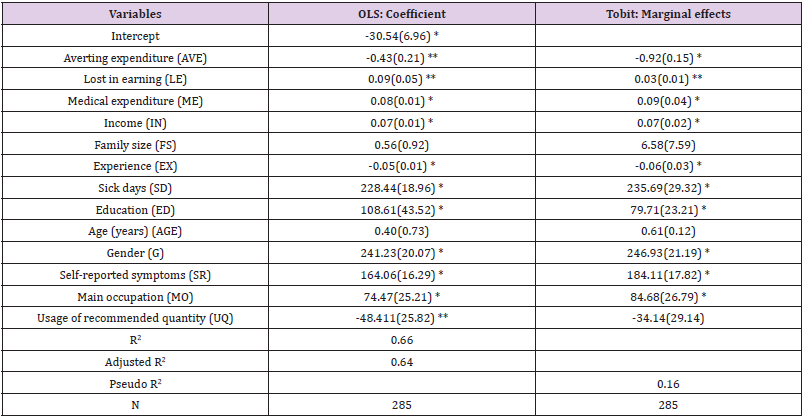
Mucor is one of the largest genera of Order: Mucorales and a number of its species are worldwide. Mucor species are common in soil and also occur organic matter. Colonies fast growing, filling the plate of malt extract agar after 3 days at 25C (Figure 2). The most common Mucor species is Mucor circinelloides, Mucor hiemalis and Mucor racemosus [11,13]. The Genus Rhizopus belonging to Phylum: Mucoromycota; Class: Zygomycetes; Order: Mucorales; Family: Mucoraceae. Colonies fast growing, filling the whole plate after 2 days on malt extract agar at 25C, at first white, later becoming pale or dark grey brown. Mycelium non-septate, occasionally septa produced in old cultures, differentiated into stolons, rhizoids arising from stolon ends and erect unbranched sporangiophores sporouting opposite the rhizoids; sporangia generally globose, apophysate, columellate, many-spored; sporangiospores short ellipsoidal, brownish, striate in many species; most species heterothallic; zygospores naked, dark-coloured; wall strongly roughened, suspensors straight, large and swollen. Rhizopus species are common in soil and soil containing various organic matter. Rhizopus species are impotent spoilage organisms. The most common Rhizopus stolonifer, Rhizopus arrhizus and Rhizopus oryzae (Figure 3) [11,13].

biomedres-openaccess-journal-bjstr

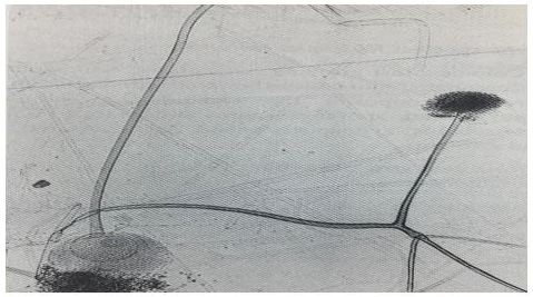
Figure 3: Rhizopus spp., different species.

The Genus Circinella as a Biotransformation Agent

Enzymatic reactions and biotransformations catalyzed by fungal enzymes and used in industry, agriculture, food technology, and medicine have increased in importance tremendously in recent years. It was later demonstrated that filamentous fungi from several genera, including Circinella, Mucor and others, are able to hydroxylate dibenzofuran, forming 1-, 2-, 3-, or 4-hydroxybenzofuran and some other more hydrophilic products [18]. Yan et al., [19], mentioned that, nine hydroxylated and glycosylated metabolites, of which two have not been reported previously, were obtained and identified after incubation by Circinella muscae. The specific 7β, 15α, and 21β hydroxylated products yielded from this bio-process by Circinella muscae. In addition, the selectively glycosylation at C-28 was another main reaction type. It was also observed that the 3β-OH group was selectively dehydrogenated into carbonyl group. These reactions may be difficult to achieve by chemical synthetic means. In vitro biological tests indicated that compounds 3β, 7β dihydroxyolean-12-en-28-oic acid; 3β, 7β, 15α-trihydroxyolean- 12-en-28-oic acid, and 3β, 7β, 15α-trihydroxyolean-12-en- 28- oic acid-28-Oβ-D-glucopyranosyl ester showed significant antiinflammatory activities, which suggested that hydroxylation at C-7 and glycosylation at C-28 by Circinella muscae had benefit effects [19]. Macromycete Circinella muscae AS 3.2695, catalyzed the oxidation of Oleanolic Acid [20]. Biotransformation of ursolic acid by Circinella muscae CGMCC 3.2695 was investigated by Chu et al., [21].

Scaled-up biotransformation reactions yielded ten metabolites. Their structures were established based on extensive NMR and HR-ESI-MS data analyses, and four of them are new compounds. Circinella muscae could selectively catalyze hydroxylation, lactonisation, carbonylation and carboxyl reduction reactions. Also, all the identified metabolites were evaluated for their anti-neuroinflammatory activities in LPS-induced BV-2 cells. Most metabolites displayed significant inhibitory effect on nitric oxide (NO) production and the results suggested that biotransformed derivatives of ursolic acid might be served as potential neuroinflammatory inhibitors [21]. Biotransformation of betulinic acid was carried out with Circinella muscae CGMCC 3.2695 and Cunninghamella echinulata CGMCC 3.970 by Chen et al., [22], yielded six previously undescribed hydroxylated metabolites and four known compounds. Circinella muscae could catalyze the regioselecitve hydroxylation and carbonylation at C-3, C-7, C-15 and C-21 to yield seven products. Cunninghamella echinulata could catalyze the C-1, C-7 and C-26 regioselecitve hydroxylation and acetylation to yield five metabolites. The structures of the metabolites were established based on extensive NMR and HR-ESIMS data analyses and most of the metabolites exhibited significant inhibitory activities on lipopolysaccharides-induced NO production in RAW264.7 cells [22].

The Genus Mucor Source of Biologically Active Compounds

Species of Mucor ramosissimus produce extracellular enzymes, such as endopolygalacturonase and lipase, and secondary metabolites, such as phytoalexin elicitor [23,24]. Lately, several studies have focused on applying Mucorales members to produce ethanol and biomass by-product. Particularly, Mucor ramosissimus has been reported as a potential ethanol-producing mold [25]. Mucor circinelloides has a great industrial potential since it started producing bioactive compounds of interest, such as microbial lipids, carotenoids or sterols [26,27]. Mucor circinelloides is able to produce microbial lipids which could be useful for the production of biodiesel using the common way to produce fatty acid methyl esters, due to the beneficial properties of these compounds, new sustainable and more environmentally friendly methods have emerged in order to increase their production [28]. Three important strains of Mucor circinelloides grown in specific media for specified period (72 h, 120 h and 168 h) under submerged fermentation conditions were investigated by Hameed et al., [29], for their potential antioxidants/secondary metabolite production and found that all mycelial extracts demonstrated effective antioxidant activities in terms of β-carotene/linoleic acid bleaching, radical scavenging, reduction of metal ions and chelating abilities against ferrous ions [29].

Antioxidant property of all three important strains of Mucor circinelloides extracts were duo to their phenolic and condensed tannin contents. Strains Mucor circinelloides MC277.49 was found to be the biggest producer of secondary metabolites under nutritional stress condition in late exponential phase. These Mucor strains prove to be rich sources of antioxidants and secondary metabolites, which could be used in the development of nutraceuticals and natural antioxidants [29]. The new cyclic heptapeptide unguisin F and the known congener unguisin E, were obtained from the endophytic fungus Mucor irregularis, isolated from the medicinal plant Moringa stenopetala, collected in Cameroon studied by [30]. The structure of the new compound was determined on the basis of one- and two-dimensional NMR spectroscopy as well as by high-resolution mass spectrometry. Heptapeptide unguisin F and congener unguisin E were evaluated for their antibacterial and antifungal potential, but failed to display significant activities [30]. Biotransformation is an important tool for the structural modification of organic compounds, especially natural products with complex structures, which are difficult to achieve using ordinary methods [31].

Mucor genus is widespread in nature and some species are used extensively in biotechnology for enzyme and useful compound production. Biotransformation can be used as a very convenient way of producing compounds, particularly when the structure is complex and they can neither be isolated as metabolites nor chemically synthesized [31]. It is safe to conclude that biotransformation by Mucor species is of great importance due to their wide-ranging use in the stereospecific production of compounds of commercial interest, and because they simplify the study of the metabolism of such compounds in order to obtain novel agents with many interesting biological activities [31]. The biotransformation of 1R-(−)-camphorquinone, achieved by growing cells of Mucor plumbeus isolated from soil. Results were found for M. plumbeus, which was only able to perform monoreduction of camphorquinone when cultivated on a glucose– peptone–yeast extract medium. Large-scale experiments were set up and the camphorquinone biotransformation products formed by Mucor plumbeus were purified by column chromatography and identified by 1H and 13C nuclear magnetic resonance (NMR). Mucor plumbeus could be of great use for the selective reduction of camphorquinone and related compounds [32].

The Genus Rhizopus Source of Biologically Active Compounds

Secondary metabolites is one of the characteristic features of microorganisms. More than 50,000 bioactive compounds have been isolated from the extracts of microorganisms with a diversified arrangement of chemical structures, which showed antimicrobial, antitumor and agrochemical activity and others [33]. The ethyl acetate extract of Rhizopus stolonifer has potent cytotoxic activity against brine shrimps, suggesting that it could serve as a lead compound for anticancer compounds. Its antifungal activity also indicates its potential for development into an antifungal product. Furthermore, its phytotoxicity against Lemna minor weed demonstrates that it contains compounds with herbicidal activity. More studies on Rhizopus stolonifer is needed to isolate and structural characterization of its constituents which could lead to the development of pharmaceutical and agricultural products [34].

From previous studies, it was concluded that the extracts of Rhizopus species have compounds which have ability to inhibit the growth of fungal and bacterial strains [35]. Chitosan was isolated from the fungus Rhizopus oryzae by using yeast peptone glucose broth medium, showed antimicrobial activity against Escherichia coli, Staphylococcus aureus and Candida albicans and reach to 20, 15 and n 15 mm as inhibition zone respectively [36]. Nanotechnology is a field that is increasing day by day, making an impact in all spheres of human life. Biological methods of synthesis nanoparticles called “greener synthesis” of nanoparticles and these have proven to be better methods due to slower kinetics, they offer better manipulation and control over crystal growth and their stabilization [37]. The extracellular synthesis of silver nanoparticles by Rhizopus stolonifer and its efficacy against multidrug resistant (MDR) strains isolated from burnt cases from hospitals at Gulbarga region, Karnataka, India is reported by Rathod and Ranganath, [38], the biosynthesized nanosilver showed excellent antibacterial activity against multidrug resistant Pseudomonas aeruginosa isolated from burnt infections. Biologically synthesized nanosilver showed zone of inhibition (mm) of two isolates about 33mm and 30.5mm in diameter strains of Pseudomonas aeruginosa respectively [38].

This study reports the extracellular synthesis of silver nanoparticles by Rhizopus stolonifer and its efficacy against multidrug resistant (MDR) Escherichia coli and Staphylococcus aureus isolated from Khwaja Bande Nawas Hospital, Gulbarga, Karnataka was carried by Banu and Rathod, [39] and synthesis of silver nanoparticles (AgNPs) was carried out by using fungal filtrate of Rhizopus stolonifer and an aqueous solution of AgNO3. Biotransformations of sesquiterpenoids by Rhizopus species have been used to provide new derivatives with potential biological activities. Fungal transformation processes by Rhizopus species, has been the introduction of hydroxyl groups into remote positions of the molecules, which is difficult to achieve by chemical means. Other reactions carried out with stereo-selectivity by these microorganisms have been: epoxidations of double bonds, hydrogenations of the exocyclic C-C double bond of the lactone rings, and reductions of carbonyl groups giving Salcohols. Several reactions have also been performed by these Rhizopus species with regio-selectivity, such as oxidations of hydroxyl groups and deacetylations [40]. The enttrachyloban diterpene ent-18, 19-dihydroxytrachylobane was biotransformed by Rhizopus stolonifer, and produced the new ent- 11β, 18, 19-trihydroxytrachylobane, and the new ent-kaurene diterpenes ent-16α, 18, 19-trihydroxykaur-11-ene and ent- 18, 19-dihydroxy-16α-methoxykaur-11-ene. The formation of derivative ent-11β,18,19-trihydroxytrachylobane, involved the first hydroxylation of C-11 of ent-trachyloban diterpene skeleton by fungus, and compounds ent-kaurene diterpenes ent-16α, 18, 19-trihydroxykaur-11-ene and ent-18,19-dihydroxy-16α- methoxykaur-11-ene were probably produced by backbone rearrangement of ent-11β,18,19-trihydroxytrachylobane, these results, verified the potential application of microbial transformation (Especially By Mucorales Rhizopus species) of natural products for the formation of new compounds [41].

Conclusion

Fungi in general are important source of unique natural products with a high level of biodiversity and also yield several compounds having different industrial applications and pharmaceutical activities, which is currently attracting scientific researches. Every study conducted on Circinella, Mucor and Rhizopus resulted in discovery of new metabolites that may have an important applications, which made these genera potential source of pharmaceuticals and attracted attention for further investigations of their important bioactivities properties. Circinella, Mucor and Rhizopus are known for their capability of producing various bioactive compounds with medical applications as antineuroinflammatory, antiinflammatory, and enzymes producers and as biotransformation agents.

For More Articles: Biomedical Journal Impact Factor: https://biomedres.us

Journal on Biomedical Imaging

A Novel 3 Dose Rabies Vaccine

Introduction

Rabies is a zoonotic viral disease that is caused by rabies virus (RABV) and claims the highest fatality among all infectious diseases. Rabies is found in more than 100 countries and territories [1]. To prevent rabies, timely and complete postexposure prophylaxis (PEP) is necessary. Currently five doses of rabies vaccine are administered on days 0, 3, 7, 14, and 28 but the studies have shown that the compliance to complete course was only 40% [2]. Therefore, an effective way to address these limitations is the reduction of PEP doses in humans and a novel vaccine with improved immunological outcomes through an accelerated PEP schedule is desirable. In this regard, Cadila Pharmaceuticals Ltd., Ahmedabad, India has developed a novel recombinant nanoparticle-based rabies G protein vaccine (Thrabis®) prepared by using Virus Like Particle technology (VLP).

Recombinant Nanoparticle-Based Rabies G Protein Vaccine

For generation of recombinant rabies G protein vaccine using VLP platform; genetic sequences encoding the rabies G protein sequence are selected. The genes are then cloned into baculovirus and the baculovirus was made to infect insect cells (sf9). The target antigens were expressed in the sf9 cells which were purified using various chromatographic techniques. The purified target antigen exists as an assembly of polypeptides that is present in multiple copies in subunit antigens in well-ordered arrays with defined orientations [3]. This can potentially mimic the repetitiveness, geometry, size, and shape of the natural host-pathogen surface interactions. Such nanoparticles offer a collective strength of multiple binding sites (avidity) and can provide improved antigen stability and immunogenicity [4,5]. Safety is a major advantage of VLPs. Like traditional vaccines, VLPs are immunogenic and excellent to control infectious diseases. VLPs cannot replicate, recombine or undergo reassortment because they do not contain infectious DNA or RNA material; therefore, VLPs are safer than traditional vaccines. VLPs possess several advantages over the products that are produced by chemical syntheses such as smaller size, which ranges from 10 to 2000 nm, availability of high-resolution threedimensional (3D) models of their structure, construction flexibility, high-production yields, and structural uniformity of each type of virus or VLP [6].

Clinical Study

A multi-centric, open label, assessor blind, centre-specific block randomized, parallel design, phase III clinical study was conducted among 800 subjects. The eligible subjects were randomized in 2:1 ratio for recombinant rabies G protein vaccine and the reference vaccine. Subjects in recombinant rabies G protein vaccine arm received 3 doses of vaccine on days 0, 3 and 7; while subjects in reference vaccine arm received 5 doses of WHO pre-qualified vaccine on days 0, 3, 7, 14 and 28. The primary objective was to demonstrate the non-inferiority of the test vaccine on day 14 after first dose relative to the reference vaccine in terms of seroprotection rate (RVNA titer of ≥0.5IU/mL). The secondary endpoints were the seroprotection rate on day 42 post first dose of the study vaccine and the frequency of solicited and unsolicited adverse events (AEs) were reported between day 0 & 180. On day 14, 99.24% in the test vaccine arm and 97.72% in the reference vaccine arm were seropositive; the difference was statistically non-significant. Likewise, on day 42, 98.69% of the subjects in the test vaccine arm and 100.00% in the reference vaccine were seropositive, the difference was statistically non-significant. A statically significant higher number of participants in the reference arm had adverse events (AEs) compared to test arm (17.2% vs 9.9%, P=0.0032). All the AEs were mild to moderate in nature, which resolved without any complications. The most frequently observed local AEs were pain, redness and swelling at the injection site. The systemic AEs were fever, headache, ear pain, urticaria, joint pain and nausea [3].

Conclusion

The novel 3 dose recombinant rabies G protein vaccine (Thrabis®) was found to be safe & immunogenic and was comparable to 5 doses of WHO pre-qualified vaccine in simulated post exposure prophylaxis. The reduced number of vaccine doses leads to a reduction in number of visits and travel cost; as well as increases the compliance, which is important to prevent rabies and ultimately help in eliminating dog mediated human rabies by 2030 [7].

For More Articles: Biomedical Journal Impact Factor: https://biomedres.us

Open Access Journals on Biomedical Research

Application of High-Flow Oxygen Therapy in Acute Pancreatitis Complicated with Acute Respiratory Dysfunction

Introduction

Pulmonary complications of acute pancreatitis (AP) characterized by increased permeability of the pulmonary microvasculature and alveolar spaces filled with leaked proteinrich exudate were frequent with morbidity about 75%, [1,2] including acute lung injury (ALI) or ARDS, atelectasis, pleural effusion, alteration in diaphragmatic function, i.e. [3-5]. It usually resulted in a need of endotracheal intubation and mechanical ventilation, in addition, the mechanisms were complex and still to be discovered [6]. It was reported that 30%-60% of death was related to pancreatitis- associated ALI and ARDS [7-9]. Furthermore, nearly one-third of death of acute pancreatitis prior to admission to hospital were associated with ALI [10]. Studies have revealed that acute respiratory dysfunction (ARD) was an independent prognostic factors for hospital mortality in severe acute pancreatitis (SAP) along with the age, chronic health situation, and organ failures, which was associated with 60% of death within first week [11,12]. Therefore, more attention have been paid on treatment of pancreatitis-associated respiratory complications for reducing early death.

HFNC, which is increasingly used in intensive care unit (ICU) and non-ICU wards was considered as an alternative to Noninvasive Positive Pressure Ventilation (NPPV) and conventional oxygen therapy (COT) with better tolerated and decreasing work of breathing [13,14]. It could provide heated and humidified air and reduce airway secretion and atelectasis with high-flow rates of up to 60 L/min as well as high FiO2 from 0.21 to 1.0 [15,16]. Previous studies have revealed that HFNC has the capability of generating low levels of positive end-expiratory pressure and decreasing physiological dead space through flushing expired carbon dioxide into the upper airway [17]. It has been applied to various diseases, including various respiratory failure, cardiogenic pulmonary edema, postextubation, postoperative patients, and infants [15,16,18-20]. However, the efficiency of HFNC in acute pancreatitis is unclear. Our research is intended to illustrate the value of HFNC in acute pancreatitis complicated with acute respiratory failure.

Material and Methods

Trial Design

All acute pancreatitis complicated with ARD admitted to ICU were included in this retrospective study from January 2014 to June 2019. This trial was approved by the ethical committee of our hospital (approval number:KY030-01) and was registered on the Chinese Clinical Trial Registry (www.chictr.org.cn/, registration number: ChiCTR2000029202) and written informed consent was obtained from patients or their families before HFNC treatment.

Patients

Computed tomography (CT) combined with supportive specific laboratory data were used in the diagnosis of AP. All AP were enrolled if they followed one of the following criteria: PaO2/FiO2 < 300 mmHg; respiratory rate > 25/min; dyspnea or accessory muscle use; asynchronous or paradoxical breathing; required >5 L/min O2 to maintain SpO2 >92%.Exclusion criteria: need for immediate intubation, central nervous system disorder, cannot answer questions, contraindications for the use of the mask (noncooperative patient).Patients were asked to grade their dyspnea score [21] (improvement; no change; deterioration) and comfort score [22] (poor; acceptable; good) 1 hour after intervention.

Study Intervention

All patients were treated with COT though face mask or nasal cannula before enrolled. In the COT group, oxygen therapy was applied continuously through face mask or nasal cannula at a flow rate up to 10 liters per minute. The rate was adjusted to obtain SpO2>92% until patients recovered or were intubated. High flow humidified oxygen (37°C and 44mgH2O/L) was delivered continuously through a nasal cannula with Optiflow (Fisher and Paykel Healthcare) with a primal flow rate of 50L/min, a primal FIO2 of 50%, and dynamic adjustments to obtain SpO2>92%. HFNC was switched to COT if SpO2 was ≥95% at ≤5 L/min O2 or the PaO2:FIO2 was at least 300. Intubation and mechanical ventilation decisions were made by the physicians with the criteria: respiratory arrest; respiratory pauses with loss of consciousness or gasping respiration; encephalopathy; cardiovascular instability; unmanageable secretions; respiratory fatigue; refractory hypoxemia (HFNC:SpO2≤ 88% with FIO2 = 100%; COT: SpO2≤88% with at least 10L/min), or respiratory acidosis (pH<7.30 and PaCO2 ≥50mmHg).

Primary and Second Outcomes

The primary outcomes are the early intubation rate, defined as percentage of intubation and mechanical ventilation in 10 days and the median time to intubation from patients enrolled. The second outcome includes changes in physiological parameters as well as arterial blood gases and grade of dyspnea, comfort score, regression of respiratory failure after 1 hour intervention, adverse events (atelectasis, pleural effusion and abdominal distension), early mortality defined as mortality in 10 days and ICU stay length.

Statistical Analysis

The Chi-square test or Fisher test was used in the comparisons for categorical variables while the unpaired Student’s t test or Mann-Whitney U test were performed for continuous variables. The analysis of the data obtained before and after intervention from each patient was made with the paired Wilcoxon test. Variables associated with respiratory failure regression were assessed by univariate and multivariate logistic- regression analyses. Variables suspected to be associated with regression of respiratory failure with a P<0.10 after univariate analysis were accounted into the multivariate analysis. All statistical analyses were performed using IBM SPSS, Version 19.0 (IBM Corp., Armonk, NY, USA) or GraphPad Prism version 6.0 (GraphPad Software Inc., San Diego, CA, USA). A p value (two-tailed) <0.05 was considered statistically significant.

Results

Patients

A total of eighty-one patients with AP were admitted to ICU. Seven were intubated immediately at admission and five did not meet the classification criteria at last. Ultimately, sixty-nine patients were included in this retrospective study. 30 out of 69 were treated with conventional oxygen through nasal cannula or face mask, while 39 were treated with HFNC. Baseline characteristics of patients, including demographic data, severity of illness (APACHE II), etiology, severity of AP (Ranson score and Balthazar score) and associated comorbidities were recorded in Table 1. Vital signs and arterial blood gases before and after enrollment (1h) were collected in Table 2. We also evaluated the incidence of atelectasis and pleural effusion in the two groups by CT.

biomedres-openaccess-journal-bjstr

Table 1: Patients Characteristics.

Note: HFNC: High-Flow Oxygen though Nasal Cannula; COT: Conventional oxygen therapy

biomedres-openaccess-journal-bjstr

Table 2: Primary outcome.

Note: Median ± SD, h

HFNC: High-Flow Oxygen though Nasal Cannula; COT: Conventional oxygen therapy

Primary Outcome

The early intubation rate was 25.6% (10 of 39 patients) in the HFNC group, 56.7% (17 of 30) in the COT group (P=0.013) Table 3, Figure 1. The median time to intubation was 64.25h in the HFNC group, 7.75h in the COT group (P<0.001) Table 3. Meanwhile, we compared the intraabdominal pressure (cm H2O) before intubation of intubated patients in the two groups (HFNC 21.7 vs COT 19.1; P=0.103; not shown).

biomedres-openaccess-journal-bjstr

Table 3: Physiological parameters and arterial blood gases at baseline and 1hour after intervention.

Note: HFNC: High-Flow Oxygen though Nasal Cannula; COT: Conventional oxygen therapy; a: Baseline VS 1 Hour in HFNC; b: Baseline VS 1 Hour in COT; c: Changes in the HFNC group VS that in the COT group 1hour after intervention.

biomedres-openaccess-journal-bjstr

Figure 1: Comparison of intubation rate between the two groups.

Note: HFNC: High-Flow Oxygen though Nasal Cannula; COT: Conventional oxygen therapy.

Changes in Physiological Parameters and Arterial Blood Gases

After 1 hour of intervention, patients treated with HFNC had decreased respiratory rate (breaths/min), heart rate (beats/min) (27 vs 21, P<0.001; 121 vs 104, P=0.008) and increased PaO2 (mmHg) (64 vs 110; P< 0.001), while PaCO2 (mmHg) and PaO2/ FiO2 (mmHg) had no changes (37 vs 42, P=0.087; 189 vs 210, P=0.539) compared with baseline. However, in the COT group, after 1 hour of intervention, respiratory rate, heart rate, and PaO2/FiO2 were similar (30 vs 26, P=0.166; 114 vs 109, P=0.486; 192 vs 201, P=0.142) with baseline, whereas PaCO2 and PaO2 were higher (38 vs 50; P< 0.001; 64 vs 83; P<0.001) Table 2. There was no difference in physiological parameters and arterial blood gases at baseline between the HFNC group and the COT group. Compared with the COT group, patients treated with HFNC had decreased respiratory rates, PaCO2 (21 vs 26, P=0.020: 42 vs 50, P<0.001) and increased PaO2 (110 vs 83, P=0.003) 1 hour after intervention. Patients showed greater improvement in PaO2 in the HFNC group than that in the COT group. (P<0.001) Table 2.

Dyspnea Grade, Comfort Score, and Respiratory Dysfunction Regression

The comfort score was similar between the two groups (P=0.596, Table 2). There was a higher proportion of patients felling improvement in dyspnea in the HFNC group (87.2% vs 56.7%, P=0.006) Figure 2. Improved in respiratory dysfunction was defined as respiratory rate <25/min and improvement in breathing effort, including accessory breathing muscle activity and/or paradoxical breathing. Finally, 34 out of 69 patients (26 in the HFNC group vs 8 in the COT group; P=0.001; Figure 3) got respiratory failure regression 1 h after intervention. In the univariate analysis the following parameters were associated with respiratory failure improvement: APACHE II score, PaO2 at baseline and Oxygen strategies. Higher APACHE II score, lower PaO2 at baseline and conventional oxygen therapy exhibited an increased risk for poorer regression of respiratory failure (OR =12.250, 95% CI 1.268-118.361, P=0.030; OR=8.000, 95% CI 1.252-51.137, P= 0.028; OR=6.429, 95% CI 1.026-40.261, P=0.047). In multivariate logistic-regression analyses, HFNC and APACHE II score were independent predict factors to regression of respiratory failure (OR =20.381, 95% CI 1.177-351.911, P=0.038; OR=36.827, 95% CI 1.529-887.083, P=0.026) Table 4.

biomedres-openaccess-journal-bjstr

Figure 2: Comparison of dyspnea score improvement between the two groups 1 hour after intervention.

Note: HFNC: High-Flow Oxygen though Nasal Cannula; COT: Conventional oxygen therapy

biomedres-openaccess-journal-bjstr

Figure 3: Comparison of respiratory failure regression between the two groups 1 h after intervention.

Note: HFNC: High-Flow Oxygen though Nasal Cannula; COT: Conventional oxygen therapy

biomedres-openaccess-journal-bjstr

Table 4: Univariate and Multivariate analysis of risk factors to regression of respiratory failure.

Adverse Events and Clinical Outcomes

No significant differences were found for adverse events between the two groups. Early mortality in the HFNC group had trend to be significantly lower than that in the COT group (17.9% vs 40.0%, P= 0.058). Patients treated with COT had much longer ICU stay length (19.5±13.4 vs 7.8±4.7, P=0.009) Table 5.

biomedres-openaccess-journal-bjstr

Table 5: Adverse Events and Clinical Outcomes.

Note: HFNC: High-Flow Oxygen though Nasal Cannula; COT: Conventional oxygen therapy

Discussion

Accumulating research have revealed some advantages of HFNC application in pneumonia-associated respiratory dysfunction, including decreased intubation rate, lower 90-day mortality, and increased ventilator-free days compared with NPPV and/or COT [23-26]. Despite this, COT with a face mask or nasal cannula is still the first-line treatment for those lacking studies about HFNC application in AP associated respiratory dysfunction. However, COT is not able to satisfy some patients with severe hypoxemia. Therefore, to our knowledge, this study illustrated the efficiency of HFNC in acute pancreatitis complicated with acute respiratory dysfunction for the first time. In this study, eliminating the effect of secondary infection on intubation, we compared the early intubation rate and the median time to intubation of the two groups in combination with the pathophysiological characteristics of pancreatitis. Severe acute pancreatitis has two representative phases. The first stage is in the first ten days, with characteristics of the systemic inflammatory response syndrome (SIRS). The second usually comes up during the second week and is marked by infectious manifestations [27]. In the end, we found patients in the HFNC group had a lower early intubation rate and a longer median time to intubation.

We also compared other outcomes, including changes in physiological parameters and arterial blood gases, comfort score, regression of dyspnea and respiratory dysfunction, adverse events, early mortality, and ICU stay length between the two groups and found that first, HFNC was superior to COT in improving dyspnea, decreasing respiratory rate and heart rate, preventing carbon dioxide retention, and had a stronger effect on improving PaO2. However, we did not find a difference in PO2/FiO2 between the two groups, indicating that HFNC could improve PO2 with higher FiO2, but it could not improve physiopathologic changes. Second, HFNC was independently associated with the regression of respiratory dysfunction. Third, the comfort score of HFNC was similar to that of COT. At last, patients treated with HFNC had shorter ICU stay length and lower early mortality. We hypothesize that the reason why the early mortality between the two groups did not reach statistical significance would be due to the limited number of patients in our study. Above all, we would come to the conclusion that HFNC is prior to COT in acute pancreatitis complicated with acute respiratory dysfunction. There are some limitations to our reach. First, this study is a single center with a small number of patients. Second, we did not compare HFNC and NPPV. Hence, in the future, we should compare HFNC, NPPV and COT in multicenter studies with large samples.

Conclusion

HFNC is a more efficient option in acute pancreatitis complicated with acute respiratory failure and may be an alternative treatment to COT.

For More Articles: Biomedical Journal Impact Factor: https://biomedres.us

Open Access Journals on Microbiology Research

How Can We Track COVID-19 Hotspots and Prevent its Spread?

The outbreak of novel coronavirus disease 2019 (COVID-19) was declared a public health emergency by the World Health Organization (WHO) on 30th January 2020, due to its spread across the globe [1,2]. Because of continued waves of severe acute respiratory syndrome coronavirus 2 (SARS-CoV-2), it is essential to introduce efficient monitoring and screening techniques Virus containment remained challenging in spite of the various advancements in the research field. The most common modes of virus transmission include droplet, contact or fomite, and fecal transmission [3,4]. Fecal transmission can be a serious risk for both humans and animals in case of aerosolization of fecal waste contaminated with the virus [5]. In diarrhea patients, severe acute respiratory syndrome coronavirus (SARS-CoV) was found stable in feces at room temperature for a minimum of 1-2 days and could survive for up to 4 days in the stool [6]. This detection increases the chance of fecal-oral transmission [7] because flushing may aerosolize fecal matter and cause airborne transmission [8]. This type of transmission could be high where toilets are shared for example in quarantine centers and hospitals. From these toilets, flushed water enters into sewerage systems. Consequently, the sewerage system becomes a carrier of this virus. Countries are trying hard to maximize the testing of the virus to avoid community spread of the COVID-19 and after around two years it looks still difficult. Therefore, it is crucial to locate the COVID-19 hotspots to plan mitigation strategies And sewerage system monitoring could be an efficient strategy for finding the virus hotspots. This probable route of virus transmission may worsen the problem of community transmission [9]. Though, this environmental surveillance, the magnitude, and duration of the virus spread may be measured in specific populations. Further, bacterial and viral community interactions can be easily studied in wastewater systems. And this model has already been successfully working for monitoring of poliovirus and Aichi virus towards elimination [10,11].

Coronaviruses can survive up to 2–3 days in sewage water and up to10 days in tap water at 23 °C [10]. Further, it was also observed that temperature, organic matter levels, and the presence of antagonistic bacteria and oxidants such as chlorine may affect the virus survival [10]. Some studies reported the presence of ribonucleic acid (RNA) of SARS-CoV-2 in sewage water [12], however, the persistence of the virus in the sewage system is not yet exactly determined [13]. Fecal matter’s chemical components are mostly organic in nature and may stimulate the extended survival of the virus in the system [14]. Previous studies on SARS-CoV and Middle East respiratory syndrome coronavirus (MERS-CoV), determined that coronaviruses (single-stranded RNAs) are less resistant and more fragile to water treatment procedures. It was found that the virus can be grown with the help of bacteriophage in cell culture media for propagation. After the RNA isolation, it was tested for SARS-CoV-2 activity by using a real-time reverse transcriptionpolymerase chain reaction (RT-PCR) assay and untreated samples tested positive. However, the treated samples showed the presence of viral RNA, it was unclear whether the virus retained infectious properties after the routine treatment [11].

Standard methods are not so far established for COVID-19 detection in wastewater, however, environmental surveillance of sewersheds helps to track COVID-19 hotspots in different areas [15]. Factors that affect the efficiency of monitoring tools include geographical location, general sanitary, climatic conditions, sampling methods like trap sampling, precipitation methods, charge-based filters, and detection methods [11]. The main challenge is that the virus’ genetic markers may easily get lost during the flow of sewage [15]. Water treatment plants perhaps impact the virus signals. Researchers are investigating the procedures to understand the data collected from sewage samples. These results help to create an accurate map of how the virus is spreading and show the emergence of the next wave of the pandemic. Sampling and sample storage is a very important part; it can be dangerous for the wastewater workers due to exposure to the virus. This would help in to make policy decisions [15] find out the virus hotspots, and identify the communities with virus carriers to prevent further spread of COVID-19, and also for surveillance purposes.

Arthropods such as cockroaches and houseflies which are major vectors of some pathogens may play a role in transmitting coronaviruses mechanically by contact with contaminated surfaces and/or with the feces of infected individuals [4]. Though, SARSCoV- 2 spread to healthy individuals through inhalation of droplets of infected individuals’ coughs and sneezes. To investigate and measure the potential role of houseflies and cockroaches in the transmission of COVID-19 is crucial in the countries with open sewerage systems because these arthropods feed on human feces, wastes, and carcasses [16,17] and can mechanically carry microbes including viruses in their moth parts and with their legs to transmit to a healthy individual. Previously, through nested RT-PCR, coronavirus was detected in surface wipe to study the spread of coronaviruses by cockroaches [18]. Hence, in an open wastewater or sewerage system, these arthropods could carry and transmit SARS-CoV-2.

Therefore, it is crucial to study the survival rate of SARSCoV- 2 in both surface wastewater and sewage water because of asymptomatic infections. For virus surveillance, existing wastewater treatment systems and evaluation methods should be improved. Consequently, the sensitivity of tools is very important, so that these can capture even the smallest amount of viral infection during the initial stages to prevent the spread of viral particles. In wastewater pre-treatment, the use of nanofiber filters especially electrospun nanofiber membranes can screen diseasecausing pathogens. Virus detection methods such as enzyme-linked immunoassay (ELISA), RT–PCR, multiplex PCR, complementary deoxyribonucleic acid (cDNA) microarrays, isothermal nucleic acid amplification-based methods, or the newly discovered paperbased device for coronaviruses could be used in these wastewater treatment facilities. Metagenomic approaches could also be helpful to study the entire microbiota of wastewater towards management strategies. Owing to the physical stability of coronaviruses in the environment, the absence of protective immunity in humans, infection control against SARS-CoV-2 remains the primary means to prevent person-to-person transmission. Further, there is a possibility of the re-emergence of SARS-CoV-2 and other novel viruses; therefore, there is a need for preparedness for the next pandemic.

For More Articles: Biomedical Journal Impact Factor: https://biomedres.us

Journals on Medical Microbiology

Cardiac Arrest with Multiple Coronary Occlusions….!!!

We report the case of 59 years-old man with history of arterial hypertension, heavy smoking and obesity. The patient has presented effort angina two weeks before he had a cardiac arrest while effort (preceded by severe chest pain). The patient immediately received two defibrillations for ventricular tachycardia and cardiopulmonary resuscitation. Then, the prehospital thrombolysis was administrated. The low flow time was 30 min. The ECG post resuscitation showed a right bundle branch block with premature ventricular complexes. In coronary angiography, double simultaneous culprit occlusion of the proximal left anterior descending (LAD1) coronary artery and the distal left circumflex (LCX 3) associated to chronic total occlusion of the proximal right coronary artery (RCA) were found (Figures 1 & 2). A double angioplasty with stenting of LAD 1 and LCX 3 was performed. Transthoracic echocardiography showed left ventricule ejection fraction of 30% with extensive akinesia of anterior wall. The post resuscitation care was continued including a therapeutic hypothermia (36°) to optimize the neurological prognosis and hemodynamic optimization with inotropic and vaso-pressive agents. The evolution was unfavorable with anoxic encephalopathy and death at 9 days after admission. The simultaneous coronary occlusions resulting in acute myocardial infarction remain rare (1.3-2.5% in PCI vs 25-50% in autopsy). Many factors are thought to be behind the multiple coronary occlusion such inflammatory response, hyper-catecholaminergic secretion, arterial hypotension, hyper-coagulopathy, coronary embolism and coronary artery spasm. An urgent angiography within 2 hours with revascularization of all culprit coronary lesions should be considered in survivors of cardiac arrest, including unresponsive survivors, when there is a high index of suspicion of ongoing infarction such as the presence of chest pain before arrest, a history of established coronary artery disease, and abnormal or uncertain ECG results.

biomedres-openaccess-journal-bjstr

Figure 1: The coronary angiogram in left anterior oblique projection 30°(A) showing chronic total occlusion of the proximal right coronary artery (RCA) and right anterior oblique projection with caudal angulation (B) showing occlusion of the proximal left anterior descending (LAD1) coronary artery and the distal left circumflex (LCX 3).

biomedres-openaccess-journal-bjstr

Figure 2: The coronary angiogram in anteroposterior cranial projection 30° showing successful angioplasty of the double occlusion of LAD 1 (white arrows) and LCX3 (red arrows) with coronary reflow TIMI 3.

For More Articles: Biomedical Journal Impact Factor: https://biomedres.us

Open Access Journals on Microbiology Research

Responses of Chronic Obstructive Pulmonary Disease (COPD) Patients to Healthy Individuals Using Cardio-Pulmonary Exercise Testing (CPET) – A Case-Control Study

Introduction

Chronic obstructive pulmonary disease (COPD) is a progressive inflammatory lung disease, which according to the World Health Organization is the third leading cause of death worldwide [1]. Common symptoms of COPD include dyspnea, cough and sputum production [2]. Multiple physiological mechanisms in COPD patients lead to them often having a reduced exercise tolerance. We aim to compare peak exercise tolerance of patients with moderate COPD against healthy individuals using Cardio-Pulmonary Exercise Testing (CPET). The purpose of our recommendations is to try and improve cardiopulmonary fitness of COPD patients with lifestyle and exercise interventions.

Methods

Study Population

The study consisted of 8 COPD patients (8 male) and 8 healthy individuals (6 male, 2 female) as a control. All COPD patients had to have a Global Initiative for Chronic Obstructive Lung disease (GOLD) grade of 2B or worse. Exclusion criteria included <93% SpO2, other medical conditions or an exacerbation in the past 6 months. The control group had no health conditions.

CPET

Both groups completed the following exercise protocol on a cycle ergometer to exhaustion.

i. 3 minutes resting sat on the cycle.

ii. 3 minutes unloaded warmup at 60 rotations per minute.

iii. Individualised ramp protocol. Ramp protocol to be terminated at exhaustion.

iv. 5-minute rest.

The variables in Table 1 (excluding age, BMI, SpO2), VO2, end tidal CO2 (PETCO2) and VE/VCO2 were measured during exercise with recordings at Anaerobic Threshold (AT) and Peak exercise. AT was measured at the point VCO2 starts increasing at a greater rate than VO2. Peak exercise was determined when the work-rate curve was at its highest. Maximal effort was regarded as reached when either Respiratory exchange ratio (RER) > 1.15 or maximal HR > 85% predicted (220 – age).

Statistical Analysis

Variable’s means and standard deviations (SD) were calculated. Normality of data distribution was assessed using the Shapiro- Wilk test. The Independent T-test or Kruskal-Wallis test was used to compare the two groups. SPSS software was used with P < 0.05 considered significant (Figures 1 & 2).

biomedres-openaccess-journal-bjstr

Figure 1: Comparing mean VO2/kg between COPD (blue) and Healthy (green) group at different stages.

biomedres-openaccess-journal-bjstr

Figure 2: Comparing PETCO2 between COPD and Healthy group at different stages.

Results

Patient Characteristics

No statistical difference in BMI between groups but a large difference in age between the populations.

Maximal Effort

Using the study’s criteria none of the COPD group achieved maximal effort compared to five of the healthy group. Peak RER values for the COPD group were significantly lower than the Healthy group. No COPD participants had an RER over 1.15 compared to five in the healthy group. Neither group reached a HR value equal to 220-age. Peak WR was significantly lower in COPD compared to the Healthy group as seen in Table 1.

Ventilatory: Significant difference with lower peak minute ventilation (VE), peak tidal volume (VT) and peak respiratory rate (RR) values in the COPD group compared to the Healthy group as seen in Table 1.

biomedres-openaccess-journal-bjstr

Table 1: CPET Results Comparing COPD to Healthy Groups.

Cardiovascular

Table 1 shows peak HR was significantly lower in the COPD group compared to the Healthy group. Mean SpO2 at rest (95.16 ± 1.92 vs97.9 ± 0.64 P = 0.015) was significantly lower in COPD groups compared to Healthy population. Rest VO2/kg values between groups were not significantly different (3.74 ± 0.53 vs 5.36 ± 2.11 P = 0.053). However, AT (10.31 ± 1.78 vs 19.75 ± 7.67 P = 0.004) and peak (12.27 ± 3.05 vs 42.38 ± 15.25 P = <0.001) values are significantly different.

Gas Exchange

Peak VE/VCO2 is significantly higher in the COPD population compared to the Healthy group as seen in Table 1. PETCO2 mean values for COPD vs Healthy at rest (30.68 ± 5.64 vs 35.32 ± 1.93 P = 0.045) and peak (112.03 ± 4.96 vs 110.95 ± 6.46 P = 0.714) were not statistically different. Whereas AT values (108.030 ± 6.59 vs 96.76 ± 4.95 P = 0.001) were statistically different.

Discussion

The inability of the COPD group to reach maximal effort is likely due to ventilatory limitations making it difficult to facilitate high gas exchange. This is also one of the mechanisms causing exertional dyspnea [3]. The lower RER and HR values in COPD patients suggest a high level of exercise intensity was not achieved [3]. Another reason it can be difficult for COPD patients to reach higher exercise intensities is that the disease causes hypoxic conditions in cells inducing reactive oxygen species production. This results in upregulation of inflammatory mediators responsible for muscle degeneration. The reduced skeletal muscle mass makes intensiveexercise difficult to maintain [4].COPD patients have an increased risk of coronary artery disease; a reduced left ventricular end-diastolic volume, stroke volume and cardiac output. Additionally, inflammation and damage to the pulmonary vasculature as well as reduced muscle mass all contribute to a low VO2 [4]. Increased amounts of CO2 are produced at higher exercise intensities due to increased anaerobic metabolism and buffering.

Ventilatory limitations seen with reduced Peak VT, VE and RR suggest reduced respiratory flow in COPD. This along with increased dead space and small airway inflammation make it harder to expel CO2 from the system [4]. The limit in respiratory mechanics results in difficulty performing exercise at higher intensities For longer. VE/ VCO2 is a measure of the ventilatory need to expel a certain amount of CO2 which has been produced due to activity. Higher peak VE/ VCO2 suggests ventilatory inefficiency, caused by lung damage in the form of bronchitis and emphysema likely to be present in more severe COPD [4]. Exercise induced secondary hypoventilation which occurs in COPD patients often results in a raised VE/VCO2. This reflects the presence of an elevated dead space volume/tidal volume ratio [4]. COPD patients need a higher minute ventilation to expel the same amount of CO2 as the healthy population. The VE/ VCO2 slope from a patient’s CPET is an indication of the severity of their COPD [3].

Interestingly the PETCO2 for the Healthy group increases after AT, following the COPD trend rather than falling which we would expect. Possibly due to random error or lower exercise intensity. The increase in PETCO2 after AT highlights ventilatory-perfusion (V/Q) limitations as you would expect PETCO2 to drop after the respiratory compensation point and VE increases in a healthy population [3]. The V/Q mismatch can also lead to hypoxia which can be partially seen with the lower resting SpO2 [5]. A limitation of this study is the differing ages and sexes of the two groups.

Conclusion

Several physiological mechanisms prevent COPD patients from exerting themselves during exercise as can be seen from CPET results. Recommendations to improve quality of life for COPD sufferers include smoking cessation, pharmacological therapy, pulmonary rehabilitation, education and nutritional support.

For More Articles: Biomedical Journal Impact Factor: https://biomedres.us

Open Access Journals on Microbiology Research

Methods for Determining the Energy Function of Mitochondria

Introduction

Energy exchange in the cell is associated with mitochondria, which play an important role in vital processes, participating not only in the formation of ATP but also in the storage and transmission of hereditary information, apoptosis and plastic processes [1,2]. Mitochondria are very mobile and plastic organelles that constantly change their shape, merge, and then separate again. The movement of mitochondria in the cytoplasm is associated with microtubules, which determines their orientation and distribution in the cell. In some cells, mitochondria form long mobile filaments or chains, while in others, they are fixed near the places of consumption of ATP [3,4]. Each mitochondrion contains highly specialized membranes that play a key rolein its activity. The membranes form two isolated mitochondrial compartments: the inner matrix and the narrow intermembrane space. Each section contains a unique set of proteins [5,2]. The outer membrane contains the protein porin, which forms wide hydrophilic channels in the lipid bilayer, resulting in a membrane-like sieve, permeable to all molecules weighing less than 10,000 daltons.

These molecules can penetrate the intermembrane space, but most of them are unable to pass through the impermeable inner membrane [1,6]. The main functional part of mitochondria is the matrix and the surrounding inner membrane. The matrix of mitochondria has a more viscous consistency than the cytoplasm of the cell. It contains enzymes, mitochondrial DNA, ribosomes, organic compounds, ions, including calcium and magnesium. Matrix enzymes are involved in the Krebs cycle, oxidative phosphorylation, pyruvate oxidation, and beta-oxidation of fatty acids [2]. The inner membrane forms a complex system of folds in the mitochondrial matrix – cristae, which significantly increase its area. For mitochondrial cristae in cells of various organs, morphological features and different enzymatic compositions are characteristic [7]. The most characteristic feature of organelles that the presence of enzyme complexes involved in oxidative phosphorylation and energy supply to the cell.

Most of the enzyme proteins are components of the electron transport chain that maintains a proton gradient across the membrane. Another large protein complex is the enzyme ATP synthase, which catalyzes the synthesis of ATP [8]. In mitochondria, oxidative metabolism takes place, the substrate for which is mainly fatty acids and pyruvate, formed as a result of glycolysis in the cytosol. These substances are transported from the cytosol to the mitochondrial matrix, where they break down into twocarbon groups, combined with acetyl coenzyme A (acetyl-CoA). In the composition of the acetyl-CoA molecule, each acetyl group is included in the Krebs cycle as a source of high-energy electrons. Electrons are transferred to the respiratory chain of the inner mitochondrial membrane, where energy is generated as a result of their transfer (Figure 1).

biomedres-openaccess-journal-bjstr

Figure 1: Complexes of the mitochondrial respiratory chain.
Note: complex I (NADH-ubiquinone oxidoreductase; NADH-dehydrogenase), complex II (succinate dehydrogenase; succinate-ubiquinone reductase), complex III (cytochrome bc1 complex; ubiquinone-cytochrome c oxidoreductase, complex IV (cytochrome c oxidase, complex V (mitochondrial ATP synthase) [9]

Disorders of energy metabolism in the cell are one of the key links in many diseases. This makes it necessary to study the work of the electron transport chain of mitochondria, both of its complexes and the entire chain as a whole [9]. The purpose of this review is to analyze and systematize literature data on methodological approaches to the study of the energy function of mitochondria.The Oxidative Phosphorylation System (OxРhoS), localized in the inner mitochondrial membrane, consists of five membrane enzymes. Four of the five protein complexes make up the “respiratory chain” and are involved in the transfer of electrons, which at three points is coupled with the translocation of protons across the inner mitochondrial membrane. The resulting proton gradient is used by the ATP synthase complex (the fifth enzyme complex) to phosphorylate ADP [10,11,1]. For a long time, a fluid-state model has been used to describe the organization of the OxPhoS system. According to this model, the complexes of the respiratory chain freely diffuse in the membrane, and the transfer of electrons occurs as a result of random chaotic collisions.

This model is based on the fact that all protein complexes of the OxPhoS system can be isolated while maintaining enzymatic activity [8,9]. In the last decade, there is more and more evidence indicating stable interactions of OxPhoS complexes in the form of supercomplexes. It is assumed that the OxPhoS supercomplexes and their single complexes coexist in the inner mitochondrial membrane. The association of complexes into super-complexes and the dissociation of super-complexes into OxPhoS complexes is a dynamic process that dependson the physiological state of the cell. Recent studies of mitochondria show that ATP synthase in mitochondrial membranes is organized into long strips of dimers and mitochondrial cristae act as proton traps, and ATP synthase can optimize its activity when there is a lack of protons [12,13]. Complex I, or NADH dehydrogenase, is the main entry point for electrons into the respiratory chain. Complex I oxidizes NAD-H, taking two electrons and reducing one ubiquinone Q molecule, which is released in the membrane. Ubiquinone Q is lipid-soluble; inside the membrane, it diffuses to complex III.

Complex I plays a central role in cellular respiration and oxidative phosphorylation, providing up to 40% of the proton gradient for ATP synthesis. During the oxidation of one NADH molecule, the NADH-dehydrogenase complex transfers four H+ protons from the matrix to the intermembrane space of the mitochondria through the membrane. The formation of reduced NADH is associated with the conversion of malate to oxaloacetic acid and glutamate to α-ketoglutarate. The transfer of electrons through the I complex is associated with the release of 3 ATP molecules [14,1]. Complex II, or succinate dehydrogenase, is another entry point for electrons into the respiratory chain, it is not associated with the translocation of protons across the membrane, transfers electrons from succinate to ubiquinone, and directly binds the Krebs cycle with the respiratory chain. In this case, succinate is oxidized to fumarate with subsequent reduction of ubiquinone Q.

Electrons from succinate are first transferred to flavinadenine dinucleotide, and then through Fe-S clusters to ubiquinone Q. Electronic transport of the II complex is not accompanied by the creation of a proton gradient. The H + protons formed during the oxidation of succinate remain in the matrix and then are reabsorbed during the reduction of the quinone. Complex II works as a carrier of electrons with the formation of 2 ATP molecules [15,1].Respiration of mitochondria and the work of complexes I and II are assessed by registering the rate of oxygen consumption in a polarographic cell using a built-in Clarke electrode (volume 3 ml, at a temperature of 25 ° C (Figure 2).To study the operation of complexes I and II, the rat brain is removed in the cold (0-4 ° C), dried with filter paper, and homogenized in an isolation medium (0.32 M sucrose, 10 mM Tris-HCl, 1 mM ethylenediaminetetraacetic acid, pH 7.4 in the ratio 1:10) using a Potter-Evelheim homogenizer with a Teflon pestle according to a modified method [16,17].

biomedres-openaccess-journal-bjstr

Figure 2: Polarographic cell for studying the respiratory activity of mitochondria
1. Cell
2. Thermostatically controlled chamber
3. Clarke electrode
4. Magnetic stirrer
5. Sealing plug
6. Channels for dosed anaerobic administration of substrates and ADP
7. A sealing ring
8. Channel for removing air and excess liquid
9. Fitting for connection to an ultrathermostat.

Mitochondria are isolated by differential centrifugation. The nuclear fraction is separated by centrifugation of the brain homogenate at 600 g for 10 min (4 ° C). The resulting supernatant is centrifuged at 8500 g for 10 min (4 ° C), the mitochondrial pellet is washed twice in the isolation medium and resuspended to a protein concentration of 35-40 mg/ml in the isolation medium and stored in a short tube on ice. Protein concentration is determined by the Lowry method [16]. A concentrated suspension of mitochondria is introduced into a thermostatted sealed polarographic cell with an incubation medium (0.17 M sucrose, 40 mM KCl, 10 mM Tris-HCl, 5 mM KH2PO4, 8 mM KHCO3, 0.1 mM ethylenediaminetetraacetic acid, pH 7.4) in an amount providing a final protein concentration in the cell of 1 mg/ml. Registration of changes in oxygen tension (pO2) in the mitochondrial suspension is carried out using an electronic recorder. The Clarke electrode is calibrated by sequentially blowing ai) and gaseous nitrogen through the cell.
Basal respiration is assessed, as well as respiration stimulated by the introduction of substrates: malate/glutamate to assess the work of complex I and succinate to assess the work of complex II. The following indicators of mitochondrial respiration are recorded: V1 – basal respiration rate, V2 – substrate-dependent respiration rate, V3 – respiration rate associated with phosphorylation (after ADP introduction), V4 – respiration rate after completion of added ADP phosphorylation. Indicators characterizing the conjugation of oxidation and phosphorylation processes in mitochondria are determined: the acceptor control coefficient (V3 / V2), the respiratory control coefficient (V3 / V4), and the phosphorylation coefficient – ADP / O. After recording the rate of basal (endogenous) respiration in the absence of a substrate (V1), respiration substrates (malate – 2 mM/glutamate – 5 mM or succinate – 5 mM) are alternately introduced into the mitochondrial suspension, and then ADP in an amount of 200 nmol/ml.
The obtained polarograms are used to calculate the respiration rate of mitochondria in different metabolic states and the coefficients characterizing the conjugation of oxidation and phosphorylation processes. The use of solutions of substrates of succinate and malate/glutamate complex makes it possible to assess the degree of functional activity of complexes I and II of the electron transport chain [16]. The central component of the OxPhoS system is cytochrome c-reductase, or complex III, which functions as a dimer. It transports electrons from reduced ubiquinone (ubiquinol) to cytochrome c, a small mobile electron carrier bound to the outer surface of the inner membrane. This multiprotein transmembrane complex is encoded by the mitochondrial (cytochrome b) and nuclear genomes [18]. Electronic transport in complex III is associated with the transfer of protons from the matrix to the intermembrane space and the generation of a proton gradient on the mitochondrial membrane. Cytochrome c is a component of the electron transport chain, the function of which is to transfer electrons between complex III (ubiquinonecytochrome c-reductase or cytochrome bc complex) and complex IV (cytochrome c-oxidase).
For every two electrons passing along the chain of transfer from ubiquinone to cytochrome c, two photons are absorbed from the matrix, and four more protons are released into the intermembrane space. The reduced cytochrome c moves along the membrane in an aqueous medium and transfers one electron to the next respiratory complex, cytochrome oxidase [19,9]. It is the only peripheral protein that interacts with the outside of the inner mitochondrial membrane.Cytochrome c is a water-soluble protein of low molecular weight (about 12,000 Da), the primary structure of which contains about 100 amino acids. Cytochrome c can catalyze hydroxylation and aromatic oxidation reactions and has peroxidase activity by oxidizing various electron donors [20]. Cytochrome c is a metalloprotein that functions in electron transfer reactions and contains heme c (or several hemes) as a prosthetic group, covalently bound to a protein molecule through one or two thioether bonds between the cofactor and the sulfhydryl group of cysteine squirrel. The ligand in the 5th coordination position of the iron ion is always histidine.
Cytochrome c is localized in the intermembrane space. All cytochromes c can be divided into four classes [10].The first class is the low-spin form of soluble cytochrome c of mitochondria and bacteria, in which histidine bound to the heme is located in the N-terminal part of the protein molecule, and the ligand in the 6th coordination position – methionine is located 40 residues towards The C-terminus of the molecule [18].The second class is the high-spin form of cytochrome c and several low-spin forms (for example, cytochrome c 556), which have heme-binding sites in the C-terminal region of the protein molecule. The protein contains four helical regions of the polypeptide chain [19]. The third class includes cytochromes containing several hemes and having a low redox potential (cytochrome c7 (three hemes), c3 (four hemes), and high molecular weight cytochrome c (hexadecahaem)), in which there are 30-40 residues per heme molecule. Hemes, coordinated by two histidines, are structurally and functionally nonequivalent and are characterized by different redox potentials from 0 to 400 mV [19,18].
The fourth class is necessary to maintain complex proteins with heme and other prosthetic groups, for example, flavocytochrome c, cytochromes cd. Cytochromes of this class are proteins containing four hemes, which in the 5th and 6th coordination positions have either two histidines or histidine and methionine. Mitochondrial cytochrome c is one of the three redox subunits of the third complex of the respiratory chain (cytochrome bc complex) [11,19]. Mitochondrial cytochrome c is anchored in the membrane by one membrane segment near the C-terminus. A water-soluble cytochrome C1 preparation can be obtained by removing the hydrophobic C-terminal region. Water-soluble cytochrome c1 cannot participate in the assembly of the bc complex [11,18]. Cytochrome c, in addition to the function of a carrier of electrons in the respiratory chain, can separate from the inner mitochondrial membrane, be transported into the cytoplasm of the cell, and trigger a chain of events in the cytosol, which accelerates apoptosis [11].
The ability of cytochrome c to exhibit various functions inside mitochondria and in the cytosol is associated with the cellular localization of the hemoprotein. Cytochromes are an important class of metal proteins involved in electron transfer and redox catalysis. Redox enzymes and metal proteins play an important role in signaling processes, are responsible for the regulation of genes and their expression, provide the conversion of energy in the processes of respiration and photosynthesis. Thus, cytochrome c in the electron transport chain of mitochondria acts as an electron acceptor for complex III (cytochrome c reductase) and an electron donor for complex IV (cytochrome oxidase) [20,21]. Determination of the activity of complex III The activity of complex III is determined spectrophotometrically at a wavelength of 550 nm by antimycin A-sensitive reduction of ferri- and ferrocytochrome with decylubiquinone in the presence of Tween 20, albumin, and sodium azide.To measure the activity of complex III, the mitochondrial suspension is diluted to a concentration of 0.05 mg/ml of protein in a medium containing 35 mM KH2PO4, 2 mM NaCN, 0.5 mM ethylenediaminetetraacetic acid, pH 7.25. The samples are then sonicated in a water bath for 30 s.
The activity of the complex is determined by the rate of antimycin-sensitive reduction of cytochrome c (550 nm, molar extinction coefficient 18,500 M4 cm4) with the addition of 60 μM decilubiquinone, 50 μM cytochrome c, and 5 mM MgCl2 to a suspension of mitochondria exposed to ultrasound (the content of mitochondrial protein in the sample is 0.01 mg/ ml). Spectrophotometric determination of the concentration of cytochromes c and b in aqueous solutions. The molar extinction coefficients for cytochrome c A vost-oxide = 18,500 M4 cm-1 at 550 nm. Recovery is carried out with ascorbate or dithionite. In the presence of cytochrome b, the content of cytochrome c was determined from the differential spectra of the forms reduced by ascorbate, minus the forms oxidized by ferricyanide. Cytochrome b is determined from the differential spectra of the forms reduced by dithionite minus the spectra of the forms reduced by ascorbate. The activity of enzyme systems seems to be the most important parameter characterizing the work of the electron transport chain of mitochondria and the bioenergetic status of the cell.
Extraction of cytochrome c from the mitochondrial membrane and the reconstruction of the respiratory chain makes it possible to assess the transfer of electrons from complex III to IV. The principle of the method for extracting cytochrome c from the mitochondrial membrane consists of the destruction of the outer membrane using detergents or hypotonic processing and extraction of proteins with saline solutions. Reagents for isolation of mitochondria include: 0.25 M sucrose solution, 0.15 and 0.015 M KS solutions, 5 mM potassium succinate solution, pH 7.4, 5 mM potassium glutamate solution, pH 7.4, 0.005 M solution 2.4 dinitrophenol, pH 7.4, incubation medium (0.15 M sucrose, 0.075 M KC1, 0.01 M potassium phosphate, pH 7.4), cytochrome c solution – 1 mg / ml and reagents for protein determination. All solutions are prepared with bidistilled water. Isolated mitochondria are suspended in 3 ml of 0.25 M sucrose solution. Three conical flasks are filled with 40 ml of solutions:
1) 0.25 M sucrose;
2) 0.15 M KC1
3) 0.015 M KC1. 1 ml of a thick suspension of mitochondria is added to each flask. The contents of all flasks are gently mixed for 10 minutes at 0 ° C and then transferred to three 50 ml centrifuge beakers.
The mitochondria are separated by centrifugation at 10,000 g, suspended in 0.5 ml of 0.15 M KC1, and transferred into three flasks containing 40 ml of 0.15 M KC1. The contents of the flasks are again stirred in the cold for 10 minutes and centrifuged again under the same conditions to separate the mitochondria. The resulting sediments of mitochondria are resuspended in 0.3-0.5 ml of 0.3 M sucrose solution. The rates of oxygen consumption are determined polarographically, using succinate and glutamate (5 mM) as substrates, and after 1-2 minutes dinitrophenol (50-100 μM) is added. For a preparation washed with a hypotonic solution of KCl, the dependence of the rate of succinate oxidation on the amount of cytochrome c added to the incubation medium (concentration from 0 to 100 μg / 2 ml, 5-6 points) is determined [15,4 ,11]. Complex IV, or cytochrome c oxidase, is the final complex of the respiratory chain. Cytochrome c oxidase catalyzes the transfer of electrons from cytochrome c to molecular oxygen, reducing the latter to water.
Complex IV is the terminal oxidase of the aerobic respiratory electron transport chain, which catalyzes the transfer of electrons from cytochrome c to oxygen to form water. Complex IV sequentially oxidizes four cytochrome c molecules and, accepting four electrons, reduces O2 to H2O. During O2 reduction, four H+ are captured from the mitochondrial matrix to form two H2O molecules, and four more H+ are actively pumped across the membrane. Thus, cytochrome oxidase contributes to the creation of a proton gradient for ATP synthesis and is part of the oxidative phosphorylation pathway [11,4]. Complex IV activity Cytochrome oxidase activity is assessed by the polarographic method according to the rate of oxygen consumption by mitochondria. It should be noted that all procedures for the isolation, operation, and storage of mitochondria must be carried out observing the temperature regime: the samples must be stored in ice, the media and the instrument must be pre-cooled to a temperature of 0-4 ° C. A suspension of intact mitochondria or mitochondria destroyed by detergent is introduced into a polarograph cell containing a medium of the following composition: 0.125 M sucrose, 60 mM KCl, 10 mM Tris-HCl, 0.1 mM 2,4-dinitrophenol, 40 μM cytochrome c.
The content of mitochondrial protein in a polarographic cell is 0.5-1.0 mg/ml. The cytochrome oxidase reaction is started by introducing a solution of ascorbic acid neutralized to pH 7.4, creating a concentration of 10 mM in the sample. Measure the rate of oxygen uptake and calculate the activity of cytochrome oxidase in nanomoles of absorbed 02 for 1 min per 1 mg of protein [17]. ATP synthase, complex V, uses the resulting proton gradient to synthesize ATP. ATP synthase is an integral protein of the inner mitochondrial membrane that carries out the reaction of ATP formation from ADP [20]. Mitochondrial ATP synthase plays an important role in stem cell differentiation, promotes the maturation of mitochondrial cristae by dimerization and specific regulation. The enzyme belongs to the alpha/beta ATP synthase family. It consists of two structural domains (F1 – extramembrane catalyst and F0 – membrane proton channel), connected by a central rod, consisting of γ, δ, and ε subunits, and together with the membrane subunit oligomer representing the rotary domain of the enzyme [15].
Determination of the content of ATP synthase (V complex) is carried out by the immunohistochemical method using monoclonal antibodies. For this purpose, after decapitation and extraction of the brain, the material is fixed in zinc-ethanol-formaldehyde at + 4 ° C (overnight), then embedded in paraffin [22]. Paraffin sections with a thickness of 5 μm are prepared using a microtome, mounted on glass slides. The preparations are processed according to the protocol of immunocytochemical reaction for light microscopy, excluding the procedure of thermal unmasking of antigens. To determine the immunoreactivity of the molecular marker of mitochondria ATP synthase (complex V, which forms ATP from ADP), primary monoclonal antibodies (Anti-ATP5A antibody) are used at a dilution of 1: 2400 at + 4 ° C, with an exposure of 20 h in a humid chamber [22]. Thus, the above methods for studying the activity of the electron transport chain of mitochondria, especially their use in combination, can significantly detail the understanding of the pathogenesis of disorders of cell energy exchange that occurs in various diseases, which will improve the prevention and correction of mitochondrial dysfunction.

For More Articles: Biomedical Journal Impact Factor: https://biomedres.us

Open Access Journals on Emergency Medicine

In Vivo Simultaneous Neurochemical, Electrophysiological and Behavioural Analysis of the Putative Antidepressant and Motor Stimulating Properties of Nociceptin/Orphanin FQ (N/ OFQ) Receptor Antagonists: A Research Proposal

Introduction

Nociceptin/Orphanin FQ (N/OFQ) is a 17 amino acid endogenous opioid-like neuropeptide [1] that activates a G-protein coupled nociceptin opioid peptide receptor [2,3], named NOP [4].The N/OFQ-NOP receptor system is widely represented throughout the rodent [5,6], primate [7] and human [8] CNS. Established neurobehavioral techniques as well as radio-imaging technologies have been applied to investigate this system in animals and humans, in particular on the role of N/OFQ in the control of feeding, body weight homeostasis, stress, depression, anxiety, and in drug and alcohol dependence [9] and for reviews [10,11]. In rodents the N/OFQ receptor system is widely expressed in cortical and subcortical motor areas [5] and is involved in the modulation of a number of biological actions [12] for a review [13]. Previous works demonstrated that Intra-Cerebroventricular (I.C.V.) administration of N/OFQ has been consistently shown to inhibit spontaneous locomotion [14,15] as well as motor activity stimulated by pharmacological agents [16,17].

Alternatively other experiments showed that Nociceptin administered Intra-Cerebroventricularly (I.C.V.) at doses of 2, 5 and 10 nmol/rat changed neither DA nor metabolites release in the shell of the nucleus accumbens or in the nucleus caudate but was able to reduce morphine-induced DA and metabolites release in the shell of the nucleus accumbens, therefore possibly acting mainly as modulator of neurochemical and behavioral influence of drugs of abuse [18,19]. Endogenous N/OFQ also inhibits motor behaviour since pharmacological or genetic blockade of N/OFQ transmission increases locomotor performance on the rotarod [20]. Early pharmacological evidence also supports a role for the N/OFQ-NOP receptor system in the modulation of mood related behaviours in rodents, i.e. two chemically unrelated NOP receptor antagonists, the peptide [Nphe’]N/OFQ(l-13)NH2 and the non-peptide J-l 13397, reduced the immobility time of mice in the forced swimming test (FST); [21] test that has been proved to be of utility to predict the clinical efficacy of antidepressants in rodents [22].

Later, these results were obtained also with the NOP receptor peptide antagonist UFP-101, i.e. in rodents administered i.c.v. it was followed by reduction of the immobility time and by increase of the climbing behaviour in rats submitted to the forced swimming test. These data were further supported with genetic observations, i.e. by challenging knockout mice [23] in the forced swimming and tail suspension tests [24]. Additionally, chronic treatment with UFP-101 produced antidepressant-like effects in rats subjected to a validated animal model of depression: the Chronic Mild Stress (CMS) [25]. The neurochemical substrate involved in the motor depressant action of endogenous N/OFQ and ii) the antidepressant-like action of NOP receptor antagonists is still matter of investigation. The motor depressant action of endogenous N/OFQ has been related to N/OFQ ability to inhibit dopaminergic transmission along the nigrostriatal pathway, since NOP receptor antagonists evoked striatal Dopamine (DA) release in the rat [20]. However, the influence of N/OFQ upon DA system is controversial as other works have shown that: a. Nociceptin administered Intra-Cerebroventricularly (I.C.V.) at doses of 2, 5 and 10 nmol/rat changed neither DA nor metabolites release in the shell of the nucleus accumbens or in the nucleus caudate [18].

b. Orphanin FQ when applied to the ventral tegmental area of anesthetized rats by reverse dialysis at a probe concentration of 1 mM (but not at 0.1 mM) significantly reduced dopamine levels sampled with a second dialysis probe in the nucleusaccumbens. In contrast, the receptor-inactive analogue, des-Phe1 Orphanin FQ (1 mM), produced a small but significant increase in nucleus accumbens dialysate dopamine levels [26].

c. In vivo microdialysis studies have shown a large increase ofdopamine release (in the order of 350-390% of control values)in striatum when treating conscious rats with nociceptin at themicro molar concentration [27].

Finally, in his review [28] reported about both facilitatory and inhibitory motor actions of N/OFQ and that both these effects were abolished in animals in which tyrosine hydroxylase (TH) activity was inhibited, indicating that endogenous DA is critical for both events [14]. In particular, [29] previously noted that the facilitatory effects of low doses of N/OFQ were abolished by haloperidol treatment, proposing a role for D2 receptors. Again, in more recent work, we have observed either increase or decrease of DA levels monitored with differential pulse voltammetry (DPV) in the Substancia Nigra (SN) of anaesthetized rats, depending on the amount of N/OFQ injected locally into the SN [30]. On the other hand, what appears more consistent as an effect of N/OFQ is the [negative] influence upon serotoninergic activities. Indeed, in earlier studies performed with DPV we have shown that microinjection of N/OFQ in the Substantia Nigra Reticulata (SNr) inhibits local serotonin (5-HT) release in anaesthetized rats [31,30].

Facilitation of serotonergic transmission in the SNr enhances locomotion in rodents [32,33] and both dopaminergic and serotonergic transmission is increased in the SNr and striatum during continuous motor execution [34,35]. Moreover, some authors have suggested that the antidepressant action of NOP receptor antagonists is due to blockade of N/OFQ inhibition of central serotonergic transmission at two different levels: at the dorsal raphe nucleus (RDN) neurons, where N/OFQ causes hyperpolarization by increasing a K+ conductance [36,28], and at cortical serotonergic nerve terminals, where N/OFQ inhibits 5-HT release [37,38]. It has been observed that local injection of N/OFQ into the hippocampus markedly decreased exploratory locomotor activity including vertical movements (rearing) in rats [39]. Furthermore it has been shown that it elicits hypo locomotion in rats submitted to elevated plus maze and in the conditioned defensive burying test [40].

Thus, our hypothesis is that continuous motor activity may cause release of endogenous N/OFQ which therefore may act upon DA and subsequently upon 5-HT release in the basal ganglia, as previously observed in the SN, resulting in impaired locomotion. To prove this hypothesis, a specific study will be undertaken in awake rats prepared for voltammetry – electrophysiology recordings as already described [41,42]. In addition , these rodents will be freely moving and therefore subjected to behavioral tests (i.e. the rotarod or the forced swimming test) by employing a telemetric system that allows to correlate on line and with high-time resolution both neurochemical and behavioural parameters [43,44]. Furthermore, concomitant voltammetric and electrophysiological changes of the dopaminergic and serotonergic systems will be performed at two different levels: cell bodies (i.e. SNr, RDN) and relevant nerve terminals i.e. amygdale, hippocampus, cortex, as previously described [40,45,46].

Project Strategy

Animals: The difference between strains of rats has to be taken into consideration as it appears that combined behaviouralvoltammetric investigations suggest that “behavioural despair” is the process interesting Wistar rats when submitted to FST while “learning to be immobile” is the process involving Sprague-Dawley CD rats [47].

Voltammetric Analysis and Behavior: Voltammetric analysis of 5-HT and DA release [48,44,31] will be performed in the SNr and dorsolateral striatum (DLS) of awake rats at rest and during performance on the rotarod. Then, the effect of selective NOP receptor agonists and antagonists (either injected into the SNr or given systemically) on nigral and striatal 5-HT and DA release will be investigated. Among agonists, N/OFQ will be tested while among antagonists, the peptide compound UFP-101 [49] and the non-peptide compound J-l 13397 [48] will be used. This will allow correlation between changes of 5-HT and DA release and motor effects of NOP receptor ligands. A telemetric system will be implemented to transmit data from the electrodes to the recording system [40,43].

Concomitant Electrophysiological Analysis performed in the cell bodiesand relevant terminal brain areas of anaesthetised rats [7] will give information on the effect of N/OFQ or NOP receptor blockade on cell firing. The feasibility of telemetric electrophysiological monitoring in conscious rats would be also assessed in order to correlate these outcomes with the biochemical data gathered in rotarod-behaving animals. In the attempt to verify the putative antidepressant action of NOP receptor antagonists the correlation between cortical 5-HT levels and the behavioural effects induced by the NOP antagonist UFP-101 will be analysed in rats subjected to the forced swimming test (FST) as previously described [50]. Briefly, in conscious rats previously prepared for voltammetric analysis in the cerebral cortex, an i.c.v. administration of UFP-101 at 1 and 10 nmol/rat will be performed 5 min before the FST. Three behavioral parameters, previously shown to be reliable and validated for the detection of antidepressant drug effects in the rat FST, will be scored:

a. Immobility time

b. Swimming time

c. Climbing time

Real time in vivo voltammetric measurement of 5-HT levels in the cerebral cortex will be assessed in rats before, during and after the FST, in order to correlate the behavioural effects induced by central administration of UFP-101 with 5-HT levels in the cerebral cortex.

Conclusion

It is known that the generation of specific agonists, antagonists and receptor deficient mice and rats has enabled progress in elucidating the biological functions of N/OFQ receptor system. Furthermore it has been shown that UFP-101 exhibits pronounced antidepressant-like effects in different species and animal models, possibly by preventing the inhibitory effects of endogenous N/OFQ on brain monoaminergic (in particular serotonergic) neurotransmission. The present experiments will possibly further support the involvement of the N/OFQ-NOP receptor system in mood modulation so that it can be proposed as another potential targets for antidepressant drug development.

For More Articles: Biomedical Journal Impact Factor: https://biomedres.us

Open Access Journals on Microbiology Research

Use of App-Based and Voice-Guided Techniques to Increase a Concentration of Children with Autism Spectrum Disorder

Introduction

Autism spectrum disorder (ASD) affects children’s lives and their development while ninterfering with their ability to communicate and interact with other children and adults [1]. This not only influences their home life but also can be particularly challenging when it comes to their success in school, especially in noisy environments such as classrooms [1-5]. Children with ASD can become passive and struggle to keep a focus on auditory stimuli like the voice of the teacher [6]. Limited abilities in selfregulating emotional and behavioural feedback may also affect their educational advance [7,8]. Children with tactile sensitivity are more likely to be inattentive and distractible in the classroom. Interventions that reduce unpredictable tactile input need further exploration and new tools [9]. One of such sensory deprivation tools might be a frequency modulation (FM) system. The first study with using of wireless listening technology in children with autism was proposed by Rance et al. [4]. The results showed that the children heard and communicated better using wireless technology.

The research indicates that FM systems are the most common assistive listening devices used in classrooms by students with hearing loss. The benefits provided by these devices have resulted in an expansion of their application to extend beyond individuals with severe-to-profound hearing loss to individuals with minimal, conductive, and fluctuating hearing loss, as well as individuals with normal hearing who have additional learning problems. All these advantages were studied and scientifically justified [10-15]. A technological approach of FM that scientific studies have proven can help children with ASD to concentrate better on the speaker’s words. The use of an FM system is not a form of therapy; it is merely a technology that makes it easier for a child to focus on what is being said. Phonak Field Study News announced that their testing showed that children who wore frequency modulation (FM) system had improved their speech understanding by an average of 53% compared to children who did not wear the technology [16].

Furthermore, several studies have reported that FM systems help achieve improving speech perception and speech recognition by 17% [4]; augment children’s attentiveness, awareness and demeanour [4]; and boost listening and concentration abilities [17]. Latter abilities have been identified as the most significant factors affecting academic performance [9]. Only handful of studies of the advantages of non-traditional use of FM systems for children with ASD have been published and reported promising results after using the FM system in decreasing withdrawal and improved behaviour in the children [4,6,9,18,19]. In 2013, the study published by Schafer et al. [18] on the use of FM system for children with ASD and children with attention-deficit/hyperactivity disorder (ADHD) showed that the application of the FM system highly improved speech recognition. Sustained use of FM listening devices can enhance speech perception in a noisy environment, aid social interaction, and improve educational outcomes in children with ASD [4].

One of the most acceptable sensory deprivation measures for children with communication and social impairment, specifically ASD, are often used educational and recreational applications (apps) within the context of their home and school settings. An advantage of app-based learning by extending the learning environment was seen in many studies [4,20-27]. The present study uses both abovementioned tools: FM system and an app. It is focused on improving concentration outcomes for children with ASD via app-based performance and social behaviour using the voice-guided system. The main objectives were to investigate the potential benefit of an FM system for attentively and concentration of children with ASD. The following research questions were addressed: the use of an FM system increases the concentration of the child and facilitate focus on specific tasks; eye contact is more frequent, and it is easier for the child to be interested and involved in activities.

Materials and Methods

Participants

The experiment was a part of the educational process at primary special schools. All procedures performed in studies involving human participants were in accordance with ethical standards and with the 1964 Helsinki Declaration as revised in 2000 (Ethical agreement R-I-002/350/2018). Teachers and parents were instructed and agreed to incorporate the technical tools we offer into their daily activities and subsequently evaluate the results. The consents of all participants were obtained. The first group of participants were 16 children (13 boys and 3 girls), diagnosed with ASD, aged from 9 to 12 years (mean age 10 ± 1.2 years) Inclusion criteria were: children should be 9 to 12 years of age, confirmed autism diagnosis; a hearing should be within normal limits. Children with ASD were excluded if they had an additional intellectual, hearing, visual, or physical impairment or were currently receiving occupational therapy to address sensoryprocessing issues. Children with cochlear implants, hearing aids, or other types of corrective hearing devices were not considered.

The second group were the parents of the children. Parents took the FM systems home and helped the child to get used to it as well as observed their child and evaluated his learning/concentration/ communication outcomes. The third group consisted of 5 teachers who taught them on a daily basis. Teachers observed the children during the tasks with and without the FM system. They presented their observations and assessments in the survey responses. Both parents and teachers were trained to use the voice-guided system according to standardized conditions as well as how to instruct the child during the task performance with it and without.

Voice-Guided System

In our study, four Phonak (Sonova, Switzerland) personal FM systems consisting of behind-the-ear Roger Focus receiver together with Roger Pen microphone were used. It offers typically developing hearing children the ability to clearly hear and focus on all relevant speech information required for his or her development and learning. A recommended real-ear measure for fitting Hearing Assistance Technology (HAT) on children with normal hearing was used. The FM system allows transmitting the teacher’s/parent’s voice directly into the child’s ear at controllable volume. However, due to the increased tactile sensitivity, some children with ASD may not accept FM receivers. And we were ready for that. We got the kids used to the headphones slowly. During the study, the parameters of the FM receivers were set to the default values set by the manufacturer: audio signal output (out of the box) 68 dBA free-field equivalent (transmitter in compression, fmod = 1 kHz, surrounding noise level <58 dB SPL (SPL – sound pressure level); volume range -8 dB to +8 dB; frequency range 100 Hz – 7.2 kHz. The examiners or parents through listening checks verified operation of the FM system before each trial.

Tablet and Drawing Application

biomedres-openaccess-journal-bjstr

Figure 1: Shape and colour options in the Drawing App.

Tablets and apps are willingly used by ASD children; therefore, it was involved in the study to design the unified and inclusive educational process. To ensure the same conditions for all participants in the study, the use of existing tablet applications was discarded. A custom new drawing app that could help formulate simple, well-understood tasks for children was created. In the app, one can choose the shape and colour; in fact, colour and shape are two very noticeable attributes of the world around us. Understanding colour and shape is recognized as a tool for learning many skills in life. Four shapes: circle, oval, triangle and square are available (Figure 1). Two simple tasks were designed: to outline the selected shape and to colour it. The response time (how fast the child reacted to the task he was given) and the task time (how fast a child performs a task) were captured automatically since the task was accomplished. Refusals to perform the task were also recorded.

Experimental Design and Procedure

The “before-after” experiment was designed (Figure 2). After children enrolment to the study, parents were introduced to appbased and voice-guided platform and asked to use FM system for 10 days at home and school as much and as long as it possible and in all activities of daily living. Parents helped kids to get used to the proposed tool, and completed a report to indicate how long the child used the app and FM system at home during the 10 days. Study tasks were included in children day-to-day learning environment as well. The children who participated in the study worked in their normal routine. And once they get used to the voice guided system the teacher worked with each of them individually. The children needed to perform two basic tasks on the tablet by listening to the teacher’s instructions aloud and using the voiceguided system: outline selected geometric figure, and after colour it. During FM system usage in the class, children not only perform tasks but also communicate with teachers. To obtain the most accurate results of the study, it was conducted by the following conditions: the tasks were identical; tasks without the FM system (no FM) and with the FM system were performed at the same time of day during lessons while the children are still accustomed to working; the task was performed by children in their classroom, at their school desk; teachers were prepared to instruct kids during the tasks’ performance in both conditions (no FM and FM) and encourage continuing and improving.

biomedres-openaccess-journal-bjstr

Figure 2: Experiment flow chart.

An app operation: first, the child must enter his name. If the child fails to do so, the teacher enters. Then the child can choose the colour. The teacher gives instructions: which figure he must choose, specifies to outline it as accurate and as quick as possible (Figure 3a). In the same order, the child is required to outline all 4 shapes – circle, ellipse, triangle and square. Upon completing this task, moving to the next – the colouring of the figures while leaving as little as possible white space (Figure 3b). The colouring area was captured. All the tasks performed under the teacher supervision and all the time instructing and encouraging. The response and task completing times were recorded automatically. The response time is the time measured from the moment when a child chooses a geometric shape to when it starts outlining or colouring it (depending on the task), from the first touch of the screen with his finger. Meanwhile, the task completing time begins from the first touch to the end of the task. Teachers were asked to complete the questionnaire composed according to the literature [18] about children concentration and communication. The first part of the questionnaire (pre-study) consisting of 8 questions was given to educators before they completed the study to find out the abilities and behaviour of children at school. The second part (4 questions) – post-study – after measures were completed. Parents were asked to complete the questionnaire of 9 main questions about the kid concentration and communication during FM system usage.

biomedres-openaccess-journal-bjstr

Figure 3: Tasks in the Drawing app:

a) To outline the shape;

b) To colour the shape.

Statistical Analysis

Collected data were analysed using Statistica 13.1 (StatSoft, USA)). The Lilliefors normality test (p < 0.05) was used to test data normality. The Cohen’s value d was calculated to evaluate effect size of FM system: d = 0.01 – very small effect size, d = 0.20 – small effect size, d = 0.50 – medium effect, size, d = 0.80 – large effect size, d = 1.20 – very large effect size, d = 2.00 – huge effect size [27]. Normally distributed data were compared utilizing the parametric statistical method, i.e., one-way analysis of variance ANOVA (with significance level α=0.05); data that were not normally distributed (α=0.05) were compared by employing a non-parametric statistical method, i.e., Kruskal–Wallis test (p<0.05). With regard to the relationship between quantitative variables the normally distributed data are represented as mean ± SD, while the non-normally distributed data are represented by median.

Results

No specific characteristics were observed among the boys and girls involved in the study, so gender will not be considered when evaluating the results. The potential differences in the amount of the intervention that participants received were not statistically significant (p>0.05). Since the parents had agreed to perform the test anonymously, children’ results were not marked by names and numbers.

Response time

The analysis of total measured response time (Figure 4) revealed a significant main effect of listening conditions (in the way of use FM systems and favouring the FM use) with p = 0.000002. The gain from the FM system yielded an effect size of d = 0.372. This effect is considered medium according to Cohen [29]. Analysis of on-task behaviour results presented in Table 1.

biomedres-openaccess-journal-bjstr

Figure 4: Total response time in different listening conditions no FM and FM.

biomedres-openaccess-journal-bjstr

Table 1: Response time, n = 16.

Note: Values presented as means ±SD, *indicates significant differences (p<0.05).

Task time

Outlining and colouring of the shape are different tasks and were differently accepted by children with ASD. Each individual case might be analysed separately since they are seen as very different tasks. Thus, the variability of the results is very high. Trying to summarise results it was determined, that outlining task is shorter exercise compared to colouring and total performance time yielded meaningful benefit from using the FM system with p = 0.0075 (Figure 5). The magnitude of the benefit of the FM system yielded effect size d = 0.43, which was considered medium according to Cohen [27]. Colouring task required children to maintain attention longer. The results did not show a significant difference between performing no FM and FM exercise, it has been observed that, by inviting children to perform the task through the FM system, they focused on the task longer and covered the larger area. One participant refused to colour the figures when tasks were performed without the FM system. However, he performed the task using the FM system. Since there was no measured colouring time at no FM condition, it was not possible to compare results. Therefore, the Figure 6 represents only results of 15 participants’ at colouring task.

biomedres-openaccess-journal-bjstr

Figure 5: The time of outlining under different listening conditions, where C – circle, E – ellipse, T – triangle, R – rectangle.

biomedres-openaccess-journal-bjstr

Figure 6: The time of colouring under different listening conditions, where C – circle, E – ellipse, T – triangle, R – rectangle.

Questionnaire

The first questionnaire was given to educators before the study to find out the abilities and behaviour of children at school. Pre-study questionnaire’s results are displayed in Table 2. After the study was completed the questionnaire for educators had 4 main questions and results are presented in Table 3. The results show, that 87.5 % of teachers estimated that children using the FM system have increased eye contact in daily activities. They noted that it was easier to communicate with the child, who immediately drew attention to what the teacher was saying. When the child heard the teacher’s words straight into the ear, he directed his attention to him, what allowed the teacher to communicate using emotions. It was much easier to get the child interested in doing a new task or leaving his comfort zone, for example, dare to go out to another class or yard. The nine questions questionnaire was given to the parents. Before the study, they answered three of the same questions as educators did (Table 2, No 1, 3 and 4). After investigation, they completed the same questionnaire plus two additional questions. All results are presented in Table 4.

biomedres-openaccess-journal-bjstr

Table 2: Pre-study questionnaire for educators, n=5.

Note: *The values are given in sample percentage

biomedres-openaccess-journal-bjstr

Table 3: Post-study questionnaire for educators, n=5.

Note: *The values are given in sample percentage

biomedres-openaccess-journal-bjstr

Table 4: Questionnaire for parents, n=16.

Note: *The values are given in sample percentage

For example, parents noticed that after using the FM system, the child became more open to communication. 56.25 % of parents noticed that children feel better in unknown environments; for example, in a grocery store the child was calmer, less stressful than ever when they used headphones and heard the voice of their parents. Total 68.75 % of parents were more satisfied that while using the voice-guided system it was much easier to communicate with the child. It seems that the child always listens and responds more often to the question or suggestion of doing household/ educational work.

Discussion

Before starting to discuss the results of the study, it is needed to go back and to make a note that five potential participants of this investigation have not accustomed to the FM receivers and dropped out. It turned out that the reasons for this were sensory disturbances. The FM receiver, which is worn on the ear, was accepted as a strong external stimulus; therefore, they simply pushed the receiver from the ear, even without having to turn it on. Previous studies also declare that children had tactile sensitivities to the receivers [4,18]. If it is desired to include sensitive children in programs or classes that deal with FM systems, first recommendations should be given to a company on possible modifications to the receiver design in response to children with ASD reactions and acceptance. Longer trials with FM system might diminish sensory disturbances of users. By compiling our research methodology and looking at the results of the investigations already done [9], we realized that the FM system alone will not be enough to increase the efficiency of the educational process and social skills of children with ASD. It is evident that a complex and balanced ,package of measures is required.

Teachers, parents, and the relevance of the tools used play an important role here [17]. We observed that it is very important the way of giving instructions during the task’s ant the frequency of it. Children with ASD may be able to avoid complex visual input by averting the eyes or narrowing the focus of visual attention and to actively avoid sources of unpredictable tactile input. Environmental auditory input, however, is more difficult to escape. It is known that children with ASD willingly used various gadgets and learned some skills [24–26,28]. We also knew that there were many different software applications available [26], so we looked for the task environment and themes to be unseen and new to them. Our measured parameters partially confirmed our expectations. The response time in different listening conditions (no FM and FM) showed a meaningful benefit of FM systems use (p<0.05) with medium effect size. We found out this reaction time was the most objective in the study. Subsequently, performing tasks children showed different patterns of behaviour with the FM system and without it.

For example, a child was asked to outline a shape without an FM system, in usual way and he thought for a long time knowing where to start a task, then he took a long time without hurrying, although he was all the time instructed to perform the task as accurately as possible and as quickly as possible. With the FM system, the task was performed faster because it was continuously and closely stimulated through the receiver to perform it. Therefore, it is not possible to judge the duration of a task as a consensus or concentration on a task. In addition, the results of task performance were not as expected. Summarized outlining time from all 16 participants showed FM system might be beneficial (p<0.05) with medium effect size d = 0.43. However, colouring time was difficult to analyse because of a large variation in the results and consequently, it did not show any significant difference in FM usage efficiency during performing a task. The different behaviour of children with ASD during a task was noted before [18]. Besides, we agree with the reflection that individual case should be considered during the educational process.

Therefore, the children did not really like this task and they needed to be instructed to do so all the time. Due to differences in methodology, it is hard to compare our measured parameters with the work of other researchers; however, we are reaching similar conclusions. Summing up, our study results suggest that the FM systems were helpful and might be beneficial for children with ASD like in previous works given results are suggesting [4,11,13,18]. However, it must be made very clear that the FM system itself is not the main child support. Parents and teachers must constantly motivate the children and help them to get involved. The results showed sensitive aspects of the study design and gave motivation for future works. The limitations of this work are relatively small sample size, nonhomogeneous experimental group. Further research needs to differentiate with what autistic spectrum disorder the FM system is most effective. One more group of participants would help to manage the results and interpret as well as to control the learning of repeated tasks and the influence of extraneous factors. The voice-guided systems should be additionally involved in the usual educational process and daily activities and compared with the preliminary results. Individual case analysis would help to find new patterns of behaviour during performing tasks.

Conclusion

After analysing the results and considering the opinion of teachers and parents, we conclude that the application of voiceguided system can serve as an effective tool for addressing children’s attention and learning concentration problems. The FM system might be recommended for children with ASD: inability to use or very rarely keep eye contact; listening to the speaker only in a quiet environment; those with hyperactivity symptoms; children who rarely answer questions for incomprehensible reasons; having involuntary, recurring movements; not interested in concentrating on the work process. Summing up, our study results suggested that the app-based and the voice-guided platform was helpful and beneficial for most children; however, an individualized approach is necessary to determine the benefit of the voice-guided system for a particular participant.

For More Articles: Biomedical Journal Impact Factor: https://biomedres.us

Open Access Journals on Biomedical Research

Estimating Health Impacts of Pesticides Use: New Evidence from Vegetable Farmers in Sri Lanka

Introduction

Agriculture is the most important sector of the economy in the world as it provides food and livelihood securities [1,2]. After Second World War with the industrialization of agriculture has favored the use of plenty of agrochemicals including fertilizer, pesticide, micronutrients and plant growth regulators [3]. Pesticides are an integral part of modern agriculture, employed in various agricultural practices to control pests, weeds and diseases in plants. It includes a wide range of herbicides, insecticides, fungicides, rodenticides, etc. From the economic point of view, the use of pesticides is based on three-legged supports of efficiency namely the increase in production of crops, the increase in quality of production and the reduction in agricultural labour and energy expenses [4]. In the world approximately 5.7 billion pounds of pesticides are utilized annually for agricultural purposes. Over the past 20 years’ global pesticide use has grown to 3.5 billion-kilogram active ingredients per year, amounting to a global market worth $45 billion [4]. Globally herbicides accounts for 42%, insecticides 27%, fungicides 22%, and disinfectants and other agrochemicals 9% of global pesticides sale [5]. Some studies in this field [6-8] found that the health risk was reported to be the most important factor for consumer choice, but consumers are uncertain about their magnitude because they have limited information about pesticide concentrations, the cumulative toxicity of repeated exposure to trace quantities and the extent to which pesticides may be remove by washing, cooking and other treatment. There is visible parallel correlation between higher productivity and higher chemical input use which has resulted in number of environmental issues and health effects [3].

Pesticides have been used for agricultural practices in Sri Lanka since the 1950s. Due to the positive trends observed via the scope and use of pesticides their import has grown over the years. Despite many advantages, there are some potential hazards and risks associated with pesticide use. In many developing countries like ours, very often small farmers are not competent enough to use highly toxic pesticides safely while protecting their health and quality of the environment. Some studies show that pesticides residues are found in food, drinking water, surface water, breast milk and urine [8,9]. Non-optimal and non-judicious use of pesticides may lead to the development of resistance in pests to pesticides in the long run and certain externalities like environmental pollution and health hazards.

In Sri Lanka vegetable growers commonly depend on pesticides, typically used in heavy doses. However, its heavy use in agriculture is likely to contaminate soils, ground and surface water and mainly increase health risk of farmers and consumers because of exposure to the pesticides used in agriculture. In Sri Lanka, studies using the cost-of-illness approach [3-10] have estimated that a farmer on average incurs a cost of around US $ 97.58 annually in handling and spraying of pesticides. There are reports of health problems such a liver disorder, cancers often attributed to long-term exposing to pesticides as well as lung disorders and skin disorders associated with short term exposure, immune suppression, hormone disruption, diminished intelligence, reproductive abnormalities recorded by local health clinics [11]. Pesticide application in agriculture has obvious short-term economic benefits. They may reduce the cost of production or reduce crop loss due to pest or disease infection. However, it also cost to society in terms of health or environmental cost indirectly. Accordingly, the economic evaluation of health cost of pesticides is required to design effective rural health policies to reduce pesticide poisoning cases among the farming population. There are only few studies that have analyzed farmers’ willingness to pay for reducing the health effects of pesticides in developing countries. In this context, this paper presents health impacts of pesticide use on vegetable farmers in Sri Lanka, while estimating the cost of illness. The objective is to assess farmers’ willingness to pay to avoid pesticides related health impacts. The results of this study will assist in the design of programs or policies to effectively reduce the negative effects of pesticides usage in Sri Lanka.

Research Method and Data

There is growing evidence to show that pesticide has negative effects on human health in crop production [3-7] In this context, some authors attempted valuing the risk of pesticides to human health and identified its short-run and long run effects. Meanwhile, other scholars [12-14] included the environmental component into cost analysis and found substantially higher environmental costs of pesticide risk than health costs. Exposure to pesticides can occur in many ways. Farmers and farm workers can be exposed to pesticides in agriculture through the treatment of crops, plants and grain stores. According to model available in the literature [15] an individual’s well-being increases with aggregate consumption (C) and leisure (L) but is negatively affected by sick days (S).

Where the utility is increasing in C and L while it is decreasing in S. X is a vector of individual characteristics capturing preferences for income, leisure and health [12]. The relationship between pesticides induced damage (D) and health outcomes (S) can be summarized into a dose-response function [8]. Assume that health outcomes is a function of pollution level (P) and averting expenditure (A).

It is expected that the number of sick days is negatively related with averting expenditure (A) while it is positively related with pesticides related damage level (D). Assume that the individual allocates his total time (T) between work (W) and leisure (L) and spend income on aggregate consumption, medical care and averting activities. Individuals choose the level of C, L and A to maximize utility subject to the following budget constraint [7].

Where Pm, Pa are price of medical care (M) and averting activities (A) respectively while w denotes wage rate [15] The price of a unit of the aggregate consumption good is normalized to one. This budget constraint assumes that individual allocate his time between work and leisure. According to the equation three, time allocation to work as well as medical care expenditure is expressed as a function of the number of sick days [12]. Using this simple utility maximization problem it is possible to identify the willingness to pay for a small change in pollution as follows:

According to Equation 4, WTP can be expressed as the product of the dose response function (dS/dD) times the marginal value of illness. The term in brackets is the marginal value of illness, broken down into its four main components [3]. Accordingly, marginal value of illness includes the values of marginal lost earnings (dW/ dS) which represents the opportunity cost of labour, marginal cost of medical expenditures (dM/dS) and of the marginal cost of the averting activities (dA/dS). In addition, WTP includes the disutility of illness (US/λ), converted into money value through dividing by the marginal utility of income [12]. This study uses this basic theoretical model to assess the willingness to pay to avoid pesticides induced illness in the study area.

The study also estimates the WTP compensation and compare it with the costs of illness estimates for the study group. Furthermore, it is also important to identify factors influencing WTP compensation. For this purpose, we use OLS and Tobit methods. A vector of explanatory variables is used in the regression analysis. This study includes variables such as medical expenditure, lost of earning, averting expenditure, income, education, age, family size and main occupation.

General specifications of the OLS and Tobit models are as follows:

The dependent variable Y indicating WTP compensation are a censored variable as some have said that there was no any harmful effects of pesticides uses to them. For example, the dependent variable is zero for household who have incurred zero costs. Xi denotes a vector of explanatory variables discussed above. Data used in the study are primary data collected directly from farmers using structural questionnaires during the May and June in 2018. A total of 300 vegetable farmers in Girandurukotte in Badulla district were randomly selected for the survey. The survey covers information on input use, management practices, output level and other socio-economic information. The survey was carried out at household level including individual responses for health status, health expenditure, cultivation practices and education on pesticides induced illnesses in the area. In addition to that various socio-economic information and other details such as medical expenditure, averting activities… etc. were gathered at the same time. Village officers were discussed for the sake of validation of data acquired at household and individual level. Data were collected through face-to-face interview of the head of the house along with any helping hands.

However, around 15 questionnaires had to be dropped out from the analysis as erroneous answers and some outliers were found. The questionnaire used in the survey was validated in a pilot survey and through focus group discussions. The final questionnaire was adjusted following the pilot survey and focus group discussions. The gathering of data was conducted by a trained group of researchers under the close supervision of the research team. The interviews took place in the interviewee’s home. The participants were informed about the purpose of the study and provided verbal consent to take part in the study. A field supervisor reviewed the quality of the data gathered and entered it into a database for analysis.

Results and Discussion

Main characteristics of individuals responding to survey are explained below. As the survey was carried out only on weekends, the response rate to every question was very high. Average age of the respondents is 45 years with a minimum of 24 and a maximum of 63 years. Majority of our sample was male respondents that accounts 83 per cent. The vegetable cultivation related employment is the main income source of the sample and 94 per cent is employed either their own vegetable farming or wage laborers in agriculture. Approximately 6 per cent of the respondents are engaged in various businesses or government sector jobs in the area. The mean monthly income of the respondents is Rs.29,300. No significant difference of income is found between vegetable farmers and other categories. This survey data reveals that 98 per cent of respondents cultivate vegetable for commercial purposes. However, of those approximately 92 per cent spray pesticides in their own farms while 8 per cent hires wage laborers for that purpose. It is evident that most respondents have obtained elementary and secondary education (27 per cent and 73 per cent respectively).

When considering the possible environmental issues of using pesticides for agricultural activities in these areas, approximately 53 per cent of respondents mentioned that the pesticides usage has severely caused to pollute the water in the area. Further, majority of respondents (76 per cent) agree that the usage of pesticides has resulted to disappear most of the environmentally friendly insects in the area. Most of the farmers (72 per cent) suggest that the Government must intervene to solve this problem through regulation while 20 per cent believe that people should get together and get it solved without third party intervention. However, around 8 per cent have mentioned that they do not have any idea about the solution. Further 84 per cent and 67 per cent of respondents are aware that the pesticides cause long term illness and even death respectively.

Table 1 shows the summary statistics of the data used for the analysis. According to Table 1 average medical expenditure was USD 2.58 (month) per person and the average lost earning was USD 2.73 per person per month. Number of pesticides varieties used by the farmers varies from 3 to 9 while number of frequencies varies from 3 to 15 per month during a cultivation season. The general information about other variables such as averting expenditure, household income, family size, number of sick days and mitigating expenditure can be significant determinants of WTP to avoid health risk. Therefore, descriptive statistics of all those variables are given in Table 1.

biomedres-openaccess-journal-bjstr

Table 1: Descriptive statistics of the survey data.

Note: Farm income includes only direct income received by selling products and it does not include the value of home consumption. Average daily wage is USD 10 male and it is 7.5 for female worker.

Effects of exposure to pesticides generally fall into three main categories namely allergic, acute and delayed effects. Asthma, skin irritation and eye and nose irritation are called allergic that some workers develop a reaction after being exposed to a certain pesticide [8]. Acute effects may appear immediately after the exposure. Oral, inhalation, dermal and eye exposures are some of the acute effects are they can be cured if immediate attention is given [11-16]. Delayed effects also include developmental, reproductive and systemic effects for the human body. These are illnesses or injuries that persist over long periods and may not appear until several years after exposure to a pesticide. These includes production of tumors, cancer, kidney failure and changes in the genes or chromosomes [17].

Furthermore, the impacts of long-term exposure may affect to the reproductive system in men as well as women which results birth defects, miscarriage, infertility or sterility in men or women and impotence in men [18]. During the survey researchers explained about those three main categories and asked respondents to explain their experience about selected several illnesses such as asthma, skin irritation, eye and nose irritation, nausea or vomiting, diarrhoea, headache, loss of consciousness, and sore throat and/ or cough if they believe as a result of pesticides exposure. Figure 1 summarises those results.

The delayed effects may also result in making blood disorders such as anemia or an inability to coagulate, nerve or brain disorders such as paralysis, tremor, behavioral changes and brain damage, skin disorders such as rash, lung and respiratory disorders such as emphysema and asthma, and liver and kidney disorders such as jaundice and kidney failure [17-19]. Some of the long-term pesticide exposure that is linked to the development of depression and anxiety, hyperactivity disorder and cancer are possible. However, reliable data on those are not available in the country (Table 2).

biomedres-openaccess-journal-bjstr

Figure 1: Different experience about the illness.

biomedres-openaccess-journal-bjstr

Table 2: Costs of illness estimates under different scenario (Rs/per month).

Note: Monthly (during a season) total costs for farmers who have different experiences are reported in this table. Zero costs mean the residents who did not have any experience of the above cases over the last 3 months.

As the first step of the analysis, we considered the variation of cost to farmers who have different experiences of illnesses due to direct exposure to pesticides. The average cost was calculated for farmers classified as serious (A- hospitalisation), moderate (B – a doctor is consulted, but no hospitalisation is required) and mild cases (C – no visits to the doctor, yet medication is taken). Only the private costs due to illnesses arising from pesticides induced illness were sought in this study. The results show that approximately 3 per cent of respondents have mentioned that they have been suffering from pesticides induced ill health and have been hospitalized over the last cultivation season. Another 9 per cent has mentioned that they have taken medicine from doctors due to pesticides related ill health. Approximately 29 per cent of respondents have taken some kind of treatment for pesticides induced health issues. However, as the issues are not serious, they have not meet doctors.

A vegetable farmer may have experienced one, any two or all three of the above. As can be seen, there is considerable variation in the costs incurred for different categories. Table 2 shows that on a typical spraying day approximately 2 per cent of the vegetable farmers interviewed mentioned that they have undergone all three experiences related to pesticides induced illnesses. Further approximately 10 percent had the experience of at least two scenarios mentioned above. These figures are consistent with some of the previous studies in this field [3-12]. Further approximately 28 per cent of the interviewed farmers said that they have suffered from some form of acute illness and incurred costs during the day of using pesticides over the last three years. However, 52 per cent of the interviewed vegetable farmers said that they have not suffered any form of illness and did not incur any form of expenditure due to exposure to pesticides during the previous cultivation season.

In addition to the medical expenditure, loss in earnings from being unable to work is a large cost to the farmers [3]. In this context, the lost earnings of the farmers with different experiences of the illness is estimated. The estimated lost earnings and mitigating expenditure for different experiences is shown in Table 3. It is clear that the high medical costs as well as lost earnings are a direct result of low levels of expenditure on averting activities. However, most of the farmers are not aware of the value of the labour costs since the market for labour does not function well in Sri Lanka. This is a problem encountered in markets that are not fully functioned (Table 4).

biomedres-openaccess-journal-bjstr

Table 3: Loss in earnings and mitigating expenditure (USD /per month).

Note:

i. Daily wage varies between USD 7.5 and Rs. 12 in different areas (it varies between male and female as well). However, USD 8.75 was used as the average daily wage rate. Accordingly, average hourly wage rate is USD 1.09.

ii. mitigating expenditure mainly includes costs incurred on wwearing protective clothing, wearing marks, wearing gloves and wearing shoes.

biomedres-openaccess-journal-bjstr

Table 4: Comparison between WTP and cost of illness (USD/per month).

Note: Cost of illness estimates and WTP are calculated only farmers who have experience in any form of costs related to pesticides use. Last column gives the ratio between WTP and cost of illness.

The monthly labour cost of the farmers who have experiences of all three incidents are approximately 7.5 hours for farmers during the previous cultivation season. However, average monthly costs for the farmers due to loss of labour hours are USD 2.73 which is almost equal to the value of one day labour supply. Average labour lost for the entire sample is 3.5 hours and its cost equals Rs. 2.82. This means that during a typical cultivation season, every month farmers are losing approximately one third value of labour due to exposure to pesticides. In the sample area, spraying takes place every 2 to 7 days. However, it is evident that the extent of the precautions taken is low for all the farmers in the study area. One of the interesting observations of the data here is that the average monthly averting expenditure of the farmers who said that they had no cost or loss due to exposure to pesticides is more than double (USD 1 .96) the average of farmers who reported any form of expenditure. This show the value of using protective measures when spraying pesticides in their farms.

One of the main purposes of this study is to estimate the WTP compensation with costs of illness estimates. Therefore, total cost of the pesticides related illness which include medical cost, lost earning and averting expenditure is estimated. Then estimated WTP under different scenarios is compared with the total costs. Estimated average WTP values to avoid pesticides induced illnesses are given in Table 4. It is interesting to see that hospitalized group, the average willingness to pay is approximately USD 4.43 per month in a typical cultivation season. The average willingness to pay of farmers who have at least one or more experiences with pesticide induced illness, but no hospitalization experience is approximately USD 1.75. Also, the results show that the WTP values are underestimating the true cost of pesticides induced illnesses. For the entire group average WTP is USD 3.28 while cost of illness is USD 5.7. This finding clearly shows that the farmers’ valuation of their own illnesses is very low which may be a common characteristic among poor farmers with low opportunity costs of their working time.

As the final step of the analysis, we attempted to identify the factors affecting WTP compensation. Estimation results of the Tobit and OLS regression model are shown in Table 5. Each slope coefficient in the OLS model is a partial slope coefficient and measures the changes in the estimated unit change in the value of the given regressor holding other regressor constant. The coefficients in the OLS model are interpreted as the marginal impact of the right-hand side variable on the dependent variable. Accordingly, this result reveals that all variables except age and family size in the WTP function have turned out to be significance. The coefficients of all the variables have expected signs in this equation. Among all variable’s medical expenditure, loss of earning, number of sick days and income have positively related with WTP while averting expenditure, usage of recommended quantity and experience related variables have negative signs. Although it is not rational to compare the Tobit and OLS results, the signs on the coefficients and their significance levels can be comparable. In addition, the relative magnitudes of the coefficients across variables in the Tobit model are comparable to the relative magnitudes of the OLS model. It is evident that almost all variables in two models have taken the similar signs while showing the similarities in terms of their significance coefficients (Table 5).

biomedres-openaccess-journal-bjstr

Table 5: Results of OLD and Tobit models.

Note: i. Standard errors are shown in brackets. * and ** Denotes the significant variables under 5% and 10% level of significance respectively.

ii. A- OLS estimators are with robust standard errors.

iii. Marginal effects on the latent dependent variable are reported for the Tobit model.

Among the significant variables under different significant level, medical expenditure and loss in earning have positive effects on the WTP while averting expenditure has negative effects on it. The negative sign of the averting expenditure variable confirms that the households who spend more on protective measures are less likely to pay as their probability of getting sick is less. Insignificant age suggests that willingness to accept for bearing existing health risks was independent from peoples’ age. When analyzing the qualitative data on peoples’ expectation about the future possible environmental risk that may be arisen from excessive usage of pesticides, it is found that majority of farmers (76%) are aware of them. Most farmers mentioned (67%) that environmental damage like water pollution and damage to environmentally friendly insects are possible due to extensive use of pesticides by farmers in the study area [20-22].

Conclusion and Policy Implication

This study calculates the amount of money which vegetable farmers are willing to pay for avoiding the prevailing level of health risk and compare the mean WTP with costs of illness estimates. Data were collected covering a sample of 285 vegetable farmers who are cultivating vegetable for mainly commercial purposes in Giradurukotte in Badulla district in Sri Lanka. In addition to estimate the WTP and costs of illness estimates, OLS and Tobit regression model estimates determinants of household willingness to pay for avoiding pesticides induced health risk. Results showed that the average monthly cost of illness which includes medical expenditure, loss earning and averting activities was USD 5.7 per person. However, we find that the monthly mean willingness to pay for avoiding existing health risk as USD 3.28 per person. It clearly shows that the, willingness to pay value underestimate the reality confirming the previous studies. However, peoples’ understanding about the exogenous factors including future expectation as well as loss earning of them can have significant impact of making divergence between those two. As a result, policy-oriented research covering some of the excluded variables in our model in this field is needed to a better understanding of the problem. We also found that medical expenditure, lost earning, averting expenditure and income are some of the significant determinants of willingness to pay. This general result about the underestimation of WTP drawn from our study follows that of the results of many other contingent valuation studies reported in the literature.

It is evident that no previous study has done this type of analysis comparing the results of WTP and costs of illness for vegetable farmers in Sri Lanka. This study provides strong evidence that pesticide use in Sri Lanka results in a very high cumulative incidence rate of acute pesticide poisonings resulting in a huge social welfare loss every year. Unlike previous studies on acute pesticide poisoning incidence, this study dealt with a number of economics issues for the farming community. The findings by descriptive analysis of the data show that the reasons for the resulting adverse health effects are weak regulation, the low hazard awareness of users, inadequate use of personal protective equipment, lack of proper care during application and the use of highly toxic pesticides.

For More Articles: Biomedical Journal Impact Factor: https://biomedres.us