Journals on Microbiology Research

Post-ACL Rupture Integrated Rehabilitative Pathway: Factors Influencing Economic Resources Absorption

Introduction

In the healthcare setting, randomized controlled trials (RCTs) usually represent the main approach to create new knowledge [1]. Nowadays the use of real-world data (RWD) is becoming more and more relevant, even from a strategic point of view, to optimize and determine the best patients’ pathway. Using RWD gives the opportunity to generate new knowledge assets, mixing points of view from different actors (i.e. hospitals or patients) together with outcome and efficiency data, [2,3] thus supporting the delivery of healthcare services improvement and the outcomes’ optimization [4]. In this growing scenario, where RWD assume more and more importance all around the world, there is the opportunity to gather different dimensions and information, to create larger and integrated databases. Subsequently, they can be elaborated and merged at different levels, and could become available for both scholars, and policy makers research, thus achieving improvements in the quality, safety, and value of healthcare services delivery.

RWD could thus represent a useful support tool to assess the consequences of disease management interventions, to design an integrated clinical pathway based on knowledge derived from real world data and facts, implementing, and improving, the healthcare services that are satisfactory, in terms of effectiveness and efficiency [5]. The use of RWD could also provide a robust support for making customized decisions, concerning the treatment of patients, and for developing strategies for the delivery of care that respect the principles of fairness and coverage, without overshadowing the dimensions of quality and services efficiency/productivity. One of the most important knowledge assets that should leverage on RWD, are the costs related to specific rehabilitative programs or integrated clinical and rehabilitation pathways, [6] that could rely on real-life and specific country-oriented evidence, always following international and national guidelines suggestions and indications, and for which poor evidence is still available.

According to the above, integrated clinical and rehabilitative pathway could be the perfect example to define the significance to observe real-world healthcare data, beyond clinical trial evidence, thus providing stakeholders with valuable information about the safety and effectiveness of rehabilitative programs in a large and heterogeneous populations. Rehabilitation is an essential part of universal health coverage along with promotion of good health, prevention of disease, treatment, and palliative care, thus being defined as “disability in individuals with health conditions in interaction with their environment” [7]. The above consideration is strengthened because literature evidence focused their attention, on the inpatient’s integrated clinical pathway, without considering the outpatients’ activities and programs, after discharge. Moving on from these premises, the present paper aims at investigating the knowledge assets that might influence the creation and the design of a proper patient integrated rehabilitative pathway, after the Anterior Cruciate Ligament (ACL) rupture, assuming the hospital’s perspective. Coherently to the above, the study addressed the following two research questions.

1. Which are the main factors influencing the efficiency of the integrated rehabilitative pathway, in terms of costs’ optimization?
2. What configurations of such factors are sufficient to generate the emergence of a certain level of efficiency, in the absorption of resources?

Theoretical Backgrounds

The deep investigation about the above research questions requires the analysis of different independent variables, and their relations with the optimization of the entire integrated clinical and rehabilitative pathway. Based on an extensive literature review, a set of original hypotheses was developed, to produce a specific framework, declaring the main characteristics of a rehabilitative pathway that affect the absorption of economic resources: patient’s age, patient’s compliance, duration of the rehabilitation pathway, level of sportiness, number of physiotherapy sessions performed, number of muscle strength and threshold tests performed were the factors investigated [8-12].

Age

De Valk stated that age is a key factor for the success of the integrated rehabilitative pathway: younger patients are more likely to achieve a better clinical outcome, with a positive impact on satisfaction [8]. Since, in other contexts, younger patients are usually associated with a lower absorption of economic resources, [13] it is possible to assume that also in this case age could be a factor able to reduce the overall costs of the clinical rehabilitation pathway. Based on the above considerations, the following hypothesis was displayed.
• HP 1: Younger age has a positive impact on rehabilitative pathway management costs.

Level of Sportiness

As reported in the study of De Valk et al., a high level of sportiness enables a fast and successful integrated rehabilitative pathway [8]. Patients who are used to practice sports at high levels (e.g. professional athletes), are more likely to fully recover knee functions, achieving levels of mobility and resistance to pre-injury efforts The level of sportiness, therefore, has a strong impact on the outcome of rehabilitation, and consequently also on the resources’ absorption as well as on the rehabilitation pathway efficiency.
The following hypothesis was formulated.
• HP 2: High level of sportiness, positively impacts on rehabilitative pathway management costs.

Compliance

Patient compliance is a fundamental component of rehabilitation activity of which the main goal is improved recovery outcome. Without compliance to the treatment regimen, the expected outcomes cannot be achieved. According to this consideration, one of the most frequent obstacles to the healthcare professionals work, is low level of compliance and adherence in all integrated clinical pathways, but particularly in physical rehabilitation.

In fact, many patients do not follow the instructions of the clinicians and physiotherapists, who plan the timing and exercises of rehabilitation sessions, and often do not respect them or do them partially. Literature estimated that sport injury rehabilitation adherence rates could be as low as 40%. [14-16]. On the contrary, being adherent to the integrated rehabilitative pathway plays a key-role in the achievement of the expected results, with a positive impact in the overall economic resources absorption [17].
Thus, the following hypothesis was proposed.
• HP 3: Compliance has a positive impact on rehabilitative pathway management costs.

Length of the Rehabilitative Clinical Pathway

The duration of the rehabilitative pathway presents a direct impact on the overall clinical results, and it is closely related to adherence [12]. In general terms, the presence of adequate clinical pathway would enhance the overall patients’ journey optimization. [18] Moving on from the above consideration, the more the patients are satisfied and adhere to the proper appointments, fewer follow-up activities would be required, with a positive impact on the possibility to free up economic resources [19]. The following hypothesis was defined.
• HP 4: The length of rehabilitation negatively impacts on rehabilitative pathway management costs.

Number of Physiotherapy Sessions Performed and Number of Muscle Strength Tests

Two other variables related to the overall duration of the integrated rehabilitative pathway, impacting on the rehabilitation management costs, are the number of sessions and the number of muscle strength tests performed. Risberg and colleagues (2004) revealed a correlation between prolonged rehabilitation and improvement in patient’s physical conditions, leading to increased costs to support intensive rehabilitation programs [20].
According to this, the following hypotheses were displayed.
• HP 5: The number of physiotherapy sessions negatively impact on rehabilitative pathway management costs.
• HP 6: The number of muscle strength tests negatively impact on rehabilitative pathway management costs.
A synthesis of the research frameworks developed, is proposed in Figure 1.

biomedres-openaccess-journal-bjstr

Figure 1: Conceptual framework for rehabilitative pathway costs.

Methods

An observational study, designed in accordance with the STROBE guidelines, [21] was conducted in two private rehabilitation hospitals in Italy, involving a total of 118 patients, to retrieve RWD, in relation to the integrated rehabilitative pathway. The informed consent forms were signed during the taken in charge of the patients, and they were informed concerning the use of data with an anonymous and aggregated methodological approach. In particular, the study involved economic data referring to all the adult patients (age ≥ 18 years old) requiring an integrated rehabilitative pathway after ACL rupture, in the year 2018, after having signed the informed consent form for dissemination activities.

Definitions of the Measures

As previously mentioned, the study was structured considering just one dependent variable, the rehabilitative pathway costs, and six independent variables, described below.

Dependent Variable

Costs: For the estimation of the integrated rehabilitative pathway average costs, the following variables were considered: the number of physiotherapy sessions and the number of procedures / tests performed during the entire programme. Economic data were evaluated, in accordance with outpatient visits and tests Regional Reimbursement Tariffs, valid for the years 2021/2022.

Independent Variables

Age: The patient’s age when rehabilitation program begins.

Level of Sportiness: It is the declared level of sportiness when the integrated rehabilitative pathway begins, defined by clinician and physiotherapist taken in charge the patient, in terms of number of training sessions performed within a week. In particular, the level of sportiness was classified as:
1. None (no training sessions performed).
2. Practitioner (at least 1 or 2 training sessions a week).
3. Amateur (at least 3 or 4 training sessions a week).
4. Athlete (at least 4 or 5 training sessions a week, with participation at sports competitions).

Compliance: The adherence/compliance to the integrated rehabilitative pathway was esteemed by attendance and participation in scheduled sessions. Patients were classified as compliant patients and not-compliant patients, depending on these two cases:
1. Patients completed the pathway prescribed at the beginning, even considering any modifications in the integrated rehabilitative pathway, and
2. Patients abandoned the rehabilitative pathway earlier.

Length of the Rehabilitation Clinical Pathway: This variable indicates the duration of the pathway, expressed as number of days occurred between the beginning and the end of the integrated program.

Number of Physiotherapy Sessions Performed and Number of Muscle Strength: This variable indicates the number of tests and sessions performed by each patient, during the whole rehabilitation pathway.

Statistical Analyses

The above variables were first analysed considering descriptive statistics, frequencies, and distributions. To ensure the relevance of the assumptions of normality, linearity and homoscedasticity, preliminary analyses were carried out. Three methodological approaches were used to answer the research questions underlying the study.
1. An analysis was carried out on the relationships among variables, verifying the existence of correlations among them. In particular, the correlation coefficient “person-product-moment” was evaluated, to verify the existence of small (from 0.10 to 0.29), medium (from 0.3 to 0.49), or large (from 0.5 to 1) correlations [22]. The exact value of -1 or +1 indicated a perfect correlation among variables.
2. In order to test the proposed hypotheses, a hierarchical sequential linear regression model was implemented (with enter methodology), that defines the predictors of the dependent variable (level of economic resources’ absorption). This approach was used to identify the impacts of the independent variables. One parameter of attention was the Adjusted R2, useful to control the explanatory power of each model. Thanks to this approach it is possible to test the hypotheses, through incremental models, to establish the specific impact of each input variable on the dependent variable. The “exclude case pairwise” option has been implemented, as it represents the preferred methodology to be applied for a small sample size, avoiding data exclusion. All the statistical analyses, referring to the coefficients test, and the development of the hierarchical sequential linear regression model were performed using the Statistical Package for Social Science (IBM SPSS Statistics Viewer – Version 22).
3. A qualitative-configurative analysis – QCA – was carried out to integrate the statistical approaches previously described [23,24]. The QCA is a comparative case-oriented methodology, useful to find, through an in-depth comparison of real-world cases, consistent configurations of causal conditions (the independent variables in our study), sufficient to determine the emergence of a specific outcome [25]. In the QCA, a symmetric relationship is disarticulated into two asymmetric analyses formalized by set and sub-set relationships [21]: one, related to the necessity of the conditions, with respect to the dependent variable, and the other on the sufficiency. This approach allows researchers to deal with the complexity of real phenomena. The QCA assumes the nonlinearity of phenomena under investigation and is based on the principle of causal complexity. This means that, in most cases, it does not make sense to isolate the effect (positive or negative), of a single independent variable on the outcome, but configurations of variables are identified, being related to the dependent variable. Moreover, several different configurations can be recognized as “causal recipes” of the same dependent variable [23].
In accordance with Vis, [26] which discussed the advantages of using the QCA technique to complement regression analyses for moderately large samples (between 50 and 100), the 118 empirical cases collected in this research were studied through the QCA to better understand the relationships between combinations of independent variables (the effects of complex interactions between causal conditions) and dependent variables, i.e. the results presented above. In QCA approach, the variables can be considered crisp or fuzzy. The crisp set variant (csQCA) is the version in which the variables of the study are dichotomous, and the empirical analysed cases are classified as alternatively “fully in” or “fully out” in the sets representing causal conditions and the outcomes. The fuzzy-set variant (fsQCA) is characterized by the fact that the empirical cases are classified in terms of membership degrees in the fuzzy sets of causal conditions, and of the outcome. In this analysis, we adopted the crisp set QCA.

Results

The Sample Under Assessment

The sample was composed of 118 patients, requiring a specific integrated rehabilitative pathway after ACL rupture. It should be noted here that all patients required an outpatient integrated rehabilitative pathway, starting the activities program immediately after surgery. The sample consisted mainly of males (66%) and is 34 years old on average. As far as the level of sportiness is concerned, only 3% of the patients in the sample had previously practiced sport at competitive level, while 64% of the patients belonged to the “practitioner” category. In general, the average absorption of economic resources associated with the rehabilitation of the patients in the sample was equal to € 3,334.07 ± 269.28. Compliant patients are associated with a higher absorption of financial resources than non-compliant patients (€ 3,634.47 vs € 2,157.50 p-value=0.026).

Hypotheses Testing

Table 1 shows that age (p-value = 0.047), compliance (p-value = 0.026), length of the rehabilitative pathway (p-value = 0.000) and number of threshold tests (p-value = 0.000) significantly influenced the integrated rehabilitative pathway costs. The great relationship between total costs and the number of physiotherapy sessions (β = 0.995, p-value = 0.000) depicts that between the two variables there is the collinearity phenomenon, thus being two aspects explaining the same concept. This is the reason why the variable “number of physiotherapy sessions” was not included in the regression model. The independent variables (age, compliance, and the length of the rehabilitative pathway) have a significant p-value < 0.05. Moreover, the same relationships emerged among the length of the integrated rehabilitative pathway, the number of threshold tests and the number of physiotherapy sessions (p-value = 0.000). After testing the correlation among all variables, a regression analysis was conducted to test hypotheses (Table 2). Older age (β=0.145, p=0.049), higher sportiness level (β=0.169, p=0.022), as well as the length of the rehabilitative pathway (β=0.550, p-value=0.000), and threshold tests (β=0.389, p-value =0.000) are antecedents of higher rehabilitative pathway management costs (Adjusted R²=0.703 and F=24.482).

biomedres-openaccess-journal-bjstr

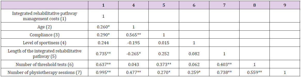
Table 1: Relationships between variables.

biomedres-openaccess-journal-bjstr

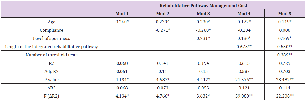
Table 2: Regression model for integrated rehabilitative pathway management costs.

Configuration of Factors Through QCA

The QCA analysis requires a preliminary step for calibration of variables transforming them in crisp ones (Table 3). After performing the calibration process, consistency, and coverage of a set of configuration variables were evaluated. Consistency is the extent to which the results are in line with statements of need or sufficiency. Coverage provides information about the empirical importance of necessary and sufficient conditions. Each configuration has a raw coverage value that “measures the relative importance of several combinations of causally relevant conditions” [23]: the proportion that a configuration covers the outcome. It is assessed by the sum of consistent scores of the configuration divided by the sum of outcome scores [27]. In the QCA analysis, the crisp value 1 is reported in uppercase whereas the crisp value 0 is reported in lowercase. According to the regression analysis, the variables that most influence the management costs are the compliance and the length of the integrated rehabilitative pathway (Table 4). In particular, the most representative recipes show that level of sportiness associated with young age affects the management, as well as the length of rehabilitation, also for the patients with a lower level of sportiness.

biomedres-openaccess-journal-bjstr

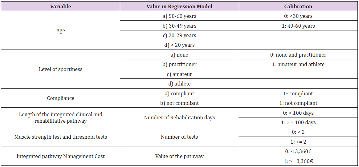
Table 3: Calibration of the variables.

biomedres-openaccess-journal-bjstr

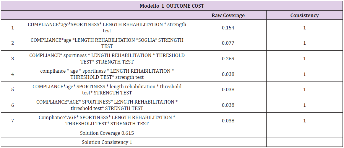
Table 4: QCA analysis for rehabilitative pathway management cost.

Discussion and Conclusion

In the current era, where the availability of large amounts of clinical data gathered during care delivery is rapidly increasing, as well as the ability to access, process, link, and analyze these data in efficient ways, alternative sources to supplement evidence from RCTs look promising. Among all the information that could derive from RWD, one of the most important topics to analyze consist of the management costs related to a specific integrated clinical or rehabilitative pathway. The knowledge of the economic resources’ absorption could contribute to fully understand the pathways as well as the choices and behaviors involved in those pathways. Economic information provides the tools for developing effective and efficient policy strategies and addressing potential tradeoffs between the goals of increasing social welfare and improving the distribution of healthcare delivery across individuals and population groups.

Within the setting of ACL integrated rehabilitative pathway, results reported that strong compliance and a high level of sportiness could be considered among the main factors that cause an increase in the total cost of managing the rehabilitation pathway for a patient with ACL rupture, confirming the information found in literature [14-16]. In fact, orthopedic surgery often requires many months of rehabilitation to achieve a successful outcome, regardless of subspecialty, with an important impact on the overall resources’ absorption [28]. For patients practicing sports by profession, the rehabilitation pathway could be more intense and costly, as it is their priority, to recover their normal motor and sports conditions as soon as possible [8]. Thanks to the QCA it is possible to integrate these results, identifying all combinations of factors that are determining the emergence of a certain results. For example, more consistent QCA analysis solutions show that compliance has an important influence on the absorption of economic resources by patients, but also higher levels of sportiness related to young age of patients, determining higher pathway costs. The results obtained and the findings of this study could give a contribute useful to enlarge the existing research, regarding the use of RWD in the healthcare sector, with particular focus on the rehabilitation programs, thus considering a larger sample and producing results that could be replicable. Although the relevance of this topic, healthcare stakeholders often do not have enough information on outcomes to take decisions, being well-informed. In this context, knowing which are the factors that have a positive or negative influence on the integrated patients’ rehabilitative pathway, could be useful to optimize economic resources, as well as for the achievement of an adequate effectiveness, thus becoming more efficient and effective [6]. This, in the Italian setting, is becoming more and more important, due to the recent re-consideration of the integrated clinical and rehabilitative pathways, after the COVID-19 pandemic, for the re-organization of the healthcare delivery of services.

For More Articles: Biomedical Journal Impact Factor: https://biomedres.us

Open Access Journals on Microbiology Research

Study on the Prevalence of Goiter and Associated Factors Among Hospitalized Patients of District Timergara, Dir Lower Pakistan

Introduction

The thyroid gland is responsible for producing thyroid hormone, which has an important role in metabolic processes. It is essential for these hormones to have adequate amounts of iodine [1,2]. Goiter can be detected clinically by palpation and physical examination when there are insufficient amounts of iodine [3,4]. In several areas of the world, goiter is considered a significant problem for public health, especially in developing countries, and is an indicator of chronic iodine deficiency [5]. Iodine deficiency disorders (IDDs) affect 30% of the population globally. There are more than 150 million IDD patients worldwide [6]. Thyroid nodules and goiters are the most common thyroid conditions due to a combination of genetic, environmental, and endogenous factors [7]. Around 15.8% of the general population develop goiter, and iodine is a significant environmental factor that influences goiter prevalence [7]. A thyroid nodule can be detected by ultrasound in 19% to 67% of cases, but only 4–7% is palpable [8]. Most thyroid nodules and goiters do not present any symptoms; however, they can be associated with other disorders, including endocrine dysfunction, impaired body composition, autoimmune thyroid disease and various metabolic disorders [9]. Coordinating basal metabolic rate and thermogenesis is a key role for thyroid hormones [10].

Researchers have linked low thyroid hormone levels with poor metabolic health in several studies, even when they are within normal limits. There are many metabolic processes regulated by thyroid hormone, including growth and development, reproduction, and enhancing metabolism [11]. The thyroid produces and releases thyroxine (T4) and triiodothyronine (T3) into the circulation system. Thyroid stimulating hormone (TSH) is released by the anterior pituitary gland and helps to regulate thyroid hormone levels, which are controlled by thyrotropin releasing hormone (TRH) from the hypothalamus. A negative feedback loop is present between TRH and TSH in the circulation system, and the regulation is carried out by T4 and T3 [12,13]. An iodine deficiency causes goiter, the enlarged thyroid glands that are greater than the terminal phalanx of the thumb, which indicates chronic iodine deficiency [14].

Goiters provide an indication of the iodine status in a region and can be used as a long-term indicator of the success of an iodine programmer. A measurement of the prevalence of goiter caused by iodine deficiency is based on indicators like urinary iodine concentrations (UIC) and total goiter prevalence (TGP) as well as the proportion of salt in households that is adequately iodized (>15 ppm) [15]. Throughout the world, iodine deficiency is the leading cause of goiter [16-18]. Iodine deficiency was reported in Pakistan by the World Health Organization in 1990 as affecting an estimated 50 million people; goiter was reported as prevalent as 80-90% in mountainous areas [19]. Cretinism is estimated to have a prevalence of about 3% in the general population [20]. Lower Dir District is situated in Malakand Division of Khyber Pakhtunkhwa province in Pakistan. There are 1.05 million people living in Timergara City, the district capital and largest city with 1,582 square kilometers. Our main objective in this study was to find out the prevalence and factors of goiter in hospitalized patients residing in District Timergara, Dir Lower Pakistan.

Methods

Study Setting, Design, and Participants

A descriptive cross-sectional retrospective study of hospitalized patients was conducted from July 2020 to June 2021 in District Timergara, Dir Lower Pakistan. There were 405 participants in the study (247 women and 158 men). The study comprised all patients admitted to the endocrinology department at the (DHQ) Timergara hospital regardless of the reason for admission. Throughout the examination, every patient’s clinical history was examined, as well as the previous case record. We collected the information using a questionnaire designed to inquire about patients’ general socio-demographic data, including goiter physical appearance, age, occupation, ethnicity, gender, residential area, economic status, educational, and clinical manifestations, among other considerations, are considered. A trained endocrine specialist, who has already examined the patients, written informed consent was obtained from all participants. In the study, patients with serious physical and mental illnesses were excluded as it was difficult to obtain data and measurements from them. The study was approved by the Ethical Committee of the Department of Surgical, Institute of Paramedical Sciences Khyber Medical University Pakistan, which followed the recommendations of the Declaration of Government District Headquarter Hospital (DHQ) of Timergara, Dir Lower.

Examination for Goiter

Using palpation techniques, a trained and experienced public health officer assessed the presence of goiters based on WHO/ UNICEF/ICCIDD criteria [21]. Therefore, Grade 0: stand for not visible, not palpable; Grade: I stand for palpable, not visible; and Grade: II stands for visible and palpable.

Statistical Analysis

In order to examine the data, the patient’s records were manually checked, sorted, categorized, and coded. Then, SPSS version 23.0 was used to analyze the results. We measured the prevalence of goiter based on the relative frequencies and ratios for all patients with goiter.

Results

In total, 405 patients were recruited to participate in this study. Here are the sociodemographics characteristics of the participants (Table 1). Out of the 405 respondents, 247 were women (61.0%) and 158 were men (39.0%), and goiter was much more prevalent in women than in men. All of the patients were of the same ethnicity, patients with no formal education had the highest prevalence of goiter, 267 (65.9%), and 138 (34.1%) patients were literate. As educated levels increased, goiter prevalence declined, reaching the lowest levels in comparison to illiterate patients. The results revealed that 278 (68.6%) were married and 127 (31.4%) were unmarried, while 129 (31.9%) were government employees, and that most of the patients were farmers with no proper job 267 (68.1%). Participants were sorted by age into 10-year age groups based on the results the prevalence of goiter was lowest among those between the ages of 14-24 (10.4%), while among 35–44-yearolds, the rate reaches its highest level (37.5%). Goiter prevalence was greater in hilly than plain areas, at 283 (69.9%) in hilly and 122 (30.1%) in plain. In hilly areas, goiter was significantly more prevalent than in plain areas. On the basis of thyroid examination, there were 176 (43.5%) patients with palpable and visible goiter (grade II), and goiter prevalence was higher in individuals aged 35- 44 years old (37.5%) (Figure 1).

biomedres-openaccess-journal-bjstr

Figure 1: Age and gender-wise prevalence of goiter among hospitalized patients.

biomedres-openaccess-journal-bjstr

Table 1: Sociodemographics characteristics of goiter hospitalized patients.

Discussion

Goiter is a common endocrine problem that affects around 300 million people globally, with more than half of those affected being unaware of their condition. The most common thyroid disorders are hyperthyroidism and hypothyroidism, which affect 1.6 billion people in more than 100 countries around the world [22-27]. Our findings revealed that females were more prone to goiter than males, as demonstrated by their ratio of (61.0%) to (39.0%), although overall goiter prevalence did not significantly differ between genders. During this study, goiter cases were classified according to age, with older patients 35-44 (37.5 %) having a higher prevalence than younger patients. There is a documented association between the prevalence of goiters and gender. Interestingly, Knudsen et al. observed that men were more likely to have goiters than women; however, women were two to ten times more likely than men to have goiters [28,29]. Studies have shown that sex hormones may play a role in thyroid volume only after puberty, suggesting that changes in thyroid volume are influenced by gender differences [30]. In addition, women are more likely than men to develop thyroid problems during their lifetime [31,32]. There was a study that found older women had a higher risk of goiter than younger women. A higher prevalence of goiter was observed with age in areas with severe or moderate to mild iodine deficiency. Other studies report an enlargement of the thyroid gland with age in areas with severe or moderate iodine deficiency [33].

According to this study, goiter was most prevalent in illiterate patients compared to literate patients because illiterate individuals were unaware of goiter. The findings of our study are in agreement with previous research findings, which found that the prevalence of goiter increased in hilly areas in comparison to plain areas. Goiter is more prevalent in Baltistan, Northeast Pakistan, where the prevalence was found to be 16.6% among the general population. In the North, it is 20.4% of males, 28.1% of females, and the South 13.9% of males, 21.2% of females [34]. In previous studies, it was discovered that northern Pakistan has become one of the first known areas to have an iodine deficiency. Goiters have been reported in only a few studies across various regions of Pakistan, ranging from the Northern region in the upper Himalaya to the lower Himalaya and even on the Punjab plains. It was estimated in 1990 that 70% of the population was at the risk of iodine deficiency related disorder, with the prevalence of goiters ranging from 55% in plains to 80-90% in the northern mountainous region [35]. According to the WHO classification of goiter based on size and visibility, 7 percent of the global population possesses visible form goiter [36], our results showed that the prevalence of goiter on the basis of thyroid examination with palpable and visible (grade II) was high frequency 176 (43.5 %), the goiter rate was higher in the 35-44 years of age group (37.5%). The prevalence of goiter was reported to be 3.4 % in women and 6.9 % in men among French citizens [37], 23.9 and 35.6 % in German citizens, 58.1 % in Italian citizens [38] and 35.7 % in Romanian citizens [38]. According to Hatemi et al., the prevalence of goiters using neck palpation was 30.5% in Turkey [39]. However, the prevalence was higher than the 16 % and 27 % reported by a report about global iodine deficiency in world and African populations [40]. Similarly, the prevalence of goiter in Ethiopia was higher than various studies which showed a prevalence ranging from 26.3% to 62.1% among children [41-44]. The main cause of goiter is iodine deficiency, and it is imperative to concentrate prevention efforts at the society level on eliminating iodine deficiencies.

Conclusion

The study conclusions suggest that goiter prevalence might depend on TSH levels specific to normal or abnormal hormonal dysfunction. The study also concluded that increased goiter risk was independently related to women’s gender and low TSH levels. The study area had an iodine deficiency problem, and goiter was linked to increased parity, older age, and illiteracy. The study area has a high prevalence of goiter. The health sector of the district should invest more efforts in spreading important messages about iodized salt and iodine-rich foods to ensure community awareness. There is a need for a systematic investigation of the epidemiological profile of patients with diffuse goiter. It is necessary to conduct further research on both the environmental and genetic interactions that lead to goiter.

For More Articles: Biomedical Journal Impact Factor: https://biomedres.us

Open Access Journals on Biomedical Research

Solid Organ Transplantation: A Glimpse into its History

Introduction

On the Basis of the Definition

According to the dictionary of the Royal Spanish Academy [1], transplanting consists of “transferring a living organ or tissue from a donor organism to a recipient organism”. For its part, the dictionary of medical terms of the Royal Academy of Medicine of Spain [2], includes a broader definition of the term transplant: “to transfer an organ or tissue from one place to another in the same individual, between individuals of the same species or between individuals of different species”, adding to the concept the possibilities in terms of types of donors and possible recipients. The term “transplantation” comes from Latin, as the sum of “trans” (beyond) + “plant(āre)” (i.e. to plant) and literally means “change of location of a plant”. The surgical use of this term has been documented in English since 1951 [2] and refers to the “operation of transplanting into a recipient organism an organ or tissue taken from a donor organism. A distinction is made between autologous transplantation, isotransplantation, allotransplantation and heterotransplantation according to whether the donor and recipient organisms are identical, univitelline twins, members of the same species or members of different species, respectively”.

Early Historical References

Although there are references to transplantation techniques dating back to antiquity, such as the legend of Saints Cosmas and Damian [3], which describes how these twin brothers amputate the leg of a deacon with a tumour (in some versions they say he had severe ischaemia) and then transplant the leg of a recently buried Ethiopian; the real breakthrough in solid organ transplantation is considered to have come at the beginning of the 20th century. This era was marked by several milestones, including the following: the refinement of the vascular anastomosis technique by Alexis Carrel [4] (who received the Nobel Prize in Physiology or Medicine in 1912), Charles Claude Guthrie, Mathieu Jaboulay and Julius Dorfler; the refinement of organ transplantation techniques in animals by Emerich Ullmann in 1902, with a kidney transplant between dogs, and the techniques described by Vladimir P Demikhov [5] between the 1930s and 1950s, which laid the foundations for surgical techniques used in humans; within the field of Immunology, the scientific breakthrough of the 20th century was the discovery of acquired immune tolerance by Peter Brian Medawar and Frank Macfarlane Burnet (who received the Nobel Prize in Physiology and Medicine in 1960) [6,7]; this was accompanied by the discovery of drugs with immunosuppressive properties_ especially from the 1960s [8,9] .

First Solid Organ Transplant in Humans in the World

Renal Transplantation: The first human kidney transplant in history took place in 1933 and was performed by the Ukrainian surgeon Voronoy [10]. The recipient was a young woman in a uraemic coma and the donor was a 60-year-old man; although the patient survived the surgery, the transplanted kidneys failed to function properly and on the third day the patient died. Voronoy performed five more cadaver kidney transplants in 1949, again without success. It was not until 23 December 1954 that Joseph Murray succeeded for the first time in performing a successful kidney transplant in humans at the Peter Bent Brighan Hospital in Boston. In this case, the donor and recipient were univitelline twins [11,12]. Murray received the Nobel Prize in Physiology and Medicine in 1990 for performing the first successful human organ transplantation. In the following years, kidney transplantation between twins continued to be performed, with subsequent good progress. At the same time, discoveries in the field of immunology and the development of immunosuppressive drugs made it possible to consider organ transplantation between immunologically nonidentical people.

Liver Transplantation: The first liver transplant was performed on 1 March 1963, at the Veteran’s Hospital in Denver (Colorado) by Thomas Starzl’s team. The recipient was a 3-year-old boy with biliary atresia, who died within hours of the transplant. Two months later, the same team performed another liver transplant on a 48-year-old man with liver cancer, who was implanted with the organ of a patient who had died of a brain tumour; this time, despite achieving adequate liver function, the recipient survived for 22 days, dying after suffering a pulmonary embolism. Like other types of solid organ transplantation, it was not until the 1980s that there was an improvement in the results of liver transplantation in relation to major advances in surgical techniques, coagulation management and the discovery of immunosuppressive drugs [13].

Heart Transplantation: The first human heart transplant was performed by Christiaan Neethling Barnard on 3 December 1967 in Cape Town, South Africa [14] on a 58-year-old recipient suffering from terminal heart failure, who died 18 days later, from Pseudomonas pneumonia. The results of the first interventions of this transplantation modality were disappointing. The explanation, again, lay in immunological problems that increased the risk of rejection and infection in recipients. As previously mentioned, the change came about after the introduction of new immunosuppressive drugs. In particular, in the case of heart transplantation, the advent of cyclosporine [15] led to a marked increase in post-transplant survival.

Lung transplantation: On 11 June 1963, James D. Hardy performed the first-ever lung transplant in a human in Jackson, Mississippi. The recipient was a 58-year-old man diagnosed with lung cancer who was in respiratory failure and also suffered from kidney failure. The circumstances of the recipient, John Russel, made this milestone even more unique as he was on death row for committing a murder in 1957. During his stay in prison, after being diagnosed with the oncological disease, the seriousness of the situation was explained to him and he was offered the chance to be the first human to undergo a lung transplant, promising, if he survived the transplant, to commute his prison sentence to the governor of the state for ‘contribution to the cause of humanity’. Russell agreed and the left lung was transplanted. However, after surviving for eighteen days with good function; he died as a result of worsening kidney failure [16]. Despite several subsequent attempts, it was not until the 1980s that good survival results were achieved in this type of solid organ transplantation, with the team at the University of Toronto and its lung transplant programme being the reference [17] (Figure 1).

biomedres-openaccess-journal-bjstr

Figure 1: Chronology: first solid organ transplants in humans.

The Particular Case of Spain and Solid Organ Transplantation

The first transplant in Spain took place in 1965 at the Hospital Clinic i Provincial de Barcelona, where a cadaveric kidney transplant was performed by the Gil-Vernet team [18]. Almost two decades would have to pass before the first transplants of the other modalities were performed. The new heyday of solid organ transplants began again in the 1980s. Thus, it was not until 23 February 1984 that the first liver transplant was performed by Carles Margarit and Eduardo Jaurrieta at the University Hospital of Bellvitge (Barcelona) on a patient diagnosed with a liver tumour [19]. The first successful heart transplant in Spain was performed on 8 May 1984 at the Hospital de la Santa Creu i Sant Pau (Barcelona) by the team led by Josep María Caralps and Josep Oriol Bonín. The latest date in Spain corresponds to the first onelung transplant, performed in 1990 by Ramón Arcas at the Hospital Gregorio Marañón (Madrid) [20].

That same year, in February 1990, the first combined cardiopulmonary transplant with long-term survival was performed [21] and it was not until 1992 that the first bipulmonary transplant was performed [22,23].

Within the history of transplantation in Spain, there are a number of notable years: In 1979, Law 30/1979 on Organ Transplantation was passed [24], which legally recognised the concept of “brain death”. Until then, in order to proceed with the extraction of an organ for transplantation (until that date it was always the kidney), the donor had to wait for asystole. After this law and its implementation by Royal Decree in 1980 [25], there was an increase in the total number of transplants carried out in Spain. Another important year was 1989, when the National Transplant Organisation (ONT), officially created in June 1980 by Resolution of the Secretary of State for Health [26], acquired a physical organisation that facilitated the achievement of its goals. The ONT was defined as a coordinating body of a technical nature, belonging to the Ministry of Health, responsible for developing the functions related to the procurement and clinical use of organs, tissues and cells. It is structured as a network, for the organisation of which three levels are established: National Coordination, Autonomous Community Coordination and Hospital Coordination. The so-called “Spanish model” [27] of transplantation and its organisational structure showed good early results, making Spain the leading country in the world in terms of the number of donations.

Conclusion

The possibility of performing solid organ transplants has meant a significant change in the lives of many people in recent decades. The great advances made in the 20th century in the field of surgery made it possible to consider this type of treatment in humans for the first time. Although initially the results in terms of survival were not favourable, the discovery of the immunological mechanisms that produced rejection, along with the knowledge of the human leukocyte antigen (HLA) system, and the subsequent appearance of immunosuppressive drugs, led to a change in the paradigm of the management of transplant recipients. Today, solid organ transplantation is a particularly relevant therapeutic option for people with diseases previously considered terminal due to severe dysfunction of one or more organs who are candidates for inclusion on the waiting list to receive a graft, since after transplantation, the recipient’s quality of life is often improved, and survival is increased.

For More Articles: Biomedical Journal Impact Factor: https://biomedres.us

Journals on Biomedical Intervention

Van Lohuizen Syndrome, A Late-Diagnosed Case in an 18 Years-Old Female

Introduction

Van Lohuizen Syndrome, or Cutis Marmorata Telangiectatica Congenita. Is a very rare birth defect involving the cutaneous blood vessels, often reported as a benign, congenital disorder of unknown etiology, epidemiology, and pathophysiology. Described as persistent cutaneous telangiectasia, and phlebectasia, these appear as reticulated streaks of the skin capillaries and venules, resulting in a marbled-looking skin (cutis marmarota) [1-3]. First described by Kato van Lohuizen, a female Dutch pediatrician, in 1922 [4]. Since then, it has been referred to under several names, including, congenital generalized phlebectasia, [5,6] naevus vascularis reticularis, [7] as well as congenital livedo reticularis [8]. Characterized by the presence of erythematous network streaks, without venectasia, which is not responding to local heating. It may occur along with port-wine stain, [9] cutaneous ulceration, and atrophy within the affected area, as well as body asymmetry, and may affect any organ, including the eyes, skeleton, kidneys, and the brain [10-12].

Case Report

We are reporting the first case of CMTC in Libya, in 18 years old girl referred to our clinic for consultation. She had persistent skin disorders in her Rt. upper limb since birth (Figure 1). She is the third of three siblings, born as a full-term baby, by uncomplicated vaginal delivery, to a non-consanguineous marriage, and there was no family history of similar lesions. A high school student, leading a normal active life, until about a year ago, when she began to feel intermittently some discomfort and numbness of the right hand and forearm, with no history of trauma. Her upper extremity skin changes were detected at birth along with contralateral DDH, which was treated conservatively. Our clinical findings revealed a network of blue-purple lines giving the skin a marbled appearance along the right upper extremity from the deltoid insertion to the metacarpophalangeal joints level dorsally, sparing the Palmer skin, otherwise, her skin temperature and texture were normal, without atrophy or ulceration (Figure 2). The lesions were fading at Direct pressure, but not affected by gravity. There were multiple irregularly shaped port-wine stain patches on the right anckle laterally, left leg posteriorly, and the right thigh anteromedially. No upper or lower limbs atrophy, cercomfrential, or length discrepancy, and her gait was normal, there was no scoliosis, or facial asymmetry. There were no sensory or motor deficits detected clinically. Here skeletal survey, abdominal and pelvic ultrasound, echocardiography, and all laboratory investigations were within normal. NCS, reported mild right CTS, Based on this medical history and clinical presentation, the diagnosis of Van Lohuizen syndrome was confirmed (Figure 3).

biomedres-openaccess-journal-bjstr

Figure 1: Cutis marmorata, Cutis marmorata, erythematous network of telangiectatic cutaneous capillaries.

biomedres-openaccess-journal-bjstr

Figure 2: The lesion extends along the right upper limb, between the deltoid insertion proximally and the Metacarpophalangeal joints level distally, sparing the phalangeal skin dorsally and the palmar skin volarly.

biomedres-openaccess-journal-bjstr

Figure 3: Multiple erregularly shaped port-wine stain patches on he Anckle A, Leg B and Thigh C.

Discussion

Van Lohuizen syndrome is a rare congenital disorder, with less than 300 cases reported worldwide to date [2,12,13]. However, since not all cases are diagnosed, it is difficult to determine incidence rates. Its origin and gender prevalence are still obscure and unclear, although, it is believed by some to be prevalent among females (64%) [14,15]. In some rare cases, CMTC may run in families [11]. However, Amitai et al. observed no familial cases of CMTC in their series, and most of the cases seem to be sporadic, with no racial predilection [16]. However, skin biopsies have revealed genetic mutations in GNA11, [17-20] and some researchers have also reported genetic mutations in ARL6IP6, suggesting its inheritance as a recessive trait [20-23]. Moreover, viral infections have been postulated, and the underlying connective tissue abnormality has been supposed. Its diagnosis is clinically based. It appears as cutaneous erythematous reticulum streaks, fading upon direct local pressure but unaffected by local heating or gravity, with no venectasia, findings that Kienast, [9] considered three of which as primary signs. While the presence of skin atrophy and ulceration, and the port-wine stains outside the areas affected by CMTC, have been considered a secondary diagnostic criterion (Table 1). For diagnosis, the presence of three major signs and two of the minor criteria is indicative [2].

biomedres-openaccess-journal-bjstr

Table 1: The major and minor diagnostic criteria, Kienast classification [2].

In most cases (66.8%), the disorder is localized, and the leasions are unilateral in 65% of the cases, more frequently involving the limbs (69%), [16] where the upper limbs are affected in (25.9%), while the hands are involved in only (4.9%), but can be generalized in about (24.5%) of cases [24]. CMTC may involve any organ in the body, including the eyes, skeleton, brain, kidneys, and others [10]. The most frequently associated anomalies, in 18.8% of patients [16] include body asymmetry, neurological, ophthalmological, and cardiovascular defects, dysmorphic features, genitourinary, and endocrinological defects [2,16,24-29]. The CMTC should be distinguished from congenital livedo reticularis, which is caused by mosaic PIK3CA gene mutations, [30] where ulceration and phlebectasia do not occur, Sturge-Weber syndrome, which is caused by mosaic GNAQ gene mutations, [17] Neonatal lupus erythematosus, [31] and Klippel-Trenaunay syndrome.

Although, histopathology may confirm the diagnosis by showing an increased number and size of capillaries and veinules in the dermal layer, endothelial swelling, and sometimes aneurysms, it is not necessary, and nonspecific [16,32-35]. Imaging, and fluorescein angiography are indicated only for the evaluation of other congenital anomalies that may accompany the CMTC.

Up to the moment, there is no definitive treatment for skin symptoms in CMTC. However, sympathectomy, and laser therapy are postulated [14,32,34-38]. In our current case, we believe that treatment is unnecessary, apart from reassurance and may be a psychological support to ease the burden of the aesthetic appearance, for although the problem involves a wide area of about (9%) of the total body surface area (TBSA), [39] it is still localized, and it is without any other associated problem apart from the described port-wine patches. She is a tertiary school student, living a normal life with no functional or intellectual issues. However, if we consider her complaint of discomfort and numbness as part of the problem or some of its long-term sequels, [40] the clinical findings and investigation results do not suggest more than the need for follow-up. Although some authors, including Kienast, state that skin leasions typically become less and often completely disappear by adolescence, [3,4,16] this is not the case with our patient [12,41].

For More Articles: Biomedical Journal Impact Factor: https://biomedres.us

Open Access Journals on Microbiology Research

COVID-19: The Impact of Emerged Omicron on Vaccine Escape

Introduction

Going to the thitd year since the emerging of Covid-19 virus and its global containment effort that ends up with the ongoing scenario of failure to halt virus spread waves or eradication until now. There are many queries raised in what went wrong with repeayed emerging waves. On the other hand, the newly emerged covid Omicron and the expected future coming variant based on the significance of dry lab sequence analysis outcomes on vaccines protuctivity or virus escape indicated fir the crucial need for web lab that will indicate the how far the current vaccines still offering enough prevention or a new vaccine version is required, this review covers the key preventive action that should be taken towards the ideal Covid eradication strategy.

The Current Covid-19 Vaccines

Principally covid-19 vaccines were approved with great expectations to protect healthy individuals from exposure to infection, as well as to enhance recovery and minimize the disease severities, reduces hospitalization, fatality, terminate infected healthy carriers and new cases among the vaccinees. Since initiated covid-19 vaccination in December 2020; and out of more than one hundred vaccine trials; currently only 14 fast track coronavirus covid-19 vaccine has been designed, tested & approved. But an unexpected different scenario took place among the vaccinee as what currently noticed that some of those fully vaccinated captured the infection in addition to the raised issues on the vaccines boosting dosage, number, timing and the best efficient way to deliver the vaccine in order to induce the best types of protective immune response/s toward such respiratory pathogens. The direction and significant of Omicron Covid variant dry-lab analysis vs wet lab results: Sequences based lab analysis known as dry lab, while the real phenotypic are wet-lab; the sequences of viral isolates analyzed in dry lab based showed presence of mutation in term of many variant including Omicron as a common expected feature in RNA viruses [1].
The virus have many structural proteins and genes of which Covid spike is the unique key player that have role on its capacity of invasions to target cell and tissue tropism, bind to receptor/ co-receptor epitope, immunogenicity, passive plasma therapy for sever disease and vaccine success or failure. Covid based sequences outcome should focus on how far its significance the new viral emerged variant such as Omicron in term of the location, number and types of mutation on spike genes S1 & S2, the 3D epitope modified conformation compared to native viral spike and how far the mutations have impact on spike epitopes affinity binding to Ace2 receptor on the fusion of viral particle to target cell and the outcome of the reported mutation on the protective capacity of new variant spike based on wet lab, using the well-known in-vitro virus neutralization test in susceptible VERO-E6 cell lines, towards sera from vaccinee received full Covid vaccines dosage [2]. This wet-lab virus neutralization result outcome remain as the most important and crucial parameter to prove and will tell the reality of legendlegend binding affinity and to give answer for the assumptions raised based on dry-lab sequence analysis about whether those mutations in Omicron spike gene will affect protectivity, partial vaccine escape or fully emerged new viral variant escape totally missmatch the current ongoing Covid vaccines. At any time the wet-lab reported failure of vaccinee sera in virus neutralization, it mean new design of covid vaccine version will urgently require to include the mutant gene or epitopes of both the new Covid variant like Omicron and to maintain the effective and protectiveness of the ongoing covid vaccines.

What Should be Ideal Vaccine Delivery Route toward Covid-19 Eradication

Although covid vaccines offers protection to vaccinees and the current need for a third boosting dose towards Omicron and the challenging pressures on manufacturers to meet the increased covid vaccine market. But still there is a major gap in covid-19 vaccine that clearly appeared on neglecting vaccine delivery route to consider the crucial issue of vaccine capabilities in blocking the virus invasion and replication in its target cell & tissue tropism at entry sites. And the role of spike specific IgA in the neutralization of covid primary replication, attach of viral spike to ACe2 cell receptor that terminates presence of healthy carrier vaccinees [2].
Therefore it is top demand for vaccine capable to induce anticovid- 19 specific mucosal immunity as the key component and most crucial effective mechanisms to halt exposure to infection through blocking early virus entry at the mucosal front line, attached to target cell Aec-2 receptors and inhibits early virus replication among the fully vaccinated population [3-5].
An ideal novel smart covid-19 vaccine need to be designed and delivered to offer the vaccinees with triple immune responses as follows; enough cellular memory and specific higher humoral immunity in term of Covid-19 IgM & IgG responses induced through parenteral immunization routes. In addition to strengthened potent, covid-19 spike-specific mucosal immunity in term of higher covid-19 IgA antibody titer to neutralizes virus inoculum on mucosal lining on upper respiratory, pharyngeal, nasal sites and lung, through mucosal (nasal spray or oral) vaccine delivery as 3rd and 4th doses following initially two parenteral doses [3,6]. Therefore, a new revised covid-19 vaccine design, downstream processing, through encapsulating carrier, mucosal delivery systems post-parenteral dosage to modulate and boost covid-19 spike specific M-cells and APC that promote adherence and transport of vaccine epitopes for triggering covid-19 spike-IgA class-switching as mucosal vaccines that is expected to terminates virus spread and game changer and initialization of pandemic eradication era [7-10]. In addition introduction of such mucosal requires re-adjusting of vaccine testing and efficiency parameters including measurement of covid-19 IgA titer in vaccine and to develop in-vitro neutralization testing protocols & assay for covid-19 mucosal samples (saliva, nasal) and viral specific mucosal dendritic & M-cell flow-cytometry [11,12].

For More Articles: Biomedical Journal Impact Factor: https://biomedres.us

Open Access Journals on Biomedical Research

Biochemical Study on the Effects of Ruzu Herbal Bitters Formulation on Wistar Albino Rats

Introduction

The development and use of traditional herbal medicine have come a long way and it corresponds to the Stone Age. In Africa, the practice of traditional healing and magic is much older than some of the other traditional medical sciences and seems to be more prevalent than conventional medicine [1]. World Health Organization (WHO) has defined Herbal medicine as an authorized medicinal herb or herbal materials or herbal preparations and finished herbal products which contain whole plants, parts of plants, or other plant materials such as: barks, leaves, flowers, berries and roots, and/or their extracts as active ingredients intended for human therapeutic use and sometimes animals [2]. It has become a common traditional practice in some cases, that a combination of plants or their extracts is used in the treatment of certain ailments with the believe by the herbalists that the individual plants contain different therapeutic agents in which when combined together will give a better therapeutic efficacy for a particular disease or multiple diseases than that of a single plant.

biomedres-openaccess-journal-bjstr

Figure 1: Ruzu Herbal Bitters.

Therefore, most herbal preparations work in synergy, hence the emergence of polyherbal mixtures. Ruzu herbal bitters (Figure 1) are polyherbal mixtures, produced by Ruzu Natural Health Product and Services, Nigeria. The polyherbal mixture is made up of three different plants: 20% Uvaria chamae (bush banana), 40% Citrullus colocynthis (bitter apple) and 40% Curculigo pilosa (squirrel groundnut). Ruzu herbal bitters are commercially available and the manufacturers concluded that the product has the following medicinal functions amongst others, as indicated in the leaflet of the product: management/treatment of diabetes, typhoid and malaria, high blood pressure, waist and back pains, fibroid, infertility (male and female), gonorrhea/staphylococcus, syphilis, pile, obesity/ stomach troubles, detoxifies the kidney and tones the liver [3]. Based on the medicinal claims of the manufacturer of Ruzu herbal bitters with paucity scientific proofs, there was need to carry out scientific investigations to ascertain its effects on wistar albino rats by assessing biochemical indices.

Materials and Methods

Materials and Chemicals

Syringes and needles, hand Gloves, incubator, glucometer, Aucku check active strip micropipette, stop watch, oven, centrifuge Model 800, cotton wool, HPLC, and GCMS. The chemicals included 10% Chloroform, xylene, hemotoxylin and eosin stains.

Collection of Herbal Formulation Samples

Ruzu Herbal Bitters a Nigerian Herbal formulation was purchased from a registered pharmaceutical shop (Cynflac Pharmacy Yenagoa, Bayelsa State). The product is a combination of several medicinal plants. As an inclusion criterion, the product was ascertained to have been registered with the National Agency for Food, Drug Administration and Control (NAFDAC number: A7- 1102L). The manufactured and expiry dates of the product were inspected and all were confirmed to be within the acceptable time frame. The Manufacturer’s seal, inspected to ascertain the authenticity of the product was intact in the bottles of the syrup purchased for the analysis and was taken to the Research Laboratory, Department of Pharmacology, Faculty of Basic Medical Sciences, College of Health Sciences, Niger Delta University, Wilberforce Island, Bayelsa State and was stored under room temperature prior to the experiment.

Experimental Animals

Animal Handling: Twenty (20) adult albino rats used for this study were purchased from the animal house of the Faculty of Basic Medical Sciences, College of Health Sciences, Niger Delta University, Wilberforce Island, Bayelsa State. The animals were kept in standard plastic rat cages in the research laboratory of the Department of Pharmacology, Faculty of Basic Medical Sciences, College of Health Sciences, Niger Delta University, Wilberforce Island, Bayelsa State. The animals were allowed to acclimatize for 7 days under standard laboratory conditions with free access to commercial grower’s mash (Delta Feeds), water ad libitum and 12h/ 12h light/darkness cycle and fresh air prior to the inception of this study. The animal experiment was conducted in accordance with internationally accepted practice for laboratory animals and approved by the Animal Ethics Committee of the Faculty of Basic Medical Sciences, College of Health Sciences, Niger Delta University, Wilberforce Island, Bayelsa State.

Administration of Poly-Herbal Formulation: The herbal formulation was administered using 5ml syringe, the corresponding dose was given to each rat based at 5, 10 and 15 mg/kg body weight dose of the herbal formulation was selected.

Experimental Design

After the period of acclimatization, the animals were randomly divided into experimental and control groups. The albino rats were grouped and herbal formulation administered as follows;

• Group 1 (n = 5) Control: Albino rats received 2ml of distilled water daily within the period of the study before sacrificing.

• Group 2 (n = 5) Albino rats were treated with 5 mg/kg body weight of Ruzu Herbal Bitters

• Group 3 (n = 5) Albino rats were treated with 10 mg/kg body weight of Ruzu Herbal Bitters

• Group 4 (n = 5) Albino rats were treated with 15 mg/kg body weight of Ruzu Herbal Bitters.

Blood Sample Collection: The animals were observed in their cages for clinical symptoms daily and at the end of the 14 days treatment, the rats were sacrificed under chloroform anesthesia and blood was collected by cardiac puncture, using 5ml syringes and 23G needles into blood sample containers. The blood was allowed to stand for 2 hours to coagulate and was centrifuged for 10 minutes at 2000 rpm and the supernatant (Serum) carefully collected for biochemical analysis.

Biochemical Analysis: Serum levels of aspartate aminotransferase (AST) and alanine aminotransferase (ALT) were determined following the principle described by [4] while the alkaline phosphates (ALP) were carried out according to the method described by [5] to assess liver function. Renal function was assessed by measuring plasma creatinine (CREA) levels and blood urea nitrogen (BUN) was assayed following the method of [6,7]. In order to assess the synthetic function of the liver, total serum protein (TP), Total bilirubin and albumin (ALB) concentrations were determined according to the principles based on the Biuret reaction [8] and bromocresol green reaction [9] respectively. Total Serum cholesterol (TC) concentrations were estimated following the method described by [10].

Statistical Analysis of Data: All data were expressed as Mean ± Standard Error of Mean (SEM). Significant differences among the groups were determined by One-Way Analysis of Variance (ANOVA) using the statistical analysis program for social sciences (SPSS 17.0).

Results

Biochemical Parameters

The Body Weight of wistar albino rats administered with Ruzu herbal bitters formulation is shown in Table 1. Table 2 shows the effects of Ruzu herbal Bitters formulation on biochemical indices of wistar albino rats at 5, 10 and 15mg/kg doses. The renal, hepatic, cardiac and lipid profile indices analyzed were albumin, total protein, urea, creatinine, Alkaline Phosphatase (ALP), Alanine aminotransferase (ALT), total bilirubin, Aspartate aminotransferase (AST) and total cholesterol and were compared with the control groups. All mean values of total protein and creatinine levels analyzed were significantly higher while albumin levels were lower when compared with the control. Higher concentrations (10 and 15mg/kg) of ALT, AST, ALP and total cholesterol were significantly higher when compared with the control. Whereas lower (5mg/kg) concentration of urea and total bilirubin levels in the experimental animals were observed to be higher when compared with the controls. Statistically, there were no significant differences of the analyzed parameters at 95% confidence level (P < 0.05).

biomedres-openaccess-journal-bjstr

Table 1: Body weight of wistar albino rats administered with Ruzu Herbal Bitters.

Note: (Mean ± SEM, n = 5)

biomedres-openaccess-journal-bjstr

Table 2: Effects of Ruzu Herbal Bitters on Biochemical indices of wistar albino rats.

Note: Data are Means ± SEM; n=5. Means of the same superscript alphabets in the same column shows no significant difference at 95% confidence levels (p<0.05).

Discussion

Herbal medicines are now receiving greater attention as an alternative to clinical therapy leading to increase in their demands [11]. In the rural communities of developing countries, the exclusive use of herbal drugs to treat various diseases is still very common and is prepared most often and dispensed by herbalists without formal training. Experimental screening method is therefore important in order to establish the active components present, ascertain the efficacy and safety of the herbal products [12]. The evaluation of the safety of these poly-herbal products is presently carried out in animals. In risk assessments, there are better correlations between rats and humans, whereas it is less predictive when mice are used for experimental [13]. Therefore, the aim of this study was to assess the effects of Ruzu Herbal Bitters formulation on wistar albino rats by carrying out biochemical parameters as marker enzymes. Study of the renal profile such as total protein, urea, creatinine and albumin levels give useful information about the poly-herbal formulation-induced renal toxicities. Animals which received herbal formulation in 5, 10 and 15mg/kg body weight doses did not show any significant changes in albumin levels. Decreased serum levels of albumin occur when there is impairment in the synthesizing function of the liver [14] therefore; the significant decrease in serum levels of albumin observed in this study may be an indication of liver impairment. However, animals at 5, 10 and 15mg/kg body weight doses showed highly significant increase in creatinine and total protein levels when compared to the control group. Creatinine levels are used as marker of kidney function.

Previous studies have shown that an apparently minor increase in serum creatinine can reflect a marked decrease in glomerular filtration rate [15]. Thus, elevated serum levels of creatinine may indicate kidney injury, with resultant reduced glomerular filtration. The results are synonymous with the findings of [16]. Urea is formed in the liver, representing the principal waste product of protein catabolism and is excreted by the kidney. In this study, urea levels at 10 and 15mg/kg doses shows significant decrease. However, the minimal increase in serum levels of urea at 5mg/kg dosage observed in the poly-herbal formulation group may be due to nephrotoxic effect of the herbal formulation, leading to reduced renal function. Liver damage tempted by herbal formulations or synthetic drugs may consist of hepatocellular necrosis, cholestasis, or a mixture of biochemical and histopathological patterns [17]. The estimation of AST, ALT and ALP is suitable in the early diagnosis of viral or toxic hepatitis and thus patients exposed to hepatotoxic drugs [18]. Generally, perturbation of parenchymal cells of the liver by xenobiotics or drugs results in elevation of both transaminases (AST and ALT) in the blood [19]. AST has both mitochondrial and cytoplasmic origin and any elevation could be taken as a first sign of cell damage that leads to the appearance of these enzymes in the serum [20]. Hence animals were tested for ALP, ALT, AST and total bilirubin levels to check for hepatic and cardiac toxicity. Therefore the increases observed in AST (15mg/kg) and ALT (10 and 15mg/ kg) activities in this study suggest that the administration of Ruzu herbal bitters did interfere with the integrity of the parenchymal cell. However the increase was only significant in the high dosage groups. The findings of this study were in agreement with the works of [21].

One of the major enzymes involved in hepatobiliary evaluation is alanine amino phosphatase (ALP). ALP levels above normal are mainly associated with blocked bile duct although this enzyme is also concentrated in the kidney and bone. The liver, via bile excretes ALP, whenever liver function is compromised the excretion of bile by the hepatocytes diminishes and this results in the increased levels in the serum ALP [22]. There was no significant increase in the ALP values at 5 and 15mg/kg body weight dosages, suggesting that Ruzu herbal bitters at the above doses did not obstruct bile excretion nor caused congestion and therefore Ruzu herbal bitters has no tendency to cause cholestasis. However, a minimal increase was observed at 10mg/kg dosage for ALP and 5 mg/kg dosage for total bilirubin when compared with the control groups. The non-significant and minimal increase observed in ALP and total bilirubin levels in this study is an indication that Ruzu herbal bitters did not impair the capacity of the liver to excrete bilirubin. Liver disease is characterized by an elevation in the serum levels of alkaline phosphatase (ALP), while hyperbilirubinaemia is seen in conditions causing hepatic liver diseases that impair the excretion of bilirubin [23]. This result is in accord with the works of [24,25]. The minimal increase in 15mg/kg dose observed in serum total cholesterol in the animal group may be due to the effect of the herbal formulation. The increase in the serum levels of total cholesterol may be attributed to the toxic effect of the formulation, leading to hepatobiliary disorders and impaired cholesterol metabolism. However the increase was only significant in the high dose group. These findings were in agreement with the works of [26].

Conclusion

In conclusion, this study has provided an insight and data on the Nigerian Poly-herbal formulation (Ruzu Herbal Bitters) which may be relatively safe at lower therapeutic dosage. However, at higher therapeutic dosages and prolong administration may result in renal, hepatic, cardiac and hepatobiliary disorders. Therefore, the creation of public health awareness becomes imperative on the safety and the impending health risk associated with the administration of the poly-herbal products.

For More Articles: Biomedical Journal Impact Factor: https://biomedres.us

Open Access Journals on Microbiology Research

Dental Arch Dimensions in a Matched Pairs Study of Hypodontia Patients and Controls

Introduction

Hypodontia is a common variation of tooth number in the population. In the permanent dentition approximately 25% of individuals have 1 or more congenitally missing third molars and some 3.5% to 7% of the population have hypodontia of other permanent teeth [1-6]. The condition is more frequent in females and approximately 90% of affected patients have less than 4 permanent teeth, other than third molars, congenitally absent. The condition can present challenges requiring careful long term treatment planning and care involving general practice, paediatric dentistry, orthodontics and restorative dentistry. Variations in tooth size and shape are well established in patients with congenitally missing teeth and may also occur in other components of the stomatognathic system [7-9]. As part of an international collaboration investigating the aetiology and clinical implications of hypodontia, this paper explores dental arch morphology in mild or moderate hypodontia. The dental arches and the dentition are two closely related components of the stomatognathic system, which develop in anatomical proximity over an extended time period from early in utero to early adulthood. The dental arches and the dentition are both complex systems, whose development is determined by multiple interactions between genetic, epigenetic, and environmental factors [10-12]. Interactions continue as development progresses through cellular, soft tissue and mineralisation stages to the emergence of the mature phenotype [9]. Hypodontia is an outcome of these complex interactions [13].
Similarly, in addition to genetic factors, the dimensions and shape of the dental arch are influenced by the configuration of the underlying basal bone and the actions of prenatal and postnatal environmental factors [14-17]. Postnatal environmental factors that have been identified include: the intraoral and circumoral musculature [18,19], sucking habits [20], postural and breathing patterns [21] and early loss of primary teeth [22]. There have been varying results in previous studies of dental arch morphology in patients with hypodontia. Woodsworth, et al. [23] found no significant differences in hypodontia patients compared to controls, Paulino, et al. [24] found greater intercanine and intermolar distances in the permanent dentition of adolescent and young adult men than in women, while Nelson, et al. [7] and Higgins [25] report the upper arch depth and chords were significantly reduced. They found greater differences in severe hypodontia. Sex differences are present in arch dimensions [26] and the degree of change in hypodontia may vary between male and female patients. Moreover, the differences may be greater in the upper arch than the lower [27] and may be influenced by the location of the congenitally missing teeth [7]. The aim of the present study is to investigate dental arch dimensions using a well validated 2D image analysis system [7,28,29] in a sample of hypodontia patients and matched controls to determine if there are any differences and, if so, how these relate to the sex of the patient, the location of the congenitally missing teeth and the upper and lower arches.

Materials and Methods

This study was approved by the Ethics Committee of the Scientific Research of the George Emil Palade University of Medicine, Pharmacy, Science and Technology of Tirgu-Mures (Approval no. 60/07.03.2018). The participants gave their written informed consent. Sixty patients with hypodontia, 40 females and 20 males, having a mean age of 15.40±2.85 years were included. The criteria for inclusion were the congenital absence of one to five permanent teeth, excluding third molars and that the formed permanent teeth were fully erupted. Diagnosis was based on dental history, clinical examination and orthopantomograms. Exclusion criteria were the presence of any other congenital conditions, syndromes, or a history of orthodontic treatment or tooth extraction. The same number of controls with complete permanent dentitions, matched for sex, age, ethnicity and exclusion criteria were also studied. Mean age in the control group was 15.48±2.87 years. In order to examine the possible influence of location of the congenitally missing teeth anterior (26 cases) and posterior (31 cases) hypodontia subgroups were formed. Anterior hypodontia was defined as missing upper and lower incisors and/or canines. Posterior hypodontia was defined as missing upper and lower premolars and/or molars, excluding third molars. For these subgroups age- and sex- matched controls were selected from the control group (Figure 1).

biomedres-openaccess-journal-bjstr

Figure 1: Image of measuring the depth of the palatal vault.

biomedres-openaccess-journal-bjstr

Table 1: Definition of measured parameters.

Alginate impressions (Ypeen Premium, SpofaDental) were taken for each individual from the upper and the lower arch. Study models were made from dental stone (FujiRock, GC). Images of the study models were taken with a digital camera (Nikon D3100, Nikon Corporation, Japan) and macro lens (Tamron SP AF-S 90 mm f/2.8). The camera was fixed above the dental cast, on an adjustable stand (Kaiser 5360, Kaiser Fototechnik, Germany) with two fixed led bulbs providing standard lighting conditions. Images of the dental arches were transferred using View NX2 (Nikon Corporation) and processed by the Image Pro Insight 9.3 software (Media Cybernetics, USA). Each image taken included a ten-millimeter scale for calibration and the measurements were made directly on the images. The 2D measurements of the dental arches were the arch circumference, arch length, intercanine width, intermolar width and the depth of the palatal vault. The definitions used for these measurements are given in (Table 1). The measurements were all carried out by the first author. Intraoperator and interoperator reproducibility was determined using the upper and lower models of 8 individuals. Three trained operators carried out the procedures separately, including image capture, calibration and measurement of selected dimensions, on 2 occasions, 2 weeks apart. Statistical analysis was performed using MedCalc (MedCalc Software Ltd). After excluding outliers, normal distribution of the data was confirmed (Shapiro-Wilk test of normality). Intraclass Correlation Coefficients (ICC) were determined to assess reproducibility of measurements. Correlations were also calculated between the number of missing teeth and the arch parameters. Significance of the differences was assessed using one-way ANOVA test, two-way ANOVA test with Bonferroni correction and Pearson’s correlation coefficient. The significance level was set to 0.05.

Results

The intra-operator and inter-operator reproducibility was excellent, with all ICC values being higher than 0.9. (Table 2).In the overall hypodontia group there were 29 patients with one congenitally absent tooth, 23 with two, 2 with three and 6 patients with four congenitally absent teeth. Lower second premolars were the most often missing teeth, followed by the upper lateral incisors, upper second premolars, lower first incisors and lower second molars. In the subgroups, for anterior hypodontia 13 female and 13 male cases were found, with upper lateral and lower central incisors missing. For posterior hypodontia 24 female and 7 male cases were found, with upper and lower second premolars and lower second molars missing. Three cases had both anterior and posterior congenitally missing teeth and were not included in either subgroup. When all hypodontia cases were compared to matched controls, significant differences were detected both in upper and lower arch parameters. Arch circumference, arch length and intercanine width values were significantly smaller in the hypodontia group for the upper arch than in controls. The more teeth that were missing, the lower the upper arch circumference was. In the lower arch intermolar width values were significantly higher in the hypodontia group than in controls (Table 3). More significant differences were seen in male patients than in female patients in the upper arch, although the interaction between the sources of variation was not significant in every case.

biomedres-openaccess-journal-bjstr

Table 2: Intraclass correlation coefficients.

biomedres-openaccess-journal-bjstr

Table 3: Mean values of measured parameters for all hypodontia patients compared with matched controls for both upper and lower arches; SD=Standard Deviation.

Intermolar width differences were significant in women (p=0.02), while in men arch length differences (p=0.008) were significant in the lower arch (Table 4). For the anterior hypodontia subgroup in the upper arch statistically significant smaller arch circumference, arch length and intercanine widths values were found in the hypodontia patients (Table 5). In the lower arch significantly greater intermolar width values were seen in the posterior hypodontia subgroup than in matched controls (Table 6). The analysis of variance highlighted differences also between the anterior and posterior case subgroups. The upper arch circumference and the upper intercanine widths was significantly lower in the anterior subgroup than in the posterior subgroup for hypodontia cases (p<0.001).Significant negative correlations were detected between the number of missing teeth and other parameters. All statistically significant results are shown in (Table 7). When correlating the upper arch parameters for all cases with the number of missing teeth, significant negative correlations with the intermolar width were seen. The higher the number of missing teeth, the lower the upper intermolar width was. On the other hand, when looking for correlations based on sex, strong negative correlations were detected only in men and only in the upper arch (Table 7). Regarding the anterior hypodontia subgroup, both the upper and lower arches showed significant correlations between the number of congenitally missing teeth and some of the parameters (Table 7).

biomedres-openaccess-journal-bjstr

Table 4: Mean values of measured parameters for females and males with hypodontia compared to matched controls; *significantly lower than values from the control group, when interpreting separately.

**significantly higher than values from the control group, when interpreting separately; SD=Standard Deviation.

biomedres-openaccess-journal-bjstr

Table 5: Mean values of measured parameters in upper arch for anterior and posterior hypodontia subgroups and matched controls; †3 patients were excluded from this section as they had both anterior and posterior hypodontia; SD=Standard Deviation.

biomedres-openaccess-journal-bjstr

Table 6: Mean values of measured parameters in lower arch for anterior and posterior hypodontia subgroups and matched controls; †3 patients were excluded from this section as they had both anterior and posterior hypodontia; SD=Standard Deviation.

biomedres-openaccess-journal-bjstr

Table 7: Statistically significant negative correlations between the number of missing teeth and different parameters.

No significant correlations were found for the posterior hypodontia subgroup.

Discussion

The validity of the study can be examining the nature and structure of the sample, the study design, the pattern of hypodontia in the subjects, the measurement techniques, the reproducibility found and the raw data. The sample is derived from a single ethnic group and is of a Dental Age [30] where the dental arches have developed to maturity in width and length [31,32]. The sample size is satisfactory as determined by power calculations [7] and the controls are matched for age, sex, and ethnicity. The matched pairs design and the pattern of congenitally missing teeth accords with previous studies [1,6,33]. The accuracy and validity of the 2D image analysis system used here has been established over a series of studies [7,12,28,33].The hypodontia patients included in the present study had significantly smaller arch circumference, arch length and intercanine width in the upper arch than controls. This agrees with the findings of Nelson et al. [7] for their mild/moderate hypodontia group; in their severe hypodontia group the differences were greater. Bu, et al. [26] report similar results.In the present study the only significant difference in the lower arch was a larger intermolar width in the hypodontia group. This has previously been reported by Hobkirk, et al. [34], but not by Fekonja [27] and Higgins [25]. These contrasting findings could have arisen from difference in measurement techniques.
A possible explanation for a larger lower intermolar width could be increased tongue pressure in the lower molar region arising from the position of the tongue in response to the narrower upper arch [34]. Moreover, if the lower second premolars are congenitally absent, the lower second primary molars may be retained, preventing the forward movement of the first permanent molars, and holding them back in a wider arch. Arch dimensions in females and males were investigated separately because differences had been suggested by Berwig, et al. [35]. The present study also showed sex differences, with greater reductions in males compared to their control group. In the upper arch the male hypodontia patients had highly significant reductions in arch circumference, arch length, and intercanine width, while females had less difference in these three parameters from their controls. The location of the congenitally missing teeth had significant impact on the dental arch parameters. In the upper arch, when maxillary lateral incisors were congenitally absent, the arch circumference, arch length and intercanine width were all significantly reduced, suggesting that the presence or absence of these teeth may have a substantial effect during upper arch development.
While the growth of the maxilla is affected by the missing anterior teeth, in the posterior hypodontia group, in which the maxillary second premolars were congenitally absent, no significant differences were found. In the lower arch the only significant difference was an increase in the intermolar width in the posterior hypodontia group in which the lower second premolars were absent. These changes in the dental arches occurred in those hypodontia patients most frequently encountered in clinical practice. A recent study of the orthodontic treatment of similar patients in a Western Australia private practice reported a trend away from space opening and prosthetic replacement to space closure over the years 2000 to 2017/18 [36]. The findings of the present basic science study suggest that within any such general trend, different treatment plans may be appropriate for individual patients. In (Tables 3-5) while the mean values for the arch circumference, arch length and intercanine width in the maxillary arch are all smaller for hypodontia patients than those of controls, the standard deviations are greater. This indicates more variation in the amount of space available, which will also be affected by the extent of reduction in the size and shape of the teeth present. In conclusion, the evidence from this study in relation to the aim shows that hypodontia does influence the dimensions of the dental arches. Different parameters in hypodontia patients are affected to different degrees: the upper arch is more affected than the lower; males are more affected than females and the location of the congenitally absent teeth is influential.
The changes are evidence of interactions between two complex adaptive systems, the dentition and the dental arches, that are components of the stomatognathic complex. They also interact with a third component, the tongue. The underlying factors in these interactions during development are genetic, epigenetic and environmental [13]. The question remains as to the relative influence of genes and genetic mutations that are common to both the teeth and the arches compared with the environmental effects arising from the congenital absence of teeth in specific locations, resulting in a lack of stimulus to bone growth. This study provides a basis for further investigations of this and other samples to examine this question further.

For More Articles: Biomedical Journal Impact Factor: https://biomedres.us

Open Access Journals on Biomedical Research

Analysis of Relationship between Recovery of Consciousness and Personal Safety after Painless Induced Abortion

Short Communication

In clinical painless induced abortion, it is obligatory that patients who do not suffer any operative pain or special discomfort should regain consciousness completely within a short time after the operation, return to their preoperative state and leave the hospital safely. Intravenous anesthesia using propofol and sufentanil has been widely used in painless induced abortion [1]. It is characterized by precise anesthetic effect, quick onset and quick elimination without significant accumulation. Patients regain consciousness completely and have no memory of the operation. It can inhibit the vagus reflex and eliminate the induced abortion syndrome, has minimal affect on circulation and respiration and is relatively safe [2]. However, dosage is frequently excessive or insufficient. Excessive dosage will delay recovery and insufficient dosage will affect the operation [3]. At present there is still no domestic uniform standard for the retention and in-hospital observation time for patients following induced abortion. In this article we carried out a survey of patient recovery of consciousness following induced abortion to provide criteria for the safe discharge of patients with relationship to their state of wakefulness.

Materials and Methods

There is no uniform standard for in-hospital observation of patients after induced abortion under anesthesia and analgesia using propofol and sufentanil. In this article, we surveyed the recovery of consciousness of patients following induced abortion to provide evidence for clinical use. One hundred and twenty four patients who volunteered for the questionnaire were at ASA I grade, with an average age of 25.8±5.6 years, weight of 58.8±9.6kg and gestation period of 63±11.6 days. With fasting and water deprivation for 12h before operation, the blood pressure (BP), electrocardiogram (ECG) and pulse oxygen saturation (SPO2) was continuously monitored in the operation room. Beginning three min before the operation, atropine 0.5 mg, 0.15ug/kg sufentanil and 2mg/kg propofol was given in succession by intravenous injection within 2 min, respectively. When patient consciousness and eyelash reflex were no longer present, the operation began. BP, heart rate (HR) and SPO2 were recorded before the anesthesia, 1 min, 2 min, 5 min, and 10 min after anesthesia and observations were made to determine any adverse reactions. The doctor filled out the unified tabular questionnaire after questioning patients. Consciousness recovery:

Retrograde Amnesia

Before the operation, patients were shown two pictures of familiar animals (dog and cat) and then asked to distinguish these among five pictures after the operation.

Orientation

Determination was made whether patients could tell the direction indicated by doctors.

Excitability

Whether the patient took the initiative in communicating with medical staff or other patients.

Motor coordination

While standing or walking, whether patient’s step was sure.

Fatigue

Whether the patient had a sense of fatigue or drowsiness. Data was statistically processed by Excel 2003. Measurement data were expressed withx ±SD using t-test.

Results

The systolic pressure decreased one and two min after anesthesia, a statistically significant difference compared with that before anesthesia (P <0.05). However, for patients without severe hypotension who needed ephedrine, there was no statistical difference in systolic pressure at other time points compared with that before anesthesia (P <0.05). There was no statistical difference in diastolic pressure, heart rate or blood oxygen saturation before or after anesthesia (P <0.05) Table 1. Eighty-nine (71.8%), 28 (22.6%) and 12 (9.7%) patients had adverse reactions of respiratory depression, injection pain and postoperative nausea, respectively. The respiratory cases showed lower respiratory frequency and apnea with a period of 36±15.8s after intravenous injection of propofol. It took the patients 3.8±1.4min to regain consciousness. Some patients had retrograde amnesia. The orientation suppression and increased excitability lasted 30 min after the operation, while motor coordination suppression and sense of fatigue lasted 2h after the operation Table 2.

biomedres-openaccess-journal-bjstr

Table 1: Changes in Patient Vital Signs before and after Anesthesia ( X ±S).

Note: Compared with that before anesthesia *P<0.05.

biomedres-openaccess-journal-bjstr

Table 2: Postoperative Recovery of Patients [Case (%)].

Discussion

In this group, patient systolic pressure was significantly decreased one and two min after the administration of drugs, which, however was within the normal range and did not significantly affect their recovery of consciousness, with no need for special treatment. It is common knowledge that an intravenous injection of propofol and sufentanil will cause apnea in most patients and so in administering anesthesia it is necessary to use auxiliary respiratory measures before hypoxia occurs [4,5]. Thus hypoxia had no influence on the recovery of consciousness in this group of cases with a blood oxygen saturation of < 96% [6]. After an intravenous injection of propofol, patients became unconscious with the disappearance of the eyelash reflex and respiration response. The inability to remember the operation is not amnesia. Retrograde amnesia within the initial 5 min following recovery of consciousness might be related to incomplete consciousness [7], but 2h later 1.6% of patients still did not have full preoperative recall, which might be relevant to propofol.
Wang Chunyan [8] thought patients undergoing non-cardiac surgery just after a general anesthesia had cognitive functional disorder. Simon [9] also thought that patients had preoperative retrograde amnesia after propofol anesthesia. As reported in most literature, propofol is not stimulatory [10]. However, it was discovered that in our target group, after regaining consciousness, patients experienced a definite degree of excitability. Different from hallucination or nightmare caused by ketamine, this was a feeling of comfort and pleasure lasting for 5 to 30 min, which expressed itself among some patients who had not liked communicating with others before the operation in that they actively began to talk about how good they felt after the operation. Individual patients displayed initiative in describing their own dreams and a few patients involuntarily swung their legs back and forth, seeming to forget they were in an operating environment. Finco G, et al. [11] also discovered that patients had a sense of well-being after gastroscopy and propofol anesthesia but did not analyze this. Dizziness and disorientation appeared for a certain period of time after the operation, but patients basically recovered within 10 min. This is similar to the observed results of Bouillon T, et al. [12].
But in the present study, we also discovered that the disorientation of a very few patients (1.6%) continued at some level 30 min after operation. The decrease in motor coordination was mainly manifested as wobbling as patients walked immediately after operation. Sometimes patients themselves described this as “weakness of legs.” In our observation, 2 patients nearly fell while walking, which was considered related to propofol and sufentanil. There were also reports of dizziness even with a single use of propofol [13]. As an opioid drug, sufentanil’s above-mentioned side effects were clearly in evidence. Currently there are no unified regulations concerning postoperative management for painless induced abortion using propofol and sufentanil [14].
In most hospitals, patients are kept under observation for 30 min, while some hospitals, especially small hospitals, don’t even have an observation room at all. Although an increase in postoperative excitement is transient, it is inappropriate for patients to leave the hospital too early due to the patient’s decreased safety awareness. Because orientation and motor coordination are affected, patients might fall or even hurt themselves if they began to walk alone immediately after the operation. Generally, patients have a sense of fatigue and some even have drowsiness. In our investigation, it was also discovered that a very few number of patients were not accompanied by family members or even returned home by themselves on bicycle after the operation due to insufficient awareness of the safety issues surrounding painless induced abortion.
The doctor should give patients safety instructions and explain to them preoperative fasting and water deprivation, the need to be accompanied by others and the prohibition against driving. Patients should be accompanied by nurses back to the observation room instead of walking alone immediately after operation. It is advisable that patients without nausea or vomiting after the operation take fluids or soft food, while fluid infusion may be considered for patients with a poor constitution who cannot eat [15]. In our hospital, an observation period of 30 to 120 min is determined based on whether a patient has had nausea, vomiting or dizziness. During this period, fluid infusion and oxygen will lead to a full recovery of patient strength and will avoid the possibility of postoperative orthostatic hypotension caused by preoperative fasting. In view of probable retrograde amnesia after anesthesia, it is necessary to register and check valuables carried by patients before the operation to avoid unnecessary issues later.
Anesthesia affected patient orientation and motor coordination and was probably responsible for a sense of fatigue and increased excitement thus leading to a decrease in patient safety consciousness or awareness. Therefore, it is suggested that patients be forbidden to drive or work high above ground for 24h after the operation. At the same time, patients must be accompanied by others when leaving the hospital. To sum up, it is not appropriate that all patients should be routinely observed for 30 min after painless induced abortion using propofol and sufentanil. The observation period should depend on the varying reactions of patients to the operation and the anesthesia and on whether or not they are accompanied by others to ensure their medical and personnel safety.

For More Articles: Biomedical Journal Impact Factor: https://biomedres.us

Open Access Journals on Medical Research

A Narrative Review of the Impact of Healthy Nutrition and Regular Exercise on Physical and Mental Wellness

Introduction

According to the World Health Organization (2017), being healthy is not defined as the absence of disease, but as a state of complete physical, mental and social well-being [1]. In this description, it is seen that health is considered to be multifactorial and well-being is underlined. It is not always possible or easy to be healthy in all areas of life due to hereditary and environmental factors, but it is emphasized that healthy living habits can be preventive for diseases [2]. Behaviors that serve to protect and increase the well-being of individuals are defined as healthy lifestyle behaviors [3]. These behaviors include responsibilities such as adequate and balanced eating habits, stress management, regular physical activity, cognitive development, and sociological development [4]. To live a healthy life and to minimize the health risks that may occur in aging, the main factors are nutrition and physical activity [5] emphasize that inadequate exercise or inactive life is a real cause of chronic diseases and death. Physical inactivity has been associated with the development of 40 chronic diseases and premature death, including major non-communicable diseases such as type 2 diabetes and coronary heart disease [6]. Blair et al. 1989 stated that cardiorespiratory condition, generally measured by maximum oxygen uptake (VO2max), is an important determinant of health. [7], showed in exercise tests that for every 1 metabolic equivalent increase, there is a 12% improvement in survival.

Exercise improves multiple factors that affect VO2max, including oxygen-carrying capacity, oxygen diffusion to working muscles, and adenosine triphosphate production [8]. Another main component of a healthy life is nutrition. A healthy diet with the appropriate balance of nutrients can prevent or delay the development and complications of not only common chronic diseases such as diabetes, high blood pressure, heart disease, and cancer but also diseases [9]. Healthy aging, maintenance of function, immunity, and healing are supported by a high-quality diet that meets micronutrients (vitamin C, A, D, E, and K; zinc; folate; calcium; iron; and B vitamins) and macronutrients (protein, carbohydrate, and fat) requirements [10]. No single food contains all the nutrients the body needs. There are different types of nutrients in each of the foods that have different properties and have different functions in body functioning [11- 13]. For optimal nutrition, it is necessary to take these nutrients in certain proportions [14,11-13]. The ratios of these nutrients in the diet affect each other’s absorption, metabolism, and requirement [11-13]. In addition, the foods consumed for optimal nutrition do not only contain essential nutrients, but also contain bioactive components called phytochemicals, which are effective in the protection and development of health and the prevention of dietrelated chronic diseases [15-17].

Discussion

Many studies aiming to reveal the importance of exercise for a healthy life have focused on improving health and increasing physical activity. A study of healthy community-dwelling older adults showed that seven to twelve months of regular exercise was associated with changes in self-efficacy and cognitive mediators, and these gains were associated with regular exercise [18]. In another study, self-efficacy was increased for those who completed an exercise prescription scheme, but not for those who left without completing a regular exercise prescription [19]. It has been concluded that positive changes can be achieved on health parameters through the nutrition education program conducted to improve the life health of CVD high-risk groups in socio-economically deprived regions [20]. Since it’s observed that in the school-age and pre-adolescent period, overweight and obesity tends to increase more frequently, it should be important for individuals in this age group to develop healthy living habits together with regular physical activity and healthy nutrition education [21-23].

Conclusion

The most important point to be emphasized about the concept of healthy life is health; not only the absence of illness and disability but also the fact that one lives in a state of complete well-being from a physical, psychological and social point of view. Being healthy all your life is everyone’s dream. In line with this goal, many factors can be mentioned in life to maintain health and become healthier, but among all these factors, a natural and balanced diet and physical activity are of great importance for a healthy life. With a better understanding of the importance and necessity of physical activity for health all over the world, a more active lifestyle and regular exercise have been adopted by the people. With the adoption of an active lifestyle shaped by physical activity, the chances of healthy aging will increase thanks to the preservation and improvement of the physical and psychological health of people of all ages. And with a natural and balanced diet, it is possible to provide all the nutrients necessary for the efficient and effective functioning of the whole body. And also, it is known that in the presence of a balanced diet, the body will be more resistant to all kinds of disease-causing factors, infections, chronic fatigue, and metabolic performance losses and a decrease in the incidence of chronic diseases such as cardiovascular diseases, hypertension, diabetes, depression, osteoporosis will be observed. And so it will be possible to increase the chances of a long and healthy life in the presence of healthy nutrition and regular exercise.

For More Articles: Biomedical Journal Impact Factor: https://biomedres.us

Open Access Journals on Regenerative Medicine

Infection of Piglets with the Porcine Respiratory and Reproductive Syndrome Virus (PRRSV): A Morphological Study

Introduction

The porcine respiratory and reproduction syndrome virus (PRRSV) forms small, enveloped particles (50-65 nm in diameter) harboring a relatively long (approximately 15 kb in size) single strand RNA genome [2]. The viral RNA (vRNA) is a positive-sense molecule with terminal cap at 5´-end and a poly-A repeat at 3´- end [3]. In the course of virus replication, the vRNA is copied as whole, when synthesized via a full length negative-strand RNA intermediate. The vRNA sequence begins with 2 (two) long open reading frames (called ORF1a and ORF1b), which together comprise about 75% of the total genome sequence [4]. This portion of the genome specifies 14 non-structural proteins (nsps) which are formed by cleavage of both translated polyproteins. Of special importance are, for example, two non-structural proteins (nsp9 and nsp12), which function as vRNA replicase, also termed RNAdependent RNA polymerase (RdRp) [5]. The rest of the genome encodes 7 structural proteins, out of which 5 are glycoproteins (designated GP2a/Gp2, GP2b/E, GP3, GP4 and GP5) along with the M (membrane) protein and the N nucleoprotein [6].
Regarding to the structure of the vRNA, the PRRSV has been classified as a member of family Arteriviridae (order Nidovirales), along with the equine arteriitis virus and the lactate dehydrogenase elevating virus of mice [7]. In the course of vRNA replication, a total length (genomic) minus strand is generated, which serves as template for the synthesis of new vRNA molecules. During viral mRNA synthesis, the negative sense RNA sequence is being formed first; then a set of positive sense nested sub genomic (sg) RNA molecules is transcribed. Finally, the full set of minus sense sub genomic (sg) RNAs is formed, which becomes a template for the synthesis of functional positive sense sg mRNAs [7]. Both strands are complementary to each other; the coterminal 3´-ends are equipped with a common leader sequence at their 5´-ends [8,9]. The viral genome reveals several (but at least two) conserved transcription regulatory sequences (TRS), which are located either in the front of ORF1a (encoding the structural protein GP2a) or before ORF2a (encoding the envelope glycoprotein Gp2b/E).
The classical PRRSV strains which were isolated in the US (VR2332) and/or in Europe (Lelystadt) differ at both, by serological as well as genome examinations [10]. Experimental infection with the PRRSV isolates can be lethal in newborn and/ or 3-week-old piglets. A key event of the infection process is the involvement of porcine alveolar macrophages, which are the most important virus target also mediating virus spread [11]. To date, at least two macrophage surface molecules are known as entry mediators: the siglec sialoadhesin and a scavenger receptor CD163 [12]. The PRRSV induced pneumonia is characterized by thickening of inter-alveolar septa due to infiltration with macrophages and by the presence of occasional inflammation and cell debris within the alveoli itself [13]. Also, alveolar pneumocytes of type II may be found PRRSV antigen positive along with the hyperplasia of peribronchial lymphatic tissue [14]. The severity of lung lesions may vary from relatively mild to quite extensive. The viral genotypes can differ in their pathogenicity, namely the Type 2 North American PRRSV induces more severe respiratory disease than type 1 European virus. Nevertheless, mild thickening of interalveolar septum can be mistaken with focal thickening of inter-alveolar septa in combination with slight infiltration of peri-bronchial connective tissue (referred to as mild non-specific interstitial infiltrate, MNSII), was occasionally seen in a proportion of non-infected control piglets and interpreted as unrelated to PRRSV infection [15]. In this paper we describe the correlation of the lung lesions as seen at histological examination slides stained with, HE on comparison to the immunohistochemical detection of viral N-protein along with the results of serological tests for N-protein antibodies.

Materials and Methods

Virus

A North American strain was cultured on the MARC-145 cell line; its titer end point (TCID50) was evaluated using a 96-well plate as described by Zhao, et al. [16] and/or Ramakrishnan (2016) [17].

Animals

Pigs (28 infected animals) were inoculated into both nostrils with 105 TCID50 of above mentioned PRRSV strain administered in a volume of 300μl culture supernatant. The negative control (9) animals were inoculated with a virus free culture medium; these animals were kept under conditions of careful isolation avoiding any contact with the virus-inoculated piglets.

Specimen Sampling and Histological Examination

At given intervals post-infection, the animals were succumbed. Blood was drawn for obtaining serum, whole the tissue samples (coming from each lung lobe, from both tonsils including adjacent paryngeal area, from spleen and liver) were removed and immediately immersed into in 10% neutral formalin for 24hr. Fixed tissue samples were rinsed in phosphate buffer, dehydrated in a series of corresponding reagents and embedded into paraffin as described Szeredi, et al. [18] and/or Stipkovits, et al. [19]. Next to sectioning, the sections were stained either by classical hematoxylin and eosin (HE) and/or treated by immunohistochemical reagents, namely using a commercial anti-PRRSV N-protein antibody mixture SDOW-17 and SR-30 in the first layer. This has been purchased from 4rtilab (SDOW17-A and SR30-A, respectively) and mixed for use in an equal 1:1 ratio.

ELISA Titer Measurements

The specific serum class IgG antibody levels against the PRRSV N antigen were determined using the INgezim PRRS 2.0 ELISA kit (purchaed from Eurofins) strictly following the procedure recommended by the manufacturer.

Saliva Collection

The pooled oral fluid was collected from each animal separately using the Civtest suis oral fluid rope IDEXX. Obtained saliva samples we examined for the presence of class IgA specific antibody to the N-antigen of PRRSV. The antibody test was performed with the Oral Fluids (IDEXX PRRS OF) kit as recommended in the manufacturer’ s manual.

Results

As documented in Figure 1A the interalveolar septi in normal lung tissue are very thin in order to ensure the diffusion of oxygen into blood capillaries, where the erythrocytes circulate. Occasionally, a few mononuclear cells (mainly lymphocytes) might be seen in the peribronchial connective tissue. Surprisingly, in 4 out of 9 uninfected (control) piglets, a slight focal thickening of interalveolar septi was noted along with the accumulation of relatively few interstitial infiltrates consisting of mononuclear cells, mainly lymphocytes (Figure 1B). As expected, staining for the N-antigen of PRRSV in the control lung tissue was negative by all control animals, including the above-mentioned areas in which the above mentioned mild interstitial infiltrate (MNSII) has been detected. The non-extensive MNSII (Figure 2A) has been also found in a few infected piglets (5 out of 28, 18 %), especially when the N-protein could not be detected (Table 1). As expected, in the majority of infected animals (23 out of 28) the lung tissue revealed a typical picture of usual interstitial pneumonia (UIP). In the latter, the interalveolar septi were thicker due to the presence of a rich mononuclear cell infiltrate. In UIP cases, the capillaries were widened along with occasional focal bleeding in result to the injury of endothelial cells (Figures 2B & 2C). High power view of such areas showed that the interstitial infiltrate in question consisted mainly of lymphocytes (Figure 2D). Occasionally (for example in piglet no. 40) the infiltrate was so extensive that it altered the original lung structure (Figure 3).

biomedres-openaccess-journal-bjstr

Figure 1: Histological picture of the lung tissue in uninfected (control) piglets.
A. In the left (piglet no. 5). The normal lung structure at low power view shows thin interalveolar septa devoid of any infiltrate; in the peribronchial (and/or perivascular) connective tissue a few mononuclear cells (mainly lymphocytes) can be seen.
B. In the right (piglet no. 2). Unlike to 1A, this Figure shows areas of thickened interalveolar septa due to the accumulation of monocellular cells (mainly of lymphocytes). Such focal mild non-specific interstitial infiltrate (MNSII) was found in the lungs of 5 out of 9 uninfected controls (Table 1).

biomedres-openaccess-journal-bjstr

Table 1: Survey of histological lesions and the N-antigen presence in infected piglets.

Note: *Mild non-specific interstitial infiltrate (in the peribronchial area and/or interalveolar septa); ** Severe interstitial infiltrate corresponding to the diagnosis of “Usual Interstitial Pneumonia” [1].

biomedres-openaccess-journal-bjstr

Figure 2: Histological findings in the lungs of PRRSV infected piglets.
A. In the left above (piglet no. 16). At low power view some areas of the lung tissue even in the infected animal showed rather less extensive thickening of interalveolar septa (infiltration by mononuclear cells referred to as mild non-specific interstitial infiltrate, MNSII); note the dilatation of small vessels (magn x100).
B. In the right above (piglet no. 16). In contrast to the area shown above, another lung area of the same reveals typical UIP with more widespread thickening of interalveolar septa and their abundant mononuclear cell infiltration (along with hyperemia, i.e. dilatation of capillaries and small blood vessels).
C. In the left below (piglet no. 25). The lung tissue of an animal who developed typical UIP shows widespread mononuclear infiltration of interalveolar septa and peribronchial connective tissue (a lymphatic follicle like structure can be seen, magn. x100).
D. In the right below, the same piglet as above (no. 25). The mononuclear infiltrate in the peribronchial area consists mainly of lymphocytes along causes thickening of interalveolar septa (magn. x240).

biomedres-openaccess-journal-bjstr

Figure 3: Extensive interstitial pneumonia in PRRSV infection.
Note: Lungs of the piglet no. 40 show severe interstial pneumonitis: the infiltration of interalveolar septa by mononuclear cells (mainly lymphocytes) is so widespread that the original lung structure can be hardly seen. In addition, the abundant hyperemia along with extensive proliferation of connective tissue is clearly visible (magn. 220x).

Staining with the anti-N antibody showed the presence of PRRSV antigen predominantly in the columnar ciliary epithelium lining the bronchial tree (Figure 4A). Details from such areas also demonstrated the presence of viral antigen in the cytoplasm of the small acinary mucous glands situated below the ciliary epithelium lining, namely in the connective tissue of bronchial wall (Figure 4B). The alveolar lining was rarely positive, though occasionally the type II alveolar cells could harbor the N-protein, mainly present in alveolar macrophages moving from the alveolar space across the thickened interalveolar septa into local lymphatic capillaries and/ or to the sinuses of regional lymph nodes (Figure 4C), where virus was finally deposited. Nevertheless, in some piglets the local lymph nodes did not stop the virus spread, which then might reach the spleen and/or liver via blood stream. The reticular cells of regional sinuses in spleen were found positive as well, and occasionally the antigen could be seen also in lymphatic follicles (Figure 4D).

biomedres-openaccess-journal-bjstr

Figure 4: Staining for N-antigen in the respiratory pathway and spleen
A. In the left above (piglet no. 34). The lungs of infected animals reveal overwhelming positive staining for N-protein, namely in the bronchial epithelium, in parabronchial mucinous glands and occasionally in the flat epithelium cells lining the aveoli (magn. 80x).
B. In the right above (piglet no. 12). The N-protein can be seen in the cytoplasm of ciliary epithelium cells lining the bronchi along with the negative goblet cells (magn. 120x).
C. In the left below (piglet no. 45). The N-protein can be seen in the cytoplasm of cells lining the alveolar wall and in mononuclear phagocytes which infiltrate the interalveolar septi (magn. 400x).
D. In the right below (animal no. 44): the spleen showing lymphatic follicles consisting mainly of lymphocytes positive for the N-protein (magn.x120).

Outside of lung tissue, the N-protein of PRRSV was found especially in the non-hornified squamous epithelium of the pharyngeal area including that over tonsil (Figure 5A). Here the virus antigen occupied the deeper layers of stratified epithelium, namely the multiplying parabasal cells as well as those in the medium layer. The virus was also found in the salivary glands as documented in submandibular gland, which acinar cells harbored the N-antigen in their cytoplasm (Figure 5B). While the tonsils and/ or pharyngeal epithelium were involved relatively frequently, the presence of the virus in salivary glands acinar cell was relatively rare. Nevertheless, the real incidence of given antigen in the salivary glands was difficult to assess, since such tissue has appeared in the sections examined just by chance. The development of antibody response in comparison with lung lesions as detected by ELISA in serum samples is documented on Table 2. This shows that the virusspecific antibodies were rarely detected on day 11 post-infection but were frequently found on day 18. This may not be surprising and can be explained by the viremia (Figure 6), which peaked on day 6 post-infection, but was absent by day 10. Interestingly enough, on next day 11, the ELISA could not detect free serum antibodies, probably because they were bound to virus particles. However, the specific antibodies were clearly detected at later intervals (i.e. by day 18), when their levels in the serum increased.

biomedres-openaccess-journal-bjstr

Figure 5: N-protein in the pharyngeal area of PRRSV infected animals.
A. In the left (piglet no. 26). In the tonsillar squamous epithelium, the N-protein is expressed mainly wthin cytoplasm of actively growing cells of the suprabasal and intermedial layers including a few basal epithelium cells (magn. 220x) B. In the right (pig no. 13). N-protein can be seen in the acini of a submandibular salivary gland as well as in the marginal sinus of adjacent lymph node (magn. 220x).

biomedres-openaccess-journal-bjstr

Figure 6: Presence of vRNA in the serum of infected pigs (viremia has been detected on days 6 and 8 post-infection).

biomedres-openaccess-journal-bjstr

Table 2: The comparison of UIP with serological response.

Discussion

The PRR syndrome in piglets is characterized with high mortality, reproductive failure (late-term abortions and stillbirths, premature farrowing, mummified pigs in pregnant sows) and a severe respiratory disease (interstitial pneumonia). The disease occurring in the nursery and among growing/finishing piglets causes significant economic losses to the swine industry worldwide. The corresponding virus (PRRSV) replicates mainly in the porcine alveolar macrophages (PAMs) and dendritic cells (DCs) [20]. The virus also causes persistent infection eliciting antibody dependent enhancement (ADE) and occasional immunosuppression. Being a member of the family Arteriviridae, it belongs to the order Nidovirales together with the Coronaviridae and Roniviridae families [21]. PRRSV was originally divided into European type 1 and North American type 2 genotypes. Later on, the East European PRRSV isolates have been found to be of the European genotype but forming different subtypes. A novel virus, namely the Belarusian strain Lena, has been recently characterized as a highly pathogenic East European subtype 3, which differs from European subtype 1 Lelystad and North American US5 strains at genetic as well as antigenic levels [22].
Numerous results suggest that PRRSV may utilize multiple strategies of replication and spread in the infected pigs, including subversion of the host innate immune response, inducing an antiapoptotic and anti-inflammatory state as well as developing ADE. The PRRSV induced immunosuppression might mediate apoptosis of infected cells, which causes depletion of immune cells and induces an anti-inflammatory cytokine response due to which the host is unable to eradicate the primary infection. The initial antibodies do not confer protection and can even be harmful by mediating an antibody-dependent enhancement (ADE), since they can facilitate the virus entry of into targets cells in vitro. To characterize the humoral immune response direct enzyme-linked immunosorbent assays (ELISA) can be used including different mainly recombinant PRRSV antigens. For example, the kinetics of antibody responses directed against nonstructural virus coded proteins (nsp) can be analysed in pigs experimentally exposed to the virus [23]. In such case, high antibody reactivities especially against nsp1, nsp2, and nsp7 were noted. Among the latter, nsp7 recombinant proteinbased ELISA showed good sensitivity and specificity most suitable for diagnostic development especially for identification and differentiation of type 1 and type 2 PRRSV. Several non-structural proteins (such as nsp1, nsp2, nsp5, nsp7 nsp9, nsp10 and nsp11) have been implicated in the induction of IFN-γ and also in the development of the cell-mediated immune response [24]. On other hand, the induction of neutralizing antibodies (NAs) may be delayed and/or their levels may remain low, which is not only the problem of early diagnostic, but is also of importance regarding effective virus elimination. NAs may protect against disease if present in sufficient quantities before infection, but they do not seem to be essential for clearing virus in blood during the course of the infection. PRRSV is able to modulate innate responses, probably through the regulation of IFN-α and IL-10 responses [25].
As described, PRRSV replicates predominantly in the lung alveolar macrophages, can induce prolonged viremia, and cause persistent infections lasting for months after initial infection. PRRSV strongly modulates the host’s immune response and changes its gene expression. Studies showed that PRRSV inhibits type I interferons (IFN-β). Regarding cell-mediated responses, development of PRRSV-specific gamma interferon-secreting cells (IFN gamma-SC) and interleukin 4-secreting cells (IL4-SC) in PBMC was examined by ELISPOT assay. Using this technic, no IFN gamma-SC was detected until day 14 p.i., whereas for IL4-SC, such differences were not seen. Concurrently with the onset of viremia and the development of clinical signs, serum haptoglobin levels and interleukin 10 (IL10) in PRRSV-stimulated PBMC-culture supernatants increased significantly. These results are compatible with the model of pathogenesis in which the immune response does not fully control the outcome of infection [26].
The PRRSV replication and its spread in the body subverts the host innate immune response as well when high jacking its lipid metabolism and inducing an anti-apoptotic and anti-inflammatory state. The latter is indicated by suppressing the expression of serine proteinase inhibitor 2 (SPI 2), IFN-α, and down-regulation of the expression of pro-apoptotic genes such as B-cell lymphoma 2 (BCL-2) antagonist/killer (BAK) and the BCL-2 associated X (BAX). Whereas BAX resides predominantly in the cytosol, BAK is constitutively localized to the outer mitochondrial membrane; both form toxic mitochondrial pores in response to cellular stress. Furthermore, the APR-1, i.e., the Adenomatous polyposis coli (APC) protein which is a Wnt signaling component along with a microtubule-associated protein SARP3 (several ankyrin repeat protein 3), may be downregulated. Both were shown to interact with all isoforms of PP1 (protein phosphatase 1). Infections of N-PRRSV viruses resulted in fever and inflammatory response, as indicated by high expression of proinflammatory cytokines and chemokines, adhesion molecules, inflammatory enzymes and their receptors, such as IL-1β, IL8, SELL, ICAM, CCL2, CXCL9, CXCL10, B2M, proteasomes and cathepsins. This was compounded by cell death and elevated expression of NFKBIA, XAF1, GADD45A, perforin, granzymes, and cytochrome C, coupled with increased ROS-mediated oxidative stress, as indicated by up-regulated expression of cytochrome b245. Taken together, the N-PRRSV infection may have resulted in an excessive immune and inflammatory response that contributed to tissue damage [27].

For More Articles: Biomedical Journal Impact Factor: https://biomedres.us