Open Access Journals on Biomedical Research

The Baseline of the Patient’s Cohort Undergoing Bariatric Surgery in Chile

Introduction

The prevalence of obesity worldwide has reached pandemic levels in recent years, mainly due to profound changes in lifestyles, among which sedentary lifestyle and high calorie density diets stand out [1-3]. The Chilean population is not exempt from this reality, according to the latest data published by the Organization for Economic Cooperation and Development (OECD), 74% of the adult population in Chile are overweight or obese, making Chile the country of the OECD with the highest rate of obesity and overweight, above Mexico (72.5%) and the United States (71%) [4]. These data are reaffirmed with the 2016-2017 National Health Survey, which show that in Chile 42% of the adult population between 30 and 49 years of age are overweight, 31.2% are obese, and 3.2% of the population is morbidly obese [5]. Currently, obesity treatments include first-line lifestyle modification diet, physical activity, and behavioral therapy, and in some case pharmacotherapy [6]. However, only some patients achieve satisfactory weight loss or resolution of comorbidities associated with obesity with these conventional treatments. When patients do not respond favorably to these treatments, BC is considered. Strictly speaking, the term ‘bariatric surgery’ (BC) is applied to all surgical procedures that aim to reduce excess weight, and actually is considered as the most effective therapy available for significant and sustainable weight loss and control of obesity-related comorbidities in morbidly obese patients, improves the quality of life and reduces mortality in obese people [7-9].

Currently, it is estimated that in Chile there are about 500.000 morbidly obese people eventual candidates for BC. However, the National Health Fund (FONASA) provides treatment consistent in comprehensive care and BC to 400 people per year, an insufficient considering the morbidly obese people that currently exist in the country. The first published cases of BC in Chile date back to 1986 when González et al. described six jejunoileal shunts [10] and, in 1999 when the first horizontal gastroplasty in Roux-In-Y was described by Csendes, et al. [11], one year later, in the 2000, the first RYGB was performed at the public hospital San Juan de Dios Hospital of Curicó. BARCO is a cohort of obese patients operated for RYGB and SG procedures at the San Juan de Dios Hospital, in Curicó, Maule district, from January 2000 to May 2018. BARCO was created to support clinical, epidemiologic, and behavioral research to patients operated by BC in public regional health. This paper aims to report the baseline characteristics of BARCO´s patients, to provide a basic description of the sociodemographic, anthropometric measurements, principal comorbidities, mental health, lifestyle habits, clinical, and post-surgical complications of patients undergoing BC.

Materials and Methods

Data Collection

For data collection, a standard review file was created that included 46 variables with sociodemographic, clinical, mental health, main comorbidities diagnosed by a doctor, complications after the intervention, and lifestyle habits of patients undergoing BC. The data were collected through an exhaustive review of physical clinical records, two members of the BC team (one physical therapist and one nursing intern) were trained in data extraction from clinical records, to standardize the information collection method, while avoiding the reduction of systematic errors in their transcription. The information was entered into a database by two surgical interns who, in case of having any doubts regarding the values or data, reviewed the clinical file again for corroboration, in case of discrepancy the chief surgical team determined the inclusion or exclusion of the data. The compilation lasted 6 months.

Patients

biomedres-openaccess-journal-bjstr

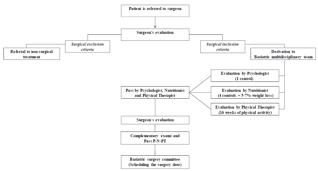
Figure 1: Diagram flow of the patients’ selection to bariatric surgery procedures. The patients’ selection to bariatric surgery procedures. Pass Psychologist = Pass P, Nutritionist = Pass N, Physical Therapist=Pass PT.

652 patients were operated on between January 1, 2000, to December 31, 2018, 3 adjustable gastric band, 5 deceased patients, and 19 patients whose clinical records were lost with the 2010 earthquake were excluded from this study. The remaining 625 patients, who were included, were older than 18 years with a BMI ≥ 35 kg / m2 in presence of at least one comorbidity, or BMI ≥ 40 kg / m2 without comorbidity, and who had undergone of RYGB or SG as the primary intervention. All patients had to be evaluated by the hospital’s BC multidisciplinary team, composed of bariatric surgeons, nurses, physical therapists, nutritionists, and psychologists (Figure 1).

Surgical Procedures Type

RYGB: For this procedure, a 5 cm gastric pouch with a 40-cc capacity was made with a 60 mm x 3.5 mm purple endo linear cutting stapler, between the 2nd and 3rd vessels of the lesser curvature, with 3 refills and 110 cm digestive loop and 150 cm biliopancreatic loop were used. SG: For this procedure, the greater curvature was dissected with a ligature from 4 cm proximal to the pylorus to the Hiss angle, and then the 36 F-calibration probe was installed under direct vision of the lesser curvature. Finally, an EndoGia 60 stapler was used for the section (5 purple loads). The Medtronic® kit was used for both procedures.

Variables

A database created included 46 variables referring to sociodemographic such as age, gender, occupation, and marital status: and the anthropometric measurements body weight, and BMI. The principal comorbidities like arterial hypertension, diabetes mellitus, insulin resistance, hypothyroidism, dyslipidemia, skeletal muscle diseases, obstructive sleep apnea, and gastroesophageal reflux, were included. Mental health including anxiety disorder, depression, bipolar disorder, and panic disorder were evaluated. Lifestyle habits such as alcohol, tobacco, and physical inactivity were also included. In the case of clinical parameters: pre-surgical exam results of upper gastrointestinal tract endoscopy and pulmonary evaluation by forced spirometry and rest EKG were evaluated. The number of RYGB and SG procedures between 2000 to 2018 years. Number of cholecystectomies during the surgical procedure and post-surgical complications. This study was approved by the Department of Teaching and Research of the San Juan de Dios Hospital of Curicó, complying with the criteria indicated in the Helsinki Declaration.

Data Analysis

Qualitative variables are presented as frequencies and percentages. The quantitative variables are presented as means, with standard deviations. It found that 34 were missing data, which were eliminated. The data observed mean in the case of continuous variables and use of the mode in the case of qualitative variables. To statistical analysis the data, the software SPSS version 2.4 was used.

Results

A total of 625 patients met the eligibility criteria of the study. SG was the most frequently reported procedure (85.6%) followed by RYGB (Table 1). Participants mean age was 39 years old and were predominantly female (85.9%). Most of the patients were homemakers (45.8%), professionals (21.3%), office workers (18.6%), 9.6% were students and 4.7% declared other kinds of occupation. Regarding marital status, 60.5% was married, 29% single, 6.1% divorced, 1.1% widows and 3.2% declared other. Regarding the anthropometric measurements, the mean of preoperative body weight was 110.6 kg and the mean of BMI was 42.2 kg / m². Most of the patients (87%) had a BMI ranged from <40 kg / m² to 49.9 kg / m², 11.4% 50 to 59.9 kg / m² and 1.6% have 60 kg / m² or more. The most prevalent comorbidities were hypertension (37.3%), type 2 diabetes (23.6%), insulin resistance (17.9%), hypothyroidism (17.5%), dyslipidemia (7.6%), the skeletal muscle diseases (3.3%), obstructive sleep apnoea (1%) and finally gastroesophageal reflux (0.6%). Regarding the total number of associated comorbidities, most of the patients (42.7%) did not present comorbidities at the time of surgery, 29.3% presented with one comorbidity, 17.6% presented with two comorbidities, 7.5% presented with three and 2.9% presented with four or more comorbidities.

biomedres-openaccess-journal-bjstr

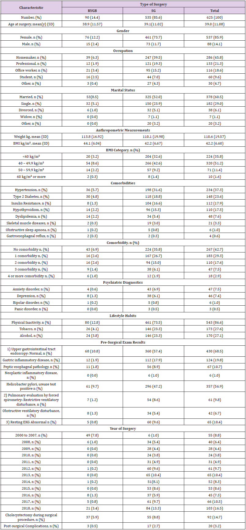
Table 1: Baseline summary of BARCO patients.

Between the psychiatric diagnostic, the most prevalent were anxiety disorder (7.5%) and depression (7.4%), followed by bipolar disorder (1%) and panic disorder (0.5%). During the clinical interview, the patients declared about their lifestyle habits, 86.6% were sedentary, 27.6% smoked tobacco and 27.1% drank alcohol. The pre-surgical exam results indicated that 68.5% of patients presented with a normal upper gastrointestinal tract endoscopy, 19.8% gastric inflammatory pathology, 10.7% peptic esophageal pathology, and 1% presented neoplastic inflammatory disease. Notably, 56.9% of patients presented with Helicobacter pylori (H. pylori), positive urease test. Pulmonary evaluation by forced spirometry indicated that 9.8% presented with restrictive ventilatory disturbance and 6.7% obstructive ventilatory disturbance. Regarding the rest EKG, 10.4% of patients presented abnormal results.

The number of bariatric procedures progressively increased, from 55 cases between 2000 and 2007, and over 100 cases in 2018. Regarding the type of surgery performed per year, between 2000 and 2007, RYGB was the most frequent procedure (49 of 55), however from 2008 to 2018, SG was the most reported procedure (528 of 568) used. During surgical procedures, 92 patients underwent a cholecystectomy, of which 55 were SG and 37 RYGB. Finally, the percentage of post-surgical complications was 3.2%, of which 2.7% corresponded to SG and 0.5% to RYGB. No patients died during the procedures (Table 1). The trends used from 2000 through 2018 are summarized by procedure types in Figure 2. The number of RYGB was higher compared to SG procedures between the years 2000 and 2008, and from 2008 onwards, the number of RYGB decreased and SG procedures had a significant increase. BMI distribution by bariatric procedure types is summarized in Figure 3. The mean BMI was 35 to 65 m/kg2 between both bariatric procedures, however, some patients operated with SG presented a BMI of over 60 m/kg2.

biomedres-openaccess-journal-bjstr

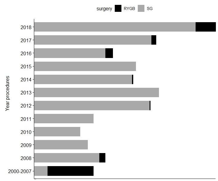
Figure 2: Bariatric surgery utilization trends over time, by procedures types.

Distribution of the number of bariatric surgery procedures from years 2000 to 2018. Roux-in-Y gastric bypass (RYGB) is show by the dark gray bars, and Sleeve gastrectomy (SG) by the light gray.

biomedres-openaccess-journal-bjstr

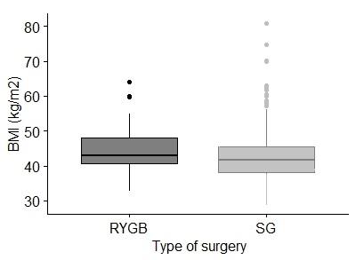
Figure 3: Body mass index by bariatric procedure types.

Box-and-whisker plots of body mass index (BMI) by surgery procedure types. Roux-in-Y gastric bypass (RYGB) is shown by the dark gray box and sleeve gastrectomy (SG) by the light gray box. The lines through the middle of the boxes are the means. The tops and bottoms of the boxes are the standard deviations.

Discussion

According to recent OECD data, Chile, a developing country, tops its list of obesity and overweight (74%), surpassing Mexico (72.5%) and the United States (71%) [4]. These data are reaffirmed with the latest National Health Survey, which shows an important increase of the obesity and morbid obesity from 21.9 % to 25.1 % and from 1.3 % to 2.3%, respectively, in the last 7 years [5,12]. Currently, it is estimated that in Chile there are about 500.000 morbidly obese people, who are eventual candidates for BC, an amount that according to projections is increasing. Although, the National Health Fund has annual coverage of 400 BC per year, it is insufficient considering the amount of morbidly obese people that currently exist in the country. It must be considered that, because BC is not routinely indicated for obese patients, the option of performing a SG versus RYGB was made according to a multidisciplinary team evaluation, considering comorbidities such as hypertension or type 2 diabetes. Effectively, as expected, SG patients had a higher prevalence of high-risk metabolic diseases as hypertension, type 2 diabetes, and insulin resistance. They also had higher rate of hypothyroidism, dyslipidemia and skeletal muscle diseases before surgery. As observed in this study, SG is the type of BC most performed followed by RYBG procedures, in public and private hospitals both in Chile [13] and in other countries [14,15].

However, in a recent article, Sun et al., indicated that RYGB was the BC type most frequently reported procedure in England followed by SG [16]. Despite these differences, it is important to note that both techniques are effective in the remission of the more prevalent comorbidities of the morbid obese, like type 2 diabetes, insulin resistance and hypertension [17,18]. In this study, it was observed that most of patients were female, which is consistent with other studies that point to a higher prevalence of surgical treatment for obesity among women [19]. This difference could be associated with the fact that in Chile, as well as in other countries such as the United States and Canada, the prevalence of obesity is higher in women than in men, and that women seek more health services compared to men, mainly because they have a greater concern for their health [20,21]. It can be assumed that this higher prevalence is due to the search for an ideal beauty and positive attributes, both associated with thinness, deriving from the many social demands that fall on women [22,23], although specific reasons for this disparity remain unclear. Considering the characteristics of the sample, it was observed that the majority ages of bariatric patients fluctuate between 39 to 46 years, are married or single, and BMI > 40 kg/m2, like other studies [13,21].

Most patients did not present comorbidities prior to surgery, and the prevalent comorbidities in patients are the same as those observed in other studies, type 2 diabetes, hypertension, insulin resistance, hypothyroidism, dyslipidemia and skeletal muscle diseases [21,24]. Interestingly, a higher prevalence of H.pylori (56%) diagnosed prior to surgery was observed, compared with similar studies that report 37.1% [25], and 22% [26] respectively. This difference may be due to the fact that the H. pylori infection is prevalent in 73% of the general Chilean population [27] and the social economic insecurity the patients face. However, the relationship between H.pylori and obesity is controversial, so further studies are needed in this area. It is known that obesity is a multifactorial disease that places individuals at risk for additional health compromising conditions such as biological, social consequences, psychological and psychopathology including depression and anxiety. In this study, it was observed that 93% of patients presented with psychiatric disorder including depression (47%) and anxiety (46%), higher than describe previously in another study of veteran women 60% (28% depression and 32% anxiety) [28].

One can also think that this higher prevalence is justified because most of patients were rural women, homemakers and sedentary. Therefore, it will be very interesting analyze the impact of BC procedure in these patients. Finally, none of the patients that died during the procedures were registered and only 3.2 % presented with immediate post-operative complications, which offers a satisfactory outcome when compared with other studies showing 5 to 10% [29]. These results obtained in the present study demonstrate that RYGB and SG BC procedures are feasible to be carried out in a public regional hospital with results comparable to those obtained in both public and private hospitals at the national and international levels. Although beyond the type of surgery applied, the therapeutic goals of surgery for morbid obesity are to improve quality of life and prolong life by counteracting the lifeshortening effect of obesity and its comorbidities, the future and ongoing investigations will provide evidence on the long-term benefits and risks of these most used bariatric procedures in current clinical practice.

This study presents some limitations, such as related to the external validity of our findings, because BARCO represents rural populations therefore, some results may not be applicable to residents of large urban areas. Here we report the baseline characteristics of BARCO´s patients, to provide a basic description of the sociodemographic, anthropometric measurements, principal comorbidities, mental health, lifestyle habits, clinical, and postsurgical complications of patients undergoing BC. In conclusion, given that BARCO is the largest cohort of the public health system patients operated of BC in Chile, these longitudinal data could be used to evaluate the effectiveness of the Chilean public policies related with the BC to treat the morbid obesity. Future and ongoing research will provide evidence on the long-term benefits, risks, and effectiveness of these bariatric procedures in reducing cardiometabolic morbidity, mortality related to obesity, and maintaining long-term weight loss. So, among the findings, the presence of Helicobacter pylori and the high prevalence of psychiatric disorders stand out as relevant factors for investigation in future studies.

For More Articles: Biomedical Journal Impact Factor: https://biomedres.us

Open Access Journals on Behaviorial Medicine

Dynamics of P Under Redox Conditions in Rice in Tropical Soils

Introduction

Rice is one of the main foods in the world, due to its nutritional content and because of the ease of handling and adaptation in different parts of the world, especially in tropical and subtropical conditions [1]. On the other hand, this crop is established under different types of management, depending on the genetic material to be managed, the type of tillage, agronomic management, fertilization plans and irrigation management [1]. Among the types of risks to be managed in rice cultivation, there is the condition of constant sheet or flooding, which has the advantage of properly managing weeds, avoiding stress in areas with high temperatures, reducing specific phytosanitary problems in each region; however, soil fertility is affected by the high and low availability of some nutrients [2,3]. Among the elements that are affected by its nutritional dynamics is Phosphorus (P), an element determined as a macronutrient and which is required to fulfill specific functions of the plant related to the production of energy (ATP) [4], in addition to the importance it has for the production of tropical agricultural systems [5].

The behavior of this element is not the same when humidity is present at field capacity, as when flood irrigation is used in anoxic conditions, this, due to the fact that the concentrations of O2 in the soil are affected, the pH and pH are also affected. the dynamics of microorganisms that affect the available forms of P, as well as the concentration of elements such as Iron (Fe+2) that is found in the vast majority of tropical and subtropical soils in the form of minerals of high or low crystallinity. which directly influences the availability of P and the adsorption and desorption processes, in its Fe-P relationship, especially in acid soils where Fe is found in high concentrations and closely related to the oxidereduction conditions known as Redox conditions, which they are subject to the activity of electrons, pH and concentration of O2 in the soil; However, for this availability of P to exist, there are other parameters that must be present in the soil, such as high fertility defined by the cation exchange capacity (CEC), organic matter (OM) and especially dissolved organic carbon (DOC ) and a texture with high clay content [6,7], all this is described later as also in (Figure 1).

biomedres-openaccess-journal-bjstr

Figure 1: Dynamics of P in acidic soils in Redox processes in rice cultivation. Created with BioRender.com.

Relationship of pH and Eh in the availability of P

Is important to establish that flood periods do not generate immediate changes in pH, but can vary from a few to several weeks, among the factors that can establish changes in pH in flooded soils are: concentration of organic matter (dissolved organic carbon), Microbial activity, temperature, Fe concentration, ammonium accumulation, among other chemical properties of the soil [8,9]. Conditions such as pH and Eh are indirectly responsible for the release of P under reduced soil conditions [10,11]. After there is reduction in the soil by agronomic management such as flooding, regardless of the pH, which generally in tropical soils is very strongly acidic (<4.5) in soils of the orders Oxisol and ultisol, it has a tendency to rise until reaching the agronomic neutrality (6.5-6.8) [12,13], this is due to the fact that processes of reduction of MnO2 and Fe2O3 occur, to the production of OH- as a result of the mineralization of organic N to NH4 + through the process of the In the same way, the increase in pH can also be attributed to the rapid microbial mineralization of labile Carbon compounds (C), generating a rapid decarboxylation of organic anions and in addition to the ammonification as already mentioned above [14].

On the other hand, there is also a decrease in the redox potential (Eh), which is defined as a measure of the reduction state because it changes with the Oxidation / Reduction ratio; Eh is affected by the formation of complex ions and by pH [15]; It can be said that Eh decreases when the pH increases, therefore, the order of the compounds in which they are reduced in flood conditions in rice can be defined, which is described below: NO3 – = MnO2> Fe2O3 (Eh Low). In a soil with high Fe2O3 content such as oxisols, Eh remains an average of + 100 mV, however, in soils with little Fe2O3, Eh can range from -100 to -400 mV where the reduction of SO4 -2 to S-2 [16,17]. In the end, there is a correlation between pH and availability of P, studies carried out by [11] established that the increase in pH in acid soils (Oxisol and Ultisol), under flood conditions in rice, which indicated that these increases in pH, facilitated the Fe (III) reduction and associated P mobilization in the acid soils evaluated [18,19]

Dynamics of P and Fe Concentration

Lower Eh initial conditions in acidic soils such as oxisols [11], can result in a strong decrease in the sorption force of Fe-OP, leading to a strong increase in the P available in this type of soils [20,21]. In relation to this, it can be said that a reduction of Fe is more likely to occur at a slightly acidic pH, when the increase in pH is occurring in reducing environments [22]. It is important to bear in mind that potentially reducible Fe can be established, through methodologies such as the determination of Fe with Citrate-Ascorbate (FeCA), which indicates that the total concentrations of Fe (III) and recalcitrant Fe (Easily reducible) control the production. Fe (II) in acid soils such as Oxisols and Ultisols [11,23]. It is important to note that Fe (II) has a lower binding force for P than Fe (III) [24]; therefore soils with higher potentials to reduce Fe, an early increase and a higher bioavailability of P can be expected [22]. The reducing dissolution of crystalline Fe (FeCA, defines Fe and low crystallinity and potentially available and reducible Fe) can be catalyzed by a high concentration of Fe (II) [25,26], resulting in a higher initial availability of P in acid soils [11,27]. In short, the increase in the availability of P in flood conditions has to do with the reduction of ferric phosphates (Fe+3) to ferrous phosphates (Fe+2), to the release of P from insoluble components of Fe and Al and to a certain dissolution of Ca phosphates when there are high levels of CO2 in the soil solution (soils with alkaline pH). The release of P through these processes can take a few weeks after the flood. This initial flux of released P can be fixed on clay particles and Al hydroxides (AlOOH), and in some soils with high amounts of active Fe and Al it can still result in a reduction in the availability of P in the soil [28,29].

Organic Matter and P Concentration

Labile organic acids are more efficient to solubilize P from acidic soils, this because there is a greater solubility of Fe and Aluminum (Al) phosphates, with increasing pH and low Eh (Reference). Therefore, a decrease in Eh, followed by a reduction in Fe (III), can generate the release of P bound in Fe minerals; The reduction of Fe (III) and the corresponding accumulation of Fe (II), strongly depend on the labile mineralization of C and OM. Labile C can define the intensity of Fe (III) reduction because it is the main source of energy for microorganisms, in addition to being a strong electron donor [30]. These processes result in the production of Fe (II) but in the same way a desorption producing available P; therefore, higher OC and Fe (II) content in soils such as oxisols, generates a drastic decrease in Eh after immersion [11], which leads to a greater consumption of microbial O2 much faster, which can generate the release of large concentrations of P low binding energy (Reference). It is important to bear in mind that microbial mineralization with rapid activities due to the presence of labile C in acid soils can reduce P sorption, a specific adsorption analogous to phosphate ions [31] or by Fe and Al chelation [32].

Conclusion

It is important for the researcher, extension worker, professor in the area of agriculture, to understand the dynamics of P in systems where Redox processes are present, since for the producer it will serve as an orientation not to carry out high fertilization of phosphate fertilizers, which can lead to high processes of contamination by eutrophication, cost overruns, currently necessary due to the costs of fertilizers that have doubled and tripled their price in the market, in addition to the need to carry out calibrations in the determination of P in the soil, since in the tropics There is determination of P by the method of Bray II (Colombia), Mehlich 3 (Brazil – Minas Gerais), Resina (Brazil – São Paulo and Rio Grande do Sul), generating ambiguity by establishing non-standardized methodologies in various regions of the tropics and by Finally, in addition to values such as pH in the soil analysis, one should work on the fractionation of P as results in the soil analyzes for all The agricultural professional (Agronomist Engineer, Agroecologist Engineer, Agroforestry Engineer, etc.) who is the person in charge of the recommendation, with this, the forms of P, labile, moderately labile and nonlabile are determined to make a much clearer and more accurate recommendation.

For More Articles: Biomedical Journal Impact Factor: https://biomedres.us

Open Access Journals on Behavioural Medicine

Wearable Self-Powered Sensors for Health Care, 5G, Energy Harvesting, and IOT Systems

Introduction

Basic theory and design of small, printed antennas is presented in [1]. However, the efficiency of small antennas is low, [2-4]. Compact printed metamaterials antennas and sensors are used in wireless communication systems and were discussed and presented in several publications in this century, [2-6]. Printed dipoles, FIPA and loop antennas, printed Slots, microstrip antennas, and other compact antennas are employed in radars, Internet of Things (IoT), 5G, and healthcare systems [2-6]. Several types of small efficient wideband wearable antennas are presented in, [2- 4]. Metamaterials are materials with periodic artificial structures. The metamaterial elements and structure define the electrical properties of the material. Metallic posts structures and periodic split ring resonators (SRRs) may be employed to produce structures with required permeability and dielectric constant as discussed in [7–11]. Metamaterials may be employed to develop efficient sensors for communication, wearable healthcare and IoT devices, [12-16]. In [6] the metamaterial antenna gain, and bandwidth are similar, to those of microstrip antennas. In [8] materials with negative dielectric permittivity are evaluated. In [9], a model and setup to simulate and measure the polarity of SRRs is presented.

The model is used to compare measured results to computed results. In [12] a transmission-line metamaterial antenna with two transmission line arms that resonate at two different frequencies, is presented. The antenna bandwidth is 3% with 2.6dBi directivity. The radiation efficiency is 65% at 3.3 GHz. The antenna gain is around 0.8dBi. Compact radiators such as printed loops and dipoles, patches, and FIPA antennas suffer from low efficiency [2- 5,16-28]. These antennas are linear polarized. Compact efficient metamaterial antennas may be important element in wearable medical and energy harvesting systems. In several 5G, energy harvesting, and medical systems, the receiving signal polarization may be horizontal, elliptical, or vertical polarized. In these cases, the antenna should be dual polarized sensors. Small efficient wearable metamaterials antennas for medical systems are presented in [15- 16]. Wearable antenna measurements in vicinity of human body are presented in [17]. Active wearable antennas for communication and medical systems are presented in [19]. Wearable healthcare devices are used to increase disease cure and prevention. Efficient medical care may be achieved by online evaluation of a continuous measured medical data of patients.

Healthcare remote monitoring system in hospitals is presented in [29]. A wireless wearable device with thermal-aware protocol is discussed in [30]. In [31-40] wearable sensors and antennas for medical applications are presented. Wearable medical sensors can monitor and check patient daily health [34-35]. Dual polarized dipole wearable antenna for medical applications is presented in [39]. In this article, metamaterials technology is used to design high efficiency sensors and antennas with harvesting energy unit for healthcare, 5G, and IoT devices. Dual polarized metamaterial antennas have significant advantages over regular printed antennas. Such as, high efficiency and gain. The antennas bandwidth is around 40%, for VSWR, better than 3:1. The gain of the antennas with CSRRs is around 7dB. The sensors efficiency is higher than 90%. The energy harvesting units connected to the sensors provides selfpowered efficient and compact sensors.

Wearable Technologies and Devices for Medical, 5G, IoT, and Sport Applications

Wireless Body Area Networks, WBANs, can measure and record several healthcare parameters such as body temperature, blood pressure, heartbeat rate, electrocardiograms, arterial blood pressure, sweat rate, and electro-dermal activity. Wearable devices will be in the next decade an important part of individuals’ daily lives. Wearable sensors may provide scanning and sensing features that are not offered by mobile phones and laptop computers. Wearable devices usually have communication links and users may have access to online information. Wireless technologies are used to process and analyze the data collected by the medical system. The collected data may be stored or transmitted to a medical center to analyze the collected data. Wearable sensors gather data which is analyzed by medical software. This analysis may send alert to the physician to contact a patient who needs urgent healthcare treatment.

Applications of Wearable Medical Systems

1. Wearable Medical devices help to monitor medical centers activities and accessories.

2. Wearable devices can help to operate and monitor companies’ activities and accessories.

3. Wearable Medical devices can assist several patients such as Diabetes patients, Asthma patients, Epilepsy patient, and Alzheimer’s disease patients.

4. Wearable devices can help to solve Sleep disorders, Obesity problems, cardiovascular diseases.

5. Wearable Medical devices help to gather data for clinical research trials and studies.

Several physiological parameters can be measured by using wearable medical devices and sensors. Each patient may have a wearable device attached to the body. The wearable device is connected to several sensors. Each sensor perform a specific task. The sensors measure heart rate, body temperature and blood pressure. Doctors may carry a wearable device, which allows other hospital personal to contact and locate them.

Measurements of Blood Pressure and Heart Rate

Usually, blood pressure and heartbeat are measured in the same set of measurements. The Blood pressure and heartbeat may be transmitted to a medical center and if needed the doctor may contact the patient for further assistant. Blood pressure indicate what is the arterial pressure of the blood circulating in the patient body. Some of the causes of high blood pressure may be obesity and stress. Blood pressure of a healthy person is around 70 by 120. Where the diastole is 70 and the systole is 120. The heart rate of a healthy person is 72 times per minutes. Changes in heartbeat can cause several cardiovascular diseases. A change in the heart rate change the blood pressure and the amount of blood delivered to the patient body. To measure and analyze the heart beat a wearable medical sensor can be connected to patient chest.

Measurements of Respiration Rate

Elderly, sick, and overweight people have difficulties in breathing normally. Measurements of respiration rate indicates if the patient is healthy and breathe normally. The measured respiration rate may be transmitted to a healthcare center and if needed the doctor may contact the patient for further assistant. It is better to use a wearable wireless medical device to measure accurate respiration rate.

Measurements of Human Body Temperature

Temperatures below 35˚C or above 38˚C can indicates that the person is sick and temperatures above 40˚C may cause death. The human body temperature may be transmitted to a medical center and if needed the doctor may contact the patient for further assistant.

Measurements of Sweat Rate

Measurements of sweat rate and PH can be used to monitor the physical activity of a person. A wearable medical device may be used to measure the sweat rate of a person when extensive physical activity is done. The wearable device may be attached to the person clothes close to the skin to measure the sweat rate. The device may be used to measure the sweat PH to analyze the patient health. When a person does extensive physical activity, glucose come out of the skin as a sweat. Sweat is a mix of water, glucose and minerals. Glucose is supplied to the body usually by sugar that is a monosaccharide that provide energy to the human body. The sweat evaporated from the skin is absorbed in the medical device to determine the sweat PH. If the amount of sweat coming out of the body is too high the body may dehydrate. Dehydration causes tiredness and fatigue. The patient sweat rate can be transmitted to a medical center and if needed the doctor will contact the patient for further assistant.

Measurements of Human Gait

Human gaits are the various ways in which a human can move. Walking, jogging, skipping and sprinting are natural human gait. Different gait patterns are characterized by differences in limb movement patterns, overall velocity, kinetic and potential energy cycles, forces, and changes in the contact with the ground. Gait measurements and analysis are fundamental research tool to characterize human locomotion. Wearable devices may be located at different parts of the human body to measure and analyze human gait. The movement signal recorded by these devices are used to analyze human gait. In sports, gait analysis based on wearable sensors can be used for sport training and analysis and for the improvement of athlete performance. The ambulatory gait analysis results may determine whether or not a particular treatment is appropriate for a patient. Parkinson’s disease is characterized by motor difficulties, such as gait difficulty, slowing of movement and limb rigidity. Gait analysis has been verified as one of the most reliable diagnostic signs of this disease. For patients with neurological problems, such as Parkinson’s disease and stroke, the ambulatory gait analysis is an important tool in their recovery process and can provide low-cost and convenient rehabilitation monitoring.

In healthcare centers, gait information is used to detect walking behavior abnormalities that may predict health problems or the progression of neurodegenerative diseases. Fall is the most common type of home accident among elderly people. Fall is a major threat to health and independence among elderly people. Gait analysis using wearable devices was used to analyze and predict fall among elderly patients. IoT systems are wireless communication devices of interrelated computing networks, personal devices, digital machines, mechanical machines, and medical sensors that have a unique identifiers UIDs).

IoT system consists of modules that use communication systems, sensors, processors, and antennas. IoT systems receive, transmit and process information received from their environments that are connected to the internet web. IoT devices are connected to an IoT gateway where the collected information is processed online or sent to data centers to be diagnosed and shared with other IoT devices. In several IoT and medical devices the polarization is not defined. In these cases, the antenna should be dual polarized. IoT has an important role in future medical centers and hospitals. IoT devices may connect several medical devices and medical data centers to improve medical treatment and to provide low-cost medical treatment.

1. Wearable sensors and IoT devices are used to automate processes, to reduce company and medical centers hardware and to reduce labor costs.

2. IoT antennas and devices can transfer information through a network without requiring human to computer interaction or human to human interaction.

3. IoT devices may have a complete control over routine services and tasks in medical centers and companies, helps people everyday life, and to work smarter.

4. IoT devices provides companies with an online observation how the company systems operate.

An application of WBANs in medical centers where medical parameters of large number of patients are constantly being monitored is presented in (Figure 1). A block diagram of IoT medical device is presented in (Figure 2).

biomedres-openaccess-journal-bjstr

Figure 1: Wearable Wireless Body Area Network, WWBAN, Health Monitoring System.

biomedres-openaccess-journal-bjstr

Figure 2: IoT Medical System block diagram.

IOT Major Disadvantages

As the number of connected devices increases and more information is shared between devices, the potential that a hacker could steal confidential information also increases.

1. If there’s a bug in the system, it’s likely that every connected device will become corrupted.

Mobile Networks Features

Cell phones began with 1G technology in the 1980s. 1G is the first generation of wireless cellular technology. First generation of mobile networks were reliant upon analog radio systems. In 2020 5G technology was developed and is used in cellular phones. A comparison of cellular phone technologies is presented in (Table 1).

biomedres-openaccess-journal-bjstr

Table 1: Comparison of cellular phone technologies.

5g Main Features

It is highly supportable to Wireless World Wide Web.

1. High speed, high capacity.

2. Provides large broadcasting of data in Gbps.

3. Multi-media newspapers, watch TV programs with the HD Clarity.

4. Faster data transmission than that of the previous generation.

5. Large phone memory, dialing speed, clarity in audio and video

6. 5G technology offer high resolution for cell phone user and bi- directional large bandwidth sharing.

Dual Polarized Wearable Passive and Active Sensor with Energy Harvesting Unit

A dual polarized metamaterial antenna with CSRR, metallic strips, and with energy harvesting unit is shown in (Figure 3a). The antenna consists of two layers with thickness of 0.16cm. The dipole matching network and the metallic strips are printed on the first layer. The radiating dipole with CSRR is etched on the second layer. The wearable antenna, with the matching network and the energy harvesting unit, dimensions are 21x4cm. The dipole with CSRR is horizontal polarized. The slot antenna is vertical polarized. The antenna is a dual polarized antenna. The resonant frequency of the antenna without CSRR is around 400MHz. The resonant frequency of the antenna with CSRR is around 330MHz. Several medical devices operate in the frequency range between to 200MHz to 500MHz. The computed S11 and antenna gain are presented in (Figure 4). The measured antenna bandwidth is around 50% for VSWR better than 3:1. The antenna radiates is in the z axis direction. The measured directivity and gain of the antenna with CSRR are around 5.5dBi as shown in (Figure 5). The feed network of the antenna in (Figure 3a) was optimized, see (Figure 3b), to yield VSWR better than 3:1 in frequency range of 180MHz to 400MHz as shown in (Figure 6). The antenna bandwidth is around 60% for VSWR better than 3:1.

biomedres-openaccess-journal-bjstr

Figure 3:

a. Dual polarized antenna with Metallic strips and CSRR with energy harvesting unit.

b. Optimized antenna with CSSR and metallic strips.

biomedres-openaccess-journal-bjstr

Figure 4: Gain and S11 of the dual polarized antenna with metallic strips and CSRR.

biomedres-openaccess-journal-bjstr

Figure 5: Radiation pattern and Gain of the antenna with metallic strips and CSRR.

biomedres-openaccess-journal-bjstr

Figure 6: S11 of the optimized dual polarized antenna with metallic strips and CSRR on human body.

(Figure 3a). Dual polarized antenna with Metallic strips and CSRR with energy harvesting unit b.Optimized antenna with CSSR and metallic strips. The dual mode energy harvesting unit is connected to the dipole and the slot feed line, see (Figure 3). The dual mode energy harvesting unit may charge the battery when the switch is connected to the harvesting module. RF AC energy is converted to direct current by using a rectifying diode. The rectifier can be a half-wave or a full-wave rectifier. The harvesting unit consists of an antenna, a rectifying diode, and a rechargeable battery, see (Figure 3).

Dual Polarized Wearable Active Receiving Sensor with Energy Harvesting Unit

The layout of a receiving active antenna with an energy harvesting unit is shown in (Figure 7). The harvesting energy unit operates as a dual mode RF harvesting system. The LNA may be connected to the harvesting unit via the switch to charge the battery. A matching network match the antenna to the Low Noise Amplifier, LNA. The TAV541 LNA, is a high linear PHMET gain block amplifier. At 1.9 GHz, the amplifier has 18dB gain and 0.5dB Noise Figure. The LNA output P1dB is 20dBm. The LNA specifications are listed in (Table 2). An output matching network match the LNA to the receiver. A DC bias network supply the required voltages to the amplifiers. The sensor dimensions are around 21x5x2cm. S11 and gain of the dual polarized antenna with CSRR and matching network is shown in (Figure 8). The active receiving metamaterial sensor gain is 12+3dB from 100MHz to 1GHz as shown in (Figure 9). The sensor noise Figure for frequencies from 100MHz to 1GHz is better than 1dB. The active receiving dual polarized sensor was evaluated with Triquint TQP3M9028 LNA. The LNA specifications are listed in (Table 2). The active receiving metamaterial sensor gain with TQP3M9028 LNA is 11+2.5dB from 150MHz to 0.9GHz as shown in (Figure 10). The sensor, with TQP3M9028 LNA, noise Figure for frequencies from 150MHz to 1GHz is better than 2dB. Comparison of measured performance of the sensors with different LNAs is given in (Table 3). The sensor with (Figure 8) LNA TAV541 has better noise Figure and higher gain. However, sensor with LNA TQP3M9028 has better gain flatness and 1dBc compression point.

biomedres-openaccess-journal-bjstr

Figure 7: Dual polarized receiving sensor with CSRR and with energy harvesting unit.

biomedres-openaccess-journal-bjstr

Figure 8: S11 and gain of the dual polarized antenna with CSRR and matching network.

biomedres-openaccess-journal-bjstr

Figure 9: Active receiving dual polarized receiving sensor gain, with LNA.

biomedres-openaccess-journal-bjstr

Figure 10: Active receiving dual polarized receiving sensor gain, with TQP3M9028 LNA.

biomedres-openaccess-journal-bjstr

Table 2: Comparison of the Specification of the S band Low Noise Amplifiers.

biomedres-openaccess-journal-bjstr

Table 3: Comparison of the Sensors measured performance with different LNA Amplifiers.

Dual Polarized Wearable Active Transmitting Sensor with Energy Harvesting Unit

The layout of a transmitting active antenna with an energy harvesting unit is shown in (Figure 11). The harvesting energy unit operates as a dual mode RF harvesting system. The harvesting unit can be part of a medical, IOT, and smartphone. The harvesting unit will charge the battery when the control unit will connect the switch to the harvesting unit input port. The radiating element is connected to the HPA via the switch to an output HPA matching network. The matching network match the metamaterial antenna to the HPA. Two amplifiers were employed to design the metamaterial sensor. The first amplifier is a HPA MMIC GaAs MESFET VNA25, The second amplifier is a HPA MMIC GaAs PHEMT HMC459. The amplifiers specification is listed in (Table 4). The active transmitting dual polarized antenna S11 parameters, computed and measured, is better than 3:1 in the frequency range from 250 to 450MHz. The antenna gain, computed and measured, is around 6dBi as shown in (Figure 12). The active sensor gain with the VNA25 HPA, computed and measured, is 13 ± 3dB for frequencies ranging from 100 to 800MHz. The active transmitting sensor S21 parameter, gain presented in (Figure 13). The active sensor gain with the HMC459 HPA, computed and measured, is 12±4dB for frequencies ranging from 0.1 to 1GHz. The active transmitting sensor S21 parameter, gain presented in (Figure 14).

The active transmitting dual polarized antenna output power is around 19dBm. Comparison of measured performance of the sensors with different HPAs is given in (Table 5). The transmitting sensor with VNA25 HPA has higher gain, better gain flatness, and lower DC power consumption. However, the transmitting sensor with HMC459 HPA has higher 1dBC compression point, higher input and output power. However, HMC459 has higher DC power consumption. Photos of the dual polarized metamaterial antenna with CSSR and metallic strips.are presented in (Figure 15).

biomedres-openaccess-journal-bjstr

Figure 11: Dual polarized transmitting sensor with CSRR and with energy harvesting unit.

biomedres-openaccess-journal-bjstr

Figure 12: Radiation pattern and Gain of the dual polarized antenna with metallic strips and CSRR.

biomedres-openaccess-journal-bjstr

Figure 13: Active transmitting dual polarized sensor gain, with HPA VNA25.

biomedres-openaccess-journal-bjstr

Figure 14: Active transmitting dual polarized sensor gain, with HPA HMC459.

biomedres-openaccess-journal-bjstr

Table 4: Comparison of the Specification of the HPA Amplifiers.

biomedres-openaccess-journal-bjstr

Table 5: Comparison of the Sensors performance with different HPA Amplifiers.

biomedres-openaccess-journal-bjstr

Figure 15: Photos of the dual polarized antenna. a. Feed network b. Radiator with CSSRs c. CSSR.

Wearable Dual Polarized Metamaterial Sensors for 5G, IoT and Medical Systems

The dual polarized antennas and sensors presented in this article may be employed in 5G, healthcare sensors, and IoT devices. The sensors S11 variation near the human body were computed by using the human body and antenna model shown in (Figure 16a). Dielectric constant and conductivity of human body tissues are listed in (Table 6), [16]. The effect of the antenna location on the human body is simulated by evaluating the antenna S11 coefficient on human body. The variation of the dielectric constant of the body tissues affects the electrical performance of the dual polarized sensor. The antenna resonant frequency is shifted up to 9%, in different locations of the sensor on the patient body. As listed in (Table 6) the dielectric constant is 5 at fat tissues, and 45 at the stomach area, and increase to 128 at the Small intestine tissues. The dual polarized sensors may be located inside a belt as presented in (Figure 16b). The belt thickness, and dielectric constant changes the antenna electrical performance. The sensors electrical and mechanical parameters were tuned to achieve the best sensor electrical and mechanical parameters. The sensors electrical performance were computed and measured for air spacing between the sensors and human body up to 20mm at different locations on the human body. Wearable sensors and antenna measurements and setup measurements are presented in [2-3,18]. RF measurements of wearable sensors and antennas are done by using a phantom with sugar, salt and water that represent the dielectric constant and conductivity of human body tissues, [2-3,18].

biomedres-openaccess-journal-bjstr

Figure 16:

(a) Model of Wearable Antenna environment;

(b) Wearable Medical System on human body.

biomedres-openaccess-journal-bjstr

Table 6: Electrical parameters of human body tissues [16,17].

(Table 7) presents a comparison between computed and measured results of sensors without and with CSRR. (Table 8) presents a comparison of computed and measured results of compact wearable antennas. As presented in (Tables 7 & 8) there is a good agreement between measured and computed results. Results presented in (Tables 7 & 8) verifies that the gain of the antennas without CSRR is lower by around 2.5dB than the antennas with CSRR. Electrical performance of passive and active patches and slot antennas, loop, dipoles, and other antennas were given in [2-5]. Smart City, healthcare, WWBAN, and IoT monitoring system with WBAN Networks is presented in (Figure 17). Patients can contact health care center and doctors from any place at any time.

biomedres-openaccess-journal-bjstr

Figure 17: Smart City, healthcare, WWBAN, and IoT monitoring system with WBAN Networks.

biomedres-openaccess-journal-bjstr

Table 7: Electrical performance comparison between wearable antennas without and with CSRR.

biomedres-openaccess-journal-bjstr

Table 8: Comparison of electrical performance of compact wearable printed antennas [2-5,15].

Energy Harvesting Module for Healthcare Sensors, 5G, IoT, and Communication Systems

As shown in (Figure 3) the energy harvesting module consists of compact dual polarized antenna, rectifying circuit, and a rechargeable battery. The energy harvesting modules and the antenna provide a self-powered sensor. The rectifier diode converts electromagnetic energy, AC energy, to direct current (DC energy). Two types of diode rectifiers are usually employed a half wave rectifier or a full wave rectifier, [41-45]. A Half wave rectifier is shown in (Figure 18). A half-wave rectifier converts only the positive voltage half cycle. It allows to harvest only one half of the RF waveform. The efficiency of the half wave rectifier is 40.6%. Only 40.6% of the input electromagnetic energy is converted into DC power and may charge the batteries. A full wave bridge rectifier is presented in (Figure 19). The bridge full wave diode rectifier circuit converts RF energy to DC energy. The bridge rectifier consists of four diodes D1 through D4, as presented in (Figure 19). During the positive half cycle voltage, terminal A will be positive and terminal B will be negative. Diodes D1 and D2 will become forward biased and D3 and D4 will be reversed biased. The rectifier output DC voltage,  
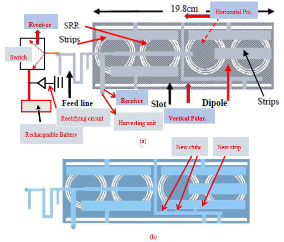
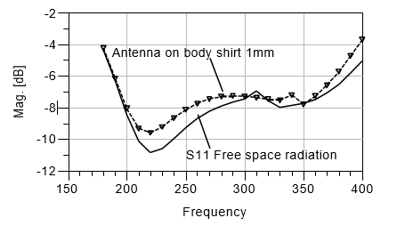
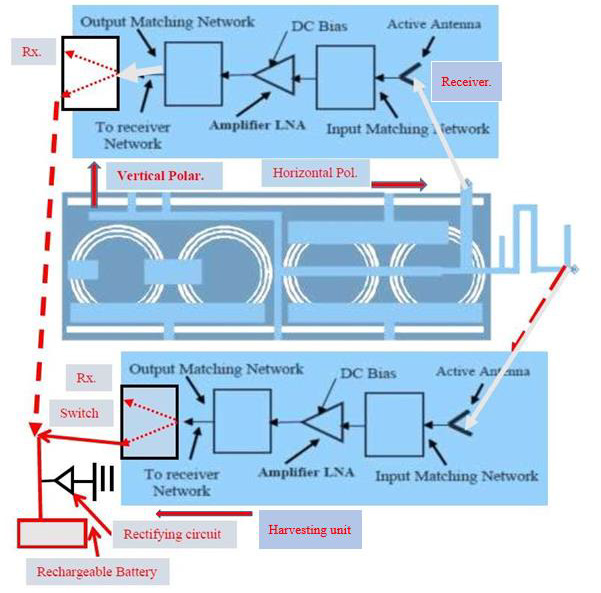
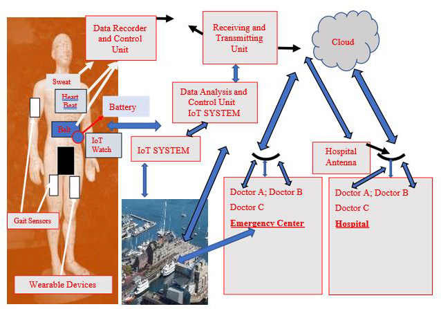
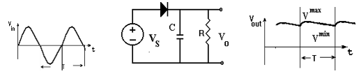
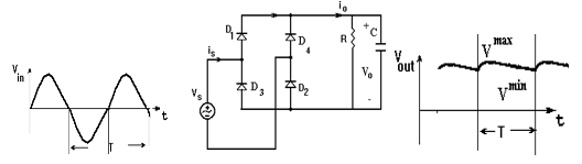
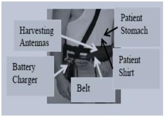
The rectifier output voltage may be improved by connecting a capacitor in shunt to the resistor as presented in (Figure 19). The full wave rectifier efficiency is 81.2%. Energy harvesting systems provide green renewable energy and may eliminate the usage of power cords and the need to replace batteries frequently. Wearable RF System with energy harvesting unit for IoT, 5G, and healthcare devices is presented in (Figure 20). The wearable harvesting module with a compact battery charger is placed on the patient shirt as shown in (Figure 20) [46,47].

biomedres-openaccess-journal-bjstr

Figure 18: Diode Voltage rectifier with a capacitor, half wave.

biomedres-openaccess-journal-bjstr

Figure 19: Diode Bridge voltage rectifier with a capacitor, full wave.

biomedres-openaccess-journal-bjstr

Figure 20: Wearable RF System with Energy Harvesting unit for IoT, 5G, and Healthcare Systems.

In 2021 every person use wireless communication devices, cellular phone, tablet and other communication devices. This result in a huge increase in the amount of electromagnetic energy in the air. The expected amount of radio wave in the air in 2020 was around 50 Exa-bytes, EB, per month. However, the expected amount of radio wave in the air in 2025 is expected to be around 165 Exabytes per month. In electromagnetic energy harvesting systems, the electromagnetic waves propagating in free space may be received by the dual polarized harvesting antennas and converted to electric energy that is used to charge batteries, wearable sensors and other wearable devices. Harvested power amount in malls and stadiums range from 1 μW/cm2 to 5mW/cm2.

Conclusion

The active and passive dual polarized antennas and sensors discussed in this article are compact, wideband, efficient, and lowcost. Energy harvesting unit is connected to the dual polarized sensors. RF waves propagating in free space may be received by the harvesting antennas and converted to DC power that may recharge the medical system batteries, wearable sensors, and other wearable modules. Development of dual polarized efficient active and passive wearable sensors and antennas are one of the most important goals in development of wearable sensors, IoT, 5G and healthcare systems. Wearable passive and active dual polarized antennas may operate in receiving or transmitting BAN, IoT, 5G, and healthcare systems. In receiving sensors, the LNA is an integral module of the receiving sensor. In transmitting sensors, the HPA, is an integral module of the transmitting channel. The output power of the transmitting dual polarized sensor is around 18dBm. Passive and active dual polarized compact sensors performance such as efficiency, bandwidth, noise Figure gain, and radiation pattern were discussed in this article.

The dual polarized metamaterial patches and sensors presented in this research may be employed in wideband wearable communication systems for IoT, 5G, sport, and healthcare applications. Metamaterial technology is used to develop efficient antennas and sensors. The resonant frequency of the dual polarized antennas without CSRR is higher by 5% to 10% than the antennas with CSRR. The directivity and gain of the dual polarized patches with CSRR is higher by 2dB to 3dB than the antennas without CSRR. Electrical computed and measured results of several efficient antennas with and without CSRRs are presented in this paper. The bandwidth of the active dual polarized antennas is around 30% to 50% for S11 lower than -6dB. The dual polarized antenna efficiency, bandwidth, gain and radiation pattern were improved by optimizing the sensor dimensions and feed network. The active receiving and transmitting dual polarized antennas gain is around 14dB. The receiving module noise Figure is around 1Db, with TAV541 LNA. The wearable active and passive dual polarized antennas can be operated as linear polarized sensors.

The dual polarized sensors and antennas discussed in this article may operate in healthcare systems that improves the daily health and the life conditions of patients. Wearable sensors and medical devices seem to be an important choice for medical organizations, medical centers, and patients. Dual polarized wearable devices support the evaluation of personal medical devices with online immediate physician response to cure and improve patients’ health. The energy harvesting units connected to the sensors provides selfpowered autonomous compact sensors. In future work more types of fractal and metamaterial compact antennas and sensors with high efficiency for IoT, and 5G communication systems with energy harvesting units will be developed. In future research metamaterial fractal linear and dual polarized efficient sensors and antennas for wireless communication systems, healthcare, IoT and 5G systems will be developed.

For More Articles: Biomedical Journal Impact Factor: https://biomedres.us

Journals on Microbiology Research

Free-Breathing 10-Min Cardiac MRI Protocol at 3.0t: Single-Center Experience

Introduction

Cardiovascular magnetic resonance (CMR) imaging has become an essential tool for the non-invasive examination of the heart. It has been used for the diagnosis, risk stratification, and prognosis of cardiac diseases [1,2]. Cine and late gadolinium enhancement (LGE) imaging are routinely included in the conventional cardiovascular magnetic resonance (CCMR) protocols in our center. Data acquisitions are typically performed with breath-holding (BH). While they work well in patients that are capable of holding their breath during image acquisition, such CCMR protocols remain challenging in patients with compromised BH capacities. In addition, the relatively long imaging time hinders the efficiency and throughput at a busy medical center like ours where there is a need to scan over 30 cardiac patients per MRI system per day. Real-time compressed sensing (CS) cine has been proved to be able to obtain high-quality images for evaluating cardiac function [3- 7]. Motion corrected (MOCO) single-shot LGE imaging techniques can also produce high-quality images without BH to detect fibrotic myocardial scars [8-9]. The novelty in this work is that both methods (CS cine and MOCO-LGE) are in corporate for a comprehensive FB CMR study. The feasibility and potential clinical utility of the proposed protocol were evaluated in patients that were unable to hold their breath during CMR imaging and in patients who could hold breath well.

Materials and Methods

Subject Enrollment

After the institutional review board approval was granted, adult patients scheduled for CCMR imaging were prospectively recruited for this study from February 1, 2017 to June 30, 2019. The inclusion criteria were as follows: in-patient at our hospital scheduled for contrast-enhanced CMR examination, a glomerular filtration rate of ³30 mL/min per 1.7m2, and no contraindications for CMR imaging. All patients who received FCMR protocols signed informed consent. A total of 148 patients were selected to complete both CCMR and FCMR protocols examination. These patients were divided into two groups,

a) Group 1, 111 patients with good BH;

b) Group 2, 37 patients with bad BH. The average age of these patients was 59 ± 16 years, with a male/female ratio of 90/58.

The CMR Imaging Protocol

CCMR and FCMR scans were performed on a 3 Tesla (T) clinical magnetic resonance imaging (MRI) scanner (MAGNETOM Skyra, Siemens Healthcare, Erlangen, Germany). The system was equipped with an 18-element body array coil and a 32-element spine array coil. Key sequences for the CCMR included:

a) BH-cine imaging with segmented, balanced steady-state free precession (BSSFP) readout;

b) BH-LGE sequence for viability imaging under breath-hold using segmented, fast low-angle shot (FLASH) readout and phase-sensitive inversion recovery (PSIR) reconstruction.

The primary FCMR protocols included:

a) Single-shot FB-CS-cine imaging with BSSFP readout, featuring a two-dimensional sparse data sampling and iterative reconstruction (SSIR).

b) FB-MOCO-LGE employs non-rigid motion-correction and averaging of multiple single-shot SSFP images with PSIR reconstruction.

c) The BH-cine, FB-CS-cine, BH-LGE, and FB-MOCO-LGE protocols comprised separate 2-, 3-, and 4-chamber long-axis (LAX) acquisitions, and a short-axis (SAX) stack covering the entire left ventricle (LV). All scans were started from running BH CCMR protocols, when the CCMR protocols were successfully finished, FCMR protocols were began. Intravenous gadolinium diethylenetriamine pentaacetic acid (Gd-DTPA) contrast agent was administered at a dose of 0.2 ml/kg of body weight. For all exams, the contrast agent was administered to each patient in one injection.

biomedres-openaccess-journal-bjstr

Figure 1: The workflow for the breath-holding (BH) conventional cardiac magnetic resonance imaging (CCMR) protocol and free-breathing cardiac resonance (FCMR) protocol. Abbreviations: HASTE, Half-Fourier-Acquired Single-shot Turbo spin Echo; MOCO, motion-corrected; LGE, late gadolinium enhancement; CS, compressed sensing.

The CMR protocol workflows is illustrated in Figure 1. Detailed information regarding the sequence parameters of both protocols is shown in Table 1. Both protocols were conducted using semiautomated cardiac day optimizing throughput (DOT) engine software including Auto Align feature to automatically prescribe the 2-, 3-, and 4-chamber views as well as the short axis stack [4,10]. Scan parameters like trigger delay were automatically adapted to patient physiology such as patient heart rate. The scanning time of every sequence and the total time of every protocol was write down, every two sequence intervals time was not included in the total time. Acquisition time, IQ were compared between the two protocols. The consistency of the two methods in diagnosis, LVF measurement and scar presence/absence was analyzed.

biomedres-openaccess-journal-bjstr

Table 1: CCMR and FCMR protocols’ imaging parameters.

Image Analysis

Image Quality: All CMR images were transferred to a workstation (cmr42, Version 5.12.1, Circle Cardiovascular Imaging, Calgary, Canada) for evaluations. For those 148 patients where both FCMR and CCMR images were acquired, FCMR and CCMR were randomly assigned to two senior doctors, Reader 1 and Reader 2, both with more than 5 years of experience in CMR reading, for double-blind evaluation respectively. Image quality scores were evaluated based on a five-point Likert score (5 = excellent, 4 = good, 3 = the presence of artifacts but acceptable, 2 = poor, and 1 = non diagnostic) [6-8].

Imaging Analyses: LVF measurements were assessed with cmr42 software. Endocardial and epicardial contours were automatically delineated on the short-axis cine images using the cmr42 software and manually adjusted as needed [11]. Papillary muscles and trabeculations of the left ventricle (LV) were included in the ventricular cavity volume measurements. Ejection fraction (EF), end-diastolic and end-systolic volumes (EDV and ESV), stroke volume (SV), and LV end-diastolic mass (LVEDM) measurements were accessed from the cine images acquired in both protocols, and the consistency of measurements between both protocols was analyzed by using linear regression analyses and Bland-Altman plots. If LGE involves the sub endocardial distribution of coronary artery, it can be identified as ischemic LGE type; otherwise, it can be identified as non-ischemic LGE [12,13].

Statistical Analyses: Statistical analyses were performed using dedicated SPSS (version 20.0, SPSS Inc., Chicago, USA) and MedCalc10.0 (Med Calc Software, Ostend, Belgium) software. Continuous data were checked for normality using the Shapiro-Wilk test and presented as the mean ± standard deviation or median (interquartile range, Q1–Q3), and compared using the T test or Mann-Whitney test. The FB-CS-cine and BH-cine imaging were compared using Bland-Altman. Intra class Correlation Coefficient (ICC) were used to evaluate the consistency of quantitative data. The Kappa statistics were used to evaluate consistency of qualitative variable. Agreement strengths for kappa values were classified as follows: <0.1, poor; 0.1 to 0.20, slight; 0.21 to 0.40, fair; 0.41 to 0.60, moderate; 0.61 to 0.80, substantial; and 0.81 to 1.00, almost perfect. P <0.05 was considered statistically significant.

Results

Image Acquisition Times

The total time of the FB-CS-cine SAX (25±5)s, FB-CS-cine LAX (8±2)s, FB-MOCO-LGE SAX (120±19)s, FB-MOCO-LGE LAX (37±6) s was significantly shorter than that of the BH-cine SAX (340±30) s, BH-cine LAX (75±16)s, BH-LGE SAX (331±29)s, BH-LGE LAX (100±9)s respectively, (all P-values < 0.001).The total time of the FCMR was significantly shorter than that of the CCMR protocol [(10.8 ± 0.6) minutes vs. (35.5 ± 2.9) minutes, P<0.001].

Imaging Analyses in the Group 1

IQ was significantly better in the BH-cine images compared to the FB-CS-cine images [5 (4-5) vs. 4 (3-4), P<0.001]. However, IQ was significantly better with FB-MOCO-LGE compared to BH-LGE [5 (4-5) vs. 3 (3-4), P <0.001]. When the cine and LGE scores were averaged, they were 4 (3-5) for the CCMR protocols, and 4 (3-5) for the FCMR protocols, no differences were seen in IQ between the CCMR and FCMR protocols (P = 0.623). Figure 2 showed images from patients with good BH cooperation acquired with both excellent CCMR and FCMR images. The comparison of LVF parameters between BH-cine and FB-CS-cine is as follows: LVEDV (ml) [ 161.7 ± 88.5 vs. 160.5 ± 90.4, P = 0.828], LVESV(ml) [ 90.8 (74.5 ± 107.1) vs. 91.6 (75.2 ± 107.9), P = 0.962], LVSV(ml) [ 70.8 ± 25.9 vs. 69.8 ± 25.7, P = 0.920], LVEDM(g) [ 161.6 ± 64.8 vs. 162.0 ± 65.3, P = 0.939], LVEF(%) [50.2 ± 18.6 vs. 49.3 ± 18.1, P = 0.685]. There was high consistency (ICC, 0.94-0.98) between BH-cine and FB-CS-cine for LVF evaluations. The Bland-Altman statistical method was used for intergroup bias analysis Figure 3.

biomedres-openaccess-journal-bjstr

Figure 2: Both conventional cardiovascular magnetic resonance (CCMR) late gadolinium enhancement (BH-LGE) images including breathing hold (BH) cine and BH and late gadolinium enhancement (LGE) sequences corresponding free-breathing cardiac magnetic resonance (FCMR) protocol including free-breathing (FB) cine and motion-corrected (MOCO) LGE sequences were excellent in patients with good breathing hold (BH).

biomedres-openaccess-journal-bjstr

Figure 3: Bland-Altman plots for left ventricle (LV) functional parameters in breath-hold cine MRI and free-breathing compressed-sensing cine MRI derived a cohort of 111 patients. A, LV ejection fraction (LVEF); B, LV end-diastolic volume (LVEDV); C, LV end-systolic volume (LVESV); D,) LV stroke volume (LVSV); and E, LV end-diastolic myocardial mass (LVEDM).SD= standard deviation.

The mean differences in LVF measurements between BH-cine and FB-CS-cine were as follows: LVEDV, 1.1 ml (95 % CI: -24.7 to 22.5 ml); LVESV, -0.8 ml (95% CI: -23.7 to 22.1ml);LVSV,1.8ml(95% CI: -5.0 to 8.7 ml); LVED mass, -0.4 g (95% CI: -17.5 to 16.8 g); LVEF, 0.9% (95 %CI: -5.7 to 4. 0 %). There was high consistency (kappa, 0.89) between BH-LGE presence 55.9% (62/111) and FBMOCO- LGE 54.1 % (60/111). All the 111 patients with suspected heart disease were diagnosed by CCMR as following: 18 patients were diagnosed as myocardial infarction; 40 patients were dilated cardiomyopathy; 23 patients were hypertrophic cardiomyopathy; 6 patients were hypertensive cardiomyopathy; 5 patients were restrictive cardiomyopathy; 1 patients were right ventricular cardiomyopathy; 1 patients were myocardial amyloidosis; 2 patients were non-compaction of the ventricular myocardium; 4 patients were myocarditis; 1 patient was pericarditis; 5 patients were atrial or ventricular septal defect; 1 patient was myxoma; In addition, 65 patients had some of the above cardiomyopathy combined with valve insufficiency.

There were 6 patients without any heart disease. All the 111 patients with suspected heart disease were diagnosed by FCMR as following: Among the 5 cases of CCMR trial or ventricular septal defect, 2 cases was positive and 3 cases were negative by FCMR. Among 65 cases with valvular insufficiency by CCMR, 39 cases were positive and 26 cases were negative by CCMR. The two diagnostic methods were completely consistent in the remaining patients. The diagnostic consistency of the two protocols was 0.56 for atrial or ventricular septal defect, 0.55 for valvular insufficiency, and 0.86 for all patients. Figures 4 & 5 show some cases with bad BH cooperation got both non-diagnostic CCMR but diagnostic FCMR.

biomedres-openaccess-journal-bjstr

Figure 4: Images of a patient with an uncertain diagnosis on conventional cardiovascular magnetic resonance (CCMR) imaging (a1-h1). IQ of 2-chamber short axis (LAX) breath-holding (BH)-cine was excellent, however, 2 chamber and 3 chamber long axis (LAX) BH-cine images (c1, d1) and BH-late gadolinium enhancement (BH-LGE) images (e1-h1) were poor. On free-breathing cardiac magnetic resonance (FCMR) imaging, FB-CS-cine images (a2-d2) and motion-corrected (MOCO)-LGE images (e2-h2) were all excellent. LGE appeared at Left ventricular free wall on the MOCO-LGE images (e2-h2).

biomedres-openaccess-journal-bjstr

Figure 5: Images show a few cases with various cardiac diseases acquired with breath-holding (BH)-LGE and corresponding free-breathing motion-corrected (MOCO)-late gadolinium (LGE). Some patients with bad BH cooperation had non-diagnostic BH LGE images (a1-d1) but diagnostic FB-MOCO-LGE images (a2-d2).

Imaging Analyses in the Group 2

In group 2, of those 37 patients with BH limitation, IQ was significantly worse in the BH-cine images compared to the FB-CScine images [3 (2-4) vs. 4 (3-4), P <0.001]. IQ was also significantly worse with BH-LGE compared to FB-MOCO-LGE [2 (1-2) vs. 5 (4- 5), P <0.001]. When the cine and LGE scores were averaged, the IQ of CCMR was significantly lower than that of FCMR [2 (2-3) vs.4 (3-5), P <0.001]. The consistency was low for the LGE presence (Kappa, 0.32), LVF assessment (ICC, 0.57-0.87) and the diagnosis (Kappa, 0.047). There were 24 patients with limited BH capabilities had inconclusive results with the CCMR protocol, but definitive diagnoses were made with the FCMR protocol. The diagnosis rate of FCMR was significantly higher than that of CCMR [94.6% (35/37) vs. 29.7% (11/37), P <0.001].

Discussion

The purpose of the study was to explore the feasibility of clinical utility of a FCMR imaging protocol at 3.0T. The results showed that FCMR and CCMR protocols had comparable image quality ratings, left ventricular function assessment, myocardial scar detection and heart disease diagnosis when both protocols were successfully obtained. The total acquisition time of FCMR including FB-CS-cine and FB-MOCO-LGE was significantly shorter than that of the CCMR including BH-cine and BH-LGE. Furthermore, our results showed that the FCMR protocols could get robust images even in patients with limited BH capabilities. In our study, the total acquisition time of FCMR was significantly shorter than that of the CCMR protocol. The CCMR imaging protocol requires multiple breath-holds to provide diagnostic image quality [14,15]. Generally, each BH takes 8-15 seconds per slice, with an additional pause that lasts 10 seconds before the next breath-hold session. So, the CCMR protocol needs longer scanning time than the FCMR protocol. Such repeated and long BH requirements can be challenging for patients who cannot hold their breath for extended periods.

Also, to achieve sufficiently high spatial and/or temporal resolutions during CCMR imaging, segmented k-space data are acquired over multiple heartbeats. Such segmented acquisition is prone to motion artifacts that could lead to repeated scans in case of suboptimal breath-holding. In our clinical setting, a few of the patients were unable to complete the CCMR examinations due to impaired BH capacity. The FCMR protocol not only removes the BH barrier which is particularly important for scanning most vulnerable patients with compromised BH capability, it also improves the scan efficiency. In addition, single-shot readout effectively eliminates breathing motion artifacts in both FB-CS-cine and FB-MOCO-LGE images [14-16]. High quality images were acquired for cine with CS acceleration, the high image quality of the CS technique translated into high agreement for left ventricular function. Also, high quality images were acquired for LGE by combining non-rigid MOCO and averaging of multiple single-shot measurements.

For patients with good BH, they can well cooperate with breath holding, high agreement between the BH and FB MOCO technique was also achieved for LGE, with a non-significant difference of LGE presence or types. Our study found that there was no difference in LVF calculation and LGE detection between CCMR and FCMR images obtained from 111 patients without BH impairment, which was consistent with previous studies [3-5,7-9]. Moreover, the consistency of the two protocols for the diagnosis of myocardial infarction and cardiomyopathy was excellent, although the consistency of the two protocols for detecting the atrial or ventricular septal defect and valvular insufficiency was moderate. However, we observed that FB-CS-cine scans sometimes lead to a little of image blurring and low spatial resolution, and the IQ in FB CS cine was lower than that of BH cine. In addition, CS cine of the FCMR protocol was worse than the segment cine of the CCMR protocol in the recognition of valve insufficiency and atrial septal or ventricular septal defect. There were some reasons for these phenomena as following [17].

First, FB-CS-cine was susceptibility for fold over artifacts, therefore, the field of view must cover the entire anatomy, and thus, some penalty in spatial resolution may occur in relation to the patient’s anatomy. Second, in some scans, flow-related artifacts occurred in the phase-encoding direction during systole because the sparsity in the temporal domain may be limited in anatomic regions of very high flow. Lastly, the contrast between myocardium and blood pool in FB CS cine images was not as good as that of the BH cine images.

Overall, FCMR imaging leads to consistent images for diagnosis in all patients, regardless of whether they could hold their breath or not. In comparison, the IQ of CCMR depends on the BH capability of a patient during data acquisition. For patients with BH impairment, CCMR images suffer from severe motion artifacts, interfering the radiologist’ ability to interpret morphologic cardiac structures, cardiac function calculations, and LGE detection. FCMR has been shown to be an effective alternative to CCMR in this study, expanding the application range of CMR imaging. There were several limitations to this study. First, the current study assessed FCMR and CCMR scans in patients with various cardiac diseases, complicating the comparison of the two protocols. Secondly, no advanced MRI sequences, such as mapping, perfusion, and flow quantification were performed in the study since they are not part of the standard CMR protocols at our institution. Finally, the consistent of myocardial strain between BH-cine and FB-CS-cine, and scar volume between BH-LGE and FB-MOCO-LGE was not assessment. The encouraging results from this study warrants future study with a larger sample size to demonstrate the clinical utility of free-breathing.

Conclusion

In conclusion, we demonstrated that FCMR imaging could be used as an alternative technique in patients with BH impairment to obtain high-quality images. FCMR significantly shortens the time needed for CMR imaging and resulted in improved image quality. We believe that the FCMR protocol will allow the fast screen of cardiac diseases in clinical practice, with the potential to increase both the throughput and robustness of CMR.

For More Articles: Biomedical Journal Impact Factor: https://biomedres.us

Open Access Journals on Microbiology Research

Lymphoma Masquerading as Chondrosarcoma: The Importance of Bone Biopsy Prior to Definitive Treatment

Introduction

Enchondromas are common, benign cartilage tumors that typically present as solitary lesions of long bones [1]. They begin growth in childhood, from chondroblasts and fragments of epiphyseal cartilage that leave the growth plate and subsequently proliferate within the marrow space. They are typically asymptomatic but can occasionally cause bone pain or pathologic fracture [2]. They carry an approximately 1% lifetime risk of malignant degeneration to chondrosarcoma [3]. When this occurs, they often transform to Grade 1 chondrosarcoma or dedifferentiated chondrosarcoma, and this diagnosis is challenging to make. Atraumatic development of deep bone pain is the most common suggestive symptom [3]. Radiographic features suggesting transformation are endosteal scalloping, cortical thinning and expansion, and intralesional lysis beside/replacing the well-calcified enchondroma [4-6]. En chondromas and Grade 1 chondrosarcomas are both treated effectively with intralesional curettage and an adjuvant to decrease local recurrence.

Due to the identical treatment methodology and the sampling error that exists when performing needle biopsy of cartilage tumors, some surgeons advocate against biopsy as a means to distinguish enchondroma from Grade 1 chondrosarcoma. Instead, they rely solely on radiographic findings and symptomatology to indicate surgical intervention [7]. Degeneration to a higher grade chondrosarcoma is suggested by frank cortical destruction, soft tissue mass formation, and extensive bone edema. Intermediateand high-grade chondrosarcomas are treated with wide en bloc excision.

Considerably less common are primary and secondary bone lymphomas. Diffuse large B cell lymphoma (DLBCL) comprises 70- 80% of all bone lymphomas and often presents with painful lytic bone lesions, which can be subtle and nonspecific. Occasionally, these lesions can be misdiagnosed as benign or malignant primary bone tumors. Therefore, clinical-radiological suspicion of bone lymphoma must be confirmed with histopathology.

Making this distinction is of utmost importance, as treatment of primary and secondary bone lymphomas differs greatly from treatment of primary bone malignancies such as chondrosarcoma [8]. Chemotherapy and potential radiotherapy are the gold standard treatments for DLBCL and can be curative [9], while cartilage tumors are surgically treated [8]. We present the case of a 58-year-old woman who presented with a painful right proximal tibia bone lesion, demonstrating a lytic area surrounding a wellcalcified enchondroma. Suspicion for secondary chondrosarcoma was high. The lesion underwent biopsy which revealed aggressive DLBCL, not chondrosarcoma, drastically changing the anticipated treatment. The patient was informed that data concerning the case would be submitted for publication, and she provided consent.

Case Presentation

A 58-year-old woman presented for evaluation of unilateral proximal tibial pain. Plain radiographs showed an intramedullary lesion within the proximal tibial metaphysis, with stippled calcification and intralesional/perilesional lysis, concerning for malignant degeneration of benign enchondroma to chondrosarcoma (Figure 1). MRI demonstrated an area of T2 hyperintensity and post-contrast enhancement surrounding the calcified tumor, with cortical thinning, further suggesting transformation of an enchondroma to chondrosarcoma (Figure 2). An open biopsy with frozen section of the right proximal tibia was performed, with the plan being to perform intralesional curettage with adjuvant treatment if low grade cartilage tumor was found. If higher grade malignancy was detected on frozen section, the plan would be to abort further excision at that time and defer to permanent section which would dictate further oncologic treatment. Frozen section indeed demonstrated a high-grade malignancy with sheets of blue cells that was not consistent with cartilage neoplasm. Additional tissue was sent for biopsy and flow cytometry, hemostasis was achieved, and the wound was closed at that time.

biomedres-openaccess-journal-bjstr

Figure 1: AP and lateral plain radiographs of right knee, demonstrating bone lesion in proximal tibia with calcifications in center, surrounded by lucency.

biomedres-openaccess-journal-bjstr

Figure 2: T1 sagittal MRI (a, left panel), T2 fat saturated sagittal MRI (b, right panel), and T1 fat saturated postcontrast axial MRI (c, bottom panel) demonstrating cartilaginous tumor surrounded by enhancing bone lesion and associated cortical thinning.

Permanent pathology indicated aggressive large B-cell lymphoma with germinal center derived phenotype, characterized by BCL6 gene rearrangement. A bland appearing hyaline cartilaginous lesion consistent with enchondroma was also noted among the biopsied tissue. A PET scan was obtained to evaluate the extent of her systemic involvement, which revealed extensive involvement of multiple lymph nodes in the axillary, supraclavicular, pelvic, and iliac regions, consistent with diffuse involvement of lymphoma (Figure 3). Six cycles of R-CHOP with radiation were planned. Her care was subsequently transferred to another medical center due to insurance issues and she was initiated on this program at the new facility. Shortly thereafter, the patient presented to the outside medical center for a bone marrow biopsy and port placement but was admitted due to hypoxia and hypercalcemia. She was diagnosed with pneumonia and cardiac tamponade and therefore underwent a cardiac window. After this, she completed her initial cycle of R-CHOP. During her hospital stay, she developed metabolic abnormalities, tumor lysis syndrome with subsequent acute kidney injury, as well as neutropenia status post chemotherapy. Soon after, she developed Pseudomonas urosepsis and shock, requiring intensive care. Her family was contacted, and comfort care was initiated. The patient passed away shortly thereafter.

biomedres-openaccess-journal-bjstr

Figure 3: Coronal PET scan image demonstrating multiple areas of intense abnormal hypermetabolism in the neck, right clavicle, mediastinum, and the nodes of the right supraclavicular, left axilla, chest wall, and pelvic/iliac basins; consistent with diffuse involvement in lymphoma.

Discussion

The differential diagnosis for an adult patient with a painful new bone lesion is broad. Plain radiographs are a quick and easy way to evaluate the patient’s condition, but this case report demonstrates the importance of tissue biopsy prior to definitive management of a bone tumor. The argument surrounding bone biopsy is not a recent development. In his study from 1982, Dr. Henry Mankin emphasizes the importance of a technically sound biopsy and highlights several pitfalls that can pose challenges to surgeons [10]. It is an inherently difficult yet invaluable procedure. Non-representative sampling can lead to errors in diagnosis, and poorly planned biopsies can lead to poor outcomes including amputation. Intramedullary tumors composed of cartilage matrix are still prone to sampling error [11].

With these considerations in mind, some surgeons today do not perform biopsies for tumors that have strong clinical and radiographic evidence to support chondrosarcoma diagnosis. Often, these cases suggest intermediate- or high-grade chondrosarcoma due to pain symptoms, an associated soft-tissue mass, endosteal scalloping, and aggressive radiological findings [12]. Therefore, these practitioners will rely heavily or exclusively on radiographic characteristics and clinical judgment, skipping biopsy and proceeding directly to wide en bloc excision. This patient’s presenting imaging studies were extremely concerning for high-grade malignant degeneration of a previously benign enchondroma of the proximal tibia; imaging showed intralesional lysis and bony destruction beside an area of calcified cartilage matrix, with marrow edema and enhancement. It is the authors’ opinion that, depending on their level of concern, many surgeons would have recommended definitive proximal tibia resection and endoprosthetic reconstruction based on these imaging findings suggestive of high-grade chondrosarcoma, and not learned of the true lymphoma diagnosis until several days postoperatively. Diagnosing this bone tumor as lymphoma was shocking and humbling to the surgeon, demonstrating that clinical suspicion can be gravely wrong and that rarities such as DLBCL arising within/ around an enchondroma do occur.

Mankin et al. supports the belief that a carefully performed and uncomplicated biopsy is essential to the operative treatment and control of disease despite potential biopsy dangers, suggesting that the benefits of biopsy far outweigh the considerable risks [10]. This case supports this argument that all suspected malignant bone lesions should be biopsied prior to definitive treatment, even those that are traditionally thought to be diagnoseable on imaging studies. If the surgeon in this case had empirically treated the patient with orthopaedic oncologic surgery based on the suspicion of highgrade chondrosarcoma, the patient would have been exposed to the considerable perioperative risks of unnecessary tibial resection and reconstruction, as well as a delay in systemic treatment for lymphoma. While chemotherapy in this patient unfortunately did not prolong life due to her comorbidities, the extent of her disease, and toxicity, the importance of biopsy cannot be understated.

For More Articles: Biomedical Journal Impact Factor: https://biomedres.us

Open Access Journals on Cancer Research

Re-Irradiation Spine SBRT in Oligoprogression Breast Cancer: A Case Report

Introduction

The clinical state of oligometastases was first described by Hellman and Weichselbaum in the 1990s [1]. They suggested that there is an intermediate tumor stage between localized lesions and the widespread metastatic disease and proposed the concept of “oligometastatic disease” (OMD). Some studies have described OMD as a maximum of five treatable lesions. Recently, Guckenberger M, et al. redefined a new OMD classification system [2]. SBRT has experienced exponential development in recent years, as its ablative capacity has demonstrated a benefit in certain patients including OM and OP patients [3]. Oligoprogression is a limited tumor progression in some tumor sites with continued response or stable disease in other sites. SBRT allows the administration of high antitumor biologically effective doses. There are different dose fractionation schemes used depending on the anatomical location, size and tumor histology, among other factors. In general, the most common fractionation used in SBRT is over 6 Gy per fraction delivered 1-5 fractions. The radical treatment of metastatic lesions includes surgery, radiation therapy and combination therapies.

Case Presentation

We present a case of a 59-year-old woman with a history of infiltrating metastatic ductal carcinoma of the right breast. The subtypes of ER negative, PR 50%, Her2+ and ki-25% were identified at the time of diagnosis in March 2013. CT revealed multiple pulmonary and hepatic metastases. Prior to the diagnoses, the patient started systemic treatment with Navelbine given orally + Herceptin. Following two years with stable disease, on 15 January, CT revealed the progression of a unique bone metastasis of the vertebral column on vertebra T3. MRI confirmed the bone metastasis on the T3 right lateral vertebral body with bulging of the medullary canal. Given the oligoprogression of the disease, a radical treatment with SBRT was performed until reaching a dosage of 18 Gy in a single fraction of the T3 vertebral body at Hospital Vithas del Consuelo. The spinal cord received a maximum dose (Dmax, 0.01 cc) of 5.7Gy. Following treatment with SBRT, systemic treatment with Navelbine given orally and Herceptin resumed. In February 2019, follow up with CT indicated local progression of the previously treated lesion on vertebra T3. An MRI was performed showing an osteolytic lesion in T3 affecting the body, pedicle, and right lamina, with an extradural tumor mass occupying the spinal canal in 50% of T3 and medullary compression/deviation from right to left (Figure 1). Based on these findings, the tumor in the spinal canal was resected, with subsequent post-operative SBRT on the surgical site.

biomedres-openaccess-journal-bjstr

Figure 1: These images represent vertebral metastases before SBRT.

a) Saggital plane MRI before treatment.

b) Axial plane MRI before treatment.

On 1 March 2019, the extradural intra-spinal tumor mass was resected, decompressing the spinal cord and separating it from the surgical site using two Teflon sheets (the sheets surrounded the thecal sac). The postoperative period was without complications. After the resection, on 25 March 2019, SBRT was performed on the surgical site of the D3 vertebra. Fixation and immobilization were carried out using a body stereotaxic frame (BodyFix®, Elekta). The simulation was performed by CT and MRI. IRMT was used with stereotaxic coordinates on the tumor bed until reaching a dosage of 18 Gy in a single fraction (Figure 2). The spinal cord received a maximum dose (Dmax, 0.01 cc) of 7.3Gy. Following treatment with SBRT, the patient resumed systemic treatment with Navelbine given orally and Herceptin resumed. As of the last follow-up on 25 October 2021, the patient remained stable, with no evidence of local or distant progression.

biomedres-openaccess-journal-bjstr

Figure 2: Patient’s planning image with dose distribution.

Discussion and Results

Breast cancer (BC) is the most common cancer in women, with 2,26 million estimated new BC cases worldwide in 2020 [4]. Bone is the most common site of metastasis for BC [5]. Nevertheless, metastasis confined to the bone have a more favorable prognosis than other types of distant metastasis [6]. As we previously mentioned, SBRT and its ablative ability offers a greater tumor control compared to conventional palliative radiotherapy (CPR) [3]. A non-systematic review has been carried out on the topic of SBRT targeting oligometastases in BC. Table 1 summarizes the publications reviewed from PubMed database within the last 10 years. The potential benefit of SBRT in oligometastatic BC has increased the evidence supporting local control (LC), overall survival (OS) and progression free survival (PFS) in these patients. After this review, 12 articles were included, four of them were randomized clinical trials (RCT) [3,7-9], 2 were prospective studies [10,11] and 5 were retrospective studies [12-17]. The sample size is remarkably different among studies ranging from 22 to 227 patients. The follow-up is also distinct ranging from 17 to 73 months. Furthermore, 6 out of 12 articles include multiple histologies [3,7,10,12-14]. LC after SBRT is achieved in 60 to 100% of the cases [10,11], and OS varies from 24 to 50 months [3,18]. When focusing on studies including only BC patients [8,15,9,11,16,17] LC varies from 73% to 100% [8,11].

biomedres-openaccess-journal-bjstr

Table 1: A review of the different retrospective and prospective trials of SBRT in oligometastases breast cancer.

OS median ranges from 28 to 50 months and it was not reached in one of the studies [8]. PFS varies from 38 to 81% [15,9,16,17] and 2-year PFS from 17,3 to 65% [3,11]. A single study included exclusively bone lesions (47% spine metastases) in BC patients [11]. This prospective study reports up to 100% LC rates and a 2-year PFS of 65% of bone metastases treated with radiosurgery (20Gy in 1 fraction) [11]. Milano M et al. demonstrated a better LC (100% vs. 73%), OS (not reached vs 38 months; p=0.002) and PFS (75% vs 42%) after the treatment of extracranial bone lesions compared to those not involving the skeleton (adrenal gland, liver, lung or lymph nodes) [8]. Other authors also report differences in terms of LC depending on the target [3]. Palma et al. reported an improvement in LC (46% vs 63%; p<0.05) and OS (28% vs 50%; p<0.05) after SBRT to all metastatic sites compared to CPR standard-of-care in oligometastatic patients. No increase in toxicity was observed after SBRT [3]. In this context, Sprave and collegues showed no detriment in the quality of life (assessed through QOLBM22, QLQ FA13 and QSC-R10) following SBRT compared to CPR in vertebral metastatic lesions [19]. Grade 3 or higher toxicity reported in the articles reviewed ranges from 0% to 9%. In patients with spinal instability, cord compression, or neurologic deficits, the standard of care is surgery followed by radiation therapy.

Some authors report excellent results with the use of SBRT in patients who have undergone surgical intervention for spine metastases [19,20]. Separation surgery, as our case, refers to providing sufficient surgical circumferential decompression of the spinal cord to create at least 1–2 mm of space between the spinal cord and disease to optimize the SBRT dose distribution. SBRT in oligometastatic patients shows favorable results. In our patient, after radical surgical treatment and SBRT, a complete response was achieved. After more than two years of follow-up the patient remains with neither local nor distant recurrence. Finally, we have to consider that the studies reviewed show heterogeneity both in the target location and treatment site (lung, liver, bone…). Owing to the limitations mentioned above, few robust conclusions can be drawn to the date. There are currently several ongoing clinical trials, such as NRG BR002 (NCT02364557), AVATAR (ACTRN12620001212943) [21] or STEREO-OS (NCT03143322) [19], that will provide more data in relation to the SBRT in oligometastatic BC patients [22,23].

Conclusion

Patients with oligometastatic or oligoprogressive breast cancer are candidates for radical treatment modalities. SBRT has demonstrated promising LC, PFS and OS in these patients with an acceptable toxicity. In this context, there are currently ongoing phase III studies in order to provide stronger evidence.

For More Articles: Biomedical Journal Impact Factor: https://biomedres.us

Open Access Journals on Microbiology

Cannabis Abuse and Suicide in Non-Affective Psychosis: A Recent Literature Mini-Review

Introduction

Schizophrenia is a severe chronic psychiatric disorder characterized by delusions, hallucinations, disorganized thought and/or behavior and negative symptoms [1]. Compared to the general population, schizophrenic patients have and increased risk of suicide [2-4]. According to the WHO, suicide is the most common cause of death among patients affected by a psychotic disorder [5]. There is a close relationship between schizophrenia and suicide [6]. Bleuler defined suicidal impulse as “the most severe of schizophrenic symptoms” [5]. It has been reported that schizophrenia reduces overall life span by approximately 10 years. Suicide is the largest single contributor to this reduced life expectancy. Suicide is the most common cause of death among people with schizophrenia [5]. There is also a strong correlation between schizophrenia and substance abuse disorder (SUD), including cannabis. In fact, cannabis is the most common substance of abuse in the world, being easily available at low cost [7]. Substance use and psychosis appear to be linked by reciprocal interactions, in terms of the development, evolution, and severity of the disorders [8-10]. According to some studies, patients with schizophrenia have an increased risk of developing a substance use disorder [11]. On the other hand, substance use can represent a trigger for a psychotic episode, or a precipitating factor for an already florid psychosis [12-15].

According to the current evidence in the literature regarding cannabis use disorder (CUD) and synthetic cannabinoids (SCs), not only they seem to be a possible risk factor for the development of a psychotic disorder, but also are likely to be involved in the progression of the disease as well as determining the severity of symptoms [16-21]. Evidence also seems to suggest that the abuse of cannabis or synthetic cannabinoids may increase violent behaviors both directed towards others and themselves [22-24]. For this reason, we decided to conduct a literature search with the aim of better clarifying the existence of a possible relationship between suicide and cannabis use or abuse, as well as explaining and evaluating nature and characteristics of this correlation.

Methods

Data source

We searched the PubMed database from 2010 to November 2021 using the following search string: “(“Suicide”[Mesh] OR suicide[tiab] OR “suicide attempt*”[tiab] AND “suicidal behaviour*”[tiab] OR suicidality[tiab] OR “suicidal ideation*”[tiab] OR “self injurious behaviour*”[tiab] OR “self-injurious behaviour*”[tiab] OR “suicide idea” [tiab] OR “self harm*”[tiab] OR “suicidal thought*”[tiab] OR “suicidal thinking*”[tiab] OR “self injur*”[tiab] OR suicidality[tiab]) AND (“Schizophrenia Spectrum and Other Psychotic Disorders”[Mesh] OR schizophrenia[tiab] OR “nonaffective psychos*”[tiab] OR “non-affective psychosis” [tiab] OR “non affective psychosis”[tiab]) AND (Cannabis[tiab] OR “Cannabis use disorder*”[tiab] OR Marijuana OR cannabinoid*[tiab] OR endocannabinoid*[tiab] OR CUD[tiab] OR “Cannabis”[Mesh] OR “Marijuana Abuse”[Mesh] OR “Endocannabinoids”[Mesh] OR “Cannabinoids”[Mesh] OR “Synthetic cannabinoid*”[tiab])”.

Eligibility Criteria

For our mini review we focused on studies regarding patients diagnosed with schizophrenia or schizophrenic spectrum disorders or diagnosed with non-affective psychotic disorder induced by the use of SCs, who used cannabinoids and attempted suicide or had suicidal ideation. We included all studies that reported the effects of cannabis on suicide risk, including ideation or attempts. We excluded those regarding self-harm that did not have suicide as a goal. We included only reports written in English. Specifically, we included all articles that reported on the effects of cannabis in general, THC, CBD, and synthetic cannabinoids. We applied the following exclusion criteria: articles in which self-injurious acts were mentioned but without suicidal intent; articles in which patients were not diagnosed with schizophrenia or schizophrenic spectrum disorders or diagnosed with non-affective psychotic disorder induced after using SCs; articles that did not include specifically cannabinoids use but included drug abuse as a general category. Titles and abstracts were screened for inclusion by three researchers (A.C., E.C. and A.P.). A fourth investigator (V. R.) was assigned for those cases in which there was no agreement about whether the manuscript met the criteria for inclusion.

Results

The research yielded a total of 49 articles which have been screened according to the inclusion criteria. Among these, 8 studies published between 2010 and 2021 were selected. Study designs distributed as follows: reviews (2), cohort studies (1), meta-analysis (1), cross-sectional analysis (2), systematic review (1); longitudinal studies (2); case reports (1). An Australian cross-sectional analysis (“Is cannabis a risk factor for suicide attempts in men and women with psychotic illness?”), performed on a sample of 1790 people diagnosed with schizophrenia or schizophrenia spectrum disorder, shows a positive correlation between cannabis use in adult males and suicide attempts. This study indicates a different gender susceptibility in response to cannabinoids regarding suicide risk, thus laying the foundations for further investigation to understand whether regular cannabis use has an influence on specific biological mechanisms that could explain the differences observed between men and women. However, the article concludes that there may be confounding factors and (depression, loneliness, homelessness and hallucinations) that need to be considered [25]. The Danish court study “Associations between substance use disorders and suicide or suicide attempts in people with mental illness”, performed on 35 patients, reports that cannabis is associated with an increased risk of suicide attempt in people with schizophrenia (HR: 1.11, 95% CI: 1.03-1.19).

As previously suggested, the associations between SUD and suicide attempts can be explained either by causal associations or by shared genetic and environmental vulnerabilities that predispose to both outcomes [26]. The longitudinal study “Suicidal behavior in first-episode nonaffective psychosis: Specific risk periods and stage-related factors” considers cannabis use, along with depressive symptoms, as the predominant risk factor for suicidal behavior over time. In particular, the article emphasizes the risk of cannabis use during first-episode psychosis (FEP), being this a phase characterized by a particular vulnerability to cannabis abuse, which appears to be an important risk for suicide attempts. Therefore, it would be important to assess the intervention on substance use during FEP as a valuable strategy to achieve the goal of reducing if not preventing suicidal risk [27]. The longitudinal study “Persistent cannabis use among young adults with early psychosis receiving coordinated specialty care in the United States” confirms the correlation between chronic cannabis use and increased suicidal ideation and risk. This article also underlines the importance of an intervention on the use of cannabis in the field of suicide risk prevention and quality of life [28]. In the Mendelian randomized study “Studying individual risk factors for self-harm in the UK Biobank: A polygenic scoring and Mendelian randomization study” polygenic scores (PSs) were generated to index 24 possible individual risk factors for self-harm, including suicide risk.

The results identify PSs, which appear to be predictors of selfinjurious acts. Concerning lifetime cannabis use, the study shows that it actually is a predictor of risk for suicidal self-injurious acts [29]. The only selected study involving synthetic cannabinoids follows the same trend as the studies before mentioned. The crosssectional analysis “Clinical characteristics of synthetic cannabinoidinduced psychosis in relation to schizophrenia: a single-center cross-sectional analysis of concurrently hospitalized patients” [22] specifically studying the effects of synthetic cannabinoids, states that a psychosis induced by this specific kind of drug exhibits a very high rate of suicidal ideation and acts. The two articles dealing exclusively with the CBD molecule show conflicting results. In the first study “The effects of cannabidiol (CBD) on cognition and the other symptoms in outpatients with chronic schizophrenia: a randomized placebo-controlled trial” [30], conducted by administering 600 mg/day of CBD for six weeks to schizophrenic patients stabilized with antipsychotics, no increase in suicidal risk was observed compared to the placebo-treated control group. Thus, this article found no association between CBD and increased suicidal risk. On the other hand, the study “A Review of Human Studies Assessing Cannabidiol’s (CBD) Therapeutic Actions and Potential”14, aiming to test the efficacy and safety of CBD, proves that the impact of this substance requires further investigation. In particular, the authors emphasize the need to understand in a more detailed way the effects of CBD on suicidal ideation, which seems to be a rare but dangerous adverse event when used in combination with anticonvulsant drugs.

Conclusion

From our review only few data have emerged regarding the correlation between cannabis use and suicide risk in patients with schizophrenia or other schizophrenia spectrum diseases, therefore we can’t draw definitive conclusions. Nonetheless these studies seem to point toward a positive correlation of cannabis use with increased suicide risk. It is not clear whether a single active component or rather a set of active metabolites is the explaining cause of the increased suicide risk, and unfortunately the data and the limited knowledge of the substance consumed by the patients included in the selected studies do not allow us to have a better understanding of the underlying phenomena. We think that greater clarification on these issues could be critical in reducing suicidal risk in patients with schizophrenia or schizophrenia spectrum disorders.

For More Articles: Biomedical Journal Impact Factor: https://biomedres.us

Open Access Journals on Medical Research

Juggling Between the Cost and Value of New Therapies: Does Science Still Serve Patient Needs

Decades of hope for a cure vanished into thin air when cost outweighed the value of the first gene therapy for thalassaemia, obliging the manufacturing company to withdraw it from Europe. This may create a precedent for other curative therapies that are currently in the pipeline after many years of research, raising questions over their future acceptance by payers and the fulfilment of their purpose: to cure as many patients as possible.

Frequent transfusions, chronic pain, absence from school and work, discrimination, mental health issues are just a few of the daily challenges that patients with thalassaemia face. Standard care with lifelong regular blood transfusions, iron chelation therapy and multidisciplinary care has achieved an increase in life expectancy [1,2]. However, a curative therapy would further allow patients to lead a new life with equal opportunities and challenges, as every other person not suffering from a severe chronic disease. The Thalassaemia International Federation (TIF), representing the united voice of people with thalassaemia and their families globally, has been striving for more than three decades to empower research on a curative approach for thalassaemia.

Haematopoietic stem cell transplantation (HSCT) offers the possibility of cure, but bears specific limitations, i.e. HLA-identical sibling matching and young age [3]. Gene therapy may overpass these challenges, covering more patients and a larger age span. Research on genome-based therapies persisted for decades and this journey has been difficult and immensely challenging until a few years ago [4,5]. The small US-based biotech company bluebird bio that undertook the improvement of a vector produced by Leboulch in 1994, finally succeeded in what predecessors failed [6], partly because it paid attention to the patients’ perspective and their everyday journey with this debilitating disease. Stakeholders always knew that an innovative and complex therapy for thalassaemia would be expensive but always supported its development. Governments and academia provided grants, the industry invested in product’s improvement, healthcare professionals and patients monitored the pipeline and hoped for access. But when the European Medicines Agency granted the gene therapy product of bluebird bio, called ZyntegloTM, a conditional market authorisation in May 2019, everyone focused on numbers and cost-effectiveness studies [7] However, no health economist would ever capture accurately the real cost of thalassaemia in terms of pain, uncertainty and fear.

The withdrawal of an authorised gene therapy from Europe will most probably slow down or even halt the access of people with thalassaemia to curative approaches, rendering the future of thalassaemia treatment gloomy at the very least. And if the developed countries of Europe cannot afford an innovative therapy, what will happen to low- and middle-income countries, where the 80% of the global thalassaemia population lives? Depriving patients from a chance to be cured is at the minimum unethical and constitutes a violation of human and patients’ rights endorsed for decades now by relevant European Union and international bodies. It is also a discriminatory behaviour against people with thalassaemia, given that patients in other disease areas do receive innovative therapies bearing a hefty price tag. It is of utmost important for all stakeholders engaged in the development of medicines, and especially the industry and academia, to seek early and transparent dialogue during the long course of drug development to identify timely safety and cost hurdles.

The developers of medicines, after having invested considerable funds in developing and commercialising a product, should not be left exposed to failure but be given the necessary space, time and motivation to mitigate problems in market access. Additionally, a central, special fund on innovative therapies should be created and managed by the European Commission to compliment national funds of Member States for novel therapies. Governments need to develop synergies and discuss pricing early on, considering the lessons of the past and the challenges to come. As countries do not have unlimited financial resources, governments may opt for joint procurements in the context of regional alliances, such as the Valletta Declaration or the BeneluxA initiative, that increase their negotiating power for the purchase of expensive therapies. Finally, treating physicians and patients should become actively and meaningfully involved in the development process from the very early stages to provide concrete information on medical and other priorities, the potential number of patients that could benefit from innovative therapies, and short and long-term plans for the access of eligible patients to such therapies. Everyone has a role to play and we all share the same responsibility for the sad decision of bluebird bio to withdraw its services (even temporarily) from Europe. Life cannot be measured using mathematical models. Science needs to be available, accessible and most importantly, at the service of patient needs.

For More Articles: Biomedical Journal Impact Factor: https://biomedres.us

Journals on Biomedical Intervention

In Vitro and in Vivo Antimicrobial Effects of Aloe Vera Fermented Hericium Erinaceum KU-1 for Food Borne Pathogens and Helicobacter Pylori

Introduction

Aloe vera is most commonly known representative of aloe which belongs to the Liliaceae family and has been used as a remedy in many cultures. It is short-stemmed perennial and has leaves with sharp thorns. Aloe contains ingredients such as aloe ulcin, alomicin, aloetin, and aloesin, so it is widely known to have effects for anti-ulcer, anticancer, antiviral, antibacterial, and antifungal [1]. Hericium erinaceus is an edible mushroom and is used for gastrointestinal diseases by regulating gut microbiota and immune system [2]. It has the rich diterpenoid compounds, steroids, and polysaccharides and is known to have the antimicrobial activities against Helicobacter pylori, a human gastrointestinal pathogen [3,4]. As such, the mixture of the mycelium of Aloe vera and Hericium erinaceus was expected to have a strong antimicrobial effect, but it has not been studied yet. Therefore, we aimed to study the antimicrobial effects of Aloe vera fermented Hericium erinaceum KU-1 for food borne pathogens and Helicobacter pylori in vitro and in vivo.

Food borne pathogens, such as E. coliSalmonella and Listeria, are major causes of gastrointestinal disease worldwide. Enterohemorrhagic E. coli O157:H7 causes hemorrhagic colitis, which is occasionally complicated by hemolytic uremic syndrome [5-7]. It is estimated that E. coli O157:H7 infection causes over 73,500 cases of illness each year in the United States [8]. Salmonella spp. are facultative intracellular organisms that grow primarily inside the macrophages of the liver and spleen. S. enterica serovar Typhimurium is the most commonly isolated Salmonella serotype, accounting for 23% of laboratory confirmed Salmonella cases among the more than 1.4 million infections each year in the United States [9]. This infection is characterized by clinical symptoms such as fever, abdominal pain, and diarrhea in both animals and humans. Listeria monocytogenes is a gram-positive intracellular bacterium that is normally nonpathogenic in healthy individuals. However, in pregnant women, newborn infants, the debilitated elderly or immunocompromised people, it may cause severe clinical disease including meningoencephalitis, septicemia, or abortion [10-15].

It is estimated that Lmonocytogenes infection causes about 2,000 to 2,500 cases of illness and as many as 500 deaths per year in the United States [16]. Helicobacter pylori is characterized by its typical corkscrew-like appearance and is at least twice as long as H. heilmannii [17], with a low infection rate of 0.08-1.0 % in humans [18,19]. Its infection has been described mostly in relation to chronic gastritis [20,21], gastric ulcers [22] adenocarcinoma [23] and MALT lymphoma [24] in men. Antimicrobial chemotherapeutic agents have been widely used to control these gastrointestinal infections. However, widespread use of antibiotics is now being discouraged due to problems including the emergence of drug-resistant strains and chronic toxicity [25]. In addition, antibiotics are often responsible for acute diarrhea due to the loss of normal intestinal microbes and the protection they provide against pathogenic organisms [26]. As alternatives, Hericium erinaceum (HE) such as KU-1 and their mycelium have been administered.

It is well known as lion’s mane or monkey head mushroom have health-promoting attributes including antimicrobial properties [27-29], immunomodulation [30-33], anti-tumor characteristics [34-36], and hypocholesterolemic effects [37,38]. These findings have caught the attention of nutrition, food, and microbiology scientists and have heightened interests to produce functional foods.  We isolated HE KU-1 and their mycelium from Pterocarpus indicus, and Cyclobalanopsis spp. This HE KU-1 was fermented in an appropriate Aloe vera broth, condensed by vacuum evaporation, and mixed with equal doses of each pathogenic strain. In this study, the antimicrobial effects of this HE KU-1 fermented condensate mixture (ALOHEM) were evaluated using in vitro and in vivo models of food borne pathogens and Helicobacter pylori.

Materials and Methods

Microorganisms

coli O157H:7 ATCC 43894 (American Type Culture Collection, Rockville, MD, USA), Salmonella enteritidis CCARM 11066 (Culture Collection of Antibiotic Resistance Microbes, Seoul, Korea), Staphylococcus aureus (KFRI 240, Korea Food Research Institute, Wanju-gun, Jeollabuk-do, Korea), and Listeria monocytogenes ATCC 19115 and ATCC 51774, were used as pathogenic strains.

Mycelial Culture

biomedres-openaccess-journal-bjstr

Figure 1: Manufacturing process of ALOHEM.

The preparation process of ALOHEM is as shown in Figure 1. HE KU-1 mycelium was maintained on solid Ko medium [39] which is composed of glucose 18.02 g, arginine 2.613 g, ammonium acetate 2.613 g, CaCl2 0.33 g, KH2PO4 8.5 g, MgSO4·7H2O 2.0 g, FeSO4·7H2O 0.02 g, ZnSO4·7H2O 0.02 g, MnSO4·7H2O 0.02 g, agar 18.0 g and distilled water 1 L at pH 4.5~5.0. A single colony of each HE KU-1 mycelium was inoculated into 10 mL of the Ko broth in a 15 mL conical tube and cultured in a shaking incubator (NB 205, N-Biotec, INC., Bucheon, Kyoungki-Do, Korea) at 200 rpm and 28℃ for 2~3 days [39] HE KU-1 mycelia were fermented in Aloe vera broth on a jar-fermenter (CelliGen 115, Eppendorf, INC., NY, USA) at 150 rpm, 28℃ for 3~4 weeks. 99% of Aloe vera extract (Aloe vera gel W®, KimJeongMoon Aloe Ltd., Jeju-Do, Korea) was tested.

Preparation of Aloe vera Fermented Hericium erinaceum KU-1 Condensate Mixture (ALOHEM)

Following fermentation period of 3-4 weeks at 25~28°C, the HE KU-1 mycelia were isolated from a single colony and tested for their inhibitory effects on various food-borne pathogens. The HE KU-1 mycelia were removed by centrifugation at 2,700 ´ g for 30 min and the fermented media was concentrated 20 fold into a solid content by vacuum evaporation. Each concentrated medium was mixed with an equal weight of Aloe vera extract and lyophilized. The final ALOHEMs were prepared by mixing each condensate of equal weight and stored at 4℃ until use.

Animals

Five-week-old specific pathogen-free (SPF) female BALB/c mice (Seoul National University Laboratory Animal Center, Seoul, Korea) for S. enteritidis infection and ICR mice (Daehan-Biolink, Chungbuk, Korea) were used for H. pylori infections. They were housed in polycarbonate cages in isolators and fed a commercial pellet diet with water ad libitum. Food and water were removed from the cages 5 hours prior to inoculation of the bacteria and/or ALOHEM. All animal experimentation was performed in accordance with the laboratory animal guidelines of Korea University (Seoul, Korea).

In vitro Assessment of Antimicrobial Effects of the ALOHEM

Each pathogenic strain was grown to an optical density (OD) of 0.8-1.0 at 600 nm. Briefly, 200 mL of each bacterium was added to conical tubes containing 10 mL of appropriate concentrations (0, 1.25, 2.5, 5, 10 %) of the AFHCM in TS broth and incubated with shaking at 200 rpm and 37℃. And 1 mL of each culture was taken at 0, 1, 2, 3, 4, 6, and 8 h post incubation and centrifuged at 5,000 rpm for 10 min. The supernatants were discarded and the bacterial cells were floated in 1 mL of fresh TS broth. Finally, the OD was measured at 600 nm. This process was repeated once. To determine whether the antimicrobial effects of the ALOHEM are due to pH, tubes containing various concentrations of the ALOHEM were adjusted to pH 7.2 by the addition of NaOH. Subsequently, 200 mL of cultured S. enteritidis was added to each tube and incubated at 37℃. OD was measured at 0, 2, 4, 6, and 8 h post incubation using the same method as the sample preparation described above. This process was not performed against the other pathogens.

In vivo Assessment of Antimicrobial Effects of the ALOHEM

For S. enteritidis infection models, 1 mL containing 5 ´ 107 CFU of S. enteritidis was added to each tube containing 9 mL of the ALOHEM at various concentrations (2.5, 5, and 10%) or of the TS broth (positive control), vortex-mixed, and used as inocula. The mice were immediately administered with 0.5 mL of each inoculum (2.5 ´ 106 CFU of S. enteritidis per mouse) via the intragastric route while the negative control mice were administered with the same volume of TS broth. Subsequently, the mice of each treated group were administered with 0.5 mL of ALOHEM of their respective concentration once a day for 4 days and the mice of positive and negative groups received 0.5 mL of TS broth. Bacteria from fecal samples were counted by plating on XLD (Difco) agar at 12 h, 1, 2, 3, and 4 days post-inoculation (pi) days and mortality was checked at 21 days pi. 

 pylori originating from pig’s stomach had been kept in the gastric passage of mice [40]. Three H. heilmannii-infected mice were necropsied by cervical dislocation and the stomachs were collected. They were carefully opened and the gastric content was removed by washing with sterile phosphate buffered saline (PBS). The stomachs were then added to a conical tube containing 10 mL of sterile PBS, electrically homogenized (Ultra-turrax T25, Ika, Staufen, Germany), and the resulting mixture was used as an infective inoculum. Then 1 mL of the infective inoculum was added to each tube containing 9 mL of 2.5, 5, and 10% ALOHEM or TS broth (positive control), respectively, and vortex-mixed. The mice were immediately administered 0.5 mL of each inoculum intragastrically while the negative control mice were administered the same volume of TS broth. Subsequently, the mice of each group were administered 0.5 mL of each concentration of the ALOHEM once a day for 2 months pi, at which time all mice were euthanized by cervical dislocation and necropsied. Their stomachs were collected, opened, and the gastric contents were removed as described above. The gastric samples were then fixed in 10% buffered formalin for 24 h, processed, and embedded in paraffin wax as standard protocol. Sections measuring 2 micrometers each were prepared and stained with hematoxylin and eosin (H&E) and modified Steiner’s silver stain (Sigma, St. Louis, MI, USA) for histopathological examination and for the detection of H. heilmannii, respectively.

Results

Antimicrobial Effects in vitro of Various Concentration of ALOHEM

The ability of the ALOHEM to inhibit the in vitro growth of food-borne pathogens was evaluated and the results are shown in Figure 2. Just before the pathogens were added to each tube, the pH levels of the solutions were measured at 4.03 (10% ALOHEM), 4.32 (5% ALOHEM), 4.96 (2.5% ALOHEM), 6.02 (1.25% ALOHEM), and 7.24 (0% ALOHEM). The growth of all strains except E. coli O157:H7 was completely inhibited by the presence of the ALOHEM at concentrations above 2.5%. All strains except L. monocytogenes ATCC 51774 began to grow in the broth containing the concentration of 2.5% ALOHEM 2 to 4 h post incubation, although the growth rate was lower than that of the control broth (0% ALOHEM) (Figure 2A-2E). However, when the pH of the broth was adjusted to 7.2, the presence of the ALOHEM at any concentration did not inhibit the growth of S. enteritidis at all (Figure 2F).

biomedres-openaccess-journal-bjstr

Figure 2: In vitro growth of E. coli O157:H7 (A), S. aureus (B), L. monocytogenes ATCC 19115 (C), L. monocytogenes ATCC 51774 (D), S. enteritidis (E) was inhibited by the presence of ALOHEM. Data show the mean value of two experiments performed. At the initial time of experiment, the pH of each broth containing 0% (-■-), 1.25 % (-▲-), 2.5 % (-ⅹ-), 5 % (-◆-), and 10 % (-●-) ALOHEM was 7.24, 6.02, 4.96, 4.32, and 4.03, respectively. Another experiment was performed to clarify the effects of pH on the antimicrobial ability of the ALOHEM (F). Each broth containing the different concentrations of the ALOHEM was adjusted to pH 7.2 with NaOH. Because the ALOHEM brings about a brown hue, optical density depended on the concentration of the ALOHEM at the start of the experiment (0 h).

Bacteria Count Evaluation by ALOHEM

Antimicrobial effect of the ALOHEM against S. enteritidis and H. heilmannii infection was assessed in BALB/c and ICR mice, respectively. Table 1 shows the viable bacterial count from the fecal samples. The number of isolated bacteria was dose-dependently less in the mice inoculated with an admixture of the ALOHEM and the bacteria than in the mice that received only the bacteria (positive control). However, bacteria were steadily isolated in the mice that received 2.5% and 5% ALOHEM over the first 4 days. Bacteria were isolated from only one mice of the group administered with 10% ALOHEM 4 days following injection. The limitation of the bacterial count was 4.00 log10 CFU/g feces.

biomedres-openaccess-journal-bjstr

Table 1: Bacterial counts from fecal samples.

aValues in parentheses represent the number of mice from which bacteria were isolated. All groups originally consisted of five mice.

Assessment of Mortality by ALOHEM

Mortality was assessed for 28 days pi. Daily administration of the ALOHEM or TS broth in each group was terminated when the first mortality occurred in the positive control group on day 5 pi. All mice of the positive control group died within 7 days of injection (Figure 3). Although all mice in the 2.5 and 5% ALOHEM treated group eventually died, mortality occurred between 9- and 11-days pi, later than that of the positive control group. Within the 10 % ALOHEM treated group, one mouse died on day 14 pi and another on day 18 pi, while the rest survived until 21 days pi (Figure 3).

biomedres-openaccess-journal-bjstr

Figure 3: Mortality of the mice dosed with S. enteritidis throughout the experimental periods; the positive control group (-□-), 2.5 % (-△-), 5 % (-ⅹ-), and 10 % (-○-) ALOHEM treated groups.

The Histopathological and Microbiological Findings

Table 2 summarizes the histopathological and microbiological findings. The results show that the use of ALOHEM did not suppress the bacterial colonization and histological lesions in the stomach at all.

biomedres-openaccess-journal-bjstr

Table 2: Histopathological and microbiological findings of the mice stomachsa.

aexpressed as number of detected mice/ number of tested mice.
bconsisted of infiltration of mononuclear cells in lamina propria and submucosa.
cassessed by Steiner’s silver stain.

Histological Lesions

biomedres-openaccess-journal-bjstr

Figure 4:  Marked colonization (arrows) of H. heilmannii in the lumen of the gastric glands of (A) a positive control mouse and (B) a mouse inoculated with an admixture of 10% ALOHEM and H. heilmannii. (C) Infiltration of mononuclear cells and (D) presence of lymphoid follicle in the submucosa of the gastric body of mice inoculated with the admixture of 10% ALOHEM and H. heilmannii. A, B, C; Bar = 250 mm, D; Bar = 62.5 mm.

Regardless of the concentration of the ALOHEM treatment, marked colonization of H. heilmannii was observed in the lumen of the gastric glands, gastric pits, mucosal surfaces, and intercellular spaces of all infected mice (Figure 4A&4B). Histological lesions such as the formation of lymphoid follicles, cystic changes, and mild inflammation in the mucosa or submucosa were randomly observed (Figure 4C&4D). No bacteria and histological lesions were observed in the negative control group.

Discussion

Many studies have recently shown that Hericium erinaceum have antimicrobial effects. The potential mechanisms by which mushrooms might exert their antimicrobial activity includes the production of hericenone, other metabolites such as hydrogen peroxide and short chain fatty acids, and specific antimicrobial compounds such as antibiotics [41,29,38]. In the present study, we have shown that the ALOHEM effectively inhibits the in vitro growth of enteropathogens such as E. coli O157 H:7, S. enteritidisS. aureus, and L. monocytogenesIn vivo experimentation also revealed that the presence of 10% ALOHEM increased the survival rate of mice infected with S. enteritidis. In addition, the survival time of the 2.5 and 5% ALOHEM groups was extended beyond that of the positive control group, even though all mice died within 11 days pi.

This might be because the presence of 2.5 and 5% ALOHEM did not completely inhibit the growth of S. enteritidis and the bacterial growth rapidly progressed after the ALOHEM administration was terminated (4 days pi). Also, the presence of the ALOHEM did not inhibit the colonization of H. heilmannii in the stomach. These divergent results made it difficult to identify the antimicrobial mechanism of the ALOHEM. We first considered the low pH due to the hericenone as the factor, because the pH of each broth was in inverse proportion to the concentration of the ALOHEM. Based on this consideration, the pH of each broth was adjusted to 7.2 by the addition of NaOH and 200 µL of an overnight culture of S. enteritidis was subsequently added to each broth. As a result, bacterial growth was not inhibited in any of the broths. This finding indicates that the antimicrobial effects of the ALOHEM might be due to low pH, which is also the reason that the presence of the ALOHEM did not inhibit the colonization of H. heilmannii in the stomach. H. heilmannii is able to colonize the stomach by increasing the gastric pH through its urease production.

Therefore, it is likely that H. heilmannii mixed with 10% ALOHEM could survive at a low pH level by producing urease and colonizing the stomach. Previous findings [41] have shown that some hericenone produce by Hericium erinaceum spp, although able to inhibit a variety of pathogenic bacteria, do not inhibit the growth of both Salmonella sp. and Vibrio cholerae, when the effect of acids was excluded. Our preliminary study also shows that long periods of ALOHEM administration did not affect clinical signs, or the gross and histopathological lesions of the mice (data not shown), indicating that its ingestion might not pose health risks to humans and in fact be effective in preventing infections due to food borne pathogens.

Conclusion

This study was investigated the antimicrobial effect of an Aloe vera fermented Hericium erinaceum (HE) KU-1 condensate mixture (ALOHEM) using in vitro and in vivo models of food borne pathogens and Helicobacter pylori. The presence of the ALOHEM effectively inhibited in vitro growth of food borne pathogens such as E. coli O157:H7, S. aureusL. monocytogenes, and S. enteritidis. However, when the pH of the broth was adjusted to 7.2, the ALOHEM did not inhibit the growth of S. enteritidis at all. The in vivo antimicrobial effects of the ALOHEM against S. enteritidis and H. pylori were also assessed. In S. enteritidis-infected mice, the ALOHEM decreased the viable bacteria found in the feces and the mortality rate. However, it did not affect the gastric colonization of bacteria and histopathological lesions in mice infected with H. pylori, which is able to colonize the stomach by increasing the gastric pH through its urease production. These findings showed that the ALOHEM might have antimicrobial ability by decreasing pH.

For More Articles: Biomedical Journal Impact Factor: https://biomedres.us

Journals on Medical Research

Invent a Removable Orthosis with the Ability to Prevent Foot Drop and Venous Thrombosis and Maintain Muscle Contraction

Introduction

From the perspective of the World Health Organization (WHO), health is the science and technology of disease prevention, providing medical services for immediate diagnosis, treatment and development to address problems. Safety is one of the most important aspects of health care systems [1]. Increasing the quality of work, consequently, increases the accuracy and improvement of the treatment process, the speed of the treatment process and also patient satisfaction [2]. Intensive care includes taking care of patients with life-threatening diseases, under the supervision of the most skilled personal, with advanced equipment and facilities, which includes all sensitive care related to the patient’s life [3,4]. A significant part of the treatment system’s effort is focused on being able to provide the best services to its patients in the shortest time and at the lowest cost. Intensive care units (ICUs) have an important place due to the high human and economic costs that they can incur for the health care system [5,6].

Patients with problems such as diabetic ketoacidosis, hypertensive emergency, non-accidental self-poisoning, heart failure, ischemic heart disease and cerebrovascular disease and respiratory conditions can be mentioned [7-9]. Numerous studies have been performed to identify the increasing mortality factors of patients admitted to the ICU. Infectious shock, age, smoking and nosocomial infections are among the factors that increase mortality in ICU wards [10,11]. A comprehensive study of UK hospitals showed that the mortality rate of patients admitted to ICU wards is about 20.6% [12] and that of Singapore public hospitals is 9.4% [13]. Complications of hospitalization in the Intensive Care Unit include foot drop, deep vein thrombosis[14], muscle atrophy [15] and so on. Venous thromboembolism (VTE), including deep vein thrombosis(DVT) and pulmonary thromboendarterectomy (PTE), is a public health problem that results in 250000 hospitalizations per year in the United States [16,17]. On the other hand, one hundred to fifty thousand people are hospitalized every year due to a benign and treatable disease, but die due to pulmonary embolism, while with prevention, the death of these patients can be prevented [18].

Risk factors for this disease are immobility, cancers, myocardial infarction, respiratory failure, surgery, trauma, obesity, use of female hormones and inherited coagulation disorders [19]. Another complication of hospitalization is “foot drop”. Foot drop or foot prolapse is a condition in which a person is unable to perform the dorsiflexion joint function properly due to weakness or paralysis of the anterior tibialis muscle or other muscles originating in the peroneal nerve. Foot drop can be unilateral or bilateral. Symptoms such as pain, weakness, and numbness are sometimes seen with this complication [20]. Diagnosis of this complication is easily possible by physical examination, but the use of imaging techniques and electromyography can also help to examine this complication more closely [21]. Today, there are various treatments to control foot drop, which considering the cause of this complication, the appropriate treatment method is adopted. Among the available treatments for prevention and correction of foot drop, physiotherapy, electrical stimulation and teaching the most common treatments using ankle foot orthosis [22].

Other complications of hospitalization in the intensive care unit include muscle atrophy [15], bed sores, etc. At present, according to the subject and results of research, one of the ways to prevent and reduce complications in hospital wards is to produce and use a mobile orthosis with the ability to prevent foot prolapse and venous thrombosis and maintain muscle contraction. In the following, we will talk about its structure. This idea has been registered in the Patent Organization of Iran under invention number 103769.

Methods

This project started with a new researcher idea Then by searching in the scientific content of articles and book, the title and proposal the project was compiled with opinion of the supervisor. Then model of the device was designed in collaboration with graphic & mechanical engineers Fianccy. The main tool was invented by the researcher. By mechanic engineer and solid work the design of this tool with professional software with academic-theorical proposal researchers (Mahmoudi and Mohammadbeik) about making a tool for the purpose of preventing foot drop, deformity of toes, muscles atrophy, improvement of blood return from distal to proximal organs and keep the function of vainvalves, assessment of vital signs and keep the power of foot solid muscles and prevention from muscels atrophy. This idea is designed by softwares and then the file of this designing was transferred to laboratory by researcher and work mechanic engineer in order to hardware designing and maquette. The structure of orthosis was discussed in terms of all angle’s morphology, measurement of figure based on standards and again after changing s figures and ,measurement was redesigned by software expert.

biomedres-openaccess-journal-bjstr

Figure 1: According to Figure 1

1. Orthotic body

2. Mechanical lock

3. Electric leads.

This mobile orthosis with the ability to prevent foot drop and keep the function of vain valve and assessing the vital signs is composed of (mechanical lock: 3×2cm, body: 40 cm length , pulse sensor: 0.5× 0.5 cm, pulse oximeter sensor: 0.5× 1 cm, inflatable inner layer R: 12cm, outer layer R: 15cm, electrical message transmitter leads: R: 1cm, insole: 25cm, removale hinge R: 2cm, connector between inner laye and air pump R:2cm. Orthosis is made up of upper and lower sections which after right locating(positioning) of foot in it, two sections will be connected to each other by mechanical lock (according to Figure 1, number 2). for motion of foot in its rang of motion (ROM), first removable hinge(according to Figure 7, number 11) is connected to monitor screen which is seprated from orthosis by a wire. all normative motion for foot’s motion(foot’s dorsal flexion, foot’s sole flexion,invertion , overtion) will be done by a program which is designed on screen and by choosing each motion, message will be transferred to removable hing(according to Figure 2, number 11) by a wire and by moving the orthosis, the foot moves in the desired direction and according to instruction given to program , the foot remains at desired posture for specified period of time (5s) and returns to its initial posture again.

biomedres-openaccess-journal-bjstr

Figure 2: According to Figure 2

1. 7- outer layer of orthosis

2. 8- inner layer of orthosis

3. 11- Removable hinges.

biomedres-openaccess-journal-bjstr

Figure 3: According to Figure 3

1. Air pump

2. LCD

3. Power on

4. Power off

5. Junction with interface.

biomedres-openaccess-journal-bjstr

Figure 4: According to Figure 4

1. 4- Connector

2. 5- Orthotics insole

3. 6-Upper part of orthosis.

biomedres-openaccess-journal-bjstr

Figure 5: According to Figure 5

1. Tens devices

2. LCD

3. Power on

4. Power off

5. Wire connection position.

Four foot motion (foot’s dorsal flexion, foot’s sole flexion, invertion and overtion) is done through a program which is programmed on the screen memory via removable hing, the duration of doing this motion, number of times, the amount of foot’s rest until next move. For preventing deep vein thrombosis (DVT) and maintaining the one way valve function, first the portable small air pump (according to Figure 3, number 1) which is available in the market is connected to the connector between air pump (according to Figure 4,number 4) and inflatable inner layer (according to Figure 2, number 8 ) by an air tube through transfering air from pump to inflatable inner layer the action of inflating and emptying takes place. Of course the number of times, intensity of wind pressure is defined by screen (according to Figure 4, number 6) available on pump’s body(according to Figure 3, number 1) and applied on foot, which this action causes standard pressure(17 mmhg) on one way valve and it causes continuity and helping blood circulation and prevents from the reduction of one way valve function.

biomedres-openaccess-journal-bjstr

Figure 6: According to Figure 6

1. 9- pulse Oximeter sensor

2. 10- Pulse Sensor.

Also electrical leads (according to Figure 1, number 3) are connected to the desired position (according to Figure 5, number5) by a wire and after connecting to tennes machine (impulse maker machine) by making impulses causes electrical stimulation of muscles and prevents muscles failure and atrophy that voltage intensity, duration of muscle stimulation, number of stimulation are controlled and done by screen (according to Figure 5, number 2) and planning is done. According to location of the sensor in the upper part of orthosis(according to Figure 6 , number 10) number of dorsal pedis pulse is measured and shown on the screen. Also by placing pulse oximeter sensor on the upper part of orthosis (according to Figure 6, number9), the amount of oxygen saturation is shown on the screen. By making movable orthosis and due to structure and muscle electrical stimulation prevents reduction of muscle contraction, atrophy and foot drop and causes maintaining valve function and the amount of oxygen saturation is studied through dorsal pedis pulse sensor and pulse oximeter sensor and can prevent the accurance of many diseases and the death caused by them.

Discussion

This research has been done in the field of medicine and medical engineering. Due to the extent of the foot drop and Deep vein thrombosis when providing medical care to the drug, the use of preventive equipment is felt to prevent this problem. Due to the innovative design of this design, Used to prevent ankle foot drop, deformity of the toes, muscle atrophy, improve blood flow from the lower to the upper and improve blood flow from the lower to the upper and monitor vital signs monitor vital signs and maintain the strength of the leg muscles. The advantages of this plan include moving the foot in four directions (flexion of the back of the foot and flexion of the sole of the foot and inversion and oversight), the ability to plan the number and time of movements, check vital signs in the lower limb, prevent reduction of contraction Muscles, preventing blood stasis and thrombosis in the lower extremities, preventing atrophy and muscles mass. Also, according to the research of Esfandiari et al (2017), a study entitled Literature Review of the Effect of Ankle-Foot Orthosis on Gait Parameters After Stroke [23] and Alnajar et al (2020), with Title Advances in neuroprosthetic management of foot drop [24] and prenton et al (2018), with Title FUNCTIONAL ELECTRICAL STIMULATION AND ANKLE FOOT ORTHOSES PROVIDE EQUIVALENT THERAPEUTIC EFFECTS ON FOOT DROP [25], and this research, the use of preventive equipment to prevent Ankle foot drop, toe deformity, muscle atrophy, improving blood flow from the lower to the upper and maintaining pigeonhole valve function and monitoring vital signs and maintaining leg muscle strength is suggested.

For More Articles: Biomedical Journal Impact Factor: https://biomedres.us