Open Access Journals on Medical Imaging

Derived Biobased Catalyst from the three Agro Wastes Peel Powders for the Synthesis of Biodiesel from Luffa Cylindrical, Datura Stramonium, and Lagenaria Siceraria Oil Blend: Process Parameter Optimization

Introduction

Over-dependence on petroleum reserves for the supply of energy, increasing demand for energy, price volatility of fossil fuel, the monopoly in the crude oil market, energy crisis associated with technological advancement are indicators that the current quantity will not meet the mandate with time [1,2]. Therefore, countries (EU, Spain, North America, South America, China, Brazil, India, Argentina, Australia, Canada, Cuba, Colombia, France, Ghana, Kenya, Sweden, Singapore, USA, UK, Zimbabwe, Peru, Pakistan, Italy, Japan, Malaysia, Mali, Mexico, Iran, Ireland, Norway, Germany, etc.) around the globe have shifted attention to biodiesel due to its excellent environmental attributes, sustainability attributes, biodegradability, non-toxic, readily available, and reduction or elimination of over-dependence on fossil fuel [3-6]. Meanwhile, biodiesel potential feedstocks come from first generation biodiesel feedstocks, which associated with the use of edible vegetable oil (beniseed oil, soyabean oil, corn oil, canola oil, palm oil, sunflower oil, coconut oil, olive oil, linseed oil, peanut oil, corn oil, papaya oil, etc.), the second generation biodiesel feedstocks, which make use of non-edible vegetable oil and animal fat (Jatropha curcus oil, pongamia pinnata oil, waste cooking oil, yellow oleander oil, cotton oil, grease, tallow, rapeseed, castor oil, karanje oil, neem oil, fish fat, pig fat, rubber seed oil, etc.), and the third generation biodiesel feedstocks, which involve the use of microalgae, algae, fungi, bacteria, latexes [5,7-15]. Nevertheless, exploiting firstgeneration biodiesel feedstock leads to a major problem especially in the present world of food shortage [16]. On the other hand, the use of third generation biodiesel feedstock requires a large amount of water for algae productivity, significant fertilizer for algae growth, high production cost using current technology, the long time needed for conversion to biofuel, contenders with regional suitability issues, lack of energy-efficient product, variations in the biofuel quality, and monoculture issue. Therefore, non-edible feedstocks in second-generation biodiesel feedstock are the only secure and viable future for all through biofuel production.
Meanwhile, it has been reported that the use of mix/blend oil tends to improve the yield and the quality of biodiesel; hence, researchers have reported the use of different blend ratios of oil for biodiesel synthesis. Khalil, et al. [17] reportedly the used oil blend ratio of 40:60 for rubber seed oil and palm oil, with NaOH as a base catalyst. The study reported by Qiu, et al. [18] adopted a ratio of 50:50 for the mixture of soybean and rapeseed oil with NaOH as a base catalyst. Milano, et al. [19], combined cooking with Calophyllum inophyllum oil in the ratio of 75:25, with KOH f as a base catalyst, while Hadiyanto, et al. [20] combined waste cooking with castor oil in the ration of 1:0, 1:2, and 2:1, respectively. Falowo, et al. [21], reported a blend of Neem and rubber oil in a 60:40 ratio with a base catalyst developed from elephant-ear tree pod husk. The work recently reported by Falowo, et al. [22], adopted the ternary blend ratio for Honne-Rubber-Neem oil, with mixed catalyst from three agro wastes. Observation from the reports showed that only Falowo, et al. [21,22] used heterogeneous catalysts for the synthesis of biodiesel production via oil blend. This was due to the heterogeneous catalytic nature such as reusability, recyclability, less water usage, non-toxic, low cost, eco-friendly, and high purity of by-product over the use of homogeneous catalysts (NaOH/KOH) [16,23,24]. To the author’s awareness so far, no single report on the use of the API gravity ratio has been reportedly used for oil blend for biodiesel synthesis. Also, no report has ever derived a based catalyst from the mixture of three green wastes of Cucurbita pepo, Musa acuminate, and Citrullus lanatus unripe peels for catalytic application. Literature survey showed that the unripe Cucurbita pepo peels contain 27.85% calcium [25] while unripe Musa acuminate peels contained 57% calcium mineral [26], the calcium content found in Citrullus lanatus unripe peels was reported to be 43% [27]. Proper processing of the peels through drying, sieving, and calcination at a higher temperature above 550oC has been established as a way of improving the content of the calcium in the peels [28-30].
Hence, this work focusses on the synthesis of a mesoporous based catalyst from the mixture of Cucurbita pepo, Musa acuminate and Citrullus lanatus peels, applied it to transesterification of Luffa cylindrical – Datura stramonium – Lagenaria siceraria oil blend to biodiesel. Detail characterization of the catalysts developed was carried out using Scanning Electron Microscopy (SEM), Fourier Transforms Infrared Spectroscopy (FTIR), X-ray Diffraction Analysis (XRD), X-ray Diffractometer (XRD), and Brunauer-Emmett- Teller (BET-adsorption) to determine its catalytic potential. Process optimization of transesterification of oil to biodiesel was carried out via response surface methodology (RSM). The quality of biodiesel was determined, and the results were compared with ASTM D6751 and EN 14214 biodiesel recommended standard.

Materials and Methods

Materials

Matured, Luffa cylindrical, Datura stramonium, Lagenaria siceraria seeds were collected from the nearby location around the rural house, Omu-Aran, Kwara State with proper permission obtained from the landowner in Nigeria. The seeds were separated from the husks by sundried for two weeks (14 days), and then oven-dried to a constant weight. The husk Datura stramonium has started splitting even before oven dried. The separated dried seeds were further obtained purely by winnowing and then milled into powders of 0.30 mm particle sizes, kept in separate cleaned containers for further processing (oil extraction). Cucurbita pepo, Musa acuminate, and Citrullus lanatus peels were obtained from the fruits. The peels were washed with distilled water twice, sundried for five days (5 days), and then oven-dried in a DHG-9101- 02 oven at 80oC for 2 h to achieved constant weight. The dried peels were milled into powders, separated into small particle sizes using a mesh strainer (mesh size: 125 mm-20 μm) to aid calcination. The fine sieved powders Cucurbita pepo peels powder (CPPP), Musa acuminate peels powder (MAPP) and Citrullus lanatus peels powder (CLPP) were kept in crucibles for further processing (Calcination/ thermal treatment). The seeds were identified by the Adepoju T. F., and the sample of the seeds have been deposited in Chemical and Petrochemical Laboratory 2, Akwa Ibom State University, Nigeria with a deposition number LC2019 for Luffa cylindrical, DS2019 for Datura stramonium and LS2019 for Lagenaria siceraria. All chemicals used were of analytical grades and need no further purification.

Methods

Oil Extraction: Oil extractions from the powders were carried out using solvent extraction in 1 L soxhlet extractor apparatus. Since the heating mantle was designed to handle three-Soxhlet extractors at once, mass extraction was carried out simultaneously. The oils were extracted from Luffa cylindrical, Datura stramonium, Lagenaria siceraria powders using n-hexane as the solvent. The procedures were as follows: the powders were put in a muslin bag and inserted in a Soxhlet extractor condenser with n-hexane at the round bottom flask placed on the heating mantle. The reaction temperature was monitored at 70oC for a complete extraction period (1 h). At the end of the reaction, the residual cakes were kept as a supplement for animal feeds while excess n-hexane in oils was recovered using the rotary evaporator. The clean oils Luffa cylindrical oil (LCO), Datura stramonium oil (DSO), and Lagenaria siceraria (LSO) were collected and kept in separate containers for further treatment. Oil qualities were ascertained by determining the physical, chemical, and other properties of the oils through AOAC (1997) standard test methods.
Oil Blend: The blend is the acts of mixing two or more substances, either miscible or non-miscible. For oils proper mix, it is worthwhile to know that the action of oil always increased by mixing several oils, nevertheless, the order in which the oil must be mixed must be factor properly. Lighter oil with smaller molecules will produce less viscous oils with high volatility, but heavier oil and larger molecules produce high viscous oils with low volatility. Hence, to obtain a low viscous, low density, and high volatile oil, there is a need for oil mix in an accurate blend ratio to increase the synergistic effect within the blended oil. One must know the nature (heavy or light) of the oil before mixing. The extracted oil is defined with API gravity, API gravity greater than 10 indicated lighter oil and the oil floats on water, the value of API gravity lesser than 10 indicated heavier oil, and the oil sinks on water. The API gravity of oil is calculated using Eq. (1) [31].

Oil Blend via API Gravity Estimate: The API gravity of the oils was estimated based on the specific gravity of the oil. The total API gravity of the oils was obtained from the API gravity of the oils obtained, the mix ratio of oil was computed using the mathematically derived Eq. (2), and the oil was properly mixed by heating at 50 oC on a magnetic shaker for 30 min.

Catalyst’s Calcination and Characterization

The fine powders (CPPP, MAPP, CLPP), with mixed powder (30 g CPPP + 30 g MAPP + 30 g CLPP) (MP) were calcined at 650 oC for 3 h to obtain the powdered catalysts [6]. After cooling, the powdered were characterized using scanning electron microscopy (SEM), to examine the surface morphology of the catalysts, energy dispersive spectroscope (EDS) to determine the elemental analysis of the samples and the quantitative composition of the catalysts, X-ray diffractometer (XRD) equipped with Kά and Cu radiation source, accelerated at 20 mA and 30 kV, to establish the angular scanning electron performed in the range of 20o <2θ <80o at speed of 2oC min-1, Fourier transform infrared spectroscopy (FTIR), to check the presence of functional group and verify the presence of characteristic absorption bands of major elements present. The surface area and the basicity of the catalysts were examined using BET isothermal adsorption and the Hammett indicator method [32].

Synthesis of Biodiesel

The mixed oil (29:50:21) free fatty acid (FFA = 0.82<1.5) was within the moderate value for transesterification of oil to biodiesel [33]. Therefore, transesterification of mixed oil (MO) to biodiesel through methanolysis of the mixed powder (MP) was carried out using the procedure earlier reported by Adepoju, et al. [29] with few modifications. A three-necked-reactor was used to carry out biodiesel production, four factors with five-level were considered viz. reaction time, calcined mixed powder (CMP) amount, reaction temperature, and methanol/oil molar ratio (MeOH/OMR). The MO was preheated at 60oC for 30 min, a known CMP was added to 40 ml of methanol in a 250 ml flask, heated at 65oC for 20 min in a magnetic shaker, the insoluble methanolic-catalyst was transferred into the preheated oil in the reactor, and the reaction was monitored at a particular temperature until the reaction reach completion. At the end of the reaction, the solid phase catalyst was separated by decantation and the biodiesel phase was separated from the ethanol phase by separating funnel. The leached catalyst in the biodiesel was removed by washing with a solution (containing a mixture of 2.0 g of NaCO3 and 40 ml ethanol thermally heated for 2 h) under agitation. The mixture was filtered, washed with distilled water three times before the separation of biodiesel through gravity settling was carried out. The washed biodiesel was then dried over anhydrous Na2SO4 and then separated by filtration to obtain pure biodiesel. These processes were repeated based on experimental runs generated by response surface methodology experimental design.

Experimental Design for Biodiesel Synthesis and Its Statistical Analysis: The four-level-five-factors used for biodiesel experimental designed with the respective units are presented in Table 1. A central composite design (CCD) was used which contains 24 non-center and 6 center points with high and low center factors, the design is a full type with 2 alpha and 1 block. A total of 30 experimental runs were obtained with central composite design replication occurred for every categorical combination. For analysis through the statistical approach, the model for response surface (biodiesel) and the interaction (variable factors) was evaluated by mean of fit summary. The model order, significant effects, and desired terms were evaluated by model effects. ANOVA was used to analyze the chosen model and view results, while diagnostic, evaluate the model fit, and also the transformation choice with the graph to interpret and evaluates the model. Moreover, process optimization was established by determining the probability value (p-value), the f-value (factor value), the degree of freedom (df), and the variance inflation factor (VIF), respectively. Linear regression parameters were obtained through evaluation of the coefficient of determination the predicted coefficient of determination, the adjusted coefficient of determination, and the adequate precision (Adeq. Prec.) to confirm the model suitability. Meanwhile, to show the relationship between two input variables and one output, three dimensional and contour plots prove to be the best representative of the correlation (Adepoju et al., 2016). The second-order polynomial model equation that further explains the relationship between biodiesel yield and the independent variables is mathematically expressed as Eq. (3).

Where FAME is the response (biodiesel) in percentage, P_0 is the intercept, P_i is the linear coefficient, P_ii is the interaction coefficient, P_ij is the quadratic coefficient terms, X_i 〖 and X〗_j are the four factors and ϵ is the residual error.

biomedres-openaccess-journal-bjstr

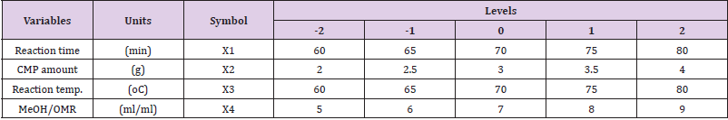
Table 1: Experimental design for biodiesel production.

Biodiesel Quality Characterization: The effectiveness and industrial application of biodiesel produce depend solely on its quality such as moisture content, density, viscosity, mean molecular mass, peroxide value, iodine value, saponification value, cetane number, higher heating value, API gravity, and diesel index. These properties were determined using AOAC, (1997) standard methods; the results were compared with biodiesel recommended standards [34,35]. All methods were performed in accordance with the relevant guidelines and regulations governing institutional, national, and international guidelines and legislation including the collection of plant material, the Experimental research and field studies on plants/seeds.

Results and Discussion

Extracted Oils Qualities and It Mixed Ratio

Table 2 showed the results of the qualities of the oils and the mixed oil obtained. From the table, it was observed that the specific gravity of the LSO (0.95) was highest, followed by the LCO (0.910), and then DSO (0.820). The higher the specific gravity, the lower the API gravity of the oil, and such oil tends to be less light in nature. The extracted oils were light oils with low free fatty acid value and low moisture content. The iodine value and the viscosity of the LSO (98.02 g I2/100g oil; 4. 10 mm2/s) appear higher than the value of LCO (96.50 g I2/100g oil; 4.10 mm2/s) and DSO (72.32 g I2/100g oil; 2.12 mm2/s). However, the acid value of the LCO (4.50 mg KOH/g oil) is higher than the acid value of LSO (1.84 mg KOH/g oil) and DSO (1.43 mg KOH/g oil), these showed that the LCO is non-edible oil. The API gravity of DSO (50) is greater than the values obtained for LCO (29) and LSO (21) which accounted for an oil mixed ratio of 29:50:21 for LCO: DSO: LSO. This mixed ratio produced oil with moderate FFA and of low density used for biodiesel production (transesterification).

Catalyst Characterization and Elemental Analysis

Scanning Electron Microscopy (SEM): Figure 1a showed the result of SEM images of morphological characteristics of the calcined mixed powder (CMP) at a different magnification of 1000x and 500x performed in the range of 20o<2θ < 80o at speed of 2oC/ min. The decomposed images after calcination at a temperature of 650oC for 3 h indicated non-uniform sizes with diverse shapes and slightly rough surface with cracks. This observation implied thermal treatment degrades organic substances in the mixed powder (MP) and also transforms the calcium carbonate (CaCO3) to the calcium oxide (CaO), owing to the gaseous form of carbon dioxide (CO2) which appears more porous, brittle and easy to ground.

biomedres-openaccess-journal-bjstr

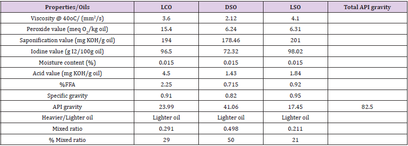
Table 2: Qualities of the oils and API gravity mixed ratio.
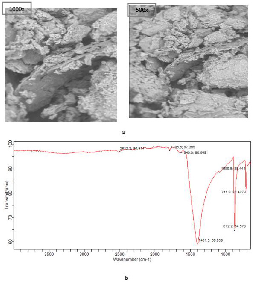
biomedres-openaccess-journal-bjstr

Figure 1:

a. SEM image of CMP at different magnifications.
b. FTIR analysis of CMP at 650 oC for 3h.

Fourier-Transform Infrared Spectroscopy (FTIR): Figure 1b shows the FTIR spectra of CMP, distinct peaks were noticed at 711.9, 872.2, 1080.9, 1401.5, 1640.0, 1796.6, and 2512.2, respectively. The band at 1401.5 cm-1 represents bending vibration of the O-Ca-O group while the band at 771.9 – 1080.9 represents a stretch of CO32- molecules to higher energy value [3,36]. The spectrum stretches from 1640.0-2512.2 cm-1 indicated the presence of functional groups such as O-H, C-H for sp3 carbon, C=O for sp2 carbon, CHO, and N-H bond. Nevertheless, the undistorted assembly of CMP catalyst transformed into a spongy like structure signifies that calcination of the mixed powder at a temperature of 650oC was appropriate for a complete transformation of CaCO3 to CaO [16]. This observation could be attributed to the presence of functional groups present in the fruits during the growth period which occurred as a result of carbon growth inhibition.
Brunauer-Emmett-Teller (BET): Table 3 represents the properties of the catalysts indicating the BET surface, total pore volume, basicity, and percentage of CaO converted through N2 adsorption-desorption isotherm Brunauer-Emmett-Teller (BET) analysis. Observation from the results shows the calcined mixed powder (CMP) has a high basic site than other calcined catalysts (CMAPP, CCLPP, and CCPPP) owing to the high CaO (75.65%) obtained during analysis in the calcined powder. The basic site density obtained for CMAPP (158. 00 μmole/m2) was higher than that of CCLPP (120.00 μmole/m2), but the value obtained for CCPPP (157.80 μ mole/m2) is approximately the same with CMAPP. However, the basic site density of the CMP (160.83 μ mole/m2) was the highest which produced the highest biodiesel yield during transesterification. The table also reflects the result of biodiesel yields, at the same process conditions, each catalyst was tested based on the CaO yield of the calcined powders (CCPPP: 62.83%; CMAPP: 65.50%; CCLPP: 58.67 and CMP: 75.65%), the yields of biodiesel based on the nature of catalyst showed the three calcined catalysts have a basic site for conversion of oil to biodiesel (83.50, 88.75, 80.32 (%wt.)), but the mixed calcined catalyst produced highest biodiesel yield (96.50% wt.), due to the percentage of CaO in the catalyst and high basic site. Hence, the catalysts could be an economic viable promising source for CaO catalyst production for industrial applications.

biomedres-openaccess-journal-bjstr

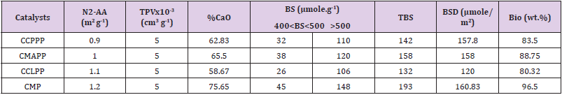
Table 3: BET-adsorption and XRD analysis of calcined catalysts at 650 oC for 3 h.

Optimization of Transesterification Mixed Oil to Biodiesel

Experimental Results Analysis: Table 4 shows the coded experimental conditions, the experimental biodiesel yield, the predicted, and the residual values of the 30 standard runs generated by CCD. The table showed the maximum experimental yield of 97.20 (% wt.) at runs 18, while the minimum yield was obtained at runs 19 with a value of 85.66 (% wt.). Based on statistical analysis, the results were transformed into a fit summary, quadratic model, analysis of variance evaluation, diagnostics and graphs modeling, the predicted biodiesel yield of 96.63 (% wt.) was obtained at the following condition: reaction time of 80 min, CMP amount of 3.53 (g), reaction temperature of 90 oC, and CH3OH/OMR of 9:1 (ml/ml), at the desirability of 95.10%. This value was validated in triplicate, an average mean value of 96.50 (% wt.) was obtained which was close to the predicted value. The result proved that the methanolysis of CMP for the transesterification of mixed oil was successful.

biomedres-openaccess-journal-bjstr

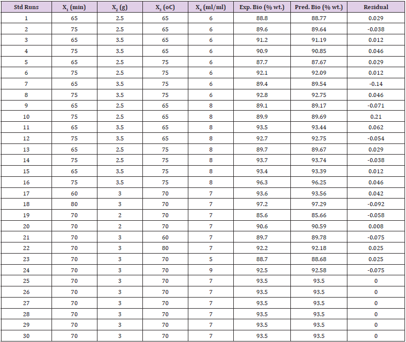
Table 4: Coded factors, experimental, predicted biodiesel yield, and residuals values.

ANOVA and Fits Statistic: Table 5 shows the analysis of variance (ANOVA) for the response surface quadratic model and the Fit statistics. Observation from the table shows that the Model F-value of 1765.55 with a degree of freedom (df) of 14, implies the model is significant with prob value >0.0001. There is only a 0.01% chance that a “Model F-value” this large could occur due to noise. Meanwhile, values of “Prob > F” less than 0.05 show variable terms are significant. In this case, X1, X2, X3, X4, X12, X22, X32, X42, X1X2, X1X3, X1X4, X2X3, X2X4, and X3X4 were remarkable significant variable terms. The coefficient of determination is the correlation coefficient, also known as R-square,which allows it to display the degree of linear correlation between two variables. The value obtained in this study is high (99.94%), indicate a high degree of correlation between the interacting variables. The “Pred. R-Squared” of 99.65% is in reasonable agreement with the “Adj R-Squared” of 99.88%. The “Adeq Precision”, which measures the signal to noise ratio. Usually, a ratio greater than 4 is desirable, the ratio of 188.58 obtained in the study specifies an adequate signal.The polynomial model quadratic equation that shows the relationship between the biodiesel yield and the four-variable factors is presented in Eq. (4).

The final equation in term of coded

biomedres-openaccess-journal-bjstr

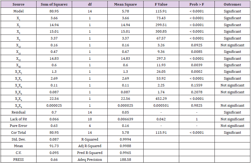
Table 5: ANOVA and Parametric Data Fit.

Meanwhile, the positive and the negative coefficients in the equation are the direct measure of the influence of variables on the response value. In this equation, the variable X2 with a coefficient of 1.23, f-value = 4796.50, with p-value<0.0001 is the most significant variable among the second-order polynomial equation in Eqn. (4).
Graphical Plots: Furthermore, the relationship between the response variable (biodiesel) and the interactive variables (X1X2, X1X3, X1X4, X2X3, X2X4, and X3X4) can be represented in contour and the three-dimensional plots as displayed in Figures 2a-2f. The observation from the graph shows Figure 2e has the highest mutual interaction between CMP amount and MeOH/OMR on the response biodiesel yield. The mutual interactive effects between reaction time and MeOH/OMR (Figure 2f) is higher than that observed in the interactive effects noticed between reaction time and reaction temperature (Figure 2b) on the response, but the interactive effects in Figure2c (MeOH/OMR and reaction temperature) is lesser than the interactive effects showed by Figure 2b, but higher than the interaction between CMP amount and reaction temperature (Figure 2d). Meanwhile, the least interactive effects were noticed in Figure 2a, which was the interaction between CMP amount and reaction time on response biodiesel produced. In all, there exist perfect interaction among the variables, which confirmed the variable factors considered in this study play an important role in biodiesel yield. The relationship between the predicted biodiesel and the experimental biodiesel yields as well as power transformation boxcox were as illustrated in Figures 3a & 3b.

Catalyst Reusability Test

For catalyst purification and reusability analysis of the reaction mixture, a built-in heating system vacuum centrifuges operated at 3500 rpm was used, the recovered catalyst was washed with alcohol to remove the impurity at the surface of catalyst that occurred during the transesterification. The alcoholic washed catalyst was oven-dried at 100oC for 1 h and then cooled to room temperature before reused. Nevertheless, the reactor wall accumulates a 0.15 g catalyst during the reaction, which tends to reduce the surface area and lowered catalyst activity. Hence, the need for catalyst reusability tests on catalytic activities. Figure 4 shows that the catalytic activities-maintained stability from the 1st cycle to the 4th cycle with little decreased from 93.6 (% wt.) to 92.20 (% wt.). However, there was significant decrease observed in 5th and 6th cycles (89.7 and 88.7% wt.), this can be associated to the continuous intermediate products formed during the reaction, such as monoglyceride and diglyceride, which obstructed the catalyst holes as well as water to oxygen reaction that occurred at the catalyst surface, which reduce the catalyst sensitivity. Hence, catalyst reusability was stopped after the 4th cycle [37-39].

biomedres-openaccess-journal-bjstr

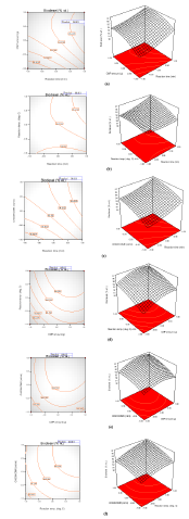
Figure 2: Contour and 3-D’s Plots.

biomedres-openaccess-journal-bjstr

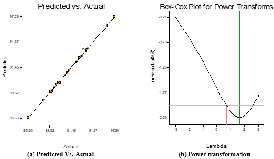
Figure 3: Diagnostics graph.

biomedres-openaccess-journal-bjstr

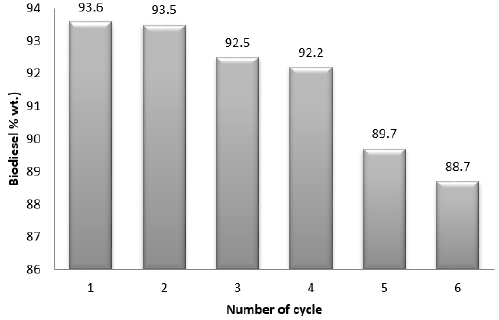
Figure 4: Catalyst recyclability test plot.

Properties of the Mixed Oil and Biodiesel

Method of AOAC were adopted to examine the properties of the blended oil and the product, the obtained results of the biodiesel were compared with the ASTM D6751 and EN 14214 biodiesel recommended standard (Table 6). It was observed that the decreased in the moisture content, density, viscosity, acid value, the iodine value, saponification, and the peroxide value of the blended oil to biodiesel was due to process transesterification. This confirmed that the synthesized product is consistent with biodiesel and the translation of blended oil complete transesterification reaction to biodiesel was achieved with insignificant resistance to flow and lessen internal drag in the engine. Further observation showed that the cetane number, the higher heating value (HHV), the API gravity, and the diesel index increased as blended oil was converted to biodiesel, this could be attributed to energy formation from viscous oil to low viscous oil. The high biodiesel yield obtained in this study could be attributed to due to decrease base consumption for neutralization. Based on cetane number, the higher the peroxide value, the better the cetane number and the decrease in ignition time [31,40]. The value of 4.34 meq O2/kg oil, can be attributed to the cetane number of 57.52 obtained. The higher heating value (HHV) of biodiesel is greater than that of blended oil which signify high heat of vaporization of water in the combustion of biodiesel. The American petroleum institute (API) gravity usually used to determine the weight of oil/petroleum in comparison with water, the value of 33.03 and 49.91 obtained for blended oil and biodiesel exhibited light oils. Diesel index which denotes the efficiency of the biodiesel as well as the ignition properties, the value obtained in this study were well within the required recommendation standard for biodiesel that can be used in I.C engine [41-44].

biomedres-openaccess-journal-bjstr

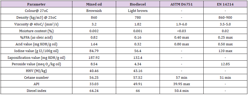
Table 6: Quality of mixed oil and biodiesel.

Conclusion

The study concluded that the ratio of blended oil produced low viscous oil, and the derived catalyst from the mixture of Cucurbita pepo, Musa acuminate and Citrullus lanatus peels powders, calcined at a temperature of 650oC for 3 h, produced high CaO- base (75.65%). Transesterification of blended oil to biodiesel was successfully carried out with maximum biodiesel yield of 97.20 (%wt.). A statistical software with second order model analysis predicted biodiesel yield of 96.63 (% wt.) at the following reaction time of 80 min, the CMP amount of 3.53 (g), the reaction temperature of 90 oC, and the CH3OH/OMR of 9:1 (ml/ ml), respectively. To validate this value, three experimental runs were carried out, and the average mean was determined as of 96.50 (% wt.) was obtained which was close to the predicted value. Analysis of variance test confirmed the significant of variables with p-value<0.0001. Catalyst reusability test was immobile at the 4th cycle due to loss of basicity that occurred due to leaching as a result of several recyclability during the reaction. Hence, the produced biodiesel conformed to biodiesel recommended standard, and the CaO catalyst could serve as promising economical feedstock for industrial application.

For More Articles: Biomedical Journal Impact Factor: https://biomedres.us

Open Access Journals on Medical Research

The Relationship of Chronic Rhinosinusitis with Nasal Polyps and PDL-1

Introduction

Chronic rhinosinusitis with nasal polyps (CRSwNP) are benign inflammatory and hyperplastic outgrowths of the sinonasal mucosa [1]. It is a disease that seriously affects the quality of life of patients by causing nasal congestion, snoring, postnasal discharge, nasal discharge, loss of sense of smell/taste. The prevalence of CRSwNP is 1% to 4% in the general population [2]. CRSwNP is observed more frequently in male patients between the 4th and 6th decades [3]. İn histologically, nasal polyposis is characterized by respiratory epithelium covering oedematous stroma infiltrated by inflammatory cells [2]. Eosinophilic infiltration is the most common finding in polyp histopathology. In addition, there is an infiltration of inflammatory cells such as lymphocytes, basophils and macrophages.
Mucosal changes, including squamous metaplasia, secretory hyperplasia and epithelial cell proliferation, are observed in polyp tissues with the effect of chemical mediators secreted from these cells. T-helper 2 (Th2) cell-directed eosinophilia develops as a result of elevated immunoglobulin-E (IgE) and interleukin-5 (IL-5) [4]. In Asians, the majority of patients with CRSwNP show features of neutrophilic inflammation in addition to eosinophilia [5,6]. PD-1 (B7-H1,CD 274) is a protein-structured receptor belonging to the immunoglobulin superfamily found on the surface of activated T cells and antigen presenting cells. By binding to the PDL-1 ligand, T cell inhibition occurs and escape from the immune system occurs. PD-L1 works like a stop sign, inactivating the immune system’s T cells. One of the ways in which sick cells escape from the immune system is to densely host the proteins called PD-L1 on the cell surface. Studies have shown that PD-1 plays an important role in immune tolerance and in the regulation of T cell function [7].
Genetic studies have found that PD-1 gene polymorphism is associated with autoimmune diseases such as systemic erythematosus and rheumatoid arthritis [8,9]. PD-1 and PDL-1 are expressed in many different tumors such as lung carcinoma, lymphoma, pancreatic carcinoma, and renal cell carcinoma. It has been reported that PD-1 expression is increased in T cells in patients with CRSwNP with using flow cytometry [10]. This checkpoint led to the development of immune checkpoint inhibitors, which are the next generation immunotherapy drugs. These drugs prevent the interaction of molecules such as PD-L1 and PD-1, thereby activating the immune system against diseased cells. Among the immune checkpoint inhibitors nivolumab and pembrolizumab act by inhibiting PD-1 receptors, while atezolizumab and durvalumab act by inhibiting PD-L1 receptors [11].
If the importance of pdl1 in nasal polyp becomes clear, these immune checkpoint inhibitors drugs can be used in the treatment. There are not enough studies in the literature regarding the presence of PDL-1 in nasal polyps. In our study, our aim is to explain the presence of pdl1 in nasal polyp by immunohistochemical method. Detecting of the PDL-1 changes in nasal polyp tissue may explain the development of chronic rhinosinusitis with nasal polyps and may help develop new methods in the treatment of nasal polyps.

Materials and Methods

In our study, 20 patients who underwent endoscopic sinus surgery due to nasal polyps and 20 patients who underwent surgery for functional and/or aesthetic reasons and whose septum and turbinate mucosa were removed were included retrospectively, in Alanya Alaaddin Keykubat University Medical Faculty Hospital between 2010-2020. Previous sinus surgery, age is under 18 and nasal malignancies were exclusion criteria. 4 micron thick sections were taken from paraffin blocks for applying immunohistochemical PDL-1 antibodies. These sections were processed with Ventana PDL-1 (SP 263) monoclonal antibody on the fully automated Ventana Ultra Benchmark XT instrument. In the evaluation of PDL- 1 antibody applied immunohistochemically, the percentage of cells showing membranous staining with PDL-1 antibody was calculated among 100 tumor cells under light microscope. IHC staining results in groups were evaluated with Mann Whitney test using SPSS software version 22 (SPSS Inc., Chicago, IL, USA). P <0.001 was considered significant. Ethics committee approval was obtained from Alanya Alaaddin Keykubat University (no:2021/09-06).

Results

There were 2 female and 18 male patients in the study group, and 4 female and 16 male patients in the control group. The mean age was 37.4±8.6 years in the study group and 35.4±8.8 years in the control group. PDL-1 positivity in nasal polyp tissues was significantly higher than normal nasal mucosa epithelium. (P=0.000) (Table 1) (Figures 1-3) The recurrence rate was higher in PDL-1 positive cases with nasal polyps, and the recurrence rate increased as the staining rate increased, and it was statistically significant. (P=0.003).

biomedres-openaccess-journal-bjstr

Figure 1: Epithelial cell of the polyp with 50% membranous staining.

biomedres-openaccess-journal-bjstr

Figure 2: Epithelial cell of the polyp with 10% membranous staining.

biomedres-openaccess-journal-bjstr

Figure 3: Control normal nasal mucosa epithelium not stained with PDL-1.

biomedres-openaccess-journal-bjstr

Table 1: Percentage of immunohistochemical staining of cells with PDL-1.

Discussion

There are some hypotheses exist for how CRSwNP develops but the aetiopathologenetic mechanism of it is still unclear [2,12]. The pathogenesis of it is thought to involve persistent inflammation of the nasal mucosa due to infiltration by eosinophils, basophils, lymphocytes, neutrophils and mast cells [2]. T lymphocytes act as the main actor in this inflammation process. PDL1 İs a member of the B7 family that regulate the T-cell response and a predictive biomarker. It is a cell-surface glycoprotein primarily expressed by antigen-presenting cells. Studies on PDL 1 in the literature are mostly about tumor cells. Different cut-off values have been reported in these studies [13-16]. However, variable cut-off levels create difficulties for researchers in making decisions during evaluation. Determining a standard cut-off value for researchers is important for the correct interpretation of the results and for the consistency between the results of the studies. In our study, we considered the presence of PDL-1 positive in staining of 1% or more. Different companies have antibodies and subclones for PDL- 1. In the literature, there are studies reporting different results with antibodies and clones of different companies in the same cell group [17,18].
We used the SP263 clone of Ventana, which is frequently used in studies in the literature, to increase the staining quality and the reliability of the results. There are studies explaining that the infectious process plays a role in the pathogenesis of CRSwNP [19,20]. İt is reported that IFN-γ secretion increases after incubation of lymphocytes with bacterial and fungal extracts [21,22]. IFN-γ is an important cytokine thas is a strong inducer of PDL-1 expression [23]. Before studies showed IFN-γ have been identified in nasal polyposis [24,25]. PDL-1 levels in other cell membranes increases by the IFN-γ charged by T-cells. Thus, it causes a negative feedback in nasal polyposis by causing down-regulation of T-cell responses. İn a study about mRNA expression of PDL-1 in patients with CRSwNP, resarchers found mRNA levels of PDL-1 was increased in nasal polypies epitel compared to healty nasal tissue epitel [26].
Although there are studies in the literature that measure PDL1 level in nasal polyp tissue and blood cells by PCR and flow cytometry, but there is not enough study in which the pDL1 level in nasal polyp tissue is analyzed immunohistochemically. In a small study in the literature using only 3 nasal polyp tissues and 3 nasal tissues as control, immunohistochemical evaluation was made and PD-1+ cells were indeed increased in the submucosa of NP tissue compared to control tissue [27]. İn our study we found that the pdl1 level in CRSwNP tissue was significantly higher than the pdl1 level in the control healthy nasal tissue, and it was was statistically significant. (p:0.01). For this reason, the use of monoclonal antibodies developed against PDL-1 as a target drug as a non-surgical drug therapy in nasal polyps is a matter of debate. Recurrence in surgical procedures is an undesirable situation for the patient and the surgeon. Different researchers used several markers for predicting nasal polyp recurrence.
Recurrence is frequently observed in nasal polyposis, and recurrence is higher in eosinophilic polyps than in noneosynphilic polyps [28]. Zhong et al. in their study, the relationship between peripheral eosinophil count and recurrence was examined and they found a significant correlation between high peripheral blood eosinophil count and recurrence [29]. In another study showed researchers that a tissue eosinophil proportion of >27% of total cells was a marker for recurrence within 2 years after endocopic sinus surgery in patients with CRSwNP [30]. In our study, recurrence rate was high, and it was found in 10 (50%) patients. The recurrence rate was higher in PDL-1 positive cases with nasal polyps, and the recurrence rate increased as the staining rate increased, and it was statistically significant. (P=0.003) These findings suggest that the presence of PDL-1 may be a predictive factor for recurrence.

Limitations

The most important limitation of our study is that it was a retrospective study, and we could not reach sufficient patient material, since the operated patients took the operation materials to an external center for histopathological consultation. It will be possible to make a clearer comment on the fact that it is a determining factor in the development of recurrence.

Conclusion

PDL-1 was significantly increased in our patients with CRSwNP compared to normal nasal mucosa tissue. Presence of PDl-1 is a predictive factor for recurrence in nasal polyposis. Since there are not many studies explaining the presence of CRSwNP and PDL- 1 in the literature, we think that our study will contribute to the literature. New studies with a larger number of patients are needed to determine the presence and importance of PDL-1 in nasal polyps.

For More Articles: Biomedical Journal Impact Factor: https://biomedres.us

Journals on Medical Research

Silver Nanoparticles for Hydrogen Peroxide Sensors

Introduction

Silver particles have been used as antibacterial agents since the 19th century, and now their uses have diversified to include many new physical, chemical and biological ones [1-4]. AgNPs are considered to be one of the most widespread NPs, with about 500 tons of annual global production [5]. More recently, AgNPs have been incorporated into industrial and surgical device coatings,dental coatings, and automotive smoke filters and textiles due to their effective properties against microbes [6]. The mechanism on this topic is currently under discussion by researchers [7]. In this review, relevant techniques of synthesis of metal nanoparticles will be discussed. After that, a detailed study of the properties and applications of AgNPs for hydrogen peroxide sensors will be presented.

Bottom-Up and Top-Down Synthesis

As a historical background for metal nanoparticles (MNPs), it was reported that many exploited the strengthening of ceramic matrices, including natural asbestos nanofibers, more than 4500 years ago [8]. Lead-based chemistry was pioneered in ancient Egypt for cosmetic preparation over 4000 years ago. Here, we look at a hair dye recipe using lead salts described in the text since Greco-Roman times. We report direct evidence for the shape and distribution of PbS nanocrystals that form in the hair during darkening [9]. Likewise, “Egyptian blue” was the first synthetic pigment prepared and used by the Egyptians using a mixture of sintered nanometer-scale glass and quartz around the 3rd century BC [10]. Egyptian Blue represents a complex mixture of CaCuSi4O10 and SiO2 (both glass and quartz). In the ancient geographic regions of the Roman Empire, including Egypt, Mesopotamia, and Greece, Egyptian blue for decorative purposes has been observed during archaeological excavations.
Natural or synthetic ways can synthesize NPs by two basic approaches, including various sub-preparation methods. The first approach is called the “top to bottom” method, including breaking down bulk solid materials into smaller pieces by applying external energy from physical, chemical, and thermal techniques [11]. The second approach, called “from the bottom up, “brings together and combines atoms or molecules of gases or liquids. The top-down approach is expensive. However, it is impossible to obtain perfect surfaces and edges due to the cavities and roughness in NPs, while a bottom-up approach can get excellent results of nanoparticle synthesis. In addition, with the bottom-up approach, no waste is formed to be eliminated, and the smaller NPs can be obtained with better control of the sizes [12-16]. However, adapting the production of large quantities of powders on an industrial scale is not simple. A significant advantage of treatment in solution is the possibility of generating encapsulated NPs using surfactants as a protective shell, which makes it possible to obtain very homogeneous and well dispersed NPs [17].
The main bottom-up approaches are the supercritical fluid synthesis, the use of the template, the spinning, the synthesis by plasma or flame projection, the green synthesis, the sol-gel process, the laser pyrolysis, the aerosol-based process, the chemical vapor deposition, and the atomic or molecular condensation. The main top-down approaches are the mechanical milling, the etching (chemical), the electro-explosion (thermal/chemical), the spraying (kinetics), and the laser ablation (thermal). The green synthesis approach is based on biological methods (accommodating plants, algae, the mushrooms, yeasts, actinomycetes and bacteria).

Types and Synthesis of Silver Nanoparticles

Among the various types of NPs, Ag NPs have been widely developed to be utilized in various applications due to their outstanding properties. Typical applications of Ag NPs include clothing and textiles, medical devices, food storage, cosmetics, sunscreens, laundry detergents [18], bandages [19], and sensors [20]. Some studies have found that Ag NPs have cytotoxicity that can induce ROS formation in cells [21]. Therefore, many products such as detergents, toiletries, etc. In addition, their synthetic or in-use personal care, whether industrial or household, produces a release of NPs, which ultimately ends up in the sewer. This untreated wastewater affects aquatic ecosystems and thus microorganisms. Recently, Ag NPs had great concerns regarding aquatic toxicology due to the difficulty of tracking these particles in the environment and accessing their effects on living organisms [22]. The fate of NPs in the aquatic environment and their interactions, the interactions between NPs with biological and abiotic components, and their potential for damage are not well understood, and these uncertainties raise concerns about related risks. These molecules impose on humans on health and the environment [23]. Based on the Scopus database [24], the publications on Ag NPs increase with time, where it started in 1990 (two reports) and reached 7105 reports in 2020.
Colloidal Ag NPs are molecules with an average diameter of 20- 40 nm and comprise 80% silver atoms and 20% silver ions. They are the best-selling nanoparticles ahead of carbon nanotubes and titanium nanoparticles and are released into the environment. The demand for Ag NPs has increased due to their applicability in multiple fields. Over the years, various synthesis techniques have been developed, and procedures have been improved to prepare small and uniform Ag NPs. Ag NPs were prepared by a chemical reduction technique, where the silver ions are reduced by sodium citrate [25]. As referenced, various kinds of Ag NPs have been utilized in various applications [26]. Specifically, Ag NPs of differing sizes and shapes have been used in a broad scope of uses and clinical gear, such as electronic gadgets, coatings, cleansers, swathes, etc. [27]. Explicit physical a nd optical properties of Ag NPs are subsequently essential factors in advancing their use in these applications. In such a manner, the accompanying subtleties of the materials are imperative to consider in their combination: surface property, size dissemination, clear morphology, molecule composition, dissolution rate (i.e., reactivity in arrangement and effectiveness of particle delivery), and kinds of diminishing and capping specialists utilized. The blend techniques for metal NPs are partitioned into top-down and bottom-up approaches. In these approaches which also include chemical and green methods, the electron transfer initiates the bioreduction through nicotinamide adenine dinucleotide (NADH)-dependent reductase as an electron carrier to form NAD+. The resulting electrons are obtained by Ag+ ions, which are reduced to elemental Ag NPs.
The physical technique usually utilized to prepare Ag NPs is the evaporation-condensation method. It is commonly performed using a tube furnace at atmospheric pressure, synthesizing various sizes [28]. Several attempts have been made. For instance, Tsuji, et al. [29] proposed a new method for synthesizing Ag NPs by a laser ablation technique with focused and unfocused laser beam irradiation carried out at 12 and 900 mJ cm−2 intensities, respectively. The radiation wavelengths used were 355, 532, and 1064 nm. This study revealed that the surface plasmon wavelength of Ag NPs irradiated using 355, 532, and 1064 nm is ~400 nm for focused and unfocused beams. The famous chemical method for Ag NPs synthesis is a reduction by natural and inorganic reducing agents. By and large, unique reducing agents, for example, sodium citrate, ascorbate, sodium borohydride, essential hydrogen, polyol measure, Tollen’s reagent, N, N-dimethylformamide (DMF), and poly (ethylene glycol)-block copolymers are utilized for the reduction of silver particles (Ag+) in aqueous or non-aqueous arrangements. These reducing agents decrease Ag+ and lead to metallic silver (Ag0), trailed by agglomeration into oligomeric clusters. These clusters ultimately arrange the metallic colloidal silver particles [30]. It is critical to utilize defensive agents to stabilize dispersive NPs during metal nanoparticle planning and ensure that the NPs consumed or tied onto nanoparticle surfaces stay away from their agglomeration [31]. Green synthesis methodologies dependent on natural reducing agents rely on different reaction parameters such as solvent, temperature, pressing, and pH conditions (acidic, fundamental, or impartial). For the union of metal oxide nanoparticles, plant biodiversity has been extensively viewed as the accessibility of successful phytochemicals in different plant separates, particularly in leaves such as ketones and aldehydes flavones, amides, terpenoids, carboxylic acids, phenols, and ascorbic acids [32,33]. These parts are fit for decreasing metal salts into MNPs [34]. The fundamental focus of such nanomaterials has been researched for biomedical diagnostics, antimicrobials, catalysis, atomic detecting, optical imaging, and marking of natural frameworks.

Applications of Silver Nanoparticles

Over the last decades, the production of NPs has been increasing rapidly for applications in electronics, chemistry, biology, and almost all our daily life applications [35]. This is mainly due to their properties of being very small, close to the biomacromolecules and providing high surface area, rapid diffusion, and high reactivity in both liquid and gas phases [36,37]. Recently, Ag NPs have attracted attention in various applications such as biological, food, optoelectronics, electronic devices for energy conversion, electron field emission sources for emission displays, and surface-enhanced Raman properties [38-41]. Accordingly, utilizing Ag NPs in sensing, mainly for the excellent physico-chemical properties, will be reviewed here. Developing fast, sensitive and selective gas sensors has received great attention in environmental monitoring, national security, and food safety applications [42]. Typically, the total performance of gas sensors strongly depends on the specific area surface of the materials utilized for detection gas. Thus nano-scale sensing is predictable to display improved sensing performance [20]. Typically, semi-conductive nano metal oxides such as ZnO and SnO2 exhibit high sensitivity and fast responses to some gases [43]. A relatively higher temperature (150–600◦C) required for maximum response is a big problem that restricts the practical applications in most areas [44]. Therefore, developing suitable nanomaterials to enhance the sensor response is highly recommended. Noble metal nanostructures are the most promising in this field due to LSPR. The LSPR is an optical phenomenon observed when the electromagnetic radiation excites the surface conducting electrons of MNPs, resulting in a coherent resonance oscillation of the particles. The location of the extinction maximum is highly dependent on the reflective index and dielectric properties of the surrounding environment and the adsorption of the molecules on the metal surface [45,46]. Therefore, the peak wavelength shift in the extinction maximum of NPs is used to fast detect moleculeinduced changes surrounding the NPs. Thus, UV-Vis spectroscopy and the naked eye can observe the changes in the absorbance of the visible and near-infrared wavelength regions.
There are many published articles on the use of LSPR sensing applications in the liquid phase to detect organic phosphorous pesticides [47] and ammonia [48]. Generally, SPR absorption of noble metal NPs is strong in the visible to near-infrared (IR) region. Moreover, SPR is particularly sensitive to its size, shape, composition, distance, and surroundings. Therefore, it displays a promising ability for various sensors. However, Ag NPs have higher extinction coefficients than gold nanoparticles (Au NPs) of the same size [49]. Due to this feature and high specific surface area, high catalytic, high crystallinity, and their electrical and optical properties, Ag NPs have been widely investigated as gas sensor applications [41,50]. This section will present the recent progress on Ag NPs as gas sensors for ammonia, methane, and hydrogen peroxide. Despite the wide use in industry, such as fertilizers, animal feed production, and manufacturing of paper and plastics, ammonia is a toxic material with a harmful effect on the human body as it can harm tissues and the immune system [51]. Ammonia is largely produced in deteriorating food and fruitbodies by various micro/macrofungi [52]. Thus, monitoring the concentration of ammonia in air and liquid in the atmosphere is extremely important. Recently, Ag NPs have been widely used to sense organic gases such as methane and ethanol [20,53,54]. Cannilla, et al. [55] successfully prepared Ag NPs in a poly-methacrylic acid (PMA) matrix by a photo-induced reduction process followed by deposition on a ceramic substrate to sense ammonia gas in resistive base sensors. To improve the conductivity of the thin films to be suitable for sensing, the as-prepared Ag NPs/PMA were loaded with multi-walled carbon nanotubes (MWCNTs). The developed sensor shows the considerable ability to work at low temperature with a wide range of detection range and represent fast response/ recovery times. Kumar et al. prepared face-centered cubic polyvinylpyrrolidone (PVP) capped Ag NPs at room temperature and a chemical reduction method [41]. The prepared sample showed an average size of ~22 nm, showing a conductive and metallic nature. As ammonium gas sensing, the synthesized NPs thin films showed the maximum sensitivity towards ammonia gas.

Hydrogen Peroxide Sensing

In the last decade, hydrogen peroxide (H2O2) monitoring has gained importance due to its widely employed in many industrial, atomic power stations, medical sectors and its application as a disinfecting agent for water pools [56-58]. However, the high presence of H2O2 can cause various biological damages, leading to aging, neurodegeneration, and cancer [59,60]. Therefore, developing a high-response, cheap method is highly recommended for medical, pharmaceutical security, and environmental protection [59]. Various techniques, such as spectrophotometry [60,61], chemiluminescence [62], and electrochemistry [63], have been utilized to detect H2O2. These methods are categorized as enzymatic and non-enzymatic methods. In the case of enzymatic, the peroxidase is an illustrative enzyme for H2O2 exposure; however, both pH and temperature are limited.
As mentioned above, noble metal NPs display strong SPR absorption with extreme sensitivity to the size, shape, composition, and surroundings suitable for colorimetric sensors in the range of visible to near-infrared region. For colorimetric assays, Ag NPs have a high ability toward the decay of H2O2 [64]. This reaction can mainly be sensed by colorimetric principles, as the colloidal Ag NPs exhibit a characteristic color from the LSPR. The alteration in both particle size and shape results from the incorporation of colloidal Ag NPs with H2O2 can be identified by determining the change in the absorption spectral at the wavelength of LSPR. For instance, Zhang, et al. [65] investigated H2O2 via colorimetric detection using three different morphologies of Ag NPs (triangular, spherical, and cubic). The Ag NPs with various shapes reacted with H2O2, and the edges of Ag NPs had been etched. The change shape transformation induced visible color change, which was used for the quantitative determination of H2O2. The triangular Ag NPs display the highest sensitivity for a quite small level of H2O2 (5 mM), while the cubic Ag NPs display lower sensitivity. Recently, Srikhao, et al. [66] prepared Ag NPs with an average particle size of 16.9 nm by using phenolic compound extracted from sugarcane leaves as a reducing agent. The results showed that 90◦C and stirring for 20 min are the optimum conditions for phenolic compound extraction. The prepared Ag NPs were evaluated as ammonia and H2O2 solution sensing by UV-Vis spectrophotometer and the naked eye. It observed that the Ag NPs sensitively detect both H2O2 and ammonia even at low concentrations.
Moreover, the prepared Ag NPs could detect these toxic agents even after two weeks. However, measuring in spectrophotometric methods requires some standard solutions and tools such as standardized cuvettes, microwell plates, and spectroscopic equipment. To overcome these limitations, Yoshikawa et al. utilized an optical technique for detecting H2O2 in their work Ag NPs deposited on a glass plate/Au NPs [66]. The Ag NPs chip diffracts incident light, and the diffraction efficiency is correlated with the amount of Ag NPs. By applying a drop of H2O2 onto the chip, the diffraction strength debilitates due to the decay of Ag NPs. A movable measurement technique of the diffraction intensity changes is assembled, and the H2O2 detection in a concentration range of 6.7–668 mmol L−1 in about 2 min by dropping the H2O2 solution onto the substrate.
The electrochemical technique is promising to detect H2O2 due to its simplicity, lower cost, ease of operation, and high sensitivity and selectivity compared to the other techniques [62,63]. Many electrochemical sensors based on metals and metal oxides-based complexes have been developed to overcome the high overpotential. Due to the high electron transfer rates, high catalytic activity toward reducing H2O2, and significantly decreasing overpotential at oxidizing and reducing agents, Ag NPs are widely utilized to fabricate these sensors. Zhan, et al. [67] decorated Ag NPs on three-dimensional graphene (3DG) via the hydrothermal process as a sensing electrode for electrochemical detection of H2O2 in phosphate-buffered solutions. The electrochemical results approved that the Ag NPs-3DG based biosensor exhibits fast amperometric sensing, low LOD, wide linear responding range, and perfect selectivity for non-enzyme H2O2 detection. Recently, Maduraiveer, et al. [68] utilized an electrochemical sensor for H2O2 by using Ag NPs introduced in a silicate matrix (APS(SG). The prepared APS(SG)-Ag NPs were deposited on glassy carbon (GC) electrode. The electron transfer behavior of the APS(SG)-Ag NPs was investigated by potassium ferricyanide ([FeCN)6]3−), methyl viologen (MV2) and ruthenium hexamine ([Ru (NH3)6]3+). The GC/APS(SG) electrode displayed a twofold increase in the peak currents and fast electron transfer kinetics toward [Fe (CN)6]3− in comparison with the GC electrode. The GC electrode modified with APS(SG)-Ag NPs significantly improved electron transfer. The GC/ APS(SG)-Ag NP electrode as an electrocatalytic sensor against H2O2 offered a reduction of H2O2 at less negative potential and showed the experimental low detection limit of 25.0 μM with the sensitivity of 0.042 μA/μM. Additionally, an APS(SG)-Ag NP-based sensor showed a fast response, good stability, and reproducibility.

Conclusion

This article presented the synthesis, properties, and hydrogen peroxide applications of silver nanoparticles in a brief detail. Despite the great role of Ag NPs in sensing applications for detecting gases and vapors of some organic compounds and detecting hydrogen peroxide in industry and biosystems, the function of this effect needs more study to be fully understood. Furthermore, the longterm stability and sensitivity should be more developed by adjusting the preparation conditions, utilizing eco-friendly stabilizing agents. Moreover, recycling Ag NPs from their applications wastes is an important issue from both environmental and economic points of view. This would help in developing Ag NP based nanomaterials more safe, biocompatible, and efficient for some vital applications in our life.

For More Articles: Biomedical Journal Impact Factor: https://biomedres.us

Open Access Journal on Medical Research

Education of a Child in the Ethics of Al-Ghazali

Of course, raising a child is not an easy task. However, the scientist emphasizes that raising children should not be the reason for not generosity and indifference of parents to others. The purpose of starting a family is to bring up a healthy generation. The upbringing of society begins in the family. Raising a child is a serious responsibility that every parent must be aware of and fulfill. Parents should be the support and guardians of their children and should do everything possible for their peace of mind, happiness and full development. Parents, raising their children, pay back their duty to their parents. Indeed, the precious heritage of parents for a child is a good upbringing. The ethical conclusions of Muhammad Al-Ghazali on the sphere of raising children are attractive, in which the ways and methods of upbringing are shown in detail. The scientist’s ethical teachings can be used as a guide by educators, psychologists and the general public.

For More Articles: Biomedical Journal Impact Factor: https://biomedres.us

Journals on Biomedical Sciences

How to Facilitate Simulation in Telemedicine in the COVID Era

Health Care Simulation is widely used to train healthcare professionals as it contributes to long-term retention of acquired complex technical, clinical and teamwork skills when compared to traditional learning methods. In addition, translational science research shows that measured outcomes transfer to improved patient care practices, and improved patient and public health [1]. Health Care Simulation follows experiential learning theory that proposes that a two-dimension cycle is necessary for learning to occur. The first dimension represents perception, where the learner begins by either grasping a concrete experience or a concept. The second dimension represents processing, where the learner transforms the experience by reflecting on it and the concept by actively experimenting on it [2]. To complete this cycle, learners and educators usually get together on site to participate in highly interactive, high-fidelity clinical scenarios using mannequins, actors, task trainers or a combination of them, followed by debriefing [3]. But the rapid development of technology-assisted learning methods, which are on the rise since the coronavirus (COVID-19) pandemic in order to help reduce the transmission risk among professionals, opens up new alternatives for distance experiential learning [4].

The best approach to carrying out clinical simulation activities in this context remains to be elucidated. Several solutions are proposed to fit the pieces in a suitable way that would allow professionals to continue with their training activities and, at the same time, to serve the clinical safety of the participants, avoiding, as far as possible the spread of the disease. They include a variety of simulation based asynchronous (e.g. recorded videos, vignettes, serious games) and synchronous (e.g. webinars, team collaboration) methods. They recreate the clinical environment with a different level of fidelity and learner personal engagement in combination with reflection strategies in combination with reflection strategies [5]. Moreover, innovations in healthcare simulation technologies can now provide the learner with opportunities to practice increasingly complex motor, decision-making and communication skills using virtual patient simulation. They recreate dynamic health conditions in a variety of clinical settings that respond to user interventions and show improvements in learning satisfaction when compared to a case-based learning approach [6].

Where to Start

The basic principle is to generate a training tool that maintains the essence of the simulation as a clinical practice that allows participants not to be in the simulation room in person in order to guarantee their safety in the midst of the COVID 19 pandemic. We have developed in Spain a model of distance clinical simulation in Valdecila Virtual Hospital, suitable for emergency physicians and we have proved in staff and also in internal medicine resident’s program [7] obtaining promising perspectives. This model can achieve positive results using distance clinical simulation combining their traditional simulation technology to address the training needs of healthcare organizations.

The Goal

Using virtual patient simulation to provide simulation-based distance learning experiences that are extremely realistic and highly interactive for the learner remains a challenge. Communicating with patients, family members and healthcare workers, performing physical examination on patients, monitoring of physiological parameters, evaluating complementary examinations in an actual clinical environment are all constricted by mathematical algorithms and software capabilities. A mix of synchronous on-site/on-line distance learning methods may address aspects of the richness and complexity of a true clinical experience and facilitate reflection on action promoting participant engagement, while limiting exposure to infectious diseases during a pandemic. Distant learning combining on site simulation technology with teleconferencing software based on experiential learning principles is feasible and easy-to-implement, and is well-accepted by participants for acute care training in the emergency room. The promising results of this new approach of “taking care of patients from a distance” can be useful not only during a pandemic, but also in our daily work because surely this new model will be able to facilitate future learning courses in the health field.

For More Articles: Biomedical Journal Impact Factor: https://biomedres.us

Journals on Medical Research

Turning the Circadian Clock Slow: A New Function of Gaseous Signal H2S

Gaseous signal molecules, such as nitric oxide (NO), carbonic oxide (CO), and hydrogen sulfide (H2S), widely exist in most tissues of the body, and exert important physiological functions. These molecules are endogenously synthesized and catabolized, can freely diffuse across the plasma membrane and regulate certain biological processes at physiological concentrations. H2S is the latest discovered among the above three gaseous signal molecules. Endogenous H2S is produced by the catalytic synthesis of L-cysteine hydrochloride by cystathionine beta synthase (CBS) or cystathionine gamma lyase (CSE). CBS and CSE belong to pyridoxal 5’-phosphate dependent enzymes, with differences in tissue distribution. CBS mainly distributes in the brain, especially in hippocampus and cerebellum. The H2S concentration in rat serum was about 46μmol/L and was 50-160μmol/L in rat brain tissues. In vivo, endogenous H2S mainly exists in two forms, i.e., free gas (about 1/3) and sodium hydrosulfide (NaHS) (about 2/3). The two forms keep a dynamic equilibrium. As a H2S donor, NaHS mainly exists in peripheral tissues, and maintains the stabilities of free H2S level and pH in vivo. Studies on endogenous NO and CO have demonstrated that both of them play important roles in the regulation of circadian rhythm in organisms. It has also been proved that H2S can change the expression patterns of clock genes such as Clock, Per2, Bmal1, and Rev-erbα by influencing the content ratio of NAD/NADH and the expression of Sirt1 in mice hepatocytes [1]. However, it is still unclear whether H2S regulate the central circadian clock In vivo. The present study aimed to test the hypothesis that H2S plays an important role in the regulation of central circadian clock located in the suprachiasmatic nucleus (SCN) by affecting the expression patterns of clock genes and clock-controlled genes.

To test the hypothesis, we performed voluntary wheel running experiments in adult male C57BL6/J mice. Animals were first entrained to 12:12h light: dark (LD) circumstance for 7 days and then subjected to constant dark (DD) condition for experiments, with circadian time 12:00 (CT12) indicated as the beginning of subjective night. Data were analyzed using the ActiView Biological Rhythm Analysis software (MiniMeter Co., USA). To observe the effects of higher or lower H2S on the circadian locomotor rhythm, intracerebroventricular (i.c.v.) injections of exogenous NaHS (50mmol/L, 4ml per injection) or amiono-oxyacetate (AOAA) (a CBS inhibitor) (50mmol/L, 4μl per injection) were performed, respectively. AOAA was dissolved in artificial cerebrospinal fluid (ACSF) composed of (in mmol/L) NaCl 119, NaHCO3 26.2, KCl 2.5, NaH2PO4 1.0, MgCl2 1.3, glucose 10.0, pH 7.4. To check whether the SCN showed a normal light entrainment, short-time (15min) light exposure (200 lux) was performed. Period length (tau) was determined by the ActiView periodogram analysis. Phase shifts were defined as the difference between the predicted activity onset times and observed activity onset times in response to NaHS or light pulses.

biomedres-openaccess-journal-bjstr

Figure 1: H2S modulation on the circadian rhythm in mice.
A. Wheel running actograms showing that H2S i.c.v. injection at CT14 induced phase delay and circadian period prolongation.
B. ACSF i.c.v. injection at CT14 (control).
C. H2S i.c.v. injection at CT20 also induced phase delay and circadian period prolongation.
D. ACSF i.c.v. injection at CT20.
E. Actograms showing phase shift and circadian period prolongation induced by i.c.v. injection of AOAA at CT14 (red asterisk), light exposure at CT15 (blue asterisk) and AOAA plus light exposure (red asterisk plus blue asterisk).
F. Statistical phase shifts at different conditions.
G. Immunohistochemical stains of SCN showing the robust circadian expression pattern of CBS.
H. RT-qPCR showing the mRNA levels of Bmal1, Per1, Per2 and Sirt1 in the SCN at different conditions.

After wheel running experiments, animals were decapitated under dim red light (4 lux), and SCN tissues were immediately harvested, liquid nitrogen frozen and total RNA were extracted as we previously reported [2]. Transcriptions of circadian genes, including Bmal1, Per1, Per2 and Sirt1, were examined by realtime quantitative PCR (RT-qPCR). Some SCN tissues were routinely developed, sectioned, and the protein expression of CBS was visualized by immunohistochemistry. Data were expressed as mean ± standard deviation (SD). Student’s t test or analysis of variance was used to perform the statistics where necessary. The criterion for statistical significance was P < 0.05. Animal use protocol was in compliance with the guidelines for the Care and Use of Laboratory Animals (NIH, revised 2011). Results showed that bolus i.c.v. injection of NaHS at CT14 (early subjective night) induced significant phase delay of the circadian locomotor rhythm in mice by about 50 ± 8 min, and also obviously prolonged the circadian period length from 23.8h to 24.3h (Figure 1A), compared with the control ACSF injection at CT14 (Figure 1B). Bolus NaHS (i.c.v.) at CT20 (late subjective night) also caused a phase delay of 30 ± 10min (Figure 1C) and a prolongation of period length from 23.8h to 24h, compared with control ACSF injection at CT20 (Figure 1D). Above results showed that NaHS (H2S donor) i.c.v. injection induced phase delay and prolongation of circadian period length, suggesting that H2S could slow down the running of central circadian clock. We thus proposed that suppression of H2S production by inhibiting CBS would lead to opposite changes of phase and period length. However, to our surprise, i.c.v. injection of AOAA at CT14 produced almost the same results as that induced by NaHS, i.e., phase delay (30 ± 10 min) and period length prolongation, but the extents of phase shift and period prolongation were obviously less than that induced by NaHS (Figures 1E & 1F).

To check whether NaHS i.c.v. injection would affect the light entrainment function of SCN, twelve days after bolus NaHS i.c.v. injection, we gave the mice a short-time (15min) light exposure at CT15. Results showed that short light exposure induced significant phase delay by about 120 ± 20min (Figures 1E & 1F). This result indicates that the SCN responded well to light signal and was not damaged by NaHS i.c.v. injection. A combination of AOAA at CT14 and light exposure (CT15) led to further phase delay (180 ± 15min) and prolongation of circadian period length (Figures 1E & 1F). As CBS is a H2S synthetase, and H2S donor NaHS affected the circadian rhythm as we observed, we thus examined whether CBS expression would show circadian rhythm, and whether inhibition of CBS would affect the expression of clock genes and clock-controlled gene Sirt1, in the SCN of mice. We detected the expression of CBS over 24 hours in mice SCN tissues by immunohistochemistry. SCN tissues were obtained at CT2, CT6, CT10, CT14, CT18 and CT22. CSB protein expression levels showed a robust circadian pattern with a cycle of nearly 24 hours (Figure 1G). The expression peak appeared at CT14, and the trough showed at CT2 (Figure 1G).

Because NaHS induced phase delay and period length prolongation as shown above, it is possible that NaHS may change the transcription of clock genes and clock-controlled gene Sirt1. We checked the mRNA levels of Bmal1, Per1, Per2 and Sirt1 before and 15min after NaHS i.c.v. injection at CT14 by qRT-PCR. Results showed NaHS significantly decreased the mRNA levels of Per2 by 0.69-fold but increased the mRNA level of Sirt1 by 1.5-fold, while NaHS did not affect the mRNA levels of Per1 and Bmal1 (Figure 1H). To clarify the major findings of the present study, bolus i.c.v. injections of NaHS at CT14 and CT20, a way thought to increase the H2S level in the SCN, induced phase delay and period length prolongation of mice free-running locomotor rhythms; deactivating CBS by AOAA to reduce the H2S level in the SCN exerted similar result as NaHS did; the used dosage of NaHS did not damage the light entrainment function of SCN; CBS expression in the SCN showed a pattern of circadian rhythm; NaHS i.c.v. injection at CT14 increased the protein expression of CBS and the transcription of Sirt1, decreased the transcription of Per2, but did not significantly affect the transcription of Bmal1 and Per1, in the SCN. These results suggest that H2S slows down the running of circadian rhythm, or in other words, H2S turns the central circadian clock slow. This study provides new evidences to support that H2S takes a role in the regulation of central circadian clock. The circadian expression pattern of CBS in the SCN is at least partially associated with the regulatory function of H2S on the central clock, and PER2 and SIRT1 may take major roles in the function of H2S.

We noticed that either increasing the H2S level by NaHS or decreasing the H2S level by CBS inhibitor induced the same results, i.e., phase delay and period length prolongation, only that the extent of NaHS-induced phase delay was greater than that caused by AOAA. Similar contradictory phenomena have also been observed in other studies. For example, Luo, et al. [3] demonstrated that both overexpression and deletion of miR-279 disrupted the circadian rhythm in Drosophila. We recently also found that either elevation or reduction of the microRNA-17-5p level in mice SCN shortened the free-running period length [4]. This unconventional regulatory algorithm deserves further investigations both in mechanism and significance. H2S has been indicated having an anti-aging effect. It is still unknown whether H2S exerts the anti-aging effect through changing the circadian rhythm. Some studies have shown that the free-running periods are shortened in senescent C57BL/6 mice, DBA mice, rats and hamsters, although some contradictory results were also reported. We show here that H2S induced phase delay and free-running period prolongation in mice, and decreased Per2 mRNA level but increased Sirt1 mRNA level in the SCN. It is possible that H2S exerts the anti-aging effect by prolonging the endogenous cycles of circadian rhythm via promoting the expression of Sirt1, because Sirt1 is considered a gene of longevity, and H2S can prevent cell senescence through SIRT1 activation. SIRT1 regulates the expression of clock genes by PER2 acetylation [5]. Higher SIRT1 accelerates the degradation of PER2 protein, thereby promoting the transcription of Clock/Bmal1. This regulatory signaling pathway may explain our finding that exogenous H2S increased the expression of Sirt1 and decreased the expression of Per2.

A question may be raised as whether exogenous NaHS (i.c.v.) would damage the function of SCN. The physiological concentration of H2S in mouse brain is about 50-160μmol/L, but the exact transient local H2S levels in the SCN are unknown. The concentration of NaHS used in the present study was 50mmol/l (i.c.v. 4ml per injection). This NaHS concentration looked much higher than the H2S level of natural brain, but the actual acting H2S level in the SCN should be much lower than the used NaHS concentration if considering the dilution and diffusion of NaHS/ H2S in the cerebrospinal fluid and brain tissues including SCN. We showed that the used NaHS concentration did not affect the light entrainment function of SCN, suggesting that SCN, or including other parts of mouse brain, is quite tolerant to NaHS/H2S. This finding suggests safety of relatively higher H2S in the treatment of related brain diseases. In conclusion, H2S can induce phase delay and circadian period length prolongation by affecting the expression patterns of clock genes and clock-controlled genes at SCN. This study suggests H2S is regulator of central circadian clock.

Acknowledgment

This work was supported by NSFC grants (31471126 and 82170523 to J.C.), Sichuan Science and Technology Program (2019YJ0472 to P.Z.), and Luzhou Science and Technology Program (2018LZXNYD-ZK44 to P.Z.).

Conflict of Interests

The authors declare that they have no conflict of interest in this work.

For More Articles: Biomedical Journal Impact Factor: https://biomedres.us

Open Access Journals on Medical Imaging

The Osseous Dissipation- Otosclerosis

Preface

Otosclerosis is a primary osteodystrophy of the otic capsule or a disorder of bone remodelling incriminating the stapes footplate or bony labyrinth of the inner ear. Otosclerosis literally implies “abnormal hardening of body tissue of the ear”. The condition is confined to the middle ear and is associated with anomalous remodelling of bone. Normally dense endochondral layer of bony otic capsule situated within the labyrinth is substituted by irregular, spongy bone. Consequently, fixation of the stapes ensues. Otosclerosis was initially scripted by Antonio Maria Valsalva in 1735 and is additionally designated as otospongiosis. Otosclerosis alters functioning of the middle ear and inner ear and is a predominant contributor to deafness in adults. Otosclerosis may engender significant disability and morbidity due to hearing loss. Antecedent disease detection with cogent therapy is associated with superior outcomes.

Disease Characteristics

Typically, otosclerosis induces lucent alterations instead of sclerotic bony modifications. Temporal bones are frequently incriminated [1,2]. Otosclerosis appears as an antecedent, adult onset disease. Generally, otosclerosis commences within second decade or third decade although hearing loss commences following the fourth decade. Disease occurrence within children is exceptional. However, the progressive condition demonstrates gradually worsening clinical symptoms and it may be challenging to ascertain precise disease onset [1,2]. A female predominance is observed with a female to male proportion of ~2:1. Otosclerosis worsens in pregnancy [1,2]. Ethnic predilection demonstrates frequent disease emergence within the Caucasian population. Otosclerosis is exceptionally discerned in Asians or Black population. Familial disease incidence appears in an estimated 50% instances. Around 85% instances are bilateral [1,2]. Otosclerosis is associated with distinctive phases such as
• Early or active otospongiosis and
• Late or inactive otosclerosis.
Of obscure and multifactorial pathogenesis, otosclerosis may arise due to genetic, viral, inflammatory or autoimmune components [1,2]. Preliminary lesions are predominantly comprised of an admixture of histiocytes, osteoblasts and osteocytes wherein osteocytes are an active cellular component. Bony circumscription of pre-existing vascular articulations is resorbed with meliorated microcirculation. Eventually, osteoblasts configure irregular foci of nascent, spongy bone, designated as “blue mantles of Manasse” [1,2].

Disease Pathogenesis

Of obscure aetiology, otosclerosis is posited to denominate a multifactorial emergence such as
• Anatomical factors wherein fissula ante fenestram is incriminated along with persisting remnants of embryonic cartilage [3,4].
• Genetic factors of otosclerosis are associated with several loci situated upon chromosomes 6p, 9p, 1q, 3q, 6q, 7q, 15q, 16q and a contemporary locus upon chromosome 7q22.1. Additionally, diverse genes as type I collagen (COL1A1 gene), transforming growth factor-β1 (TGF-β1) with BMP2 and BMP4 genes, angiotensin II with AGT M235T and ACE I/D genes may induce otosclerosis [3,4]. Sex hormones, autoimmune reaction, human leucocyte antigen (HLA), inflammatory or regulatory cytokines, parathyroid hormone, parathyroid hormonerelated peptide receptors and oxidative stress may initiate otosclerosis [3,4].
• Hereditary factors wherein an estimated 50% subjects with otosclerosis demonstrate a family history. Individuals with hereditary otosclerosis are associated with an antecedent disease onset. Majority of instances depict an autosomal dominant mode of inheritance accompanied by reduced penetrance in nearly ~40% subjects and variable expression of disease [3,4].
• Viral infection is implicated in the emergence of otosclerosis. Ribonucleic acid (RNA) of measles virus may be discerned within footplate of stapes upon ultrastructural examination and immunohistochemistry. Vaccination for measles virus may appear as a safeguard for occurrence of otosclerosis [3,4]. Additionally, factors such as menopause, trauma, bone dyscrasia or major surgery may initiate or aggravate otosclerosis [3,4].

Clinical Elucidation

Otosclerosis is commonly confined to the temporal bone and engenders fixation of footplate of stapes within the oval window. Therefore, inability to transmit sound waves manifests as conductive hearing loss [5,6]. Generally, otosclerosis engenders conductive deafness and appears in association with a normal tympanic membrane. Contingent to focal incrimination of the bony labyrinth, otosclerosis can be asymptomatic or represent with neurosensory decimation [5,6]. Frequent clinical representation of otosclerosis is hearing loss accompanied by tinnitus and vertigo. Gradually progressive bilateral hearing loss is a common symptom which typically commences in one ear and progresses to contralateral ear [5,6]. Inability to adequately discern low-frequency sounds as a whisper can emerge as an initial clinical symptom. Hearing may be meliorated in a noisy environment, a feature denominated as “paracusis willisii” which is indicative of conductive deafness. Incriminated subjects adopt a low-volume, monotonous tone of voice.
Extensive otosclerosis is associated with worsening tinnitus. Mild dizziness appears and deteriorates with disease progression, simulating Meniere’s disease [5,6]. Otoscopy may be normal or depict minimal modifications. However, around 10% instances of active otosclerosis or cochlear otosclerosis are accompanied by enhanced vascularity of the promontory which can be discerned through the tympanic membrane and is designated as “Schwartze sign” [5,6]. Of indolent clinical course, otosclerosis manifests with frequently bilateral, conductive hearing loss although sensorineural or mixed deafness may ensue. Hearing loss can be exacerbated in pregnant subjects [5,6]. Otosclerosis is comprised of distinctive subtypes denominated as
• Fenestral or stapedial variant which constitutes of a majority (~80%) of instances and incriminates the oval window along with stapes footplate. Frequently, conductive hearing loss is associated with thickening and fixation of stapes [5,6].
• Retrofenestral or cochlear subtype which is composed of nearly 20% instances. Incriminated cochlea is associated with demineralization of cochlear capsule. Sensorineural hearing loss emerges, possibly due to uncertain mechanisms [5,6].
Retrofenestral otosclerosis is usually accompanied by fenestral variant and the conditions are contemplated to represent a disease continuum [5,6].

Histological Elucidation

Upon gross examination, foci of otosclerosis appear as chalky white, greyish or yellowish. Active centric zone and rapid disease progression is associated with reddish lesions due to enhanced vascularity. Upon macroscopic examination of dissected tissue, specimens of un-altered head and crura of stapes are obtained [5,6]. Microscopically, foci of initial bone resorption and circumscription of vascular articulations by cellular, fibrovascular tissue are observed. Subsequently, deposition of immature bone, perpetual bone resorption and remodelling is exemplified. Gradually, deposition of bone, enhanced collagen and decimated ground substance ensue with consequent emergence of densely sclerotic bone demonstrating prominent cement lines [5,6]. Dense, endochondral layer of otic capsule appears to be constituted of spongy bone. Immature, active lesions of otosclerosis are imbued with abundant bone marrow, vascular spaces and numerous osteoblasts admixed with osteoclasts. Significant quantities of cement substance are deposited within the lesion [5,6]. Mature foci of otosclerosis are mildly vascular and composed of abundant bone and fibrillar substance with minimal cementum. Localized foci of bone remodelling and bone resorption of the otic capsule are followed by subsequent bone deposition.
Otoclerosis predominantly arises due to replacement of normal bone with sclerotic or spongiotic bone [5,6]. Osteolytic osteocytes are configured upon growing perimeter of the lesion. Sheets of connective tissue displace the bone. Delayed stage of otosclerosis depicts dense, sclerotic bone associated with zones of previous bone resorption. Consequently, disorganized bone is articulated, osteocytes are quantifiably enhanced and expansive bone marrow spaces are incorporated with vascular articulations and connective tissue [5,6]. Bone marrow spaces are eventually substituted by dense, sclerotic bone imbued with narrow vascular articulations and minimal Haversian canal system [5,6]. Preliminary and delayed stage of otosclerosis occurring within a singular temporal bone engenders pleomorphism. Antecedent lesions may adjoin fissula ante fenestram and expand through abutting vascular channels [5,6]. Majority of lesions are confined to anterior oval window and appear associated with calcification of annular ligament or incrimination of stapes, thereby engendering conductive hearing loss [5,6] (Figures 1-8).

biomedres-openaccess-journal-bjstr

Figure 1: Otosclerosis exhibiting sclerotic bone of footplate of stapes and bony labyrinth of inner ear [9].

biomedres-openaccess-journal-bjstr

Figure 2: Otosclerosis enunciating spongiotic and sclerotic bones replacing foot plate of stapes [10].

biomedres-openaccess-journal-bjstr

Figure 3: Otosclerosis exemplifying bony growth within footplate of stapes and spongiotic bone at the junction of incus and stapes [11].

biomedres-openaccess-journal-bjstr

Figure 4: Otosclerosis exemplifying bony sclerosis at footplate of stapes with an admixture of spongiotic bone, osteocytes and minimal Haversian system [12].

biomedres-openaccess-journal-bjstr

Figure 5: Otosclerosis delineating bony sclerosis of footplate of stapes with configured spongiotic bone and associated conductive hearing loss [13].

biomedres-openaccess-journal-bjstr

Figure 6: Otosclerosis demonstrating bony sclerosis and spongiotic bone replacing footplate of stapes with intermingled osteocytes and fibro-connective tissue [14].

biomedres-openaccess-journal-bjstr

Figure 7: Otosclerosis displaying spongy and sclerotic bone replacing footplate of stapes intermingled with abundant osteocytes, osteoblasts and fibro-connective tissue [15].

biomedres-openaccess-journal-bjstr

Figure 8: Otosclerosis exhibiting bony sclerosis of the inner ear labyrinth with several osteocytes, decimated Haversian canal and fibroconnective tissue [16].

Differential Diagnosis

Otosclerosis requires a segregation from conditions such as
• Osteogenesis imperfecta where otic capsule demonstrates lucencies simulating otosclerosis. Osteogenesis imperfecta is associated with non deformed, fragile bones and blue sclera. Severe variants of osteogenesis imperfecta are devoid of an organized bone trabecular pattern. Crowding of osteocytes within bone occurs due to decimated collagen synthesis. Enlarged foci of woven bone are delineated. Minimally severe instances depict crowding of osteocytes associated with attenuated lamellar bone [2,4].
• Paget’s disease of bone predominantly occurs in elderly individuals and demonstrates bony expansion. Acute stage is primarily composed of woven bone admixed with focal, mosaic configuration of lamellar bone associated with irregular cement lines. Osteoclasts are preponderant upon the bone surface. Osteoclasts appearing within osteolytic phase may depict around ~100 nuclei [2,4]. Chronic stage is comprised of thickened bony trabeculae and thick bones which are imbued with finely fibrotic bone marrow [2,4].
• Osteoradionecrosis arises due to vascular injury with consequent emergence of bony ischemia. Osteoradionecrosis is associated with distinctive phases denominated as
• Pre-fibrotic phase demonstrating foci of chronic inflammatory cell exudate
• Phase of organized fibrosis predominantly displaying aberrant fibroblastic activity along with an inadequately organized bone matrix. Aforesaid regions appear adjacent to foci of aging fibroblasts disseminated within a minimally cellular, fibrotic, densely sclerotic matrix [2,4].
• Fibro-atrophic phase is composed of dense hyalinization and fibrosis along with absence of bone marrow cells. Focal, reactive stratified squamous epithelium may layer a fistulous tract. Patchy, secondary infiltration of chronic inflammatory cells may occur. Lesions may display necrotic or sclerotic bone along with empty osteocyte lacunae. Fibrosis of bone marrow is admixed with irregular bony trabeculae devoid of osteocytes within the lacuna. Foci of bone marrow necrosis may arise due to emergence of a preliminary stage of oedematous, fibromyxoid disease [2,4]. Additionally, otosclerosis requires a segregation from conditions which are associated with conductive deafness such as serous otitis media, adhesive otitis media, congenital fixation of the stapes, Meniere disease, tympanosclerosis, attic fixation of head of the malleus or ossicular discontinuity [2,4].

Investigative Assay

Upon otoscopy, features indicative of otosclerosis are minimal to absent. However, severe instances with cochlear incrimination can engender hyperaemia of the cochlear promontory, a feature designated as “Schwartze sign” [7,8]. Otosclerosis can be appropriately investigated with the tuning fork and demonstrates a negative Rinne’s test. Weber’s test appears lateralized to the ear and is indicative of a severe conductive hearing loss [7,8]. Pure tone audiometry demonstrates low frequency loss of air conduction with normal bone conduction. Pure tone audiometry exhibits a characteristic decimation of bone conduction, especially at higher frequencies [7,8]. Mixed hearing loss can be observed wherein preliminary otosclerosis is associated with normal tympanometry [7,8]. High resolution computerized tomography (CT) of temporal bone is optimal in discerning otosclerosis besides recognizing and segregating associated causes of deafness. An estimated 80% instances depict fenestral foci which are situated anterior to the oval window. Also, thickened stapedial footplate and incriminated round window can be discerned, features which regulate appropriate therapy [7,8]. Retrosternal focus emerges as a “double halo” sign, a feature encountered with cochlear otosclerosis. Otosclerosis can be appropriately graded with CT [7,8].
Pertinent imaging depicts representations such as • Fenestral otosclerosis which is the commonest variant and demonstrates site of incrimination just anterior to oval window while configuring a miniature cleft denominated as fissula ante fenestram. Besides, bony overgrowth may engender fixation of the stapes [7,8].
• Retrofenestral otosclerosis incriminates the niche of round window and usually accompanies fenestral disease. Nevertheless, isolated round window otosclerosis may be occasionally discerned. Predominant site of disease emergence appears as focal or circumferential bone circumscribing the cochlea. Circumferential bony implication engenders a “fourth turn” or “double ring” sign [7,8]. Imaging features are contingent to phase of the disease and appear as
• Otospongiotic phase composed of demineralization and configuration of spongy bone. Pertinent phase manifests as decimated attenuation or lucent zone within homogeneously dense perimeter of otic capsule [7,8].
• Otosclerotic phase is associated with enhanced attenuation within disease specific region. As it may be challenging to discern otosclerotic bone from encompassing normal bone, features such as thickness of otic capsule or aberrant convexity of contour of otic capsule cortex anterolateral to anterior margin of the oval window may aid the distinction [7,8]. Severe instances are associated with comprehensive impaction of oval window or round window with a dense, bony plate associated with complete fixation of stapes [7,8]. Thin-slice computerized tomography (CT) through the temporal bone is a preferred imaging modality for adequately exemplifying anatomy of inner ear and emergent, subtle, preliminary modifications of otosclerosis. Upon computerized tomography, otosclerosis is categorized as
• Grade I which is singularly comprised of fenestral, spongiotic or sclerotic lesions which appear as thickened footplate of stapes along with decalcified, narrowed or enlarged round window or oval window [7,8].
• Grade 2 is constituted of patchy, localized cochlear disease along with the presence or absence of fenestral otosclerosis and is subdivided into~grade 2A which implicates basal cochlear turn~grade 2B which implicates middle or apical cochlear turn~grade 2C which implicates basal turn and middle or apical cochlear turn [7,8].
• Grade 3 is comprised of diffuse, confluent involvement of cochlea and otic capsule along with the presence or absence of fenestral involvement [7,8]. Magnetic resonance imaging (MRI) of retrofenestral otosclerosis exhibits a distinctive pericochlear and perilabyrinthine soft tissue intensity upon T1 weighted imaging with contrast enhancement. Besides, enhanced signal intensity upon T2 weighted imaging may be observed [7,8].

Therapeutic Options

Medical management of otosclerosis is required to circumvent or arrest disease progression. However, optimal, efficacious medical therapy which alleviates otosclerosis remains lacking. Administration of sodium fluoride in order to decimate disease progression is debatable [7,8]. Bisphosphonates are antiresorptive and induce osteoclastic apoptosis. Contemporary bisphosphonates, commonly employed to treat otosclerosis, appear promising [7,8]. Bilateral hearing aids can be utilized singularly or in combination with diverse therapies [7,8]. Fenestral otosclerosis can be appropriately treated with stapedectomy along with the employment of stapes prosthesis. Optimal and recommended surgical treatment of otosclerosis is stapedotomy or stapedectomy or correction of fixation of foot plate of stapes. Surgical intervention is commonly followed by implantation of prosthesis [7,8]. Therapeutic outcomes of surgical intervention in otosclerosis appear to be superior, regardless of surgical manoeuver adopted [7,8]. Though surgical intervention is beneficial, hearing aids may be required in certain subjects following surgery [7,8]. Untreated otosclerosis may engender significant hearing loss although complete deafness is uncommon [7,8].

Although infrequent, surgical intervention for treating otosclerosis can induce comprehensive sensorineural deafness within the operated ear. Surgical manoeuvers may induce facial nerve injury or tinnitus. An unpleasant taste may arise for a brief duration [7,8]. Prosthetic implantation is associated with granulomatous inflammation and erosion or necrosis of the incus [7,8]. Following surgical intervention, an estimated 90% subjects demonstrate significant amelioration of hearing ability. Hearing remains unaltered or declines in occasional instances [7,8]. Laser techniques and vein grafting is associated with superior outcomes. Additionally, reoccurrence of conductive hearing loss may ensue due to displacement of prosthetic implant. Original position of prosthesis is altered on account of collagen contraction within the neo-membrane configured between prosthesis and bony labyrinth [7,8]. Revision surgery may exhibit ambiguous outcomes and is usually adopted for treating associated clinical symptoms as facial nerve palsy, persistent vertigo or failure of meliorated hearing [7,8]. Nevertheless, revision surgery may be accompanied by inferior outcomes [7,8]. Strict monitoring is required in subjects with a family history of otosclerosis. Hearing loss in pregnant subjects may appear due to otosclerosis [7-16].

For More Articles: Biomedical Journal Impact Factor: https://biomedres.us

Open Access Journals on Medical Research

Contrast Medium Controlled Injection in the Navicular Bursa and the Following Bursography in 23 Cases

Introduction

Equine palmar foot syndrome is a common cause of front limb lameness in the horse [1]. The podotrochlear apparatus is a special anatomic feature in the horse due to the deep digital flexor tendon needing a pulley before the insertion on the distal phalanx. The navicular bone (navicular bursa: NB), distal impar ligament (DIL), collateral sesamoidean ligaments (CSL) and the deep digital flexor tendon (DDFT) are part of the podotrochleat apparatus [2]. Alliterations of one of these structures alone or combined lead to pain [2]. Magnetic resonance imaging (MRI) is a favorable method to diagnose the different lesions [3–5]. Ultrasound is also an option to diagnose changes in the DIL and DDFT [6,7]. Due to lesions in the soft tissue, X-ray is sufficient to diagnose equine palmar foot syndrome [8]. Contrast radiography with palmaroproximal-palmarodistal oblique views seem to be helpful in diagnosing changes in the DDFT and the flexor surface cartilage [9,10]. Intrabursal medication is one of the widest use practices in navicular disease [11,12].
There are many ways to inject the bursa. The distal palmar approach to the “navicular position,” defined as a point on the lateral hoof wall, 1 cm distal to the coronary band, and halfway between the most dorsal and most palmar aspect of the coronary band [12], seems to be very successful. In cadaver limbs, 23/25 (92 %) were injected successful with 1.1 redirection per injection. However, there was also contrast medium (CM) in the distal interphalangeal joint (DIP) in 1/25 (4 %) and palmar to the DDFT in 1/25 (4 %) legs. There is also the possibility to show alliterations in navicular apparatus in cadaver limbs with bursography, especially with the dorsoproximal-palmarodistal oblique view [13]. There is a reported success of 14/17 for NB injections (82.35 %) in living horses [14]. There is no information currently about dorsoproximalpalmarodistal oblique radiographic projections with CM in the NB in living horses. Thus, this case study will show the first results of this technique in living horses.

Materials and Methods

Thirteen horses were chosen for a reason not connected to the study for injection of the NB. Patients were presented in the hospital from 2015 to 2020 and were all warmbloods. Nine horses were injected in both front feet. Only one foot was injected in each of four horses. The NB was injected twice in one horse. There was complete lameness examination data available in seven horses, including a positive distal digital nerve block. The data were collected retrospectively. All horses underwent a distal digital nerve block and a sedation with detomidine and butorphanol. The skin between the heel bulbs was shaved or clipped and washed for 5 min with antibacterial soap and swabbed several times with alcohol. The foot was placed on a standard DP 65° wooden block and held by staff, the same way as for the DPr-PaDiO radiographic projections. An amount of 3 ml Iomeprol 300 mg ID/ml (Imeron 300M© Bracco Imaging GmbH, Konstanz, Germany) and 5mg triamcinolonactenonide (TriamHexal©, Hexal AG, Holzkirchen, Germany) were injected directly into the NB using a spinal needle (BD© Spinal Needle, 0.9 *90 mm, Becton Dickinson GmbH, Karlsruhe, Germany) and a standard 5 ml syringe under sterile conditions.
The landmark for needle insertion was a point just proximal to the central sulcus of the frog, with the needle directed toward the apex of the frog and parallel to the ground surface of the hoof [10]. The correct placement of the needle was controlled with a latero-medial view. If the needle was not in place, it was redirected and also checked with an latero-medial view until it was correctly placed. The CM was then injected and another latero-medial view was taken to see whether the bursa was filled with CM. Holding the leg still in position, the DPr-PaDiO radiographic projection and a palmaroproximal-palmarodistal oblique (PaPr-PaDiO) radiograph on a standard block were made. All radiographs were acquired with a mobile X-ray tube (TR 90/30, Gierth®, Riesa, Germany) and a direct X-ray detector system (Mark 3 Sound-Eklin® Carlsbad, CA, USA or CXDI-801C, Canon®, Tokyo, Japan).
Findings in the DPr-PaDiO CE were a change in dimension of the proximal or distal border, which represents an adhesion formation. The rough shape of the NB might come from a chronic bursitis with hyperproliferation of the synovium. Additionally, there should be no CM abaxial to the border marked by the end of the navicular bone, which would be evidence of a rupture of the NB. A dorsal tendon tear would lead to a long, straight and radiopaque line. Therefore, if there is an irregularity in the surface of the DDFT communicating with the NB, CM would be able to fill the defect in the tendon. A defect in the flexor cortex (cartilage) would manifest as a round, radiopaque area. These lesions are typically roundshaped. An adhesion would be represented by a radiolucent area. As described above, there is a small amount of CM between the navicular bone and the DDFT. If there is an adhesion between those structures mentioned, the dye column will be interrupted. The radiological density of the scar tissue is like soft tissue, therefore, there will be radiolucency.

Results/Observations

Twenty-three legs were injected. The NB was successfully injected only in 20/23 (86.96 %). In the other three legs, the CM was injected completely palmar to the DDFT (Figure 1b). The NB and the DIP were filled with CM in one horse (4.35 %) (Figure 1a). The DPr-PaDiO CE showed alliterations in 7/9 (77.78 %) legs with a clinical history of lameness. There was CM palmar to the DDFT (Figure 1a) and the NB was filled with CM in 5/20 (25.00 %) legs. The findings are summarized in Table 1. There were two DDFT lesions, one full flexor cortex lesion, three suspected ruptures of the NB and five NB adhesions.

biomedres-openaccess-journal-bjstr

Figure 1:

a. There is contrast medium (CM) in the navicular bursa and the distal interphalangeal joint, and some CM palmar to the deep digital flexor tendon (DDFT)
b. The injection of the navicular bursa has failed. The CM is completely palmar to the DDFT.

biomedres-openaccess-journal-bjstr

Table 1: Table 1 shows the information about the horses, results of the injection and radiographic evaluation. + = positive and – = negative, n.a. = not available.

Special Cases

Horse 3 was presented at the hospital because of an acute onset of lameness in the right front two weeks previously. The referring vet made a DPr-PaDiO X-ray. There was a central radiolucent area in the navicular bone that was suspected of being an osseus cystlike lesion (OCLL). The lameness was alleviated by a distal digital nerve block in the clinic. The DPr-PaDiO was repeated and an additional PaPr-PaDiO X-Ray was done. The OCLL was seen again in both views. An intrabursal administration of corticosteroids was recommended and a systemic treatment with bisphosphonates as therapy. The DPr-PaDiO CE radiographs showed a radiopaque area exactly in the middle of the suspected OCLL. The proximal and distal border of the NB also seemed to be very irregular (Figure 2a). The finding could be revealed in the PaPr-PaDiO CE (Figure 2b). The diagnosis was a full thickness flexor cortex lesion including the subchondral bone and bursitis of the NB. The prognosis given was poor. The horse was three month better with the therapy. It was euthanized after three months because of the poor prognosis and the recurrent lameness.

biomedres-openaccess-journal-bjstr

Figure 2: Contrast-enhanced (CE) X-rays of horse 3; lateral is to the left.
a. Dorsoproximal-palmarodistal oblique (DPr-PaDiO) CE view; in the middle of the suspected osseus cyst-like lesion (OCLL) is a round-shaped radiopaque shadow arising from an accumulation of CM at the level of the navicular bone. The proximal border of the NB is very irregular, which is suspected of being a massive adhesion between the collateral sesamoidean ligament (CSL) and the DDFT. There also seems to be an adhesion on the lateral sight of the distal border.
b. DPr-PaDiO CE: the suspected focal pool of CM is in the middle of the navicular bone and, thus, proof of the suspected OCLL. There is an irregularity in the border of the CM medial to the ridge of the navicular bone. This finding is indicative of a lesion in the DDFT.

Horse 11 has a chronic right front limb lameness (3/5). The distal digital nerve block was positive. The radiographs and ultrasound of the tendon in the heel region (not transcuneal) were without significant findings. The owners reported a history of stumbling. Because they did not want to make an MRI, the NB was injected and the additional CE views were obtained. There were several longitudinal (proximo-distal) straight and radiopaque lines in the DPr-PaDiO CE (Figure 3a). The horse was diagnosed with a DDFT tendonitis. Additionally, there was an irregular proximal border of the dye column. The horse was given a poor prognosis for riding but a guarded prognosis for a pasture retirement. A special shoe for DDFT tendonitis was advised. The horse was sound for two months and returned with recurrent lameness after three months. The suspected tendon tear had enlarged enormously (Figure 3b) and was not visible in the ultrasound of the pastern region. The prognosis given was now poor. A neurectomy was performed even with the assessment that the chance was very poor and the tendon tear would be progressive. Three months later the horse was euthanized because of lameness when walking.

biomedres-openaccess-journal-bjstr

Figure 3: CE X-rays of horse 11, right front, lateral is to the left;
a. DPr-PaDiO CE view from May 28, 2019; there are several longitudinal (proximo-distal) straight radiopaque lines at the level of the NB. These lines were identified as dorsal DDFT lesions. The proximal border is very narrow to the proximal end of the navicular bone This seems to be a adhesion between the CSL and DDFT.
b. DPr-PaDiO CE view from August 21, 2019; the same findings as in a) but the lateral lesion of the DDFT has become much larger, reaching proximally an area where it could be easily seen in ultrasonography.

Horse 12 was lame (1-2/5) in the right front limb. After the positive distal digital nerve block with a shift of lameness to the left side and no findings in ultrasound and X-ray, a bursography was performed. The bursography showed an accumulation of CM medial to the navicular bone in both views (Figures 4a & 4b). In the DPr-PaDiO CE view, there was also a little bit of pooling of CM lateral to the ridge of the navicular bone. After the injection, the horse had acute lameness in a few steps (4/5). This lameness was gone after four hours and a single dose of flunixin-meglumin. The horse was diagnosed with a suspected traumatic rupture of the NB. The contralateral limb was also injected and showed no findings. It was given a good prognosis. After eight weeks rest and rehabilitation training, the horse was complete sound. There was a very clear end of the proximal dye column in the lateral and the DPr-PaDiO view (Figures 5a & 5b) in 4/23 (17.39 %) legs compared to the other legs (Figures 5c & 5d). In the lateral view, there was also a radiolucent area in the bursa, which seems to be air from the injection. Therefore, the air presents the proximal border of the NB. Three out of these four legs received 10 mg/1ml triamcinolonacetonide.

biomedres-openaccess-journal-bjstr

Figure 4: CE X-rays of horse 1,2 right front; lateral is to the left
a. The DPr-PaDiO X- ray shows that the proximal border of the NB has two outer pouches with normal invaginations, which represent the normal synovium of the NB. It is nearly the same in the distal border of the NB. Except for the pooling of CM on the medial site of the navicular bone, this seems to be a normal NB. The CM medial to the bursa has to be a small rupture of the NB;
b. The finding is reproducible in the DPr-PaDiO X-ray. In addition, a small pooling of CM lateral to the ridge of the navicular bone is visible

biomedres-openaccess-journal-bjstr

Figure 5:

A. a) DPr-PaDiO X- ray of horse 4; lateral is to the left. This shows a nearly horizontal and smooth proximal border of the CM;
B. b) lateral Image of the same horse as in a). The proximal border is also very sharp and nearly parallel to the ground. There is a radiolucent area in the estimated proximal border of the NB. The CM and the medication (1 ml/10 mg triamcinolone) are arranged in two phases. This makes an evaluation of the proximal border of the NB impossible.
C. c) and d) The same projections of a cadaver limb injected with 3 ml CM without medication. The proximal border has two outer pouches and is round-shaped in the lateral view.

Discussion

The injection of the bursa is a crucial part of treating navicular disease [11]. There has been a lot of discussion about whether it is necessary to inject the bursa because medication of the DIP joint will diffuse to the NB anyway [15,16]. Therefore, whether to inject the bursa or not should be considered every time [17]. There can be a case of DDFT tendonitis after injecting the bursa [18]. If the decision to inject the bursa has been made, a contrast study [12] for a successful injection seems to be necessary. In our study, the success rate in living horses with an X-ray-guided injection was successful in 20/23 (86.96 %) cases, which is nearly the same as that reported previously: 14/17 (82.35 %) [14]. The reason for the difference between 23/25 (92 %) [19] and (96/96) 100 % [13] seems to be the movement of a living horse. The horse is able to move in the time between making the lateral X-ray to check the needle placement and the injection. Even with the foot placed on the block, the lifted leg can fluctuate and the connected movement of the DDFT can move the needle.
The bursography revealed a diagnosis in 6/7 cases. In two cases (horse 3 and 11), the prognosis based on the bursography was correct, even though one of these cases did not show marked alliterations in the radiographs. In these cases, a MRI was not necessary and saved the owner money and stress for the horse. The communication between the NB and the DIP joint, as in horse 1, is also a finding which is perhaps not visible in the MRI. The same applies to the right front leg of horse 12. The rupture of the NB may only be visible in the MRI with CM [20]. The mixture of the CM with another medication could be a problem in evaluating bursographies, as shown in Figure 5. The CM based on iodine had a higher density than other drugs. In addition, it does not seem to be dilutable in other drugs. Therefore, there can be two phases in the bursa: distal the CM and proximal the medication (e.g. triamcinolone), and perhaps air. But no problems seem to occur when mixing the CM with local anesthetics and injecting them into synovial structures [21,22]. One horse in this study developed an acute onset of lameness after the injection. No horse had a septic inflammation. The injection of up to 6 ml in the bursa seems to be safe [20].

This is the first study on the DPr-PaDiO CE in living horses. No validated statistics could be made because of the low number of cases and the absence of a gold standard. The findings were compared with results from cadaver limbs [13]. Further research should be made to ensure these first results. This study shows that the injection of the NB should always be controlled with CM to be sure if the injection was successful. The bursography could lead to a diagnosis, a safer prognosis and a successful control for intrabursal treatment especially in cases where MRI or ultrasound examinations are not possible.

For More Articles: Biomedical Journal Impact Factor: https://biomedres.us

Open Access Journals on Medical Imaging

A New Vision to Efficacy, Comparative Effectiveness and Cost-Effectiveness Research in Nursing

Introduction

Research possesses an important role in nursing practice development which can provide an opportunity to develop new knowledge and improve nursing care [1]. Recently, there is an increased focus on research conducted to compare clinical approaches [2]. Because the results of these studies are more patient-centred and generalizable [3]. Also nursing profession requires knowledge transfer from research to clinic [4] in order to utilize the obtained evidence regarding to implementations and interventions in practice. An important aim of clinical interventions research is to respond health care problems or needs of patients. Which can lead to produce clinical evidence and knowledge by identifying the effectiveness of the desired intervention [5]. However, nursing research activities have fewer focus on development of nursing interventions, while finding effective solutions to patient problems should be the primary motivation of nursing and its research community [5]. Therefore, it is necessary to mention on a more detailed information rather than effectiveness itself, that in which individuals and what circumstances the intervention is effective. Because it can develop clinical knowledge produced by interventions research and increase the level of theoretical knowledge related to different interventions [5].
In this study, two types of research that compares implementations, interventions and nursing cares, are referred to as efficacy and effectiveness research. They are both classified as intervention-based research that determine the impact of an intervention [6]. The two fundamental words of efficacy and effectiveness are different and should not be used mistakenly instead of each other [6]. Therefore, the research question determines the type of efficacy or effectiveness studies [2]. In terms of research design, to determine the impact of an intervention descriptive, correlation or observational designs, and to test the efficacy or effectiveness of an intervention the experimental or semi-experimental research designs are appropriate, respectively [6]. And the most used designs are clinical trials [6,7], which are widely welcomed by researchers, policy makers, stakeholders and journals in comparing clinical approaches. In the meantime, comparative effectiveness research and cost-effectiveness research provide opportunities to make change in health care delivery and improve patient outcomes [8]. Therefore, they can be considered as national health policies and provide the opportunity to identify the best evidence-based interventions which works in their target communities and settings [9]. Because the lack of comparative clinical data on effectiveness, costs of treatment, and care delivery models can be a major challenge in optimizing health care value [8]. The purpose of this study was to determine a new vision to efficacy, comparative effectiveness and cost-effectiveness research in nursing by an integrative review and Delphi technique in combination.

Method

This study was conducted in two phases, an integrated literature review and a Delphi technique to confirm or emphasize on the collected data in the review.

Phase One – Integrative Review

The framework of Whittemore and Knafl [2005] for integrated reviews was used to conduct the review [10]. This approach allows the review to contain diverse primary research methods, thus provides a comprehensive review needed for the topic of interest [10]. The Science Direct, Pub Med and Google Scholar databases were searched from 2000-2021 to identify the primary papers. The PubMed search strategy was ((((“Comparative Effectiveness Research”[Mesh]) OR “Cost-Benefit Analysis”[Mesh]) OR [“Treatment Outcome”[Mesh] OR “Therapeutic Equivalency”[Mesh])) AND “Nursing”[Mesh]). In other databases a full search strategy using the following terms were conducted: effectiveness research/study, comparative effectiveness research/ study, cost-effectiveness research/study, efficacy research/study and nursing/nurse led. Inclusion criteria consisted of any research or paper in the nursing field without methodological limitations about efficacy, comparative effectiveness, and cost-effectiveness research, with full text access in English or Persian. To access more papers and keep the maximum amount of data available, the publication time limit was not considered. Critical Appraisal Skills Program checklists were used to assess the quality of included papers [11] for their trustworthiness, relevance and results [12].
Papers were excluded if they were conducted using efficacy or effectiveness methods or a review of studies conducted with these methods, did not explicitly mention their use of these methods, and if they did not address the features of these types of research. The initial search result included 17096 papers and 6 additional records from other sources. Sixty-seven duplicate papers were removed. One of the authors screened the title and abstract of remaining 17033 papers based on inclusion criteria and excluded 16460 papers as irrelevant, abstract only, and inadequate quality. A review full text for eligibility resulted in 548 more excluded papers and 3 papers that were removed following critical appraisal. The outcomes were discussed with other authors at any stage and there was a full agreement with no conflict between them. Finally, 24 papers met the eligibility criteria to include in the integrative review that all were published in English (Figure 1).

biomedres-openaccess-journal-bjstr

Figure 1: The most feasible position of fetoscope.

Phase Two – Delphi Technique

As review conducted, a summary of the interested research definitions was prepared and a questionnaire consisting of 10 questions was designed to ask nursing experts about efficacy and effectiveness studies using Delphi technique. The Delphi technique was used to collect a variety of data from nursing experts who were distanced and dispersed throughout Iran, whose perspective to research was very important due to their familiarity with a variety of research methodologies used in nursing studies. Nursing experts including editors in chief of nursing journals and research assistants of their nursing schools were the research population. The list of “Medical Journal Commission” of Iran was used to obtain the nursing journals which resulted in 31 journals. The email addresses of the 3 journals were invalid, so 28 journals were listed. The email address of the editors and research assistants were also searched separately. Finally, 27 editors in chief and 21 research assistants were directly enrolled in the study. Based on snowball sampling method, two international nursing experts with high citation rates who were introduced by one of the participants were included, too. Because the number of nursing experts was limited, all individuals in the research population including a total number of 50 people enrolled in the study. The questionnaire was sent to the journals in two occasions and once directly to editors in chief, the research assistants and two international nursing experts, respectively. The results of Delphi technique were used to confirm or emphasize on extracted data from integrated review of literatures.

Results

Phase one: Integrative Review

A total of 24 papers were enrolled in the review including articles, editorials and reports, published in English between 2000 to 2021. The Federal Coordinating Council [FCC] [13] and Institute of Medicine of The National Academies [IOM] [14] reports both were published in 2009. Also, the American Recovery and Reinvestment Act [ARRA] of 2009 and the patient protection and Affordable Care Act [ACA] of 2010 have increased the considerations to comparative effectiveness type studies widely [15], Which resulted in more research conducted in effectiveness scope since 2009. In this integrative review findings are presented based on the research area, objectives, application, significant features, appropriate/ common research design, and the study classification in two tables. (Table 1) [ 6-7,16-18] indicates a comparison of efficacy and effectiveness studies. (Table 1) [ 6-7,16-18] Also considering two types of studies in effectiveness research scope that can play an important role in determining health policies at national levels, (Table 2) [6-9,13-14,19-32] presents comparative effectiveness and cost-effectiveness studies separately.

biomedres-openaccess-journal-bjstr

Table 1: A comparison of Efficacy and Effectiveness Studies.

biomedres-openaccess-journal-bjstr

Table 2: Comparative Effectiveness and Cost-Effectiveness Studies.

Phase Two: Delphi Study

A total of 7 nursing experts [14%] participated in this study, including 5 editors in chief, 1 managing editor and 1 executive manager. In spite of follow-ups, other experts did not reply to the e-mails which was justified by the coincidence of data collection period with the second semester’s examinations and the limited time of study duration. Therefore, outcomes of the conducted single round Delphi, which resulted in mindful comments and answers of participated experts, was used to confirm or emphasize on extracted data from integrated review of literatures. The Delphi phase data are classified in 6 finding areas about Efficacy, Comparative Effectiveness and Cost-Effectiveness research.

Knowledge of Experts About Efficacy, Comparative Effectiveness and Cost-Effectiveness Research

The methodology of these three studies is not mentioned explicitly in the curriculum and are not taught in detail, which results in poor knowledge and awareness of students, faculty members and nursing experts about differences and the nature of these studies (Participants No. 6 and 7). “… This lack of knowledge represents a training deficiency for nurses, journals and even the teachers …” (P.No. 6). On the other hand, the general information of individuals indicates that all three types of research usually take quantitative methods (P.No. 5) and are often conducted as a trial which assesses an impact, so differentiation between them may be difficult ([P.No. 2). Therefore, if the type of study was not explicitly mentioned, regard to the title or inappropriate use of vocabulary due to assessing the impact of an intervention or care, it may mistakenly be considered as effectiveness and usually comparative form of studies, while the underlying nature of the study is efficacy (P.No. 6 and 7).

Perspectives of Experts on Efficiency Studies, Comparative Effectiveness and Cost-Effectiveness Research

Considering the fact that nursing is an evidence-based discipline, and all of these three studies play an important role in evidence production, there should be a focus on the studies providing opportunities to produce accurate evidence in a comparative effectiveness or cost-effectiveness manner (P.No. 1, 2, 3 and 6). These studies possess high importance and offer applicable solutions as appreciated and valuable evidence that is capable of implementing an intervention with comparative accuracy and lower cost (P.No. 2 and 4).

Ideology of Experts About Nursing Research Priorities

Demonstrating the cost-effectiveness or cost-effectiveness of nursing, especially registered nursing is an imperative (P.No. 5, 6 and 7). Efficacy, comparative effectiveness, cost-effectiveness, or cost benefit of different implementations, interventions and nursing cares are appropriate priorities for nursing research (P.No. 1). Also, nursing beyond the acute care seems necessary (P.No. 5). Therefore, a wide range of actions can be mentioned for research conductions. For example, music therapy, heat/cold therapy, mental imagery, massage, etc. and comparing modern and traditional practices are notable (P.No. 1 and 2).

Perspectives of Experts on Obstacles and Strategies in Conducting Effectiveness Research

Effectiveness studies have considered throughout the world alongside efficacy studies (P.No. 5), but in Iran, despite the wide range of feasible research methods, yet they are not considered enough (P.No. 2), Which results due to the existing obstacles leading to obscurity and limited conducting of effectiveness studies, or even insufficient quality of conducted studies (P.No. 1). The mentioned obstacles, included lack of participation or inadequate power of nurses in healthcare policymaking (P.No. 3 and 6), inter and intradisciplinary conflicts or lack of cooperation (P.No. 1), unknown influence of journals and editors on research (P.No. 6), research criticizing efficaciously by stringent controls (P.No. 6 and 7), lack of research outcomes influencing the practice (P.No. 1), staff shortage either faculty members or clinical nurses (P.No. 3)and financial and time limitations (P.No. 1). “… In many medical universities, the lack of appropriate funding and, more importantly, the lack of time lead to less tendencies to conduct these types of studies timeconsuming studies. … Faculty members are busy with educational duties, so they do not have enough time to conduct these studies or because of the academic years increase in postgraduate students, such studies are not welcomed by colleagues or students. (P.No. 1).” The suggested strategies to reduce the existing obstacles, included providing clinical nursing specialties, Doctor of Nursing practice [DNP] and nurse practitioners, to achieve more power and position in healthcare (P.No. 2), making interests on studying health management alongside clinical services (P.No. 3) and providing information and support among colleagues (P.No. 1 and 4),

Ideology of Experts about Motivating Factors to Conduct Effectiveness Studies

In order to motivate nursing researchers to conduct effectiveness studies, it was recommended as necessary to increase the level of professional independence in nursing and raise the interdisciplinary coordination and partnership, which requires time and effort for nursing to reach its main position (P.No. 1 and 2). Other mentioned motivators included providing enough time and budget (P.No. 1), receiving support and information from colleagues (P.No. 1 and 4), more training in health economics and better collaboration with health economists [P.No. 5 and 7], and leading research projects and theses to effectiveness methodologies by nursing professors to make the administration context managerially (P.No. 3). “I think these studies would be suitable for PhD level study not undergraduate and masters as they will take a long time and require expertise and training not available at undergraduate level (P.No. 5).” The importance of effectiveness research could be identified by the implementation of the theses (P.No. 1-7). Also training workshops (P.No. 4) and qualitative methods (P.No. 2), emphasized on theses conductions. Despite this importance, research projects with more feasibility and less time limitations could make better research outcomes in effectiveness scope (P.No. 3).

The Role and Importance of Effectiveness Studies in Nursing Professional Development

Initially nursing professional development needs to be studied more and deeply (P.No. 4). In spite of this necessity, conducting effectiveness studies, can lead to nursing professional development if their outcomes are used in practice to solve nursing care problems (P.No. 1). These projects improve the quality of nursing care, which is the main fundamental feature of service providing professions such as nursing (P.No. 3, 6 and 7). Otherwise conducting effectiveness studies because of their possible distinctions or compulsory being, without interest and lack of adequate regulatory standard monitoring systems, reduces the motivation to use the results in practice (P.No. 1), Which is modified by ethical considerations, creation of advisory groups in health centres for researchers and assessment of clinical trials conducting process that can increase the quality of studies (P.No. 1). On the other hand, it should be noticed that effectiveness may be well represented, but there is a threat about weak point of alternative models which tends to need further exploration and search despite the proven positive results (P.No. 5).

Conducting such studies can express the profession, especially if they are published in valid journals (P.No. 2). Definitely there is no limitation for effective studies publication if they are conducted in a high quality with powerful research designs and well reported (P.No. 1-7). Among these three types of research, it seems that comparative effectiveness is preferable because of tangible evidence production, but the quality of study determines the publication priorities than methodology itself (P.No. 1-4). Publishing these types of strong studies in Iranian nursing journals can validate them, too (P.No. 4). Therefore, conducting more powerful studies by nursing professors can provide an opportunity to improve clinical conditions and create science production settings over time (P.No. 6 and 7).

Discussion

This study was conducted to determine a new vision to efficacy, comparative effectiveness and cost-effectiveness research in nursing. For this purpose, an integrative review in combination with Delphi technique was conducted. The emphasized and important role of efficacy and effectiveness research in producing clinical evidence [6,13,14,16,17], mentioned by nursing experts, considers the evidence-based nursing values. So, it is an imperative to transfer knowledge and evidence to practice [4], which results in leading the research to real practice settings as clinical outcomes research. Other possible results include making strong and reliable evidence, conducting any type of research considering the needed priorities for each nation based on health policies and validating the existing body of knowledge or making new knowledge infrastructures. The integrative review indicated that evidence made by efficacy; comparative effectiveness and cost-effectiveness research are complementary to each other, so more training workshops are needed to make theoretical sensibilities in nursing research. The efficacious intervention in an Ideal, selective and controlled setting [6,16,17] should be translated to a real-world setting to prove their effectiveness [6,13,14,16,17] in comparison to the routine care [8,13,14,16,31] in real community populations [13,14,16] with the least possible exclusion criteria [17]. This is a research process to confirm evidence and make them applicable in nursing practice as evidence-based nursing cares.
The patient-centred evidence resulted from comparative effectiveness research [3,16,21,22,25] can increase the cost, because it considers the effectiveness independently without noticing its costs [19]. While increased costs of healthcare services which results in financial burden for both patients and their families and the government itself, makes the healthcare policies to determine effective evidence with lower costs; that is also mentioned by nursing experts. So, using cost-effectiveness analysis is indeed to reduce the healthcare costs [7,19,31]. But the usefulness of comparative effectiveness research is agreed [15], and in serious diseases even cost-effectiveness research recommend the effective intervention without considering its costs while this is vice versa in less serious diseases and costs of intervention is prior to their effectiveness [19]. So, everything about making clinical evidence depends on the purpose of researcher and existing policies, also efforts on nursing professional development with increased influencing power on healthcare policies are noticeable. Nursing profession as an independent discipline needs its own strong body of knowledge. As expectations increase for comparative effectiveness research, more gaps appear in body of knowledge and research infrastructures that should be addressed in order to achieve informed decisions and better outcomes [14]. But despite the increased use of this patientcentred research method, controlled trials are more published [33] while the clinical outcome research can provide a stronger body of knowledge in nursing [2,6]. Also nursing experts claimed that there are a lot of gaps in nursing, but the existing obstacles fetter these studies. In order to modify the research obstacles, it is necessary to introduce methodologies in detail, which will motivate the faculty members, researchers and students conduct more patient-centred research to make strong body of knowledge for nursing. Therefore, there is a need for standard rules and monitoring systems to conduct high quality studies. Moreover, the effort to strengthen the nursing profession tends to take part in healthcare policy making. Generally determining that which evidence-based intervention, in whom and where is more effective [9], needs more patient involvement in the study [21], according to the community drive, healthcare policies and stakeholders’ penetration [9]. And it totally is dependent to the purpose of knowledge translation to practice, because inter and intra-disciplinary collaborations guarantee the research support, funding and evidence-based nursing practice. Yet the least information of nursing experts about the existing differences between efficacy, comparative effectiveness and costeffectiveness research is a challenging issue. And it is still possible to use these two words mistakenly [6]. Therefore, providing information especially for faculty members and nursing journals is so imperative.

Limitations

The study limitations include lack of access to some databases, limited access to full text of all papers, failure to achieve sufficient with methodological-focused approaches especially in efficacy scope and emerging effectiveness studies in health and nursing in recent decade since 2009. In order to reduce the impact of limitations abstract only papers were reviewed, to prepare their full text if they were appropriate to be enrolled in the integrative review, but none met the eligibility criteria. Instead, the editorials and reports which emphasized on methodologies enrolled to the review.

Conclusion

The efficacy and effectiveness research are important in producing complementary evidence to improve patient care outcomes. Recently, due to the need for comprehensive and accurate information to be more applicable in clinical practice, comparative effectiveness research is more considered and seems to be more important across the world. The effectiveness outcomes origin in the real clinical practice settings and can be extracted using diverse study designs depended on research question and aims. Therefore, the efficacious implementations or interventions should be studied for their effectiveness in real-world settings to become applicable. The obtained evidence needs to be reviewed to make a body of knowledge. The health policies should provide opportunities to test the new knowledge in implementation research in practice. The main conclusion of completing this research process leads to reduce the gap between theory and practice in nursing and its professional development. In spite of the increased consideration to effectiveness research, both in terms of comparative effectiveness and cost-effectiveness, still efficacy studies are more welcomed than effectiveness studies in Iran which demonstrates a controlled impact of an intervention in an ideal setting which is not necessarily applicable in real-world settings. But according to the economic constraints and the high costs of health services, cost-effectiveness studies are increasingly considered in Iran; because costeffectiveness research, determines both costs and effectiveness of two or more interventions correspondingly.

For More Articles: Biomedical Journal Impact Factor: https://biomedres.us

Journals on Medical Research

Effects of Low Electromagnetic Waves on Thymus Size, Testis and Body Weight and Therapeutic Role of Vitamin C in Mice

Introduction

During last years, increased use of devices producing electromagnetic wave, has attracted researcher’s attention to possible effects of them on human health. Electromagnetic waves have different shapes, they are classified according to their frequencies and wavelength [1]. Today, effects of electromagnetic waves with low frequency (ELF) have been attracted researcher’s attention [2,3]. Electromagnetic waves with different intensities influence on prevalence of embryo evolution disorders, infertility, neuronal/sleep disorders, gastro-intestinal, heart disorders and several neoplasms including hematopoietic, lymphoma [4,5]. Thymus is behind of sternum between lungs, often it contains 2 parts. Each part of thymus is divided to smaller parts. Cortex contains compacted lymphocytes. Medulla constructed from sporadic epithelial and lymphocytes. Epithelial cells in thymus produce its hormones, possibly it incorporates in T lymphocyte evolution; however, its effect is not clear. Thymus is large in childhood but during growth it will be replaced by connective and adipose tissues. During maturation, thymus will be smaller, but it incorporates in T cell evolution. Thymus produces many hormones like thymosin, thymic humoral factor, thymopoietin, mentioned hormones induce proliferation and differentiation of T cells. Some evidence reveals that thymus hormones can delay aging [6].
Testis produces hormones and spermatozoon. Testes are wrapped by tunica albuginea. Tunica albuginea thickens at posterior of testis and constructs mediastinum which is a fibrous connective tissue diffuses to gland and divides it to pyramid structures called testis lobules. These walls are not complete, often there are connections between them. Each lobule contains 1-4 seminiferous tubules. They locate in a fragile connective membrane which is full of blood vessels, lymphocytes, neurons and leydig cells. Seminiferous tubules produce male reproductive cells, spermatozoon. While leydig cells produce androgenic hormones, sperms are produced in seminiferous tubules. Seminiferous tubules are connective tissues contain a basal and a germ epithelial layers. Fibrous tunica properia encompasses seminiferous tubules and has several fibroblast layers. Innermost layer attached to basal layer is constructed from semi-muscular flat cells. These cells function as smooth muscles. Leydig cells occupy most part of space between seminiferous tubules. Epithelial seminiferous tissue has 2 cells; sertoli or supportive cells and cells which construct spermatogene antecedent cells. Spermatogene antecedent cells are distributed in 4-8 layers, they produce spermatozoids. Process of producing spermatozoids is called spermatogenesis. This includes cells divisions via mitosis/meiosis and final differentiation of spermatozoids which is called spermiogenesis [7].
Vitamin C or L-ascorbate is a vital micronutrient for many species like human, monkeys and few number of other mammals, especially guinea pigs and some bird/fish species. Amount of vitamin C is a critical index for commercial value of fruits and vegetables. Vitamin C is a solid, white, water soluble, safe component which has s circular ester, it is hydrolyzed in aqueous medium and convert to acid [8]. This vitamin which attacks to electrons in body reactions, is most important antioxidant in neutralizing free radicals and inhibiting oxidative stress [9]. Antioxidant mechanism of action: free radical/reactive oxygen species production is an inevitable issue in metabolism process. These compounds because of high potential in damaging biological macro molecules like fats, DNAs function as major agents of ageing and cause several diseases. Organisms have various mechanisms to defend and neutralize these reactive species including defensive anti-oxidant immune systems. These systems have enzymes like superoxide dismutase, catalase. Glutathione peroxidase and macro molecules like albumin, ceruloplasmin, ferritin and micro molecules like carotene, alphatocopherol, ascorbate, methionine, uric acid, bilirubin, and reduced glutathione (GSH) [10].
There are several reports about role of this vitamin in prevention DNA damages and occurrence cancer or heart diseases [11]. Large amount of vitamin C aggregates in ovaries and other endocrine tissues. In ovaries, vitamin C aggregation occurs in granulosa, luteal and theca cells, these cells relate closely to fertility. Studies on luteal granulosa cells demonstrated that vitamin C can increase progesterone production/concentration [12]. this relation is strong negative feedback, where high levels of progesterone reduces vitamin C concentration and its metabolism is inhibited [9]. Fritze, et al. [13] exposed rats to 900 Hz electromagnetic waves, they observed that these waves increased Hsp7z mRNA replication at brain cortex [13]. In Louis studies, pregnant CD-1 mice exposed to 2.45 GHz waves for 100 minutes from 1-17 pregnancy days, rats were dissected at 18th day and their embryos were evaluated in terms of disorders. Results showed that weight of experimental embryos was less than control [14]. In another study, few Spagu- Dawley rats were exposed to microwaves from 6-20 pregnancy day. Weight of test group was less that control [15].
Occonnor exposed mice to 2450 MHz and found that weight of embryos was reduced significantly because of maternal thermal stress [16]. Kolomiytseva, et al. [17] investigated muridae, they reported that unionizing electromagnetic waves caused lipid storage in adipose tissue and weight gain. Results of several studies on different effects of electromagnetic waves reveal that change of field intensity, even milli tesla has various biological effects, therefore investigation on electromagnetic effects can be done in very wide range [18]. In this research, amount of these effects on biomass, thymus, testis and therapeutic role of vitamin C in improvement of changes caused by electromagnetic field, have been studied.

Material and Methods

This experiment was done in research electrophysiology laboratory of biochemistry-biophysics department of Mashhad Azad University. In order to evaluate effects of 50 Hz electromagnetic waves on biomass, testis, thymus and therapeutic effect of vitamin C on mature male mice, 3-2.5 month Balb/C mice with average weight 20-25 grams were used. Mature mice have been stored in animal room with 60-70% humidity, 23±1 centigrade temperature and 12 hours light/12 hours overnight. Room light was adjusted alternatively by automatic electrical timer and temperature was controlled by radiators and cooler at winter and summer respectively. Cages were cleansed every other day. For smoothness of cage, wood chips were used. Mice feed was special prepared pellets (Khorasan Javaneh dam company), water delivered by flasks. Mice were purchased from Razi Vaccine and Serum research Institute of Mashhad, for being adapted with new environment and eliminating stress of changing environment, mice were kept one week in animal room before experiment. Electromagnetic producing machine includes 35 cm diameter and 60 cm length PVC tube equipped with 1900 copper coils which coiled 3 times around tube. This machine could produce 0.5-4 mT/25-100 Hz electromagnetic field. In order to evaluate therapeutic role of vitamin C, commercial 250 Osveh vitamin C tablets (chewable scored tablets) were used. In order to inject tablet, it was dissolved in 50 cc physiologic serum. Intraprotaneal vitamin C was injected by disposable syringe.
Study groups in this 15 day research were 15 day old ( 24 mature 2.5-3 month mice) distributed in 3 groups:
1) Control: 8 mice stored in animal room with normal conditions,
2) Exposed to electromagnetic waves group: in this group, 8 mice exposed to 50 Hz/4 mT electromagnetic waves for 15 days, 4 hours per day(12-16 hours),
3) Exposed to electromagnetic waves receiving vitamin C: in this group, 8 mice exposed to 50 Hz/4 mT electromagnetic waves for 15 days whom were treated with 0.5 cc intra protaneal vitamin C 7 times. This injection was done weekly, 3 times per week. During experiment, mice were weighted 3 times weekly by 0.01 resolution scale and their weights noted. In order to be sure about weighting, this process repeated 3 times. After required time, mice were anesthetized by chloroform, following careful dissection, organs splited over. Then these organs exposed to physiologic serum for 3 seconds, dried on paper for 3 seconds. Organs were located separately on glass to weight. Statistical analysis was done by Spss16 and plots were illustrated by Excel. Data were represented by mean±SEM. Comparison between control and test was done by statistical T-test and P˂0.05 was considered statistical significant difference (Figures 1-4).

biomedres-openaccess-journal-bjstr

Figure 1: Weighing of mice.

biomedres-openaccess-journal-bjstr

Figure 2: Dissection of mice.

biomedres-openaccess-journal-bjstr

Figure 3: Weighing mice organs.

biomedres-openaccess-journal-bjstr

Figure 4: Intra-protaneal vitamin C injection.

Results

Evaluation of Thymus Weight

Mean weight of thymus in control group is 0.055 ±0.0120 and in exposed electromagnetic fields is 0.115±0.0066, there is significant difference P˂0.01 between them. Mean weight of thymus in exposed electromagnetic receiving vitamin C group is 0.0131±0.0081 which is not significant difference to exposed field group.
• Diagram 1: comparison weight of thymus between control, electromagnetic field exposed and electromagnetic field exposed receiving vitamin C groups.
++ refers to significant difference between control and electromagnetic field exposed groups with P˂0.01.
NS refers to un-significant statistical difference (Figure 5).

biomedres-openaccess-journal-bjstr

Figure 5: Evaluation of thymus weight.

Evaluation of Testis Weight

Mean weight of testis in control group is 0.0916 ±0.0079 and in exposed electromagnetic fields is 0.045±0.0104, there is significant difference P˂0.01 between them. Mean weight of testis in exposed electromagnetic receiving vitamin C group is 0.068±0.0102 which is not significant difference to exposed field group.
• Diagram 2: comparison weight of testis between control, electromagnetic field exposed and electromagnetic field exposed receiving vitamin C groups.
++ refers to significant difference between control and electromagnetic field exposed groups with P˂0.01.
NS refers to un-significant statistical difference (Figure 6).

biomedres-openaccess-journal-bjstr

Figure 6: Evaluation of testis weight.

Evaluation of Body Weight

Mean weight of body in control group is 33.615±0.8700 and in exposed electromagnetic fields is 23.542±0.4068, there is significant difference P˂0.001 between them. Mean weight of testis in exposed electromagnetic receiving vitamin C group is 26.422±0.8551 which is significant difference to exposed field group with P˂0.05.
• Diagram 3: comparison weight of body between control, electromagnetic field exposed and electromagnetic field exposed receiving vitamin C groups.
++ refers to significant difference between control and electromagnetic field exposed groups with P˂0.001.
*refers to significant statistical difference between electromagnetic field exposed and electromagnetic field Balb/c male mice exposed receiving vitamin C groups with P˂0.05 (Figure 7).

biomedres-openaccess-journal-bjstr

Figure 7: Evaluation of body weight.

Discussion and Conclusion

Analysis the Results of 50 Hz/4 Mt Electromagnetic Wave Effects on Thymus Weight of Mature Balb/C Male Mice

According to this research, 50 Hz/4 mT electromagnetic waves reduce thymus weight of mature Balb/C male mice which shows significant relation.

Analysis the Results of 50 Hz/4 mT Electromagnetic Effects on Thymus Weight of Mature Male Balb/C Mice Receiving Vitamin C

According to this research, 50 Hz/4 mT electromagnetic waves did not change signifantly thymus weight of mature male Balb/C mice receiving vitamin C.

Analysis the Results of 50 Hz/4 mT Electromagnetic Wave Effects on Testis Weight of Mature Balb/C Male Mice

According to this research, 50 Hz/4 mT electromagnetic waves reduce testis weight of mature Balb/C male mice which shows significant relation. Studies on total weight of testis and epididymis showed significant weight reduction after exposure to electromagnetic field which is in agreement with current study and reveals the discharge of sperms at seminiferous tubules or epididymis; because a semi-muscular layer surrounds seminiferous layer in mice which has erectile property. Electromagnetic waves produce electrical current in animal body, this current changes cellular function directly/indirectly, therefore contraction/sperm discharge will be increased. In addition, local secretion of oxytocin after exposure to electromagnetic waves has been demonstrated. This protein factor increases contractile property in seminiferous tubules [19].

Analysis the Results of 50 Hz/4 mT Electromagnetic Effects on Testis Weight of Mature Male Balb/C Mice Receiving Vitamin C

According to this research, 50 Hz/4 mT electromagnetic waves did not change signifantly testis weight of mature male Balb/C mice receiving vitamin C.

Analysis the Results of 50 Hz/4 mT Electromagnetic Wave Effects on Body Weight of Mature Balb/C Male Mice

According to this research, 50 Hz/4 mT electromagnetic waves reduce body weight of mature Balb/C male mice which shows significant relation.
In Louis experiments, CD-1 pregnant mice were exposed to 2/45 GHz waves from 1-17 day of pregnancy for 100 min/day, they were dissected at 18th day and fetus were analyzed in terms of abnormalities. Louis results showed that experimental fetus weight was lower than control [20]. In another study, few Spagu- Dawley rats were exposed to microwaves from 6-20 pregnancy day. Weight of test group was less that control [21]. Occonnor exposed mice to 2450 MHz and found that weight of embryos was reduced significantly because of maternal thermal stress [22]. Some researchers investigated neonate weight of female physiotherapists and concluded that their weights were less than control. They also explained thermal stress of electromagnetic waves for this weight reduction, increased temperature not only kills fetus but also delays fetus development. This phenomenon explains less weight of test neonates than control in mentioned study [23].
Dasdage, et al. [24] investigated mobile phone waves on rats which showed decreased weight of exposed fetus [24]. In another study, invertebrate embryos exposed to low frequency electromagnetic waves lowered fertility in females and inhibited embryo development at bi cellular phase [25]. In another study, it was reported that exposure of embryonic cells to electromagnetic waves decreased cell cleavage and in harsh cases, stopped cleavage, the reason was chromosomal damage induction by electromagnetic waves [26]. It was reported that exposure of Inner Cell Mass to electromagnetic waves inhibited mitosis and pluripotency in these cells. Reason of this was free radical production in these cells [27]. mIn study by Rahbarian and Sadughy, it was demonstrated that development percentage of fetus exposed daily to 50 Hz and 200 Gauss electromagnetic waves was reduced significantly in comparison to fetus exposed to 10 Gauss waves [28].
Balanejad reported inhibitory effects of low frequency electromagnetic waves with 400 Gauss intensity on angiogenesis at chorioallantotic membrane of chick embryo. Balanejad believes that high intensity electromagnetic waves can reduce weight of chick embryo in early phases of growth [29]. Huuskonen claimed that exposure of pregnant wistar rats to low frequency electromagnetic waves leads to sever body weight and occur of abnormality in motor organs, in addition, high intensities lead to mortality of rats [30]. Canseven exposed embryonic cells of guinea pigs to 50 Hz with1,2,3 T electromagnetic waves for 5 days (4 to 8 hours/day) and investigated their development. Results showed that electromagnetic radiation degenerated embryonic cells by DNA damage, influencing on membrane enzymes and changing its permeability [31]. Cieslar studied development phases of embryonic heart cells of rats (in vitro) exposed to 50 Hz with 78.3 Gauss electromagnetic waves for 30 minutes. Results showed that length, diameter and size of heart in samples exposed directly to electromagnetic waves decreased significantly in comparison to control [32].
In a study Valles showed that low frequency/high intensity electromagnetic waves could affect cell cleavage and mitotic spindle orientation, even they could inhibit cell division by damaging mitotic spindles [33]. Low frequency electromagnetic waves decrease adrenal weight and inhibit sympatho-adneral system in hypertension rats [34]. According to Jelodar and Beizai studies, leakage waves of microwave oven reduced growth, increased T3, T4, cortisol, triglyceride and HDL levels [35].

Analysis the Results of 50 Hz/4 mT Electromagnetic Effects on Body Weight of Mature Male Balb/C Mice Receiving Vitamin C

According to this research, 50 Hz/4 mT electromagnetic waves increase body weight of mature Balb/C male mice which shows significant relation.

For More Articles: Biomedical Journal Impact Factor: https://biomedres.us