Open Access Journal on Medical Research

RNAi and CRISPR: Promising Tool for Gene Silencing

Introduction

The emergence of multiple genome editing technologies in recent years has revolutionized molecular biology research and enhanced the promises of modification of gene expression according to need of experiment. Functional genomics determine the relationship between genotype and phenotype on a genomic scale. The forward genetic approaches through random mutagenesis or viral transposons are difficult to perform on a large scale for modifying gene function. Development of RNAi and genome editing tools like ZFN (Zinc finger nuclease), TALEN (Transcription Activator-Like Effector Nucleases) and CRISPR (Clustered, Regularly Interspaced Palindromic Repeat) has overcome the technical barriers and has become popular reverse genetic tools for gene expression modification.

Fire and Mello discovered that injecting double-stranded RNA into C. elegans silenced gene sequence and produce phenotypes that revealed gene function Fire, et al. [1]. The repression of gene expression by double-stranded RNA is known as RNAi mechanism. Over the past decade, RNA interference (RNAi) has dominated various research experiments of transient gene expression repression at the post-transcriptional level through double stranded RNA Unniyampurath, et al. [2]. Clustered Regularly Interspaced Short Palindromic Repeats (CRISPR) and the CRISPRassociated protein 9 (Cas9) (CRISPR/Cas9) system of prokaryotes has been used for stable genetic modifications Boettcher, et al. [3]. CRISPR/Cas9 system has the ability to introduce heritable site-specific insertions and deletions in the eukaryotic genome Unniyampurath, et al. [2]. The increasing popularity and superior capabilities of CRISPR/Cas9 system can make it able to occupy the position of RNAi in field of molecular biology research. Each of these two loss-of-function technology has their own advantages and limitations, so choosing the right tool for the job mainly depend on the requirement of specific experimental design. Both RNAi and CRISPR-Cas9 have significant milestones in field of molecular biology. In this review along with determining superiority, the history, mechanism and application of these two technologies are described (Figure 1).

biomedres-openaccess-journal-bjstr

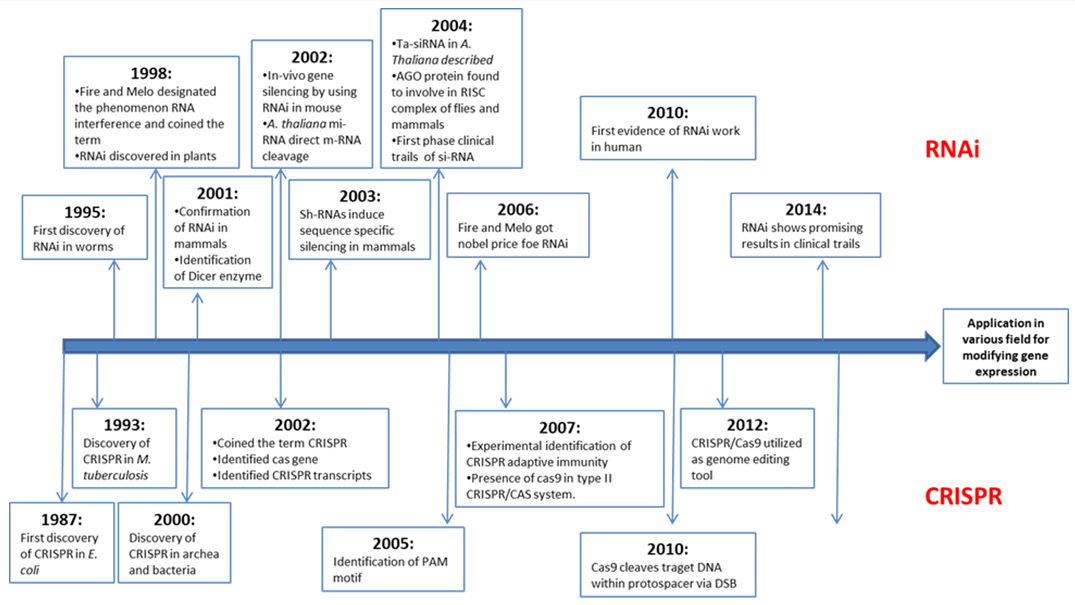
Figure 1: The timeline of milestones in RNAi and CRISPR.

RNA interference (RNAi)

In C. elegans, fire and his colleagues (1998) first coined the term RNAi. It is a new and strong method for halting mRNA gene expression. In mammalian cells, the Dicer which is a family of RNase III endonuclease cleaves longer dsRNA precursors to produce siRNAs. Dicer is a large endoribonuclease that processes double-stranded RNAs (dsRNAs), produces approximately 20 bplong small interfering RNAs (siRNAs) that act as effectors during RNA interference (RNAi) and excises microRNAs (miRNAs) from the precursors molecules that is assemble into RISC complex and direct this complex to complementary mRNA targets. Once binding with small RNA, RISC complex silence the targeted genes.

Components of RNAi

Dicer

Dicer enzymes are often less diversified in animals Meister, et al. [4] The RNase III enzyme family includes dicer enzymes, which recognise the ends of lengthy dsRNAs and cleave the RNA approximately 21 nt from the end Treiber, et al. [5]. Dicer-like enzymes have two catalytic RNase III domains that cleave both strands and leave two nucleotides 3′ overhangs due to their location on the dsRNA.

Arganoute

AGO proteins are highly specialised sRNA-binding modules that are important components of RISCs in silencing pathways. Small RNAs created during the initiation step are loaded into AGO proteins to direct sequence-specific regulation of gene expression, and AGO proteins perform the effector phase of silence.

RNA-Dependent RNA Polymerase (RDR) Proteins

Third component of RNAi pathway is RDR (RNA dependent RNA polymerase) which convert ssRNAs to dsRNAs, which are then processed by DCLs resulting in a new cycle of RNA silencing. RDRs are required for the synthesis of phased and repeat-associated siRNAs in sRNAs, but miRNAs and hairpin-derived sRNAs are RDRs independent RNAs. RDR activity was originally investigated in Chinese cabbage in 1971 and tomato cDNA was obtained in 1972. RDRs are the first components of plant small RNA biogenesis pathways to be identified and they are distinguished by a unique RNA-dependent RNA polymerase catalytic domain (RdRp).

Mechanism of RNA interference

RNA interference (RNAi) is a type of gene silence caused by the processing of double-stranded RNA (dsRNA) into short interfering RNAs (siRNAs). Both post-transcriptional and transcriptional gene silencing can be induced by RNAi. An enzyme resembling RNase III called Dicer which produces a 19–21 nucleotide duplex Hutvagner [6] siRNA with two nucleotide projections at its 3ʹ end via the cleavage of long dsRNAs. In RNAi pathway, the siRNA guide strand directs RISC to perfectly complementary RNA targets, which are then degraded. RNA degradation is induced by the PIWI domain of the Ago protein. siRISC identifies a completely complementary mRNA, resulting in Ago-catalyzed mRNA cleavage at a single duplex site. The functional siRISC is regenerated after cleavage, while the mRNA fragments are degraded further. Simultaneously transcribed RNAs (siRNAs) can recognise targets with poor complementarity.

RNAi as a Anti Viral Defence Mechanism

Plant antiviral biotechnology is now using RNA interference (RNAi). Artificial miRNA (amiRNA) is a robust biotechnology utilised in plants for gene silencing, and amiRNA engineering has been widely employed for the targeted down-regulation of endogenous genes in a variety of plants. The endogenous precursor of miRNA produced from the host has been widely exploited to substitute 21-nt long miRNA sequence with a region complementary to the target viral genome sequence. Multiple-target miRNAs can also effect multiple viruses at the same time. miRNA precursors which containing complementary sequences with Turnip yellow mosaic virus (TYMV) and the transgenic Arabidopsis expressing the recombinant miRNA precursors displayed specific resistance to these viruses Niu, et al. [7]. Based on the structure of the rice osa-MIR528 precursor. Sun, et al. [8] created three dimeric amiRNA precursor expression vectors that target the 3-proximal region of the CP genes of RSV and Rice black streaked dwarf virus (RBSDV). At a low temperature, the transgenic rice plants demonstrated great resistance to both RSV and RBSDV infection Sun, et al. [8].

RNAi as a Functional Genomics Approach and its Therapeutic Applications

In several animals, RNAi technology can be used to silence genes. In C. elegans, RNAi has been widely utilized for functional genomics. The majority of the 19,000 genes have been investigated at a high-throughput level. Ahringer and his colleagues created an RNAi library that contained 86 percent of C. elegans genes. This method has been tried and tested in a variety of additional model organisms. Mammalian cells have also been effectively treated using RNAi Hu, et al. [9]. In mammalian cells, many approaches for siRNA knockdown of specific genes have been used. In mammalian cells, DNA-vector-mediated RNAi silences genes selectively, while alternative expression methods are used for long-term silencing. For stable silence, the promoters of RNA polymerase (pol) II and III (U6 and H1, alone or together) have been employed.

CRISPR: (Clustered Regularly Interspaced Short Palindromic Repeats)

CRISPR is an adaptive immune mechanism of prokaryotic organisms. They are a family of DNA sequences used to detect and destroy the foreign invading DNA of bacterial infecting viruses (bacteriophage) with the help of CRISPR asoociated nucleases thereby preventing infection. Consequently, utilizing this innate immune mechanism, so called the CRISPR technology, allows the generation of desired modifications on specific target sequences in a genome. This modification is utilized to introduce mutations barring the need for traditional breeding programmes. The mutations are induced by exploiting the DNA repair pathways of the cells either through homology dependent repair (HDR) or non homologous end joining (NHEJ) pathways. The mutations so induced changes the Open reading frame (ORF) which in turn creates, gene knock down and knock-offs thereby creating variation. The advanced utilization of CRISPR is the ability to alter one specific nucleotide into another which is known as base editing. Types of CRISPR and associated nucleases used in CRISPR cascade:
1. Type I: The Type I systems are encoded by a single operon and consists of a signature cas3 gene which codes for a large protein with a helicase activity. They possess a single stranded DNA stimulated ATPase activity associated with the unwinding of both DNA-DNA duplex and RNA-DNA duplex Sinkunas, et al. [10].
2. Type II: The signature gene for this type of CRISPR system is cas9 which encodes for a multi- domain protein combining all the functions of effector complexes and cleavage of target DNA Jinek, et al. [11]. This system also has the ability utilize the cellular RNAse III and tracrRNA for processing the pre-crRNA Deltcheva, et al. [12]. Usually this system contains two domains in its nuclease namely the Ruv-C and the HNH domains which are both required to cleave the DNA in its targeted site.
3. Type III: The signature gene associated with this system is cas10 which encodes for a multi- domain protein. The protein consists of a palm domain which is similar to polymerases and cyclases of the polB family Anantharaman, et al. [13]. This type of CRISPR systems do not encode their own cas1 and cas2 genes but rather make use of crRNA produced from CRISPR arrays associated with type I or type II systems Nickel, et al. [14].
4. Type IV: This type of CRISPR system is generally fond in the plasmids of bacterial genomes. They lack cas1 and cas2 genes and also are not involved with any of the CRISPR arrays. This has been identified as the only CRISPR/cas system with no detectable CRISPR cassettes in the genome Makarova [15].
5. Type V: This type of CRISPR system comprises of rRNA and cas12a. The Cas12a protein contains a Ruv-C endo-nuclease domain which cleaves the non-target strand and the targeting strand to form a DSB Gao, et al. [16].

Advantages of CRISPR

To start with, this technology has been deemed to be more accurate and target specific for editing the sequences within the genome, thereby becoming one of the most widely used tool to create variations and mutations Zhang, et al. [17]. It is also efficient and fairly easy to use as compared to other editing tools like TALENs (Transcription activator-like effector nucleases) and ZFNs (Zinc finger nucleases) which are expensive and difficult to handle due to its simplicity in CRISPR/cas programming Qiu, et al. [18]. The efficiency of inducing mutation is highest through CRISPR. It is possible to target more than one target site from a single CRISPR cassette also known as multiplex CRISPR Sakuma, et al. [19].

Challenges in CRISPR Technology

Even though there has been advancement in scientific technologies, bioethical values play a huge role in the technology linked with genome editing technologies Brokowski [20]. Even though the efficiency of creating mutation in targeted sites are highest in CRISPR as compared to other gene editing tools, the efficiency of developing mutations through HDR is fairly low in comparision to the NHEJ pathway. Using CRISPR technology often triggers cell apoptosis on the subjected tissue intended for editing the genes, due to DSB’s Hu, et al. [21]. During the designing of gRNA, optimum care should be taken so that there is no off- target effects when subjected to editing the target tissue.

RNAi vs. CRISPR

RNAi is the post-transcriptional gene silencing mechanism generally found in eukaryotes. Whereas CRISPR is the new age genome editing tool that naturally serves as defence barrier found in prokaryotes. Because of its simplicity and wider capability, the type II CRISPR/Cas9 system has been widely used as a robust and versatile tool for gene editing now a day. RNAi, the major dominating genetic tool ruling the molecular biology laboratory over a decade, has some advantages and disadvantages over CRISPR technology. The CRISPR/Cas system has been used to induce gene modification by knockout via DSBs and NHEJ repair, transcription repression (CRISPRi/ CRISPR Interference) and transcription activation (CRISPRa/ CRISPR activation) Boettcher, et al. [3]. RNAi is the mechanism of post-transcriptional repression of gene expression by ds-RNA. CRISPR/Cas system and RNAi are occupying several overlapping domains of molecular biology research, raising the possibility that in near future one may selectively dominate the other. The fundamental difference between RNAi and CRISPRCas9 is that RNAi causes knockdown of gene expression at the post-transcriptional level by targeting mRNA, whereas CRISPRCas9 causes knockout of gene expression by targeting DNA. The main determinable factor for choosing the right tool is the aim of experiment.

In some cases like slow protein degradation and absence of rate limiting factor transcript levels—as caused by knockdown approaches may not produce a loss-of-function phenotype Boettcher, et al. [3]. In that case complete knock out of gene expression is the need of the experiment. When temporary reduction of gene expression is the primary need of experiment, RNAi can be advantageous over CRISPR as stable modification of the genetic code is undesirable and si-RNA may be lost after some generation Moreira, et al. [22]. Sometimes the complete elimination gene function is detrimental to the cell rather than partial loss Boettcher, et al. [3]. CRISPR/Cas system makes it possible to eliminate all variants of transcripts of a gene by using sg-RNA Wang, et al [23]. The CRISPR/Cas9 technique is a precise genome editing which leads to sequence-specific desirable modifications. The ontarget efficiencies of sgRNAs and shRNAs are found to be similar in a large-scale screen of CRISPR and RNAi (4). Off-target effects are less in case of CRISPR than RNAi (4). Due to the small length of si-RNA (24 nt), it can silence non-target mRNAs with only limited sequence complementarity (7). RNAi can only cause post-transcriptional or transcriptional gene silencing, while variations of CRISPR can be involved in gene knockout, knock down, transcrioptional activation or repression Boettcher, et al. [3]. Unlike CRISPR, RNAi targets RNA transcripts in the cytoplasm not genomic DNA in the nucleus. RNAi can be used in species for which only transcriptome data exists [24- 29].

Conclusion

CRISPR/Cas9 and RNAi both are powerful and useful tools for gene manipulations. CRISPR/Cas9 is more flexible than RNAi due to its versatile application in induction of InDels, repression or activatation of gene expression, and causing heritable and nonheritable genomic changes Unniyampurath, et al. [2]. The discovery of Cpf1 enzyme has revolutionized the CRISPR mediated genome engineering. RNAi is having a unique space in diverse genetic applications. But the increasing advantages of CRISPR/Cas9 may dominate over RNAi in near future. CRISPRi is the biggest threat to RNAi. However, wider application of CRISPRi system is yet to be established at a genome scale. In near future predictabily, CRISPR/ Cas9 will rule molecular biology lab for modifying gene expression, while RNAi will likely to be cornered with restricted domains of applications.

For More Articles: Biomedical Journal Impact Factor: https://biomedres.us

Open Access Journal on Medical Research

Pontocerebellar Hypoplasia Caused by De Novo Mutation in PAFAH1B1 (LIS1) Gene

Introduction

The advance of gene sequencing techniques has made it possible to determine the genetic origin of an increasing number of central nervous system malformations which previously did not have a defined etiology. Pontocerebellar hypoplasia (PCH) is a robust example of great variability of phenotypes associated with a specific group of malformations, characterized by atrophic changes of the cerebellar vermis and hemispheres, the ventral portion of the pons and inferior olivary nucleus, often associated with defects in cortical development and derived from mutations in a wide range of genes. To date, at least thirteen subtypes of pontocerebellar hypoplasia with distinct genotypes and phenotypes have been described, but none of them were caused by variants on plateletactivating factor acetylhydrolase IB subunit alpha gene (PAFAH1B1), related to lissencephaly [1]. Lissencephaly is a spectrum of cortical development malformations, characterized by neuronal migration defects, which comprises agyria, pachygyria and subcortical band heterotopia [2,3]. PAFAH1B1, also known as Lissencephaly 1 gene (LIS1), was the first gene identified as being related to lissencephaly, followed by X-linked doublecortin gene (DCX) [4].
Classical lissencephaly (or type 1 – lissencephaly), characterized by the presence of a thick cortex (composed of four abnormal layers) and the absence of other associated brain abnormalities (e.g severe congenital microcephaly, agenesis of the corpus callosum, or cerebellar hypoplasia) [4], is caused by mutations in some specific genes: PAFAH1B1, DCX (in males; in females, DCX mutations are associated with subcortical band heterotopia) and Aristaless-related homeobox, X-linked gene (ARX), in this case, characterized by a three-layered cortex [4]. There are other phenotypes of lissencephaly, associated with microcephaly (called “microlissencephaly”), agenesis of the corpus callosum or even cerebellar hypoplasia. However, until the present moment, there has been no record of mutations in PAFAH1B1 presented with pontocerebellar hypoplasia without cortical malformations, as we describe in this report.

Case Report

The proband is an 8-year-old male, first-child of nonconsanguineous parents, who was born after an uneventful pregnancy, labor and perinatal period (Birth weight: 3,960 g; occipitofrontal circumference: 33 cm). He presented micropenis and surgically corrected bilateral cryptorchidism. He has presented severe global developmental delay, a failure to thrive and deceleration of occipitofrontal circumference growth. Currently he has profound intellectual deficiency, inconstant eye contact, bilateral strabismus, inability to maintain his head up and presents spasticity and dystonia. His occipitofrontal circumference is 47.5 cm (z-score<-3) and fundoscopy is normal. Brain MRI (Figure 1) at 18 months of age disclosed a classic PCH with reduced white matter but normal cortical gyration pattern.

biomedres-openaccess-journal-bjstr

Note: Brain MRI at 18 months of age. Coronal FLAIR (A) and T1 inversion–recovery (B) images disclose a butterfly-type cerebellum, characterized by severe proportional hypoplastic vermis and cerebellar hemispheres. FLAIR image also shows diffuse cerebellar hyperintensity. A sagittal T1-weighted image (C) demonstrates thinning of the corpus callosum, attenuation of the pons, which is almost flat, and a small hypoplastic cerebellar vermis. Axial T2-weighted image (D) shows bilateral reduction of cerebral white matter with unremarkable cortical gyration pattern.

Figure 1: Neuroimaging of Pontocerebellar Hypoplasia.

Genetic Study

Whole exome sequencing was performed in order to identify genetic abnormalities that might be responsible for the clinical and radiological phenotype. No deleterious variants were detected in genes previously associated to PCH, but the patient harbors a missense heterozygous variant p. Arg273Gln (c.818G>A, NM_000430.3; Chr17:2,577,500) in exon 8 of PAFAH1B1, a highlyconserved (PhyloP>2) region and classified by SIFT and Polyphen as deleterious. This variant was neither present in 123,115 individuals from the Genome Aggregation Database (gnomad. broadinstitute.org) nor had been reported before. The variant c.818G>A was confirmed by Sanger sequencing in the index case and his parents were also examined, but it was not present in them.

Discussion

Pontocerebellar hypoplasia (PCH) is inherited as an autosomal recessive or X-linked trait, and it is characterized by profound congenital size reduction of the pons and cerebellum. Several genes have been implicated in PCH, including the autosomal transfer ribonucleic acid (tRNA) splicing endonuclease subunit 2 (TSEN2), TSEN15, TSEN34, TSEN54, O-phosphoserine t-RNA selenocysteine tRNA synthase (SEPSECS), cerebellar atrophy with progressive microcephaly (CLAM), arginyl-tRNA synthetase 2 (RARS2), vaccinia related kinase 1 (VRK1) and the X-linked Calcium/Calmodulin- Dependent Serine Protein Kinase (CASK) [5,6]. Up to now, no dominant inheritance has been associated with PCH. Herein, we report a case of classical PCH, associated with decreased white matter volume, although there is no cortical migration defect. However, molecular testing revealed a novel de novo heterozygous mutation in PAFAH1B1 (LIS1). This gene has been associated with laminar heterotopia and lissencephaly, occasionally combined with PCH [7,8]. In a comprehensive investigation of a large series of PCH, only 60% of cases have their molecular basis unraveled [5]. PAFAH1B1 product plays a critical role in neuronal migration during brain development [7,9].
Haploinsufficiency of PAFAH1B1 leads to neuronal migration defects of variable degrees of severity of the lissencephaly spectrum (OMIM # 607432), including Miller-Dieker syndrome (OMIM#247200). The p. Arg273Gln occurs in the hinge between two of the seven Water Displacement 40th Formula (WD40) domains, which are supposed to form a ring propeller-like structure. No missense mutations in any of the hinge regions of WD40 domains have been reported so far (The Human Gene Mutation Database). The variant Arg273*, leading to premature stop codon, has been reported several times associated with lissencephaly. De novo mutations in coding regions leading to protein structural change occur on average once at every generation [10]. Finding this type of change in a highly conserved 6 gene that is active during central nervous system formation strongly supports its association with PCH. As WES becomes more widespread, the number of genes associated with PCH will probably increase, and PAFAH1B1 might be one of these newcomers. A “new” phenotype for an “old” gene, or an “old” phenotype with a “new” gene?

For More Articles: Biomedical Journal Impact Factor: https://biomedres.us

Open Access Journal on Medical Research

Outcome of Fetoscopic Laser Coagulation in 61 Pregnancies with Twin-Twin Transfusion Syndrome in Viet Nam

Introduction

Given the increasing implementation of assisted reproductive technologies in the reproductive medicine, the incidence of twin pregnancies has been continuing to rise [1]. Thirty percent of twin gestations have monochorionic placentas which are susceptible to complications because of their unique placental architecture [2,3]. Twin-to-twin transfusion syndrome (TTTS) is one of the most severe complications of monochorionic twin pregnancies, accounting for 15% of twin pregnancies [3,4]. When transfusion syndrome occurs, the two fetuses share different portions of the same placenta and connect with each other through placental superficial vascular anastomoses [2], leading to the poor prognosis for both two pregnancies. Approximately 90% of twins acquired TTTS result in intrauterine demise of one or both fetus or perinatal death without any intervention [5]. About 50% of surviving twins are likely to leave irreversible neurological sequelae such as brain white matter necrosis, ventricular hemorrhage, cerebral dilation, cerebral palsy, neurocognitive impairment, etc [6-8]. The donor twin experiences the reduction of circulatory volume, anemia, decreased diuresis, oligohydramnios, hypotrophy. Meanwhile the recipient twin is characterized by circulatory hypervolemia, cardiomegaly, heart failure, fetal edema. Consequently, twins are at high risk of intrauterine demise or irreversible nerve damage after birth.
Treatment for TTTS includes methods such as amniocentesis, opening the amniotic septum between the two fetuses, fetoscopic laser coagulation (FLC) of the blood vessels connecting the two fetuses, or clamping the umbilical cord. Until now, the FLC and umbilical cord clamp are the only solutions to solve the pathogenesis of the TTTS. FLC was firstly conducted by De Lia at al. in 1990 under laparotomy and general anesthesia [9]. Subsequently, FLC has been improved and implemented in TTTS treatment worldwide [10]. It is demonstrated to be a beneficial therapy over serial aminoreduction in numerous population-based study as well as randomized controlled trial [4,10,11]. FLC has been also presented to reduce the long-term neurocognitive impairment in twins [12,13]. In Vietnam, Tam Anh Hanoi General Hospital is currently the only hospital that can perform FLC. However, there is no any study discussing the outcome of FLC in TTTS in Vietnam. Therefore, we conducted this study to evaluate the safety and therapeutic efficacy of FLC and other related factors in the outcomes of TTTS.

Methods

This is a prospective analysis study of pregnancies diagnosed with TTTS at the 16-26 week of gestation treated by FLC at Tam Anh Ha Noi General hospital, Vietnam. We included 61 consecutive pregnancies with moderate to severe mid trimester TTTS treated in our hospital of FLC between August 2018 and August 2019. This study protocol was approved by the Ethical Committee of Tam Anh Ha Noi General hospital on 1 August 1998. During the research period, all participants having TTTS confirmed by ultrasound and classified as the stage of II-IV (Quintero criteria) were extensively explained detailed risks and benefits of FLC. We only included participants accepting conducting the FLC and signing informed consent [14] [Box 1].

All procedures were performed under local anesthesia and ultrasound guidance. Firstly, the physician identified the position of fetoscope which is the most feasible position to approach the boundary line between the 2 amniotic chambers on the placenta surface from the amniotic fluid of the recipient fetus (Figure 1). In detail, this position has to allow the fetoscope parallel with the vertical axis of the donor fetus. Moreover, the attach positions of umbilical cords to placentas have to be observed clearly so that the conjunctive vessel will be detected more easily. Later then, the fetoscope (0°telescope, STORTZ) was introduced into the amniotic cavity of the recipient fetus and all the detected conjunctive vessels were coagulated along the whole vascular equator with a 0.4-0.6 mm fiber and laser system (20-40 watt of laser diot or laser YAGYttrium luminum Garnet).

The primary outcome was survival rate after 2 weeks of surgery and the live birth rate of one or both two fetuses. We also identified gestational age at delivery, the birth weight of neonate, the APGAR score and the weight of neonates seven day after birth as the secondary outcome variables. Gestational age in weeks was calculated based on the mother’s registration of her experienced due date. We divided gestation age at birth into very preterm (28- 31 weeks), moderate preterm (32-33 weeks), late preterm (34-36 weeks), early preterm (37-38 weeks), and full term (39-41 weeks). There was no post-term infants in the present prospective study. The birthplace at the Tam Anh hospital was only 21 pregnant women the remaining 40 pregnant women at the other 15 health facilities located in 14 provinces/cities nationwide.
Continuous variables are reported as mean and standard deviation (SD), categorical variables as absolute numbers and percentages. The differences in weight (percentage, %) was calculated by the following formulation ((weight of blood receiverweight of blood donor) *100/weight of blood receiver)) at the intervention time and the giving birth time, the end of gestation. Two-sided p<.05 were considered as significant using a t-test and odds ratio and 95% confidence interval. All statistical analyses were conducted with STATA Version 10.0.

Results

We recruited successfully 61 pregnant aged from 20 to 39, of which over 50% aged 25-29. Among them, 37 (60.7%) cases have reported having the first pregnancy, 17 (27.9%) cases having second pregnancy; 47 cases have no history of abortion and remaining 14 cases reported having abortion at least one time; 24 women have alive children and 37 other women with the first pregnancy. There was 52 women (85.2%) having history of caesarean section; 57 women (93.4%) have natural conception. 40.1% of case had anterior placenta, whereas 59.9% ones had posterior placenta. In included pregnancies, there were 26 cases having velamentous cord insertion. According to the Quintero staging classification, there was 19, 27, and 15 of pregnancies categorized to be stage II, III, and IV, respectively (Table 1).

Cervix length (mm) was 0-24 (eight cases), 25-29 (11 cases), 30-34 (21 cases), and 35-44 (21 cases). The difference (percentage) in weight (g) between two gestations was 1.33%-20% (12 cases), 20.1%-40% (32 cases), and 40.1%-62.23% (17 cases). The age at FLC (in week) was 16-20 (27 cases), 21-23 (24 cases), and 24-26 (10 cases). The number coagulated vascular anastomoses was 2-9 places in 34 cases and 10-23 places in 25 cases (Table 2). The estimated average (mean +/- standard deviation (SD)) of surgery time (minutes) was 35.3+/-11.1; of the volume (ml) of drained amniotic fluid was 1,210.7+/-817.6; of the percent (%) of differences between two fetal in weight was 30.9+/-13.6. At the intervention time, the estimated weight (g) of donor was significantly lower (280.5g+/-140.9g) than recipient (415.0g+/- 204.2g), (Appendix Table 1).

Among 61 women, 15 cases were miscarriage after the intervention within 14 days after performed FLC, remaining 46 cases being followed up. Nine cases reported having neonatal mortality before or after delivery. There were 37 cases having babies survived for 7 days, at the last study follow-up, Figure 2. Among 37 cases having successful live birth, Quintero II, III, and IV was 10 cases, 17, and 10, respectively. The proportion of participants who have no survivors was gradually decreased by increased Quintero staging, 47.4% (9/19), 37.0% (10/27), and 33.3% (5/15) for the Quintero II, III, and IV, respectively, non-statistically significant, p=0.394. The gestation age (in weeks) at delivery was full term of 39-41 (3 cases), earlier preterm of 37-38 (4 cases), late preterm of 34-36 (9 cases), moderate preterm of 32-33 (8) and severe preterm of 28-31 (13 cases), Table 3.

All 61 pregnant were healthy by the last follow-up time and 37 of them (60.7%) having at least one liveborn neonate, of which six cases gave the single birth of the donor fetus, eleven cases gave the single birth of the recipient fetus, and 20 cases had successful birth of both twins. Newborn survived for 7 days was 57 (46.7% of 122 gestation). There were not significant differences in birth weight between 6 single survived donors (mean 1,858.3g) and 11 single survived recipients (mean 1,954.5g), but significant differences in birth weight, among 20 survived twins, between donor twin (mean 1,410.0g) and recipient twin (mean 1,782.5g) in 20 cases of twins’ delivery, Table 4. The average weight (g) of the neonate was significantly lower in both the group of the gestation donors (1,918.8g versus 1,333.3g, p=0.012) and recipients (2,260.0g versus 1,763.5g, p=0.041) when compared the categorized Quintero II to the categorized Quintero III-IV, Table 5.

When compared the indicators of the first ten performed cases to the last-ten cases, from the first to the last 61 participants consecutively recruited, the average surgery time was longer (41.4 versus 33.0 minutes) but the average number of coagulated vascular anastomoses vascular anastomoses was lower (6.3 versus 10.7). Among 20 surviving twins, the estimated differences (in percentage) in weight between donor and receiver gestations have continuously getting worse and increased in two cases, from 25.09% and 6.83% (at the treated time) to 43.75% and 21.74% (at the end of gestation), respectively. This difference in weight between donors and recipients has decreased in the remaining 18 cases.

Discussion

The successful FLC in our study was over 60% after seven days of birth with severe TTTS. The performed intervention was the first time successfully treated in Viet Nam and the achievements and main findings are significantly contributing to the develop the treatment technique of FLC for further better managements of TTTS in Viet Nam and at low- and medium economy countries. The proportion of Quintero stage IV in our study population was two time higher than the previous four studies combined (24.6% (15/61) versus 12.3% (46/373)) [15-18], indicating big challenges to manage maternal and neonatal health in the present project. The higher proportion of the late stage of Quintero might be due to lack of established health facilities and trained physicians in the rural province in managing TTTS in the country. Therefore, the pregnant came to our hospital at the advanced stage of TTTS. Moreover, due to limited participants’ resources and poor quality of transportation from other regions in the South and Central areas to the Hanoi city, only one third (21/61) participants have completed the recommended monitoring and following-up at our hospital that might cause a poor outcome of the present pilot intervention project. Therefore, social-determinant factors of maternal and child health were possibly related to the present intervention study.
The average age at the intervention time (week) in the present study was similarly to the other five previous studies [15-18], from 20 to 22 week. The estimated average surgery time in the present study (35 minutes) was longer than the other recent studies in Germany and France (27-29 minutes) [18,19]. Due to local limited quality of disinfection, some pregnant women were suffering from infection after completed intervention (2/61) resulting the premature ended of the gestation. This unwanted outcome was not seen in the other studies [20]. The average gestation age of newborn of 37 cases in the present study was approximately 4 weeks lower when compared to the previous 953 cases of nine studies combined (29 versus 33 weeks) [15,16,20-26]. After the intervention, these pregnant often came back to follow their pregnancies at the local hospitals at disadvantage areas that might be related to the shorter period of gestation age. The deficiency of well-trained physicians in managing of TTTS could contribute to the high rate of preterm birth in our study. Furthermore, as in the low economic status, the hard labor, the malnutrition in these pregnant could be also the explanation for this. The overall survival of at least one surviving gestation in the present study (60.7%) was lower than previous four studies (69.2%-84.0%) [11,21,27-29].
Because there was not significant difference in weight (g) between single surviving donors and single surviving recipients but it was observed among 20 surviving twins, the possible explanation is that the nature of limited amount of blood due to unknown related factors in the donors or the velamentous cord insertion involvement. Fifteen participants have no survivor (all had miscarriage within 2 weeks of treatment) and most of them among the first half time of recruited participants due to our limited experiences. The other 9 cases had neonatal death might be due to poor taking care by other local health facilities outside of our hospital in Hanoi city. In our study, the mean estimated surgery time in the first 10 cases is shorter than the last 10 cases. After some first cases, our team gradually got more experience and spent shorter time to find the vascular anastomoses positions and do the coagulation. Treatment technique in TTTS would be transferred from the established health facilities of developed countries to the other in the low- and middle economy countries and this work would be coordinated by an international association and institution in taking care better maternal and child health. We assume that there is an estimated 3.4% of pregnancy is twin, of which about 33% is monochorionic; about 66% of monochorionic has the blood vessels connecting the two fetuses and about 15% of them developing TTTS [30], among about 140 million pregnancy in 2020 worldwide, there was about 155,509 TTTS globally.
In order to improve maternal and child health, global project to manage TTTS is highly needed. With about million newborn per year, Viet Nam will have about 1,111 pregnancy that will be suffering from TTTS and the findings of the present project is timely and that will contribute greatly to manage TTTS in the country. The present study certainly has limitations including a lack of followup for about two-third of participants due to social-economy difficulties, limited local health facilities. Another limitation is the lack of neurodevelopment follow-up of delivered babies. In spite of these limitations, during 13 months running of the project, we just managed for only about 5.5% of the estimated 1,111 pregnancy with severe TTTS and the work would be improved for further better management and outcome of TTTS in Viet Nam in particular and in other low- and median economy countries.

For More Articles: Biomedical Journal Impact Factor: https://biomedres.us

Open Access Journal on Medical Research

Oral PEY Treatment Induces Hair Regrowth and Improves Hair Quality in Patients with Alopecia

Introduction

Egg-yolk-based supplement have proven several biological beneficial effects and can be found in the market divided into unfertilized (commercialized) and fertilized. Scientific data demonstrated that eggs contain active compounds that may have a role in the therapy and prevention of chronic and infectious diseases. Although there is no currently scientific evidence to demonstrate it, eggs has been traditionally recommended for skin and hair problems and people have reported significant benefits after using eggs in their masks. However, the use of egg-yolk as a treatment of hair problems has stopped being used due to the appearance of more specific drugs and procedures to prevent hair loss and to improve hair regrowth. Among the most used procedures to combat hair loss nowadays, the transplant of hair with the FUE (follicular extraction unit) technique, is an innovative micrograft in which the hair follicles are transplanted together with all the perifollicular structures (including the blood vessels, the sebaceous gland or the piloerector muscle, among others) [1]. Furthermore, the major part of currently available conventional treatments are non-specific broad immunosuppressants administered locally or systemically, including topical and intralesional corticosteroids [2]. These treatments however are associated with multitude side effects such as skin atrophy, striae and telangiectasias, and even adrenal suppression, insomnia and glucose intolerance [3]. Other randomized controlled components under study include JAK inhibitors, such as tofacitinib, rollatini and baricite; fumaric acid esters; inhibitors of phosphodiesterases like apremilast; PGF2 analogs as Bigatures and Sterilant [4]. Nevertheless, in general, treatment of hair loss is often difficult and frustrating to patients and clinicians owing to the limited efficacy, the slow growth rate of hair and sometimes the adverse side effects.
In this study, we want to test the putative beneficial effects of our recently patented complement, PEY, in hair loss. PEY consists of an egg preparation obtained through a patented process from the fertilized egg-yolk [5]. A potent anti-inflammatory effect of PEY treatment has been recently reported in vivo [6]. Among the multitude of PEY components, highlight the presence of growth factors such as connective tissue growth factor (CTGF), platelet-derived growth factor (PDGF), and insulin-like growth factor (IGF-1) [6]; all of them with proven beneficial effects in the growth of hair follicles [7,8] Also, PEY exhibits a differential lipidomic and metabolomic profile with lipids clustered among different sphingolipid-related and immune-related pathways, and metabolites present in pathways related to omega-3 fatty acids, among others [6]. In addition to test PEY administration alone, we also want to analyze the putative efficacy of PEY to act as a booster with the most common-used treatments for alopecia.

Methods

All procedures and visits were conducted in accordance with the Guideline for Good Clinical Practice of the Institute Vila- Rovira. The study duration was around 4 months within which each volunteer (total of 45 patients) with alopecia was orally administered with 6 pills of PEY once a day. One group of patients (21 patients) only received PEY, whereas in the other group (24 patients) the PEY treatment was administered together with their usual treatment for allopecia. The PEY treatment consists of an egg preparation obtained through a patented process [5]. It comprises a mixture of yolk and white extracted from a fertilized egg that has been incubated for a short period, which then is lyophilized to obtain a commercial product called Excelvit. Qualitative analysis of the efficacy of PEY administration was determined by changes in global photography and with a satisfactory survey for each patient.

Results

Forty-Five Patients with a Clinical Diagnosis of Alopecia Were Enrolled in the Study

In the first group of patients (21 patients) PEY was administered alone to evaluate the efficacy of the product in alopecia. From this group, a total of 14 patients from the 18 that complete the treatment (77.7%) experimented a substantial improvement ranging from the stop in the hair loss to a better quality of the hair and skin. Notably, this effectiveness was higher in women than in men (86.41% vs 80%) (Table 1). In the second group of patients (24 patients) PEY was administered together with the usual alopecia’s treatment of the patient to analyze whether PEY can act as a booster to complement other treatments. In this case, from the 17 patients that complete the study 12 (70.58%) experimented an improvement of their condition ranging from hair regrowth Figure 1, stop of the hair loss Figures 2-4 and better quality of the hair Figure 3. Again, the efectiveness reported was higher in women than in men (76.92% vs 50%) (Table 1 and Figures 1,3,4). No side effects were detected by any of the patients in any treatment regimen along the study.

biomedres-openaccess-journal-bjstr

Table 1: Demographic and treatment variables of the study.

biomedres-openaccess-journal-bjstr

Figure 1: Patient demonstrated significant hair regrowth after the FUE transplant and PEY administration. This beneficial effect progressively continued up to 10 months of PEY treatment.

biomedres-openaccess-journal-bjstr

Figure 2: Patient after 3 months of treatment with PEY administered together with his conventional treatment for alopecia (PRP + Trichosol + Minoxidil 5%-F1% + Vit + Dutasteride). The patient experimented a reduction of hair loss, capillar regeneration and a faster hair growth. Also, the treatment provokes and increase in body weight.

biomedres-openaccess-journal-bjstr

Figure 3: Patient after 3 months of treatment with PEY administered together with her conventional treatment for alopecia (PRP + CAP Vitamin + Minoxidil 5%-F1%). The patient experimented a significant sttoping of the hair loss, capillar regeneration, a faster hair growth and a better hair texture.

biomedres-openaccess-journal-bjstr

Figure 4: Patient after 3 months of treatment with PEY administered together with her conventional treatment for alopecia (PRP + CAP Vitamin + Iraltone AGA plus). The patient experimented a complete sttoping of the hair loss.

Discussion and Conclusion

Our findings demonstrated that PEY treatment has a role in the treatment of alopecia. This treatment is effective when administered as a complement to conventional treatments for alopecia, but interestingly it has also a potent regrowing effect when administered alone. The anti-inflammatory effects already described for PEY, together with the higher CNTF and IGF-1 expression observed in cells treated with this compound, pointed to this molecule as potential candidates below the beneficial effects observed in patients (Cunill et al. Nutrients 2020). Indeed, both CNTF and IGF-1 has been linked to hair follicle growth and preservation [7,8]. Another highlighted point of this study is that the 73.3% of patients enrolled are woman, contrary to most of the studies of new drugs for alopecia that are focus exclusively or majority on men. Interestingly, the effects are proportionally higher in women than in men indicating that the hormonal control is one important aspect to take into account in the generation of new drugs. Remarkably, in contrast to major part of actual treatments for alopecia [2], no side effects were observed in any patient, probably due to the 100% natural origin of PEY.
In conclusion, this is an important study pointing towards the natural nutritional component PEY as a promising oral treatment for alopecia in both women and men and without any adverse side-effect. This study is in line with a growing data indicating that the food complements (like omega-3, curcumin, honey…) have a therapeutic effect through their anti-inflammatory and regenerative actions [9-13]. In this sense, it is essential that this compound will be introduced in the clinical practice, to increase the efficacy of human treatments, as will improve the general antiinflammatory state of the patients leading to a better resolution of medical interventions.

For More Articles: Biomedical Journal Impact Factor: https://biomedres.us

Open Access Journal on Medical Research

Evaluation of an Active Butanol Fraction Effects of Blighia Unijugata (Sapindaceae) Leaves on Some Biochemical Blood Parameters in Male Wistar Rats

Introduction

Blighia unijugata (Sapindaceae), a species widespread in tropical and equatorial regions of Africa has been widely used for its multiple therapeutic properties [1]. In Côte d’Ivoire, Nigeria and Congo, this plant is abundantly consumed as vegetables and also used in the treatment of fever, nausea and vomiting, leprosy, eye pain, cough, migraines, rheumatism, kidney pain and joint stiffness, dizziness and especially high blood pressure [2,3]. However, despite the increased use of this medicinal plant, data-t-on its efficacy and safety are not yet available, thus exposing populations to all kinds of dangers. However, scientific studies and the rational evaluation of plants commonly used in traditional medicine could guarantee their best use, reduce the risk of accidents and allow the establishment of specific treatments for its poisonings [4]. Blighia unijugata has been the subject of several scientific publications due to its multiple uses in traditional medicine [5,6]. However, to our knowledge, very few scientific studies have been undertaken to establish the risks of toxicity of this plant on biochemical blood parameters. Previous work in our laboratory has shown that the butanolic fraction of Blighia unijugata (BFBu) has a significant hypotensive effect in rabbit of the species Oryctolagus cuniculus (Leporidae) compared to the other four fractions from the total ethanolic extract of this plant [7]. This study was therefore carried out to evaluate the effects of this active fraction on serum biochemical parameters of male Wistar rats.

Materials and Methods

Materials

Plant Material: The plant material consisted of powdered leaves of Blighia unijugata. The fresh leaves were collected in Abidjan (Cocody) in June 2009. This species was identified by us using available herbaria and a book written by [8]. The confirmation was made by the National Floristic Center (NFC) of Félix Houphouët-Boigny University (Abidjan, Ivory Coast) where a herbarium specimen has been kept under voucher no165.
Animals: The experiments were carried out on 24 albino rats (Ratus norvegicus), healthy male, of Wistar strain, aged three months. Their average body weight was 148.00±7.07 g. These animals came from the animal facility of the Institut Pasteur de Côte d’Ivoire located in Adiopodoumé (Abidjan) and were reproduced in the animal facility of the Laboratory of the Ecology Research Center of Nangui Abrogoua University (Côte d ‘Ivoire) at an ambient temperature of 25±3 °C. The photoperiod was 12 hours of light and 12 hours of darkness. Water and pellet food (Ivograin®) were fed ad libitum to the rats before the experiment began. These animals were treated according to good laboratory practices [9].
Technical Material : The extraction equipment for the total ethanolic extract and fractions of Blighia unijugata consisted of an electric grinder (Culatti, France), an electronic scale (Mettler Toledo, PB 153-L, Switzerland), a magnetic stirrer (Stuart SB 162, UK), Buchner funnel, cotton wool and Wattman no1 filter paper, Erlenmeyer flask, 500 mL conical flask, rotary evaporator (Büchi R110, Germany) and an oven (Retsch, Germany). The subacute toxicity study was performed using a gastric tube suitable for gavage of rats, pastor pipettes, and dry tubes. The analysis of the biochemical parameters was carried out using an automatic device (KENZA MAX Biochemis Try, France).
Solvents : The total ethanolic extract, hexanic, chloroform, ethyl acetate, butanolic and aqueous fractions of Blighia unijugata were obtained by extractions with different solvents (distilled water, 96% ethanol, hexane, chloroform, ethyl acetate and n-butanol).

Methods

Preparation of the Total Ethanolic Extract of Blighia Unijugata : The total ethanolic extract of Blighia unijugata leaves was prepared according to the method of some authors [10]. The fresh leaves were thoroughly washed with tap water and then dried at room temperature. They were sprayed using an electric grinder (Culatti, France). Cold maceration was carried out with 100 g of powder in 2 liters of 96% ethanol, for 48 h, with magnetic stirring. The resulting solution was first filtered through Buchner and then through Wattman No1 filter paper. This operation was repeated twice on the same powder residue. The filtrates obtained were added and concentrated under reduced pressure at 45 °C using a rotary evaporator (Büchi R110, Germany). The concentrated product, obtained after evaporation, was collected in a container and dried in an oven (Retsch, Germany) at 45 °C for 48 h according to the method described by [11]. The 13.3 g paste obtained corresponds to the total ethanolic extract (TEE) of Blighia unijugata. It was stored at -5 °C in a hermetically sealed jar.
Preparation of Fractions of the Total Ethanolic Extract of Blighia Unijugata : Different fractions of the total ethanolic extract (TEE) were obtained by successive liquid-liquid extractions, with four solvents of increasing polarities (hexan, chloroform, éthyl acétate and n-butanol) according to the methods of [12,13]. Ten grams of the TEE was dissolved in 200 mL of hot distilled water (45 °C). The whole was homogenized by magnetic stirring for 15 min and then it was filtered. Aqueous filtrate obtained was exhausted for 10 min at 27±2 °C with 200 mL of hexane, thus giving two phases after decantation (hexan phase and residual aqueous phase). Residual aqueous phase was again treated for 10 min at 27±2 °C with 200 mL of chloroform to in turn give two phases (chloroform phase and residual aqueous phase). Same operation was successively treating the residual aqueous phase with ethyl acetate to also give an ethyl acetate phase and residual aqueous phase, then with n-butanol to finally obtain butanol phase and residual aqueous phase after settling. Each organic phases and the residual aqueous phase obtained was recovered and concentrated under reduced pressure throught a rotary evaporator (Büchi R110, Germany). Various concentrated products obtained were collected in containers and dried in an oven (Retsch, Germany) at 45°C for 24 h according to the method of some authors [14]. Thus, hexan (0.6 g), chloroform (1.1 g), ethyl acetate (1.6 g), butanol (3.18 g) and aqueous (3.12 g) fractions were obtained. These end products were tested on rabbit blood pressure in the laboratory. Of these five fractions, butanol fraction coded BFBu had a greater hypotensive effect compared to the other four fractions. BFBu was therefore chosen for this study. The extractions were repeated several times in order to obtain a sufficient amount of extract to perform the tests. BFBu was stored at -5 °C in a tightly closed jar to prevent spoilage.

Methods of the Study of Subacute Toxicity

Preliminary Tests : The results of the acute oral toxicity study were used as the basis for the selection of the doses of BFBu that were administered to the rats. The initial dose level, selected on the basis of this orientation study, is well below the Maximum Tolerated Dose (MTD). At the end of these tests, the doses of 50; 500 and 1000 mg/kg bw were chosen to be the doses to use during this study.
Distribution and Treatment of Rats : Animals were divided into 4 homogeneous batches of 6 rats. This homogeneity of the different batches is a function of the body weight of the rats. The tests were carried out on a control batch and 3 batches of treated animals. According to the method described by [15], each rat from the control group received by gavage distilled water at 10 mL/ kg bw. Butanolic fraction (BFBu), diluted in distilled water was administered, by gavage, to each of the rats of the 3 test groups, respectively at doses of 50; 500 and 1000 mg/kg bw. Volume of FBu administered to the rats of each batch was less than 2 mL. Rats were given distilled water or BFBu daily by gavage every morning between 7 and 8 hours AM. GMT for 28 days.
Blood Sample : Days 7, 14, 21 and 28 of treatment, blood samples were taken on an empty stomach from the rats previously anesthetized with ether for 2 to 3 minutes, by puncture at the level of the orbital sinus of the eye, at the level of the eye using pastor pipettes according to the technique described by [16-18]. Thus, 2 to 4 mL of blood from each animal of the 4 batches were collected in dry tubes.
Determination of Biochemical Parameters : Automatic device (KENZA MAX Biochemis Try, France) was used for determination of the biochemical parameters. Blood collected in dry tubes was centrifuged at 3000 rpm for 10 min and the resulting serum was stored at -20 °C until assaying for biochemical blood parameters. Creatinine, aspartate aminotransferase (AST) and alanine aminotransferase (ALT) were assayed by cinetic method [19]. Urea was determined by enzymatic method [19,20]. HDL (High Density Lipoprotein) Cholesterol Assay Method in VITROS HDL Plate is based on a non-HDL cholesterol precipitation technique followed by enzymatic detection [21]. Measurements of serum concentrations of total proteins, total cholesterol, triglycerides, bilirubins (direct and indirect) were carried out by the colorimetric method [19-23]. Glucose present in the serum was determined according to the glucose oxidase colorimetric method with the “Glucose Trinder GPO-POD” kit [20,24]. Finally, the electrolytes (magnesium, calcium, sodium, potassium and chlorine) were also determined by the colorimetric method [25-28].

Evaluation of the Atherogenicity Index

Atherogenicity index of rats was determined according to the formula of [29].
Atherogenicity index = total cholesterol / HDL − cholesterol

Statistical Analysis

Statistical analysis of data and graphical representations were performed using Graph Pad Prism 5.01 software (San Diego, USA). The results obtained were expressed as the mean followed by the standard error of the mean (M±ESM). Difference between the mean values of the biochemical parameters was determined by one-way analysis of variance (ANOVA 1) and supplemented by Tukey’s test for the comparison of the means of the parameters of the different test groups compared to the control group. The significance level was set at p<0.05.

Results

Effects of Bfbu on Biochemical Parameters

Effect of Bfbu on Creatinine Level : The mean creatininemie of the treated batch with BFBu at 50 mg/kg bw is lower than that of the control from D7 to D21 with the exception of D28 where it is higher than that of the control. D7, the creatinine level of the treated batch with BFBu at 500 mg/kg bw is lower than that of the control. However, from D14, this rate increases until the end of this study with a significantly (p<0.05) high rate with a value of 8.17±0.48 mg/L on D28. Only the creatinine level of the batch treated with BFBu at 1000 mg/kg bw increased throughout this study. This increase of 8.67±0.33 mg/L is significant (p<0.01) on the 28th day of treatment compared to the creatinine level of the control batch which is 6.17±0.60 mg/L (Figure 1A).

Effect of Bfbu on the Urea Rate: In the treated batches with BFBu at 50 and 500 mg/kg bw, the urea levels decreased compared to the control during this study, with the exception of D28 where there was an increase in this level. However, this decrease remains insignificant throughout this study. Moreover, from D7 to D14, in the treated batch with BFBu at 1000 mg/kg bw, a significant drop (p<0.05) in the urea level was noted compared to the control. Rate decreases and passes respectively from 0.21±0.02 g/L to 0.15±0.0 g/L and from 0.45±0.01 g/L to 0.37±0.02 g/L. However, from the 21st day of treatment, the amount of urea increased significantly (p<0.01-0.001) in this batch compared to the control batch. The urea rate increases and goes from 0.33±0.03 g/L to 0.54±0.01 g/L on D21 and from 0.35±0.04 g/L at 0.54±0.04 g/L on D28 (Figure 1B).
Effect of Bfbu on Total Cholesterol Level: Total cholesterol level of the treated batch with BFBu at 50 mg/kg body weight is lower than that of the control on D7 and D14. However, from D21, this rate undergoes a non-significant increase compared to the control. Moreover, the treated batches with BFBu at 500 and 1000 mg/kg bw, the total cholesterol level decreased during this study with the exception of the treated batch with BFBu at 500 mg/kg bw where this rate increased on D21. This decrease in mean cholesterolemia is significant (p<0.01-0.001) on D7 and D14 respectively compared to the mean value of the cholesterol level in the control batch, but it normalizes from D21. The cholesterol level decreases from 1.05±0.03 g/L to 0.85±0.03 g/L on D7 and from 0.99±0.03 g/L to 0.83±0, 03 g/L on D14 (Figure 1C).
Effect of Bfbu on Hdl-Cholesterol Levels: The mean HDL cholesterol values of the batch of rats treated with BFBu at 50 mg/ kg bw remained lower than that of the control during this study with a significant decrease (p<0.05) from a value of 0.27±0.01 g/L on D14. As for the HDL cholesterol level in the batch of rats treated with BFBu at 500 mg/kg bw, a decrease was noted during this work with the exception of D21 where its mean value is similar to that of the control. The mean value drops significantly (p<0.01) from 0.33±0.01 g/L to 0.27±0.01 g/L on D28 at this dose. Finally, in the batch treated with BFBu at 1000 mg/kg bw, the level of HDL cholesterol decreased throughout this study with a significant drop (p<0.001-0.01) in this value which went from 0.35±0.01 g/L at 0.30±0.00 g/L and from 0.33±0.01 g/L to 0.28±0.01 g/L respectively on D21 and D28 after administration of BFBu (Figure 1D).
Effect of BFBu on LDL-Cholesterol Levels: In the treated batch with BFBu at 50 mg/kg bw, the LDL-cholesterol level did not undergo any significant variation from D7 to D28 compared to that of the control batch In the treated batches with BFBu at 500 and 1000 mg/kg bw, the level of LDL-cholesterol decreased during this study. This drop is significant (p<0.05-0.01) on D7 and D14 in these two batches. From D21, BFBu does not cause any significant variation in the LDL cholesterol level with the exception of the treated batch with FBu at 500 mg/kg of bw where a significant drop (p<0.05) in this level was recorded on D28 with a value of 0.28±0.05 g/L (Figure 1E).
Evaluation of the Atherogenicity Index of BFBu: At a dose of 50 mg/kg bw, BFBu did not cause any significant increase in the atherogenicity index compared to that of the control group during this study. In the treated batch with BFBu at 500 mg/kg bw, there was a non-significant decrease on D7 followed by a significant decrease (p<0.05) in this index on D14 with a value of 2.77±0.16. From D21 to D28, the atherogenicity index of this batch remains similar to that of the control batch. Finally, in the treated batch with BFBu at 1000 mg/kg bw, the atherogenicity index remained low throughout this study compared to the mean value of the control batch. This drop is significant (p<0.05) on D14 where the mean value is 2.76±0.12 (Figure 1F).
Effect of BFBu on Triglyceride Levels: Repeated administration of BFBu at 50 mg/kg bw causes a non-significant decrease in the triglyceride level on D7 followed by an increase in this level from D14 until the end of this study. In the presence of BFBu at 500 mg/kg bw, the triglyceride level increases during this study with a significant increase (p<0.01-0.05) respectively on the 7th and 14th day of treatment. The respective average values recorded are 0.66±0.05 g/L and 0.50±0.01 g/L. In the treated batch with BFBu at 1000 mg/kg bw, a significant increase (p<0.01-0.001) in the level of triglycerides was noted respectively on D7 and D14 where the average levels are 0.64±0.01 g/L and 0.56±0.02 g/L. This increase is followed by a non-significant drop in triglyceridemia compared to the control batch on D21 and D28 (Figure 2A).
Effect of BFBu on Total Protein Level: Total protein level of the treated batch with BFBu at 50 mg/kg bw remained almost lower than that of the control during this study with the exception of D28 where this rate increased from 46.80±0.95 g/L at 48.70±1.05 g/L. Variations caused by BFBu at this dose are not significant (p≥0.05) compared to the control batch. In the treated batches with BFBu at 500 and 1000 mg/kg bw, an increase in this rate was noted throughout this work. This increase is significant (p<0.05) in the treated batch with BFBu at 500 mg/kg bw from D14. The increase in total protein level caused by BFBu at 1000 mg/kg bw is only significant (p<0.001) on the 14th day of treatment where its value is 61.50±0.43 g/L (Figure 2B).
Effect of BFBu on Alanine Aminotransferase Level: Alanine aminotransferase (ALT) level of the treated batches with BFBu at 50 and 500 mg/kg bw is lower than that of the control batch on D7. However, from D14 until the end of this study, the level of ALT increased in these batches compared to that of the control batch. The increase in this level in the treated batch with BFBu at 500 mg/kg bw is significant (p<0.001) from this period, whereas the increase in this rate is only exceptionally so on the 14th day in the lot treated with BFBu at 50 mg/kg bw where its value is 138±6.63 IU/L. In the treated batch with BFBu at 1000 mg/kg bw, the level of ALT, which remained almost the same to the control on D7, underwent a significant increase (p<0.001) from D14 to D28 with values of 165±2.63 IU/L (J14), 213±3.62 IU/L (J21) and 265±9.56 IU/L (J28) (Figure 2C).
Effect of BFBu on Aspartate Aminotransferase Level: On D7, all the doses of BFBu tested caused a significant decrease (p<0.05- 0.001) in the level of aspartate aminotransferase (AST) compared to the control. The mean AST value of the control batch is 188±7.92 IU/L. The AST levels recorded are 128±6.94 IU/L, 145±11.00 IU/L and 155±2.79 IU/L respectively for doses of BFBu of 50, 500 and 1000 mg/kg bw. However, from D14, an increase in the AST rate was recorded in all the batches treated with BFBu. This increase was not significant in all of the batch test from D14 to D21. On D28, in the treated batches with BFBu at 500 and 1000 mg/kg bw, a significant increase (p<0.01) in its mean value was recorded compared to that of the control batch (Figure 2D).
Effect of BFBu on Total Bilirubin Level: All tested doses of BFBu, the mean values of the total bilirubin level underwent a nonsignificant decrease (p≥0.05) compared to that of the control lot during this study with the exception of the lot treated with BFBu at 1000 mg/kg bw on D7 where this rate, a value of 9.50±0.34 mg/L, remained almost identical to that of the control (Figure 2E).
Effect of BFBu on Conjugated Bilirubin Level: With regard to the level of conjugated bilirubin, the mean values in the presence of all the doses of BFBu tested are lower than that of the control during this study. At 50 mg/kg bw, a significant drop (p<0.01-0.001) in the conjugated bilirubin level was recorded on D14 and D28, respectively, but this decrease was not significant on D7 and D21. At 500 mg/kg bw, the conjugated bilirubin level underwent a significant decrease (p<0.05-0.001) from the 14th day of treatment with the exception of D7 where this decrease is not significant with a value of 0.55±0.08 mg/L. Finally, at 1000 mg/kg bw, the level of conjugated bilirubin decreases significantly (p <0.05 – 0.001) during the treatment except on D21 when a non-significant drop in its value compared to the control was recorded. (Figure 2F).

biomedres-openaccess-journal-bjstr

Figure 1: Effect of Bfbu on Some Biochemical Blood Parameters and Atherogenicity Index.
Note: * p < 0.05; ** p < 0.01; *** p < 0.001; n = 6 : Differences were significant when values of treated groups with BFBu were compared to that of control group, at the same corresponding day.

biomedres-openaccess-journal-bjstr

Figure 2: Effect of Bfbu on Some Biochemical Blood Parameters.
Note: * p <0.05; ** p <0.01; *** p <0.001; n = 6: Differences were significant when values of treated groups with BFBu were compared to that of control group, at the same corresponding day.

Effect of BFBu on Glucose Level: No significant variation in blood glucose was recorded in the treated batches with BFBu at 50 and 500 mg/kg bw compared to the control during this study. On the 7th and 14th day of treatment, the mean values of the blood glucose level underwent a significant increase (p<0.05) in the treated batch with BFBu at 1000 mg/kg bw compared to the control batch. The average blood sugar values are 1.00±0.06 g/L (D7) and 0.85±0.02 g/L (D14). However, from the 21st to the 28th day of treatment, a significant decrease (p<0.05-0.001) in the blood glucose of the rats was observed in the treated batch with FBu at 1000 mg/kg bw compared to the mean value of the glycemia of the rats of the control group (Figure 3).

biomedres-openaccess-journal-bjstr

Figure 3: Effect of Bfbu on Glucose Levels.
Note: *p<0.05; ** p<0.01; ***p<0.001; n = 6: Differences were significant when values of treated groups with BFBu were compared to that of control group, at the same corresponding day.

Effects of BFBu on Electrolytes

Effect of BFBu on Sodium Level: Repeated administration of BFBu at doses of 50, 500 and 1000 mg/kg bw did not impacted with sodium levels during this study with the exception of the batch treated with BFBu at 1000 mg/kg bw where an increase significant (p<0.05) was recorded on the 28th day of treatment compared to the control batch. The sodium level is 143±1.31 meq/L at this dose (Figure 4A).
Effect of BFBu on Potassium Level: At 50 mg/kg bw, BFBu induces a drop in the potassium level with the exception of D28 where this rate increased compared to the control. These variations are not significant at this dose. At a dose of 500 mg/kg bw, the potassium level is lower than that of the control during this study. The drop in this rate is significant (p<0.001) on the 14th day of treatment in this batch where a value of 3.30±0.10 meq/L was recorded. However, on D21, the potassium level is higher than that of the control. As for the treated batch with BFBu at 1000 mg/kg bw, the potassium level remained lower than that of the control during this study with significant decreases (p<0.05-0.001) noted respectively on D7 and D14 but not significant of D21 to D28 compared to the control batch (Figure 4B).
Effect of BFBu on Chlorine Level: Chlorine level of the treated batch with BFBu at 50 mg/kg bw decreased significantly (p<0.05) on D14 compared to the control batch. As for the treated batches with BFBu at 500 and 1000 mg/kg bw, the chlorine level increased during this study with exceptionally significant increases (p<0.01-0.001) in this rate on D28 compared to the control batch. The respective values recorded at these doses are 122±3.53 meq/L and 119±1.50 meq/L. However, on D21, the chlorine level is almost identical in all the batches treated with BFBu (Figure 4C). Effect of BFBu on Calcium Levels: The calcium level of the treated batches with BFBu at 50, 500 and 1000 mg/kg bw did not undergo any significant variation (p≥0.05) during this study compared to the control (Figure 4D).
Effect of BFBu on the Magnesium Level: On D7, the magnesium level of the treated batches with BFBu at 50 and 500 mg/kg bw is higher than that of the control. However, from the 14th day of treatment, this rate decreased in these batches compared to the control. Only the magnesium level of the treated batch with FBu at 1000 mg/kg bw continuously decreased during this work. All the variations caused by BFBu at the doses tested remained insignificant (p≥0.05) compared to the control during this study (Figure 4E).

biomedres-openaccess-journal-bjstr

Figure 4: Effect of Bfbu on Electrolytes.
Note: p> 0.05; n = 6: non-significant difference compared to the control batch
* p <0.05; ** p <0.01; *** p <0.001; n = 6: Differences were significant when values of treated groups with BFBu were compared to that of control group, at the same corresponding day.

Discussion

The various biochemical parameters examined in this study are useful indicators for assessing the toxicity of plant extracts in animals [30]. Analysis of blood parameters is relevant in risk assessment since changes in the hematologic system have a very high predictive value for toxicity in humans [31]. The significant reduction in the level of transaminases (AST and ALT) observed at the start of the experiments indicates that the absorption of BFBu does not cause cytolysis but rather a protective effect in the liver. BFBu does not harm the liver within the first week of administration. However, at the end of this study, the increase in these parameters following the administration of BFBu could be due to the appearance of necrosis in one or more organs, namely the liver, heart and kidneys, thus causing an increase in the level of these enzymes in the blood. Indeed, according to [32], damaged tissue is generally associated with the release of specific enzymes from the tissue, or the affected organ, into the bloodstream. The consequence is an increase in the activity of such enzymes in bodily secretions. ALT is a cytoplasmic enzyme found in very high concentrations in the liver, and an increase in this specific enzyme suggests liver cell damage while AST is less specific than ALT as an indicator of liver function. The increase in the level of transaminases (ALT and AST) has also been observed in liver disorder [33-36]. These results are in agreement with those of [37,38] who reported that the ethanolic and methanolic extracts of Alchornia cordifolia leaves cause an increase in the activity of transaminases. These authors therefore suggested that these extracts exhibit hepatotoxic effects at doses of 250 to 500 mg/kg bw and 800 to 1600 mg/kg bw, respectively. BFBu would therefore be, at the tested doses of 500 and 1000 mg/ kg bw and in the long term, probably poorly tolerated by the liver. According to [39], the increase in the serum total protein level is an indication of tissue damage while the significant decrease in the total protein content of the liver is a reflection of hepatic nontoxicity. The significant increase in the total protein content in the batches of rats treated with BFBu at 500 and 1000 mg/kg bw could therefore indicate an increase in protein stores and therefore suggest hepatic toxicity, which would again confirm the detrimental effects of BFBu at 500 and 1000 mg/kg bw. Bilirubin, a metabolic breakdown product of heme derived from senescent red blood cells, is also one of the most commonly used tests for liver function. Its concentration could indicate the condition of the liver and the type of liver damage [40,41]. The significant reduction in total and conjugated bilirubin levels could be provided by a deficiency in the secretory function of these proteins. Certain substances present in FBu could therefore imply inhibitions at the level of the hepatic cells. This reduction can also affect the functional activity of the liver. Doses of BFBu of 50 and 500 mg/kg bw did not significantly affect blood sugar. Only the dose of 1000 mg/kg bw increased it on D7 and D14 and decreased on D21 and D28. These results suggest that this fraction has hyperglycaemic activity followed by hypoglycaemic activity at high doses. At the end of this study, the hypoglycaemic activity could be attributed to the existence of the molecules demonstrated in this fraction. Indeed, flavonoids from different plants have shown a promising hypoglycemic effect in diabetic animal models [42-44]. Saponins are triterpene, steroidal or alkaloidic glycosides. Authors have shown a hypoglycemic activity of triterpene glycosides [45,46]. It is therefore possible that the presence of flavonoids and saponins in FBu could be responsible for the hypoglycemic effects of this fraction. Some authors reported a significant decrease (p<0.05) in the fasting blood glucose level of rats made diabetic with alloxane when given the ethanolic extract of Ficus microcarpa, orally for two weeks [47]. Finally, these results are in agreement with the studies conducted respectively on the seed cotyledon and leaves of Chrysophyllum albidum at doses between 250 and 1000 mg/kg bw [48,49] on the one hand, and on the other hand on Bixin (10 mg/kg bw) extracted from the seeds of Bixa orellana [50]. These authors have shown that these extracts significantly decrease the glucose level in normal and diabetic rats induced by alloxane. However, the notable increase in blood sugar observed in this study could be linked mainly to the stress undergone by the animals at the time of sampling as suggested by [51] during work with Momordica charantia in rabbits. However, a real hypoglycemic effect could be attributed to this fraction by carrying out further study on suitable models. The significant (p<0.01) increase in mean serum creatinine was seen in the lots that received the highest doses of FBu (500 and 1000 mg/kg bw). This result suggests that BFBu is toxic to the kidneys. Creatinine is the major catabolic product of muscles and is secreted by the kidneys. Serum creatinine levels are used as an indicator of kidney defects [38,52,53]. The significant drop in the urea level observed at the start of treatment in the batch treated with FBu at 1000 mg/kg bw would be a sign of the good functioning of the kidney and the lack of renal toxicity due to this fraction. However, the increase in this level in this batch would be an indication of azotemia. According to Nduka [54], the high urea level is associated with the increased catabolism of tissue proteins. The excess protein in the blood collapses and urea excretion decreases, increasing its level in the blood. The increase in this level would also be a sign of damage to the kidney, which can no longer extract nitrogenous excretion products from the blood and concentrate them in the urine, thus confirming the renal toxicity caused by BFBu. Other indicators of kidney function such as sodium, potassium and chlorine were significantly affected by BFBu except calcium and magnesium which were not. The urea, sodium and chlorine levels significantly increased in the treated batches with BFBu at the highest doses during this study. These results probably suggest nephrotoxicity of BFBu at high doses, which would support the hypothesis made about the increased creatinine level. However, the lack of a significant effect of BFBu on serum calcium and magnesium concentrations in animals suggests that the secretory capacity of the kidney and normal organ function related to these parameters were not affected. According to the work of [41] on the aqueous extract of Felicia muricata at doses of 50, 100 and 200 mg/kg bw, the calcium and magnesium levels of the rats were not significantly modified. The hypokalaemia caused by repeated administration of BFBu may be due to an alteration in tubular reabsorption of potassium. Similar effects have been reported by [55] with the aqueous extract of Arctotis arctotoides on the decrease in potassium concentration. These authors suggested that this plant would not cause any damage to the cardiovascular system, which could also be the case with BFBu. Changes in the concentration of major lipids such as total cholesterol, HDL-cholesterol, LDL-cholesterol, and triglycerides, as well as the atherogenicity index may provide useful information on lipid metabolism and heart predisposition, atherosclerosis and its associated coronary heart disease [29,56-58]. In this study, the decrease in serum cholesterol level could be explained by the deterioration of cholesterol biosynthesis. This has also been suggested by [59] who showed that the aqueous extract of Chrysocoma ciliata at doses of 50, 100 and 200 mg/kg body weight decreases the cholesterol level in rats. Elevated cholesterol concentration is an important risk factor for cardiovascular disease [60]. Therefore, lowering cholesterol at the doses of BFBu tested could be clinically beneficial as this fraction is unlikely to be associated with cardiovascular risk. Triglycerides are the storage forms of fatty acids. The decrease in the level of lipids such as HDL and LDL cholesterols as well as the decrease in the atherogenicity index may be an indication that FBu would not predispose animals to the risk of atherosclerosis and associated coronary heart disease. According to several authors, the total cholesterol/cholesterol- HDL ratio constitutes a revealing index of arterial and especially coronary risk. This ratio would indicate an increased risk if it is greater than 4.4 [29]. However, according to the results obtained during this study, the atherogenicity index of each animal was lower than this value. BFBu could therefore be protective of heart tissue. Furthermore, the decrease in serum lipid parameters investigated in this study suggests that the lipid metabolism of animals was disturbed by the administration of BFBu. This disturbance could be an indication that BFBu would probably not predispose the animal to atherosclerosis and associated coronary heart disease despite the notable elevation (p<0.001) in triglyceride levels during the first 14 days in batches treated with BFBu at 500 and 1000 mg/ kg bw.

Conclusion

This study showed that butanol fraction of Blighia unijugata (BFBu) produced changes in biochemical parameters following repeated administration. BFBu caused disruption of the carbohydrate, lipid and protein balances. At high doses, BFBu caused hepatic and renal toxicity. However, the fraction tested at dose of 50 mg/kg bw, is non-toxic. This fraction has hypoglycemic effects and could be used in the management of diabetes, respectively. BFBu would not predispose rats to the risk of atherosclerosis and associated coronary heart disease.

For More Articles: Biomedical Journal Impact Factor: https://biomedres.us

Open Access Journal on Medical Research

Kaizen Concept or Methodology to Improve the Performance in the Health Care and Patient Safety Business and its Advantages

Introduction

Kaizen is a Japanese term Investopedia [1] meaning “change for the better” or “continuous improvement”. It is a Japanese business philosophy regarding the processes that continuously improve operations and involve all employees. Kaizen sees improvement in productivity as a gradual and methodical process. The concept of kaizen encompasses a wide range of ideas. It involves making the work environment more efficient and effective by creating a team atmosphere, improving everyday procedures, Investopedia [1] ensuring employee engagement, and making a job more fulfilling, less tiring, and safer. Kaizen (改善, かいぜん), the Sino-Japanese word Wikipedia kaizen [2] for “improvement”, is a concept referring to business activities that continuously improve all functions and involve all employees from the CEO to the assembly line workers. Kaizen also applies to processes, such as purchasing and logistics that cross organizational boundaries into the supply chain. It has been Wikipedia kaizen [2] applied in healthcare, psychotherapy, life coaching, government, and banking (Figure 1). A true Japanese development philosophy, Appvizer [3] kaizen is composed of two words, kai 改, and zen 善 which means “change” and “better”. The Kaizen approach consists of 5 founding elements
(1) Teamwork
(2) Personal discipline
(3) Improved morale
(4) Quality circles
(5) Suggestions for improvement. Chi va piano va sano ! (Slowly but surely!) (One step at a time!).
Toyota has been using kaizen to
(1) Optimize: productivity, safety (reduce risks), quality, deadlines, costs, working conditions.
(2) Value collective intelligence and Appvizer [3] DE compartmentalize skills,
(3) Reduce waste (to improve inventory management).

biomedres-openaccess-journal-bjstr

Figure 1: (Jenniffer M [20]).

There are several coping strategies Kosta S, et al. [4] to help health practitioners implement kaizen for the benefit of their organization and employees. Strong managerial support appropriate use of kaizen tools, and a greater sense of team cohesion, among other factors, can influence how effectively hospital teams implement kaizen. To reap the benefits of kaizen hospital managers should promote the exchange of opinions across hierarchy levels, allocate the necessary resources in Kosta S, et al. [4] terms of personnel and infrastructure and show nurses how the technique can help them improve their workplace. There is a need to combine kaizen practices Pamela M, et al. [5] with improvement and innovation practices that help staff and managers to address complex issues, such as the improvement of clinical care processes. The limited focus on sociotechnical aspects and the partial compliance to kaizen templates may indicate a limited understanding of the entire kaizen process and of how it relates to the overall organizational goals. Pamela M, et al. [5] this in turn can hamper the sustainability of kaizen practices and results.
Frontline provider participation in the Kaizen event Kimberly D, et al. [6] uncovered barriers to care and identified the root causes of ineffective communication and system process inefficiencies. Engaging key stakeholders from multiple care areas in a candid context was a novel approach to process improvement within our department. The Kaizen methodology is fundamental to developing sustainable quality improvement practices, creating momentum for a continuous improvement culture to engrain quality improvement in practice. Kimberly D, et al. [6] The success of Kaizen will shape the format of future ED improvement projects. Lean management and Lean six sigma methodology Ahsan Ali Siddiqui [7] have shown promising results to improve the quality of health care system. Lean six sigma methodology works by reducing 8 wastes including overproduction, inventory, waiting, Motion, Transportation, rework, over processing and non-utilized talent. Lean management is an approach to manage an organization that support the concept of continuous improvement. The 5 principles of lean model include value, value stream, flow, pull and perfection. Lean methodology Ahsan Ali Siddiqui [7] relies on three simple ideas such as deliver value, Eliminate waste and continuous improvement.

Methods

The Author of this article has chosen literature review methodology of random research articles and websites about “Kaizen concept or methodology to improve the performance in the health care and patient safety business and its advantages”. In total 25 – 35 PubMed articles and other Kaizen websites are searched and referenced to read and learn Kaizen methodology and its benefits. Kaizen, Methodology, Kaizen principles, kaizen tool, Quality Kaizen words are used to search on internet to find the articles about KAIZEN analysis tool.
The Four Main Kaizen Principles are Kaizen websites [8]
(1) Visualize: tools that will help you visualize how your change process will look like. It can be used as a simple brainstorming tool or an elaborate storyboarding system.
(2) Measure: Being consistent is key to making improvements. It’s important to measure your progress and assess your progress every few weeks. Your efforts will be more effective if you stay consistent with your goals.
(3) Improve: The principles of kaizen are split into four categories: process, product, people, and environment.
(4) Repeat: Repeat the process.
Consistently making improvements is the key to (Kaizen websites, 2021) success. There are 5 Fundamental KAIZEN Principles KAIZEN INSTITUTE [9] that are embedded in every KAIZEN tool and in every KAIZEN behavior. The 5 principles are: (1) Know your customer,
(2) Let it Flow,
(3) Go to Gemba,
(4) Empower People and
(5) Be Transparent.
The implementation of those 5 principles in any organization is fundamentally important for a successful Continuous Improvement culture and to mark a turning KAIZEN INSTITUTE [9] point in the progression of quality, productivity, and labor-management relations (Figure 2). Seven benefits of Kaizen methodology (A lean journey, 2021) include
(1) Kaizen simplifies the job,
(2) Kaizen changes things up.
(3) Kaizen keeps things from being a total pain in your rear,
(4) Kaizen improves job safety records.
(5) Kaizen improves everyone’s productivity,
(6) Kaizen improves the quality of your products and
(7) Kaizen saves (A lean journey, 2021) your money and it saves you valuable time.
The following are some of the most Tony ferraro [10] significant benefits that you will see using Kaizen in your facility.
(1) Improved Productivity,
(2) Safer Facility,
(3) Improves Quality,
(4) Cost Reduction,
(5) Improved Communication,
(6) Employee Morale high,
(7) Higher Customer Satisfaction.
(8) Ongoing Kaizen Benefits: The initial benefits that are implemented will lead to further improvement opportunities down the road.

biomedres-openaccess-journal-bjstr

Figure 2: (Ricardo singh [3]).

When done properly, Kaizen just keeps going through the cycle of identifying improvement opportunities, Tony ferraro [10] coming up with solutions to those opportunities, implementing the solutions and finally testing them 18 of the articles including the quality management Ahsan Ali Siddiqui [11] websites agrees on the use of 5S and 7 Muda methodology. Different organization around the globe are using 5S and 7 Muda methodology to get benefits for improvement of their health care system. The step-bystep process of 5S and 7 Muda methodology is smart way to start, Ahsan Ali Siddiqui [11] monitor, finish and follow up the broken health system in several countries. Over the past 4 years the Kaizen Program Gregory H J, et al. [12] at Vanderbilt has been widely and frequently used within the ED. It has resulted in over 400 changes in our adult ED system and has met the challenge of using CQI to drive ED improvements. There are limitations to this study, including the fact that its impact on patient outcomes remains unknown. However, this Kaizen Program may be an excellent tool for other departments to assist with Gregory H J, et al. [12] quality improvement and should be studied with a multicenter prospective approach (Tables 1 & 2).

biomedres-openaccess-journal-bjstr

Table 1: 22 websites & journal articles discusses about 10 principles of kaizen and agrees to the following 10 points: (Kanbanchi.com [23]).

biomedres-openaccess-journal-bjstr

Table 2: 22 websites & journal articles discusses about 10 benefits of kaizen and agrees to the following 10 points.

Measure and Statistical Analysis

Results

There are two tables such as
1. Articles discussing about 10 principles of KAIZEN and agrees to the 10 points.
2. Articles discussing about 10 benefits of KAIZEN and agrees to the 10 points. There are 2 SPSS Diagrams shows the frequency of the 10 each points representing the 10 principles of Kaizen, 10 benefits of Kaizen. The theme is to discuss the way to implement the KAIZEN tool in the organization and to show the 10 major benefits of KAIZEN analysis tool (Diagrams 1 & 2).

biomedres-openaccess-journal-bjstr

Diagram 1: SPSS Diagram regarding showing frequency of 10 principles of kaizen and agrees to the following 10 points.

biomedres-openaccess-journal-bjstr

Diagram 2: SPSS Diagram regarding showing frequency of 10 Benefits of KAIZEN and agrees to the following 10 points.

Understanding cultural logics Susanna M, et al. [13] provides the clues for problem recognition at the collective level. But these logics moreover provide the tools for solving the problems. Like in the Kaizen approach, an intervention that takes cultural logics into account needs to be process and people oriented. Kaizen methodology includes making changes and monitoring results, Susanna M, et al. [13] then adjusting. Through continuous and incremental improvement, in dialogue with communities, the mycelium can slowly be reshaped and better mushrooms can grow (Figure 3). Lean’s Value stream mapping- VSM tool is Neha V, et al. [14] amended to energy VSM so that the value-added and nonvalue- added approach can be used to determine the energy use and waste. In order to identify the maximum rejection or rework at a workstation, an entropy based mathematical model is developed and a Lean-Energy-Six Sigma Value Stream Mapping (LESSVSM) is presented. The model can be used in manufacturing at product level where temperature and energy can be measured from input to output. The proposed LESSVSM model will help to industry to Neha V, et al. [14] become more sustainable as it can minimize energy and waste in the case of rework and rejection.In order to succeed with Lean healthcare, it is Erik D, et al. [15] important to understand and recognize the differences in job characteristics between Lean manufacturing and healthcare.

biomedres-openaccess-journal-bjstr

Figure 3: THE 5W AND 1 H OF KAIZEN: (Kanbanchi.com [23]).

This paper provides insights into how Lean implementation changes the roles, responsibilities, and job characteristics of healthcare staff and the challenges and implications that may follow from this. Teamwork, value flow orientation, and companywide involvement in CI were associated with positive effects on the organizations’ Erik D, et al. [15] working environment, staff development, and organizational performance.

Discussion

The Kaizen–Kata methodology Manuel FB, et al. [16] contributed significantly to improving issues involving delays, customer complaints, process reworks and extra-cost, among other effects of operational problems. Six specific drivers were identified when applying the Kaizen–Kata methodology. Furthermore, the impact on the levels of implementation of the Kaizen–Kata Manuel FB, et al. [16] methodology in each of the improvement teams studied was also identified. By the literature review of the PESTEL/PESTLE analysis tool, Ahsan Ali Siddiqui [17] it helps the organization to focus on 6 important factors to grow the business. 6 important factors include political focus on health care, economic challenges, social factors, technological factors, legal factors, and the environmental factors. The government policies, Inflation and unemployment, demographics (age, gender, race, family size), new ways of producing and, Ahsan Ali Siddiqui [17] distributing goods, carbon footprint targets and health and safety rules are very important. The study holds value in its clarification of Hisahiro I, et al. [18] meaningful ways to disseminate and encourage the sustainability of the 5S-KAIZEN-TQM approach in Egyptian public health facilities. Moreover, officials from the Ministry of Health and Population and hospital managers in Egypt can use the findings to plan and disseminate this approach nationwide. It also seeks to encourage effective and efficient Hisahiro I, et al. [18] of the 5S-KAIZEN-TQM approach into the health sectors of low- and middle-income countries.
The main target of kaizen in the area of Masaharu Kumashiro [19] occupational health and safety in Japanese manufacturing is the improvement of inadequate working posture followed by the improvement of work for transporting and lifting heavy objects. Unfortunately, the kaizen activities undertaken by most Japanese companies are still focused on improving productivity and quality. The know-how for promoting kaizen activities that integrate the three Masaharu Kumashiro [19-24] aspects of IE, occupational health, and ergonomics is not being accumulated, however.

Conclusion

To sum up, during the current recession economy around the globe it is wise to choose the quality tools such as KAIZEN for the growth of organizations. Toyota motors, BMW Germany, Ford motors USA, other companies are using the KAIZEN tool to organize their organization, reduce the waste and increase the profits. Author has discussed in the article the principles of KAIZEN to implement in organization, and the major benefits of KAIZEN. Developing economies and under developed countries and their economic ministries should implement quality tools such as KAIZEN tools to reduce the waste and increase the profits of the organization.

For More Articles: Biomedical Journal Impact Factor: https://biomedres.us

Open Access Journal on Medical Research

Combined Mobile Unit for Supercritical CO2-Extraction of Bio-Active Components

Introduction

General information on methods for extracting active ingredients. The isolation and extraction of juices and oils have been known since pre-historic epoque. A large number of scientific and patent-commercial literature is devoted to extraction methods [1-21]. Simplistically, they are divided into traditional and new (advanced, innovative, etc, (Table 1) [1-8].

biomedres-openaccess-journal-bjstr

Table 1: Methods for the isolation of valuable components from plant materials.

Let’s Take a Quick Look at the Same Existing Methods

Cold pressing can be considered one of the oldest and most proven methods. It was used in ancient civilizations. And since then, only the materials of the auxiliary parts have changed (plastic and chrome-plated instead of wooden). It is very effective for easily squeezed plant products.
Hot pressing remains an applied method in the food industry. Water-steam extraction is still considered the simplest and most effective method. According to Kasyanov [2], “distillation of volatile components with (100°C) allows to extract substances with low boiling point (150–250°C) from plant raw materials. The advantages of the steam distillation method include the availability of its implementation even in field conditions and the low cost of producing essential oils. But at a high temperature of distillation of volatile substances, denaturation and modification of a number of valuable components occurs. In addition, some essential oils require refining due to the presence of decomposition products.” The variant of method is a vacuum-distillation, which is more progressive than conventional atmospheric distillation, is used for refining crude oils [6,17].
Extraction with oils. It is relevant to this day, which is confirmed by the emergence of new patents on this topic [3]. The extraction method for extracting oils can be used in pure form (for soybean raw materials), but more often – in combination with prepressing (for high-oilseeds). Direct extraction of high-oil crops is difficult due to the high caking of such materials after grinding. When combining the extraction method with pre-pressing, at the first stage, up to 80–85% of the oil is extracted, which facilitates the second stage – extraction. The final products of the extraction are oil and fat-free material – meal [2-6].
Extraction with solvents depends on their polarity. As is known [2], according to polarity, solvents are subdivided into low polarity, medium and high polarity. And almost all vegetable oils dissolve well in non-polar solvents (hexane, gasoline, benzene, dichloroethane, etc.). Polar solvents (methanol, ethanol, isopropanol) are mixed with oils in limited quantities at room temperature; when heated, the solubility of oils in them increases [2]. In the practice of extraction of vegetable oils, the most widespread are aliphatic hydrocarbons -extraction gasolines etc. [2]. When using hydrophilic solvents (acetone, glycerin, ethanol), extracts are obtained that are soluble in water, but contain a significant amount of dyes and other undesirable substances. Hydrophobic solvents (hexane, dichloroethane, СCl4) extract fat-like substances better, but not carbohydrates, resins, gums [2]. When extracting with solvents, the method of drying. New effective methods – micro-wave or vacuum drying, allow maximum preservation of useful substances. These drying methods are used in the production of dietary supplements and fine synthesis products [2]. The disadvantages of solvent extraction are obvious and unavoidable: fire hazard, ecotoxicity (of most solvents), loss of quality of extracts when distilling off the solvent [1,2]. The main disadvantage is that solvent does not always ensure a sufficiently complete extraction of aromatic and flavoring substances from raw materials [2].
Pre-critical extraction. The disadvantages of solvent extraction are eliminated when extracting from plant raw materials with liquefied gases – butane, propane, liquid carbon dioxide, freons, etc. [1-5]. This explains the popularity of pre-critical gas extraction methods. A new method of extracting oils from oilseeds can be used in pure form (for soybean raw materials), but more often – in combination with prepressing (for high-oilseeds). Direct extraction of high-oil crops is difficult due to the high caking of such materials after grinding. When combining the extraction method with prepressing, at the first stage, 80–85% of the oil is extracted, which facilitates the second stage – extraction. The final products of the extraction are oil and fat-free material meal [1,2].
Processing by a pulsed electric field, have next advantages – nonthermal and energy-efficient processing of raw materials by short high-voltage pulses and the possibility of destruction the integrity of the surface of the membranes of plant cells [7]. But this method has not yet found widespread use in the processing of oilseeds, due to insufficient the studied mechanisms of mass transfer, energy calculations and electrical parameters of the processed medium.

Supercritical Extraction Method (SCE)

Super-critical extraction as a method has developed in the last 20 years, replacing traditional methods [1-8]. Supercritical technologies are a promising alternative to many existing technological processes, the functioning of which requires the creation of new equipment and corresponding tooling operating under high pressure [1,2]. In turn, supercritical extraction, with its intensive development, came to replace many traditional methods, displacing them [1-8]. SCFE is based on the property of a gas under the influence of pressure and temperature to pass into a fluid phase, which has the ability to permeate gas and the solubility of an inherent liquid. The development of supercritical fluid (SCF) technologies dates back to the early 80s of the last century and today is characterized by increasing interest. Analysis of literature data shows that over the past 20 years SCF-technologies have passed the stage from laboratory research to large-scale industrial production. At the same time, both technical problems and environmental problems of the modern world are being successfully solved [2].
Cryogenic crushing\supercritical extraction – has made it possible to obtain extracts, the main characteristics of which are purity, absence of solvents and preservation of the active principles of herbal plants. Which has become an unconditional qualitative superiority over traditional extraction processes [1]. The SCE takes place at the critical point (achieved with a special combination of temperature and pressure) when a homogeneous phase is formed with the properties of a liquid and a gas simultaneously [1-8]. Due to its low viscosity (gas-like), supercritical fluid is able to extract well any components with high efficiency. The method of extracting valuable components from raw materials with liquid CO2 in a subcritical state (critical point P = 7.4 MPa, t = 31.1°C), the main advantage is eco-purity, selectivity, absence of residual solvent, fireand explosion safety, chemical inertness (without catalyst), high diffusion, low viscosity and surface tension. And also – the general availability and low cost of carbon dioxide itself.
An important advantage is the possibility of one-stage production of the most valuable components that cannot be extracted by other methods – or are extracted costly and in multiple stages (for example, during hydrodistillation [8]). SCE let extract a valuable components, that are contained in small quantities (1- 3% of dry mass) which is very difficult separates from the natural matrix. Such raw materials primarily include spicy\aromatic, essential oil, waxes, aminacids, proteins and medical compounds [1-10]. This new method is in the stage of formation. Today, the advantages are so far compensated by the immobility of most production facilities. So, the equipment here is rather bulky and expensive – but you need a mobile one (for moving to the place of growing raw materials). There are problems in logistics – since in countries with a rich resource base there is no infrastructure, but there are long distances [1,2,8]. A lot of dissertations, patents and monographic works [4,8,13-15,21] are devoted to the solution of these problems. A good comparison of pre- and supercritical extraction methods is seen from Table 2.
It can be seen from it that it is the SCE method that makes it possible to extract the most difficult to access valuable components. Moreover, its efficiency increases with increasing pressure (Table 2). The properties of supercritical CO2 as a solvent can be controlled – with an increase in pressure and temperature, its dissolving capacity increases sharply [2-4]. For CO2, these parameters are – critical point P = 7.4 MPa and T = 31.1°C [2-4]. In the technical aspect, SCFE installations are significantly more expensive due to the technical complexity of the design and manufacture of the necessary elements and equipment assemblies. CO2 in a fluid state [1-10] has a number of advantages, such as: eco-purity, selectivity, absence of residual solvent; fire and explosion safety; chemical inertness (without catalyst); high diffusion, low viscosity and surface tension; general availability and low cost. An important advantage is the possibility of one-stage production of the most valuable components that cannot be extracted by other methods – or are extracted costly or in multi-stage [8-10]. Unfortunately, these advantages are compensated for by the lack of mobility of most of the plants produced, which requires new engineering solutions and investments [1-2].

biomedres-openaccess-journal-bjstr

Table 2: Possibility of CO2-extraction in various methods [1,2].

The widespread use of mobile extraction plants is limited by technical problems and the corresponding high cost. CO2- extracts are obtained on universal extraction modules, which are hermetically sealed metal cylinders interconnected by technological pipelines equipped with shut-off and control valves and pressure gauges. New technical solutions and additional capabilities formed the basis of the extraction equipment manufactured by world market leaders (Table 3). Noteworthy is the concentration of SCFE manufacturing companies in Europe, North America, Asia and the Middle East. The Asian market is developing dynamically thanks to the manufacturing companies of China, South Korea and India. using a modular approach thanks, among other things, to a huge resource base. The main focus in India is on the use of SCFE in the production of food ingredients. China has a priority in phytopharmaceuticals. The main manufacturers of CO2 equipment in Russia are cited in south regions (Krasnodar, Rostov) [7,9,13].

biomedres-openaccess-journal-bjstr

Table 3: Major global manufacturers of supercritical equipment.

The geography of location of manufacturing companies covers all continents, which are concentrated mainly in industrialized countries. It should be noted that there is a tendency for the commissioning of SCFE plants for plant raw materials with a processing capacity of 2000t / year in the Polish segment [8,16] of this high-tech market. Modern SCF extractors have a constant trend of improvement and modernization. Laboratoric extractors is also in great demand, (as example HPEP, NOVA-Swiss, Effretikon, Switzerland), but its use is limited only to research use. With regard to mobile solutions for SCFE plants, two “good mentions” are known. The successfully implemented project of a pilot mobile supercritical extraction plant SuperEx – by group from New Zealand [19] installed a pilot extraction system in a standard sea container. Which opens up prospects for expanding the SCFE equipment line for operation in non-stationary conditions, including directly in the zones of raw material growth. The another example is authomobile\ biodiesel extractor from Sh.Yamada [20].

Our Combined Model of Scf-Extractor

Our group has experience in the creation and service of machines,devices and equipment for various fields (Industrial/ geological [22],field equipment [23],prosthetics [24-,25]. Our CO2 extraction unit includes the following interacting systems: a) Mechanical, which provides the process of CO2-fluid circulation, control and monitoring mechanisms in the system, b) Thermal, which ensures the transition of CO2 from the gaseous to the liquid phase and vice versa, c) Electronic, which provides settings for control and monitoring of technological parameters of the extraction process, d) Electrical, ensuring the operability of the compressor, refrigeration equipment and heating elements. Thus, an urgent task is the development of mobile equipment for deep processing. Which implies the creation and application of technologies that allow the wasteless use of all components of valuable raw materials to obtain environmentally friendly products with high added value.

Main Technical Characteristics of “CarbEx 40-i \ 500”

a) Extractor: 0.5 l, 1 l, 2 l, 5 l / 50 MPa, 10 l, 24 l / 40 MPa, 40 l / 320 MPa, for the extraction of both solid and liquid materials. He is equipped with a jacket for circulation heating with temperature control.
b) Separator: 0.3-24 l / 30 MPa, 40 l / 16-22 MPa, equipped with a jacket for circulation heating with temperature control.
c) High pressure pump: 20 / 40L MPa (double plunger); 50l / 50 MPa (two-plunger with frequency regulation); 400l / 40 MPa, three-plunger with frequency regulation.
d) Cooling system: equipped with a compressor.
e) Heat exchange and temperature control system: In accordance with the technological requirements, it is supplied with an extractor and a separator. Provides temperature control up to 80 0C. A digital display is used to monitor the fluid. Temperature control accuracy ± 0.1 0C.
f) Pressure control (safety system): Equipped with an electrical contact pressure sensor at the outlet of the high pressure pump. Also, he equipped with a pressure relief valves of the high pressure pump (extractor, separator), in accordance with their maximum working pressure to automatically release pressure in case of excess. The extractor is equipped with a system of back pressure valves at the outlet, which control a stable pressure and ensure its accurate adjustment. The accuracy of pressure control in the flow is ±0.1 MPa.
g) Flow indicator: Metal rotary flow meter with remote digital display to monitor instantaneous flow and operating time.
h) Piping: All tanks, taps, fittings and pipelines in contact with the CO2-fluid are made of stainless steel.
The universal equipment is designed for the extraction of complete and fractional fluid supercritical CO2 extracts from solid and liquid matrices of biological origin (roots, flowers, seeds, leaves, etc.). Scopes of equipment are: pharmaceuticals, food industry, cosmetics and chemical industries.
A number of original technical solutions have been introduced into the experimental mobile installation, which provide fast molecular purification of the extraction system when switching to another type of raw material within a short preparatory period:
 Flexible technological solutions have been applied: 1 extractor – 4 accumulators, 2 extractors – 3 accumulators, 3 extractors – 2 accumulators, 4 extractors – 1 accumulator (readjustment for the processing of raw materials 10-40 dm3);
 Adjustable position of extractors and storage extractors: vertical (for solid matrices), horizontal (for liquid matrices);
 The original lifting mechanism with a swivel boom for the “reductor gear-shutter-glass” system;
 The original design of the reducer of the upper gate of the highpressure reactor for accelerated unloading and loading of raw materials;
 Automated control and monitoring system;
 Modularity of installation. Possibility of connecting additional high-pressure reactors, co-solvent blocks, rectification columns;
 Possibility of uninterrupted (round-the-clock) work in the cycle “work – stop – work”. Modernization of equipment by processing raw materials in special containers. Retrofitting of equipment for obtaining various extracts during the transition from one raw material to another.
 The original tangential introduction of the extraction flow into the cyclone-cup of the accumulator, provides a solution to various technological processes: micronization, microencapsulation, microgranulation, impregnation, sterilization, molecular purification of products for various purposes, etc.;
 Original attachment mechanism “cup – shutter” to optimize operations for loading and unloading raw materials and unloading the product;
A number of new original technical solutions have been introduced into the design of the mobile supercritical fluid CO2 extraction unit – in particular, the design of the throttle valve, mounted in an assembly with a storage device. With the help of a mechanism that opens the shut-off needle of the valve, the fluid extract of the raw material is supplied from the extractor with a pressure variation P≥40 MPa (400 atm.) in the accumulator up to 10MPa. As a result of the extraction, the temperature, pressure and flow CO2-rate in the extractor and storage tank are monitored on the basis of preliminary experiments. Thus, the optimal dimensions of the flow cross-sections of the valve and high-pressure pipelines are selected, for the predicted technological cycle of the extraction process of the feedstock. The valve design is a rather complex system for moving the shut-off needle from the high-pressure chamber (extractor) to the low-pressure chamber (accumulator).
The design also includes a safety mechanism that has the ability to block the failure of the stem and shut-off needle in case of exceeding the working pressure (when the operating characteristics in the system are exceeded). In the event of a change in the volume of the extractor and storage device during the technological processes of processing the feedstock, the design of the valve changes in dimensions and flow sections, based on the barometric indicators of the processing process. The design of the valve allows the use of shut-off needles with various shapes: conical, parabolic, etc. The proposed technical solutions will be reflected in detail in the corresponding patent documents. Economically, SCFE technologies are justified in the case of processing high-value raw materials with a high content of biologically active substances (biologically active substances), especially plants, which are the halo of growth, which is a given territory. In our scientific arsenal there are developments in processing technologies: seeds of flax, thyme, amaranth, millet, rose hips, and a method for obtaining extracts from plant raw materials has been worked out [21].
The extraction process is determined by the following main indicators:
extraction time, temperature and pressure in the extractor, temperature and pressure in two separators, gas phase flow rate. Development of optimal technological modes of fluid extraction of raw materials is carried out according to such indicators: pressure, temperature, cycle time, rational product yield, flow rate, modulation of CO2 flow and pressure, optimal humidity, etc.

Main Technical Characteristics

a) Maximum working pressure – 500 kg / cm2;
b) Maximum operating temperature – 800С;
c) The maximum number of fractions – 4;
d) Extractor volume – 10 dm3;
e) Storage volume – 10 dm3;
f) Volume of the storage extractor – 10 dm3;
g) The volume of the filter-drier – 10 dm3;
h) The volume of the extraction cup – 8 dm3;
i) The volume of the storage cup – 7 dm3;
j) The volume of the filter drier cup – 8 dm3;
k) Installed electric power – 10 kVA
Distinctive features and advantages of the equipment developed at TOV KarbEx in the Institute of Geochemistry and Mineralogy of the National Academy of Sciences of Ukraine, over similar similar equipment of foreign production are:
1) Possibility of readjustment of equipment for another type of raw material, depending on market requirements, within a short time (1-2 hours), additional installation of a parallel circulation arm, which ensures uninterrupted operation of the system.
2) Original design of an automated CarbEx control system, which allows smoothly regulating the CO2 consumption, which contributes to a change in the density of the fluid. It let to obtain extracts of various composition and quality, as well as, depending on the type of raw material, and accordingly program the extraction control process. 3) The SCFE process with CO2 solvent requires small production areas (25 to 50m2).
4) Low power consumption of equipment – from 2 to 15 kW. Thus, the developed SCF-extraction unit fully complies with the main criteria of environmental friendliness, efficiency, safety for processing plant raw materials in accordance with the standards for natural food ingredients in its mobile version. The presented installation expands this class of equipment and allows you to extract numerous types of plant raw materials and to compete with known analogues. In our arsenal there are the possibilities for the production of SCF equipment and optimization of the technological cycle for extracting various types of modes of SCF technology for the qualitative and quantitative composition of extracts.

Samples of Extracts

With the help of the declared equipment, we have obtained a number of fatty and aqueous solutions of bio-extracts. (Figures 1-3) shows that their chromatograms have 2-6 characteristic peaks corresponding to the yield of the basic components of the extract. The most successful versions of ekstraction products are designed in the form of commercial series (Figure 4).

biomedres-openaccess-journal-bjstr

Figure 1: Schematic diagram of a laboratory fluid CO2-extractor:
1. CO2 ballon-cylinder; 2. Refrigeration equipment; 3. Heat exchanger; 4. High-density compressor; 5. Heater; 6. Extractor; 7. Throttling valve; 8. Flow-changer in throttling valve when the high-density is dropped; 9. Shut-off valves; 10. Separator; 11. Container for receiving the extract; 12. Filter.

biomedres-openaccess-journal-bjstr

Figure 2: Industrial SCFE CO2 extractor “CarbEx 40-i \ 500” (a – side view; b – top view).

biomedres-openaccess-journal-bjstr

Figure 3: Chromatogramms (left to right) of wax, sea buckthorn and grape seed extracts.

biomedres-openaccess-journal-bjstr

Figure 4: Templates of our obtained bio-extracts.

Conclusion

a) Manufacturers of equipment for SCF CO2 extraction are highly technically developed countries with huge industrial potential and engineering infrastructure, which have their own companies with a closed production cycle.
b) The proposed model of the CO2 extraction plant has a number of advantages over the known analogs and creates competitive opportunities for prospective use as an alternative mobile equipment in world markets.

For More Articles: Biomedical Journal Impact Factor: https://biomedres.us

Open Access Journal on Medical Research

Prevalence of Gastrointestinal Parasites in Buffalo and Cow Calves in Rural Areas of Rawalpindi, Pakistan

Introduction

Buffaloes are predominantly used for farm power in the cultivation of rice as well as production of curd [1]. Rearing of cattle in the country is catering for draught power, milk production, and meat production [2]. Animals’ gastrointestinal tracts (GIT) are home to a vast range of parasites, mostly helminthes, which cause clinical and subclinical parasitism [3]. These parasites have a negative impact on animal health and result in significant financial losses for the cattle sector [4]. Parasitic diseases caused by intestinal parasites constitute a major impediment to livestock production [5]. All ages of cattle are affected by a diverse set of intestinal parasites. These infections are rarely associated with high mortality of cattle [6]. However, their effects are usually characterized by lower outputs of animal products, byproducts, manure, and traction, thereby affecting the contributions of cattle in ensuring food security, especially in developing countries [7]. Helminthiosis is a well-recognized problem in free-ranging animals [8].
One or more helminthes parasites are usually infected cattle, buffalos, sheep and goats [9]. The differences in the distribution of parasitic intensity depends upon the topographic, pasturing, immunological & nutrition of the host, the intermediate host and number of infective stage or eggs ingested by the animals [10]. In the development of a profitable livestock industry worm infestation is one of the major constraints [11]. In the alimentary tract gastrointestinal helminthiosis syndrome is always caused by a mixture of species of helminthes parasites [12]. Effect of helminthes on the production are well documented all over the world [13]. The reduction in feed intake and anorexia, loss of blood and plasma proteins in gastro-intestinal tract, alterations in protein metabolism, decrease in levels of minerals, enzymes and diarrhea, all contribute to loss in weight gain [14]. Parasitic infection is common in Pakistan [15], costing the cattle business roughly 26.5 million rupees each year [16]. Gastrointestinal parasites in calves cause lower growth and are a persistent hindrance to the development of Pakistan’s livestock economy [17].
Facing water buffalo herding many obstacles of facing the economic side for educators such as by internal parasites in addition to hit the same species that infect cows such as (Eimeriaspp ,Cryptosporidum spp ,Toxocaravitulorum, Fasciola spp and Trichostrongylidae) as cause economic loss and symptoms manifested in poor growth [18], loss of appetite and digestive symptoms [19]. Although no specific number for economic losses is known [15]. It is certain that millions of rupees are wasted owing to lower milk yield, rejection of meat and edible offal’s, devaluation of hides, delayed age of maturity and mortality, especially in calves, and high production costs due to medication usage [20]. Infection with gastrointestinal parasites among the important factors contributing to increased calf mortality [21]. Sub-clinical nematode parasitic infection cause great economic losses and dam milk production [22], because it affects the availability of nutrients, the development of the digestive tract and (the appropriate development of the immunity system against some diseases such as parasitosis [23].
As a result, it is critical to reduce GIT parasites by improved management [24], just as it is in developed nations, and information of the parasite’s prevalence is required [25]. The frequency of GIT parasites and related predisposing factors in buffalo and cow calves in Rawalpindi rural districts are described in this research.

Materials and Methods

Ethical Approval

The research was conducted after approval of Institutional Ethical Committee.

Study Area

The study was conducted in the Rawalpindi region of Punjab’s rural areas (Chakri, Taxila, Kallar Syedan, Kahuta). It is situated in between latitude 33° 37’ 33.8052’’ N and 73° 4’ 17.1912’’ E with an average height of 602 m above mean sea level. Rawalpindi has a subtropical climate. During the study period, Rawalpindi received an annual rainfall of 989 mm. The temperature varied from -6°C to 48.8°C. The relative humidity varied from 18% to 89%.

Study Animals

The study was conducted on n=300 calves (150 each of buffalo and cow) were randomly picked for this purpose. Animals were categorized according to sex, i.e., male and females and age, i.e., animals of 1-6 months and 7-12 months. Age, species, sex, and management information (deworming, feeding method, housing conditions, mortality rate, and diseases problem) were all documented.

Collection of Faecal Samples

Fresh fecal samples of 300 calves (150 each of buffalo and cow) were collected randomly from different localities of Rawalpindi during February 2021 to September 2021. The faecal samples were taken directly from the animals’ rectums in self-sealed sterilized polythene hand gloves and were processed. Floatation technique was used for demonstrating nematode and cestode egg, as well as oocyst of coccidia and sedimentation technique, was used for detecting the trematode eggs. The ova/eggs of parasites were identified from their morphological characters. Eggs/oocysts per gram (EPG/OPG) of infection were determined by modified McMaster technique. The information gathered was evaluated using the chi-square approach and provided in tabular form.

Data Analysis

The data gathered from the study for the prevalence of parasitic infection were analyzed by Chi-square test.

Results

Predisposing Factors

It was discovered that most calves, particularly male buffalo calves, were ignored at the farmer’s level, and that they were feed / fodder that lactating animals refused. The majority of the calves developed pica, which resulted in worm infection. Although most farmers in the study region were aware of deworming, adoption was quite low. Only 35% of the farmers had their calves dewormed. The dosing rate and quality of the dewormed, on the other hand, were both problematic. It was also discovered that the farmers’ animals were not all dewormed at the same time. Calves letting down milk was a widespread behavior in the research region. The teats of the cows / buffaloes were not cleansed prior to being let down, and the calves consumed all of the excrement / dirt that adhered to the teats, which might lead to worm infection. The dewormers are both pricey and useless, according to the farmers’ complaint. It was common practice to use Kamila as a dewormer.

Gastrointestinal Parasite Prevalence in Buffalo and Cow Calves

Gastrointestinal parasites were found in 68.67% of buffalo calves (103 out of 150). Overall, nematodes were shown to have the highest prevalence, followed by mixed infection and cestodes. Trematodes were not discovered in any of the calves. Gastrointestinal parasites were observed to affect calves up to 6 months of age more (79.27%) than calves 7-12 months of age (55.88 %) (Table 1). Male buffalo calves were more impacted than female buffalo calves when it came to gastrointestinal parasites. (Table 2) Grazing calves were more likely than stall fed calves to be infected with gastrointestinal parasites. Buffalos and Cow calves (75.61%) and (73.68% ) is maximum as compare to stall feeding of Buffalos and cow calves 60.29 % and 21%, respectively, were discovered infected with nematodes and cestodes (Table 3). In contrast buffalo calves had 62.90% and 12.90% of stall-fed calves tested positive for nematodes and cestodes, respectively (Table 4). Male buffalo calves 70.73% were more impacted than female buffalo calves when it came to gastrointestinal parasites 66.18%. Strongyloides pappillosus, Toxocara vitulorum, Haemonchus contortus, Ostertagia ostertagi, Bunostmum phleboyomum, Oesophagostomum radiatum, Trichstrongylus spp., Nematodirus spp., Cooperia spp., Trichstrongylus spp., Moniezia benedeni and Moniezia expensa, regardless of the frequency of each species.
About 77 cow calves were discovered to have gastrointestinal parasites out of 150. Calves between the ages of 1-6 months had a greater incidence of 67.11%. However, worm infection was discovered in 35.14% of calves aged 7-12 months (Table 1). Male cow calves 55.26% were more impacted than female cow calves when it came to gastrointestinal parasites 47.30% (Table 2). Furthermore, grazing cow calf 73.68% were more affected then stall feeding 28.38% (Table 3). Nematodes had the highest prevalence, followed by mixed infection and cestodes (Table 4).

biomedres-openaccess-journal-bjstr

Table 1: Prevalence of Age wise GIT parasites in buffalo and cow calves.

biomedres-openaccess-journal-bjstr

Table 2: Prevalence of gender wise GIT parasites in buffalo and cow calves.

biomedres-openaccess-journal-bjstr

Table 3: Prevalence of Feeding system wise GIT parasites in buffalo and cow calves.

biomedres-openaccess-journal-bjstr

Table 4: Prevalence of various types of GIT parasites in buffalo and cow calves.

Discussion

In the world many research carried out on infect the buffaloes and cow with parasites because, the buffaloes community is assumption at 185 million, in Asia are 179 million of them [26]. The argument of pathogens is reliant on the number of it and the nourishing status of the infecting buffaloes [27]. A serious parasitized of internal parasites in animals basically leads to loss of production [28]. These animals are considered among the most productive domestic animals in the poor tropical countries and are the major source of quality milk, with unique feed conversion capacity, and they produce milk more cheaply than cattle [29]. Although the parasite does not harm the host to a greater extent to cause a serious problem but in heavily infected and small aged animals the parasite could prove harmful to the host by utilizing the hosts digested food not only resulting in malnutrition but also makes host weak and more susceptible to other diseases by decreasing its immunity [30]. The distribution of gastro-intestinal parasites was 74% [31].
Most common one was protozoa, and then nematode and trematod, and the cestodes were the smallest infection rate among all [32]. In current study the trematodes were not discovered in any of the calves. Grazing calves were more impacted (73.68%) than stall fed calves (28.38%). Further investigation revealed that nematodes, cestodes, and mixed infection of nematodes and cestodes were found in 62.71, 7.14, and 32.14% of grazed cow calves, respectively, whereas nematodes, cestodes, and mixed infection of nematodes and cestodes were found in 60.97%, 14.63%, and 38.09% of stall fed calves, respectively. Strongyloides papillosus, Oesophagostomum radiatum, Bunostomum phlebotomum, and Ostertagi ostertagi were the most common species discovered, regardless of their percent frequency [33]. The greater incidence of endo parasites in this study might be linked to farmers’ carelessness in calf raising and their failure to follow suggested calf management procedures [34].
It was further reinforced, that calves are a neglected class of animals at the farmer’s level [35], and that they are fed lowquality food that nursing animals avoid [36]. The high frequency of parasitism in calves in the field is mostly due to a lack of preventative measures, such as frequent deworming using a quality dewormer at the prescribed dose [37]. The results of this study are consistent with those of [38] and [39], who found that buffalo calves have a greater incidence of gastrointestinal parasites than cow calves. The different in prevalence of gastrointestinal helminthiasis from different parts of world could be due to the physiological status, age, animal spp, climatic conditions [40] and the existing Managemental practices at farm [41]. And the reflection of global climate change that has been experienced over the last several decades, which has altered distributions of organisms worldwide [42]. Also the might be due to the variation in the sampling area and the number of samples studied. The eggs found in this investigation were nearly identical to those found in previous studies [43].
In cow and buffalo calves, they found Haemonchus contortus, Ostertagia ostertagi, Bunostmum phlebotomum, Oesophagostomum radiatum, Trichstrongylus spp. Nematodirus spp. Cooperia spp. Moniezia benedeni, and Moniezia expensa eggs [44]. In buffalo and cow calves, [45] discovered a 64.43 % frequency of gastrointestinal parasites. Oesophagostomum radiatum, Mecistocirrus digitatus, Bunostmum phlebotomum, strongyloides spp., and haemonchus contortus were the most common species found [46], which accord with the conclusions of this study. The disparity in occurrence might be attributable to varying geo-climatic conditions and management strategies in the studied area [47]. However, greater rates in buffalo calves compared to cow calves may be due to variations in the two species’ eating patterns and sanitary surroundings [48]. The greater occurrence of gastro-intestinal parasites in calves up to 6 months of age may be due to the calves’ proclivity to lick other animals, dirt, and manure, among other things [49]. The responders agreed with this reasoning, stating that newborn calves commonly lick the mud, causing worm infection [50]. The technique of letting down milk via a calf is prevalent in the field, but teat cleaning is done after the milk is let down, and all the manure, urine, and other waste that sticks to the dam’s teats is consumed by the calf, resulting in worm infection [26].
The increased incidence of worm infection in buffalo male calves might be related to farmers’ disregard of buffalo male calves’ management and preference for female upbringing, as heifer farming was the prevalent practice in that area [46]. Farmers were engaged in agriculture activities in the study area, and the use of cow bullocks was common [51]. The low prevalence of worm infestation in males could be due to a caring attitude [52]. Control of gastrointestinal parasitic infections in animals requires a comprehensive knowledge of the disease epidemiology and understanding of the pasture management, farm management practices, and agro climatic conditions such as temperature and rainfall [53]. The numbers of parasitic eggs and coccidial oocytes developed inside the host animals vary depending on the parasite species, level of host susceptibility, the health status of the animal, and immunological status [54]. During this study, parasitic stages of five different parasites were detected in the fecal samples. The identified parasitic stages were eggs of hookworms (Bunostomum spp.), whipworms (Trichuris spp.), amphistomes, cestodes (Moniezia spp.), and oocytes of protozoans (coccidians). These observations comply with the past records [54].
The greater worm infection rate in grazed calves compared to stall fed calves might be due to calves taking up worm eggs produced by diseased animals through faeces while grazing. [55] Backed up this argument by pointing out that pasture contamination is only due to eggs lost by adult animals during grazing. However, most of the cattle in non-treated farms were open grazing animals and they were almost never treated for any GI infections [27]. Grazing often encourages entering of different parasitic stages into the digestive tract of cattle through oral ingestion [16]. Fecal egg counts are highly important as an indicator to decide the period that the cattle have to be given deworming treatments [27]. This can also be used after deworming treatments to investigate the effectiveness of a particular anthelmintic [10]. Therefore, unnecessary costs of veterinary services and drugs can be reduced [2]. When using fecal egg counts, there are some limitations to determining the significance of the prevalence of flukes [4].
The number of parasitic eggs per gram of feces is influenced by the fecal consistency, total amount of feces produced, and time of the day feces were collected [11]. When the feces are dried, the parasitic eggs within the feces will be more concentrated [39]. The severity of gastrointestinal parasitic infections can be due to the vulnerability of animals to internal parasites and the poor immunity [20]. The prevalence rate and clinical diseases may vary, based on different environmental factors in different areas. The high prevalence of gastrointestinal nematodes and coccidian oocytes has been reported in tropical regions including Sri Lanka, with prevalence rates ranging from 20 to 96% [27]. Adult animals are mostly immune, however calves became infected due to a rise in the quantity of infectious larvae [31]. The presence of dams and their calves on the pasture is likely to cause pasture contamination, which is hazardous to the calves [14].

Conclusion

At the farmer level, the increased frequency of worm infection in cow and buffalo calves is mostly owing to the failure to follow recommended calf management procedures. Prophylactic procedures including deworming, hygiene measures, and food control can help to alleviate the condition. Future studies are required to evaluate the economic impact of GI parasites in the study area.

For More Articles: Biomedical Journal Impact Factor: https://biomedres.us

Open Access Journal on Medical Research

Influence of Gabapentin and Atorvastatin, or its Association, in Mechanical Hyperalgesia and Motor Function Assessed on Crushing Nerve Injury Model

Introduction

Peripheral nerve lesions are frequently observed in the medical clinic and are a global health problem, which results in functional deficits in approximately 2.8% of all trauma patients1 [1] and are the main reason for absenteeism in Brazil [2]. These patients suffer from muscle weakness, loss of touch sensation, and increased likelihood of developing neurotrophic ulcers. In most cases surgical repair of the injured peripheral nerve is necessary for functional recovery and, in the United States, this represents an annual expenditure of $7 billion [3]. The main consequences of nerve damage are sensory changes, including neuropathic pain, as well as functional loss related to motor changes. Neuropathic pain leads to common symptoms such as pain in shock or burning, tingling and altered sensations [4,5], and its neurobiological mechanism is quite different from that observed in acute pain, been related to central and peripheral sensitization mechanisms, from which the sensory neurons are sensitized by different compounds and present a decrease in their activation threshold for the nontoxic stimuli [6].
These sensitizing compounds are produced by different cells, both centrally and locally, leading to a paradigm shift in the understanding of neuropathic pain, which has been based on the perception that neurons are not the only type of cell involved in the etiology of this condition. Thus, immune cells such as macrophages, neutrophils and lymphocytes, as well as glial cells such as microglia, astrocytes and endothelial cells, contribute to sensitization in pain pathways, both in the periphery and Central Nervous System (CNS) [7]. Inflammation also contributes to the painful phenomena during the neuropathy, being able to generate the phenomena of allodynia and hyperalgesia; allodynia being the pain that arises after mild stimuli that do not usually cause pain, whereas hyperalgesia refers to the intensification of pain before a painful stimulus [8,9]. In this context, pro-inflammatory or anti-inflammatory cytokines, as well as neurotrophic factors exert influence on painful phenomena and nerve regeneration, which in turn influence the motor capacity of the affected limb. Among Interleukins (IL), IL-10 is an antiinflammatory cytokine that can inhibit the production of other proinflammatory cytokines, and its expression is increased in the distal portion of the sciatic nerve seven days after crushing [10,11].
Sciatic nerve crushing model is a model of axonotmosis, produced by the application of a compressive force to a given nerve, in which the myelin sheath and axons are interrupted, but the basal lamina of Schwann Cells (SC) remains intact [12]. The fibers distal to the lesion undergo degeneration 48 to 96 h after nerve transection, through a process known as Wallerian Degeneration, the macrophages enter the endoneurium to remove cellular debris, and recovery occurs when the axons send growth cones down from the basal lamina tubes originally preserved toward the final organ [13,14]. In this type of model, evaluations of histomorphometric and functional parameters (muscle contraction force) as well as sensory parameters (allodynia and mechanical hyperalgesia) are common [15]. These aspects may be influenced by factors such as Brain-Derived Neurotrophic Factor (BDNF) [16], and Insulin-like Growth Factor-1 (IGF-1) [17], which contribute to nerve regeneration by having functions such as cell-cell adhesion, cytoskeletal reorganization, neurite growth, synaptic plasticity, neuronal survival and neurogenesis, among others [18].
The use of gabapentin (GABAP) to treat painful conditions associated with nerve damage is a consensus in clinical practice, and it has been shown that the drug has improved aspects of nerve regeneration in an animal model of crushing followed by stretching in rats [19]. On the other hand, atorvastatin (ATORV) also promoted neuroprotection in different animal models [20]. Despite this, the possible benefit of the association of these two drugs in different outcomes associated with nerve injury by crushing in mice has not been investigated in preclinical studies. In view of this, the objective of the present study was to evaluate the possible influence of the treatment with GABAP and ATORV, alone or in association, on different aspects observed in a sciatic nerve crush model in mice.

Material and Methods

Animals

All experiments were conducted using male Swiss mice (25-35 g), housed at 22 ± 2°C, under a 12 hrs light/12 hrs dark cycle (lights on at 6:00 a.m.) and with free access to food and water. Mice were acclimated to the laboratory for at least 1 h before the tests that were carried out between 8:00 and 12:00. Animals were used only once throughout the experiments. All animal care and experimental procedures were carried out in accordance with the National Institutes of Health Animal Care Guidelines (NIH publications No. 80-23) and were approved by the Ethics Committee for Animal Use (CEUA)-UNISUL, under protocol number 16.026.5.01.IV. The number of animals and intensity of noxious stimuli used were the minimum necessary to demonstrate the consistent effects of treatments. Experimenters were blinded to treatment conditions.

Chemical Reagents

GABAP (1-[Aminomethyl]-cyclohexaneacetic acid) and caffeine (1,3,7-trimethylxanthine) were purchased by Sigma Chemicals Co. (St Louis, MO, U.S.A.) and were dissolved in sterile saline solution (0.9%), immediately before the experiments. Atrovastatin was purchased by Medley (São Paulo, SP, Brazil) and dissolves as GABAP.

Crushing-Induced Nerve Injury

Mice were kept anesthetized by the inhalation of 2% isoflurane plus 100% oxygen. To obtain nerve injury, the sciatic nerve was located through a 2 cm long rectilinear cutaneous incision on the lateral side of the thigh, from the major trochanter to the knee. After exposure, the nerve was crushed in an area of 5 mm in length proximal to its trifurcation, using a clamp specially made for this purpose, calibrated for an approximate static load of 5.0 g. After this, the nerve was reattached to its bed of origin and the surgical wound was closed by planes with isolated 5-0 gauge nylon stitches (Ethicon, Edimburg, UK). In the control group, the nerve was exposed but not crushed (sham group).

Drug Infusions

From days 2 to 21 after crushing of the nerve animals were treated with GABAP (10, 30 and 100 mg/kg, intragastric [i.g]), ATORV (3 or 10 mg/kg, i.g) or sterile saline (0.9% NaCl solution) and mechanical hyperalgesia or grip force were evaluated at different time points after its administration. Sham animals were subjected to the same procedures but were only treated with saline (0.9% NaCl solution).

Study Outlines

The main outcomes of this study were the mechanical hyperalgesia evaluated by the von Frey test and the grip force. For the evaluation of mechanical hyperalgesia, mice were individually placed in clear Plexiglas boxes (9 cm × 7 cm × 11 cm) on elevated wire mesh platforms to allow access to the ventral surface of the right hand paw. The withdrawal response frequency was measured following 10 applications (manually, duration of 3 s each) of a 0.6 g von Frey hair (VFH, Stoelting, Chicago, IL, USA), responsible to produce a mean withdrawal frequency of about 20% [21]. Data are presented as the percentage of the animal’s response in 10 applications, with 100% being 10/10 and 0% being 0/10 responses. A significant increase in the number of responses, at the different periods of times as mentioned above, was interpreted as mechanical hyperalgesia. Indirect evaluation of motor function was performed by the grip force test of the limb subjected to crushing. The apparatus consists of a force transducer connected to a small metallic support, from which the difference (Δ) of the force (in g) that the animal exerted during the holding of this support with the member that suffered the injury was recorded to the member that has not undergone the crushing procedure. The apparatus was calibrated prior to each experiment and the animals were habituated to the experiment room for at least 1 h prior to the test.

Evaluation of the Effectiveness of the Association of GABAP + ATORV on Hyperalgesia and Motor Function in the Sciatic Nerve Crush Model of Paw Edema

In order to confirm the antihyperalgesic effect of GABAP in this model, different animals were submitted to the nerve injury procedure as described above and were treated daily between day 2 and day 21 after the sciatic nerve crush procedure with i.e. vehicle (saline solution, 10 ml/kg) or GABAP administration at doses of 10, 30 and 100 mg/kg for selection of the dose to be used in this study. These doses of GABAP and observation periods were chosen from a study conducted in our laboratory [22], also considering the minimum time required for the installation of the neuropathic pain process in the model. After chosen better dose of GABAP to be used in this study, the possible benefit of ATORV alone (3 mg / kg) or in combination with GABAP (30 mg/kg) was investigated at doses of 3 or 10 mg/kg, these doses were selected from previous work [23- 28]. At days zero (basal), 7, 14 and 21 days after injury, the animals were evaluated in relation to mechanical hyperalgesia and grip force, as previously described.

Comparison of the Levels of Anti-Inflammatory Cytokine (Il-10) and Neurotrophins (BDNF and IGF-1) from the Sciatic Nerve in Animals Under Different Treatments

The next protocol was carried out to investigate, if possible, changes in the levels of cytokines and neurotrophins in the nerve could be related to an increase of the late hyperalgesia (neuropathic pain) observed in the model, as well as possible influences of the different treatments on these markers. On the 7th day after the induction of the nerve injury by crushing, animals were euthanized and the sciatic nerve was removed by incision. The distal portion of the nerve was homogenized with PBS containing Tween 20 (0.05%), 0.1 mM phenylmethylsulfonyl fluoride, 0.1 mM benzomethonium chloride, 10 mM EDTA, and 2 ng/ml aprotinin A. The solution formed was centrifuged at 3,000 x g for 10 min at 4°C, and the supernatant was stored at -80°C for a maximum of 2 weeks. Cytokine or neurotrophin levels were determined using specific ELISA kits (R&D systems) according to the manufacturer’s recommendations.

Statistical Analysis

Results are presented as the mean ± standard errors of the mean (S.E.M.) for each group. A statistical comparison of the data was performed by two-way ANOVA followed by Bonferroni’s test or one-way ANOVA followed by Newman-Keuls’s test when appropriated. P-values less than 0.05 were considered to be statistically significant.

Results

In the present study crushing of the sciatic nerve induced a behavior of mechanical hyperalgesia in relation to the sham group, already observed from the 7th day after the procedure (Figure 1, Panel A). On the other hand, the treatment of animals with GABAP at doses of 30 and 100 mg/kg (intragastric) promoted an antihyperalgesic effect when compared to the vehicle group. The antihyperalgesic effect of GABAP was observed on the 7th day after the nerve injury (Figure 1, Panel B), remaining until the highest evaluation time used in this study, 21 days after the injury (Figure 1, Panel C). In this last observation period, the percentage of inhibition of the hyperalgesic effect induced by the lesion after treatment with 30 mg/kg GABAP (12.50 ± 5.26% response) in relation to the vehicle group (65.71 ± 8.41% response) was 81.0%. On the other hand, in the present study GABAP, at doses of 30 and 100 mg/kg, did not influence the effect of reduction of grip strength of the affected limb induced by nerve injury in the periods (Figure 2, Panel A). On the 21st day after sciatic nerve injury (Figure 2, Panel B), the mean values of the right hind paw grip strength difference compared to the left hind paw for the different groups were sham: 0.76 ± 1.23 g, vehicle: -5.19 ± 0.92 g, GABAP 10 mg/kg: -4.48 ± 0,69 g, GABAP 30 mg/kg: -5.01 ± 2.08 g and GABAP 100 mg/kg: -4.78 ± 1.00 g.

biomedres-openaccess-journal-bjstr

Figure 1: Influence of GABAP on the hyperalgesia observed in the sciatic nerve crush model in mice. Data are expressed as the mean ± standard error of the mean (n= 8 animals). (*)represents the comparison with the sham group; (#)represents the comparison with the vehicle group. Two-way (Panel a) or One-way ANOVA (Panels b and c), followed by the Dunnets’s test; P  0.05.

biomedres-openaccess-journal-bjstr

Figure 2: Influence of GABAP on the grip force in mice. Data are expressed as the mean ± standard error of the mean (n= 8 animals). (*)represents the comparison with the sham group. Two-way (Panel a) or One-way ANOVA (Panel b), followed by the Dunnets’s test; P  0.05.

When animals were treated with a combination of GABAP (30 mg/kg) with different doses of ATORV (3 and 10 mg/kg), as shown in (Figure 3, Panel A) there was no difference statistically significant on this antihyperalgesic effect in the tests performed on day 7 (Figure 3, Panel B). On the 21st day after the injury, the percentages of inhibition of the hyperalgesic effect induced by the lesion after the different treatment groups were sham: 8.57 ±4.04 % of response, vehicle 76.00 ± 11.66% of response, GABAP 30 mg/kg: 26.67 ± 4.21% of response, ATORV 3 mg/kg: 36.67 ± 10.85% of response, GABAP+ATORV 3 mg/kg: 8.57 ± 4.04% and GABAP+ATORV 10 mg/kg: 10.00 ± 4.47% of response (Figure 3, Panel C). Regarding motor function, ATORV alone (3 mg/kg), as well as GABAP 30 mg/kg) did not influence the effect of reducing the grip force of the affected limb induced by the nerve injury in the evaluated periods (Figure 4, Panel A). On the other hand, as can be seen in Figure 4, on the 21st day after sciatic nerve injury (Panel B) the mean values of the right hind paw grip force compared to the observed at paw of the animals treated with ATORV 3 mg/kg+ GABAP 30 mg/kg (-1.29 ± 1.22 g) or ATORV 10 mg/kg+GABAP 30 mg/kg (-1.72 ± 1.74 g) were no longer statistically significant in relation to the sham (2.00 ± 1.38 g) and vehicle (-4.19 ± 0.57 g) groups.

biomedres-openaccess-journal-bjstr

Figure 3: Influence of ATORV, alone or in combination with GABAP on the hyperalgesia observed in the sciatic nerve crush model in mice. Data are expressed as the mean ± standard error of the mean (n= 8 animals). (*) represents the comparison with the sham group; (#)represents the comparison with the vehicle group. Two-way (Panel a) or One-way ANOVA (Panels b and c), followed by the Dunnets’s test; P  0.05.

biomedres-openaccess-journal-bjstr

Figure 4: Influence of ATORV, alone or in combination with GABAP on the grip force of the affected limb at different time periods in mice. Data are expressed as the mean ± standard error of the mean (n = 8 animals). (*)represents the comparison with the sham group. Two-way (Panel a) or One-way ANOVA (Panel b), followed by the Dunnets’s test; P  0.05.

The results shown in Figure 5 demonstrate that the levels of BDNF (105.30 ± 12.53 pg/mg protein) and IGF-1 (399.60 ± 61.30 pg/mg protein) were increased in animal groups (11.51 ± 3.69 pg/mg of protein and 25.84 ± 4.91 pg/mg of protein, respectively) submitted to the nerve injury by crushing (vehicle groups). In addition, for BDNF (Figure 5, Panel A) treatment with either GABAP (34.92 ± 5.92 pg/mg protein) or ATORV alone (33.77 ± 4.20 pg/ mg protein), or in combination (30.33 ± 5.88 pg/mg protein), promoted a reduction in the levels of this neurotrophin in relation to the group with nerve damage (data above). Despite this, no statistically significant differences were observed in the mean values of the three treatment groups. On the other hand, in the case of neurotrophin IGF-1 ±, while the treatment of animals with GABAP (388.30 ± 38.57 pg/mg protein) or ATORV (306.50 ± 16.72 pg/mg of protein) alone did not change in relation to the group that suffered the nerve damage (data above), the association of the two drugs caused a statistically significant reduction in relation to this group (210.40 ± 23.92 pg/mg protein). The percentage inhibition of IGF-1 levels in the group treated with drug combinations was 47.35% relative to the vehicle group. Finally, no changes in IL-10 levels were observed between any of the study groups (Figure 5, Panel C).

biomedres-openaccess-journal-bjstr

Figure 5: Influence of ATORV, alone or in combination with GABAP on the levels of IL-10, BDNF and IGF-1 in the sciatic nerve. Data for BDNF and IGF-1 are expressed as mean ± standard error of the mean (n= 8 animals), while for IL-10 are presented as median ± interquartile range. (*) represents the comparison with the sham group; (#) represents the comparison with the vehicle group. One-way ANOVA followed by Dunnets’s test; P  0.05.

Discussion

The mechanisms responsible for nerve regeneration and chemical factors are not completely elucidated. Some patients already use some medications of continuous use, including statins in older patients which can be associated to other drugs to relieve pain and prevent neuropathies, such as gabapentinoids. The effects of these drugs and their combinations are under invetigated, justifying the interest in assessing the effect of the association of ATORV with GABAP on parameters of sensory and motor activity in a model of crushing-induced nerve injury. Our data that GABAP was antihyperalgesic on crushing model agree with the literature, since the GABAP also showed reduction of sensorial information in other studies involving thermal hyperalgesia or motor function. Besides of this, it broadens those in the literature, since no records were found for the effects of GABAP on mechanical hyperalgesia and grip strength in mice after sciatic nerve crush injury. In respect to that observed to ATORV in the present study, it was observed that this drug alone promoted antihyperalgesic activity in the model between days 7 and 21 after the injury. The association of ATORV to GABAP did not alter the antihyperalgesic effect of GABAP alone on day 7, but the increase in antihyperalgesic action on the 21st day was evident.
These findings reinforce the benefit observed with the treatment of animals with ATORV in models where their neuroprotective action has been investigated; in rats with ischemia and multiple sclerosis, the drug protected the neurons from the injury caused by free radicals or limited the secondary damage after spinal cord injury. Regarding the possible mechanisms associated with this effect of gapapentinoids on hyperalgesia and motor recovery, it could involve aspects of nerve regeneration. For ATORV, it is known that statins, particularly ATORV and sinvastatin, have been extensively studied in vivo for their positive neuroprotective effect on different models [25-27]. Thus, a synergistic effect between these two drugs may be suggested, especially if we evaluate our other findings for the association of ATORV to GABAP on the grip strength and BDNF levels, as discussed below. Statins are also related to pleiotropic effects such as anti-inflammatory, antiproliferative, antithrombotic properties and antioxidants.
In the present study, ATORV alone promoted an antihyperalgesic effect, this being another fact that had not yet been reported in the literature. However, since the mean values of hyperalgesia for both ATORV and GABAP, alone, reached values close to the control, it was not possible to observe a possible synergistic effect between both regarding this parameter. However, although in the grip force test the association of ATORV to GABAP did not promote a statistically significant improvement over this parameter, a rather important trend in this sense was noted by the fact that in this group the mean values of loss of grip strength decreased significantly in relation to the GABAP group alone and were very close to the values of the control group. The almost significant effect of the association of ATORV with GABAP on motor recovery is striking, since in addition to the actions of these two drugs on nerve protection mentioned above, another action that could add to this would be a possible control of the inflammatory process; this idea is reinforced especially if we consider that the antihyperalgesic effect of both GABAP and ATORV are already observed on the 7th day after the injury, that is, a period in which the inflammatory component is more present.
Studies have shown that the suppression of the release of inflammatory cytokines after experimental injury of the peripheral nerve reflects a positive effect on neurological recovery. Despite this, it should be remembered that some researchers have also reported that statins may be toxic to neurons; in these cases, the toxicity seems to depend on factors such as the structural characteristics and the concentration of the agent employed, as well as the cholesterol content of the affected neural cell. In the present work, a finding that reinforces a possible anti-inflammatory action of the drugs ATORV and GABAP is that the levels of BDNF and IGF-1 in the nerve, increased after the nerve injury by crushing, after treatment with both drugs, alone or in association, have been reduced. The determination of the possible influence of the treatments with these two agents on these two mediators is justified, since studies focusing on the speed of axonal regeneration in the distal part of the nerve have so far focused on three groups of factors, including trophic factors such as IGF-1, IGF-2 and BDNF, as well as the neurotrophic factor derived from glial cell line and cytokines (IL- 6 and IL-10). It is known that BDNF and IGF-1 are induced in the distal part of the injured peripheral nerves.
Thus, the fact that in the present study, the levels of BDNF in the nerve have reduced after treatment with ATORV and GABAP may suggest that by treatment with them there is a control of the inflammatory process that decreases the need for expression of this factor, even occurring for IGF-1 when treated with the combination of ATORV and GABAP. Confirmation for a possible anti-inflammatory effect of treatments in the present study was not possible since no changes were observed in the levels of IL-10 for any treatment in relation to the control group (sham). This is an anti-inflammatory cytokine that can inhibit the production of other proinflammatory cytokines, and its expression is increased in the distal portion of the sciatic nerve seven days after its crush injury.

Conclusion

the findings of present study demonstrate the beneficial effect of GABAP or ATORV, alone, on mechanical hyperalgesia in the sciatic nerve crush model in mice. In addition, an important effect for the association of these drugs was observed on the grip strength in this model, and the influence of the two agents on the inflammatory process following the nerve injury may serve as a possible mechanism of action for the observed effects. As a perspective, it is suggested that the association of substances of different pharmacological classes, such as GABAP and ATORV, in products aimed at the treatment of comorbidities associated with nerve lesions can be evaluated in humans, mainly because they are already used in the population that is more neuropathic conditions, and epidemiological studies such as clinical trials may confirm the potential benefit raised here.

For More Articles: Biomedical Journal Impact Factor: https://biomedres.us

Biomedical Open Access Journals

Chitosan Derivatives – Promising Sustainable Biopolymers for Quality Healthcare Textiles

Introduction

The consumers are now increasingly aware of the hygienic lifestyle and there is necessity and expectation for high quality healthcare textiles. That’s why it is one of the most important, continuously expanding and growing fields in technical textiles. Healthcare textiles represent structurally designed and accomplished for a new health and hygiene-related textiles products for the well-being of mankind which applications is diverse, ranging from hospitals, hotels to personal care products. Because of their importance, a number of chemicals have been employed to impart antimicrobial activity to textile materials. These chemicals include inorganic salts, organometallics, phenols and thiophenols, onium salts, antibiotics, heterocyclic compounds with anionic groups, nitro compounds, urea and related compounds, formaldehyde derivatives, amines and synthetic dyes [1]. However, with the public’s enhanced awareness of eco-safety, there has been considerable debate about their use, because majority of such agents are toxic to humans and are not environmentally friendly. In addition, another big concern is that some of these agents are being increasingly resisted by microbial pathogens. Therefore, the role of textile finishers has now become increasingly demanding and has strengthened the interest in alternative ecofriendly and biodegradable finishing agents [2-5].
Chitosan is the deacetylated derivative of chitin that is the second most abundant polysaccharide found on earth next to cellulose. When chitin is deacetylated over about 60% it becomes soluble in dilute aqueous acids and is referred to as chitosan. Chitin is the main component in the shells of crustaceans, such as shrimp, crab [6-9]. Huge amounts of crab and shrimp (prawn) shells have been abandoned as wastes by worldwide seafood companies. This has led to considerable scientific and technological interest in chitin and chitosan as an attempt to utilize these renewable wastes. The applications of chitosan include uses in a variety of areas, such as pharmaceutical and medical applications, paper production, textile dyeing and finishing, fiber formation, wastewater treatment, biotechnology, cosmetics, food processing, and agriculture [10,11]. There is a greater demand for antimicrobial finishes on textile goods. Chitosan, a natural biopolymer, has many chemical attributes, especially its cationic nature, to make it an interesting candidate for these applications. However, the major problems of chitosan are its loss of the antimicrobial activity under alkaline conditions due to its loss of the cationic nature. But chitosan derivatives will overcome that problem due to retain antimicrobial activity in wide range of pH that could have a strong economic, social, and environmental impact, especially in our country.
In view of these ecological, environmental concerns and to overcome the drawback of chitosan, we are going to explore chitosan derivatives which will in turn help to develop next generation healthcare textiles. This will be followed by a focus on some recent developmental works pertaining to antimicrobial finishing of textiles using various “green chemistry” approaches in order to provide safe and novel antimicrobial textiles for aesthetic, hygienic, and healthcare applications in the near future. The specific objectives of the present project are: preparation of sustainable chitosan derivatives from prawn shell waste, modification of cotton fabric with chitosan derivative and antimicrobial assessment and different characterization of modified cotton fabric as for healthcare textiles. The prepared chitosan and its derivatives will be applied on cotton fabric and the modification will be confirmed using FTIR, TGA and DTA techniques. As quality healthcare textile, antimicrobial test, water vapor transmission rate, swelling test, and tensile strength of finished and unfinished cotton fabric will be investigated.

Experimental

Materials

Shrimp shell was collected from Mongla (near Sundarban forest), Khulna, Bangladesh that are waste of shrimp processing area. Cotton fabrics were collected from Keya spinning mills Ltd., Bangladesh. Escherichia coli and Staphylococcus aureus bacteria are collected from “Biochemistry and Molecular Biology” department of Rajshahi University. All chemicals that were used in the present investigation were of reagent grade.

Methods

Processing of Prawn Shell Waste: Prawn shell was collected, washed, dried, and ground to 40-60 mesh using a hammer mill. The ground prawn shell was then ready for a series of chemical treatment for extraction of chitin. Through a set of chemical treatments demineralization, de-protenization and decolouration of chitin was obtained. Later deacetylation of chitin gave chitosan [12,13].
Preparation of TMC: TMC was synthesized through nucleophilic substitution with CH3I as a reagent, sodium iodide as catalyst and sodium hydroxide (NaOH) as base with N-methyl pyrrolidone (NMP) as solvent [14].
Preparation of CMTMCC: CMTMCC was synthesized by reductive carboxymethylation of TMCC that was accomplished by a chemical reaction between TMCC and monochloroacetic acid in the presence of sodium hydroxide. An amount of 0.1 g of dried TMCC was suspended in 10 mL of NMP or isopropanol as solvent and left to stir overnight at 45°C. After 20 min, under continuous stirring, 0.5 mL of 50% NaOH was slowly added to the reaction mixture that was stirred for another 45 min. Subsequently, 0.12 g of monochloroacetate (molar ratio 1:3 to TMC) was added for about 25 min and reaction was left for 3 h at 60 °C under a nitrogen atmosphere in order to avoid TMC degradation. Afterwards, pH was adjusted to neutral with 1M HCl followed by filtration, washing with 70% ethanol and subsequently with anhydrous ethanol. CMTMC precipitate was dried under vacuum, dissolved in distilled water and dialyzed for 3 days against water, changing dialysis medium twice per day. The dialyzed CMTMCC was lyophilized and later analyzed [15].

Results and Discussion

Yield Percentage of Chitin, Chitosan and its Derivatives

The percentage yield was calculated from the ratio of the dried raw sample and the weight of obtained product. This obtained percentage yield is shown in Table 1. The yield percentage of chitosan, TMCC and CMTMCC was 66.5, 102 and 93 % respectively.

biomedres-openaccess-journal-bjstr

Table 1: Yield % of chitosan and its derivatives.

FTIR Analysis

In the FTIR spectrum of the prepared chitosan shown in Figure 1a, there were two absorption peaks at 1631 cm-1 and 2919 cm-1, which correspond to the N−H bending of the primary amine, and the presence of hydroxyl groups respectively. Obvious changes of the FTIR spectra are observed after quaternization of chitosan with methyliodide. In the FTIR spectrum of TMCC, a characteristic peak at 1460 cm-1 indicates the C-H bending of trimethyl ammonium group thus shows evidence of the quaternary ammonium salt group. It should be also noted that the N−H bending (1630 cm-1) of the primary amine disappeared due to the change of the primary amine to the secondary aliphatic amine [16]. A new peak at 1740 cm-1 was also attributed to the quaternary ammonium in TMCC which is shown in Figure 1b. In addition, the spectrum shows a broad band at around 3400 cm 1, probably due to the increased number of hydroxyl groups. The FTIR spectrum of the CMTMCC is shown in Figure 1c. The characteristic peaks of amine (N-H) vibration appeared at 1594 cm-1 for chitosan. Two new absorption peaks at 1401 cm-1 and 1610 cm-1 were attributed to the methyl group of ammonium and carbonyl group in CMTMCC.

biomedres-openaccess-journal-bjstr

Figure 1: The FTIR Spectra of, (a) Chitosan; (b) TMCC; (c) CMTMCC.

The profiles of FTIR spectroscopy of the chitosan, TMCC and CMTMCC synthesized from chitosan are almost similar but have different characteristic peak which shows evidence of the conversion of chitosan to TMCC and CMTMCC.The FTIR spectra of unmodified, chitosan, TMCC modified and CMTMCC modified cotton fabrics were shown in Figure 2. The obtained spectra of both types of fabrics were mostly similar except the new additional peak in the modified cotton fabrics. In case of chitosan modified cotton fabric a new peak at 1631 cm-1 was appeared due to the N-H bending of primary amine. Similarly, characteristic peaks at 1460 cm-1 (C-H bending of trimethyl ammonium group) and 1610 cm-1 (carbonyl group) were attributed to TMCC and CMTMCC modified cotton fabric.

biomedres-openaccess-journal-bjstr

Figure 2: The FTIR spectra of, (a) Unmodified; (b) Chitosan modified; (c) TMCC modified; (d) CMTMCC modified cotton fabric.

Surface Morphology

Surface morphology of the unmodified and modified cotton fabric were evaluated using three dimensional images getting from scanning electron microscope Figure 3. Unmodified cotton fabric exhibits regular surface as well as no adherence. Besides modified fabrics were overlaid with mentioned modifier which reveals little bit irregular, or microstructure roughness helps to increase surface area as well as antimicrobial functionality [17]. Conglomerated granule was evidently visible on the fiber surface which proofs the attachment of applied modifier on the fabric.

biomedres-openaccess-journal-bjstr

Figure 3: The SEM images of, (a) Unmodified; (b) Chitosan modified; (c) TMCC modified; (d) CMTMCC modified cotton fabric.

Antimicrobial Activity

Minimum Inhibition Concentration (MIC) describes the minimum concentration of the modifier in which concentration the multiplication of the bacteria is inhibited. For being different in structure, different modifiers have variation in antimicrobial activity. The Table 2 & Figure 4 show that antimicrobial activity of chitosan, TMCC and CMTMCC treated modified fabric. Chitosan have antimicrobial activity against both E. coli and S. aureus, because of the positive charge on the C-2 of the glucosamine monomer below pH 6. The exact mechanism of the antimicrobial action of chitosan, and their derivatives is still unknown, but different mechanisms have been proposed. Interaction between positively charged chitosan molecules and negatively charged microbial cell membranes leads to the leakage of proteinaceous and other intracellular constituents. Chitosan acted mainly on the outer surface of the bacteria. At a lower concentration the polycationic chitosan does probably bind to the negatively charged bacterial surface to cause agglutination, while at higher concentrations, the larger number of positive charges may have imparted a net positive charge to the bacterial surfaces to keep them in suspension [18,19]. Chitosan interacts with the membrane of the cell to alter cell permeability. But the problem is that it has lower solubility above pH 6 and also its lower attachment capacity on cotton fabric [20]. For TMCC, the mechanism of antimicrobial activity using on cotton fabric is almost similar to chitosan. The slight difference in antimicrobial activity of chitosan and TMCC is due to having positive charge in nitrogen atom and its higher solubility in water than chitosan.

biomedres-openaccess-journal-bjstr

Table 2: Antimicrobial activity of chitosan, TMCC and CMTMCC treated modified fabrics.

biomedres-openaccess-journal-bjstr

Figure 4: Antimicrobial activity of unmodified and modified cotton fabric against S. aureus and E. coli stain.

The antimicrobial action is believed to occur when the compounds are absorbed onto the bacterial cell surface, increasing the permeability of the lipid cell membrane and causing death through the loss of essential cell materials. In addition, these derivatives of chitosan are generally more active against grampositive and gram-negative bacteria than chitosan. This effect is believed to be due to adsorption of the polymers onto the bacterial cell surface and membrane with subsequent disruption of membrane integrity. Antimicrobial activity generally increases as the content of the quaternary ammonium moiety increases comparatively than chitosan. CMTMCC has better antimicrobial activity than chitosan and TMCC. Here the mechanism is approximately same to them but the difference is that the attachment capacity of CMTMCC (MIC=5.8mg/L for E. coli and 3.3 mg/L for S. aureus) is more than chitosan and TMCC on the cotton fabric.

Wicking, Moisture Absorption, Moisture Liberation and Swelling Test Result

Wicking Test: Wicking is the spontaneous flow of a liquid in a porous substrate, driven by capillary forces. Because capillary forces are caused by wetting, wicking is a result of spontaneous wetting in a capillary system. The wicking test results for untreated cotton fabric, chitosan, TMCC and CMTMCC modified cotton are given in Table 3. Untreated cotton fabric shows lower wicking height than the treated cotton fabrics due to the absence of the coating of chitosan, TMCC, and CMTMCC having channel like structure on cotton fabric. Chitosan and its derivatives show higher wicking height (cm). TMCC and CMTMCC have also higher wicking height (cm) comparatively than chitosan treated fabric. The reason for this is TMCC and CMTMCC have channel like structure same as chitosan and also have the presence of positive charge which helps to attract water molecules.

biomedres-openaccess-journal-bjstr

Table 3: Wicking, moisture absorption, moisture liberation and swelling test result of unmodified and modified cotton fabric.

Moisture Absorption: The properties of cotton fiber are strongly affected by moisture content because it is a hygroscopic fiber and absorbs or desorbs moisture from the surrounding atmosphere. In general, the fibers that absorb the greatest amount of moisture are the ones whose properties change the most. The main types of properties affected are: dimensional, mechanical and electrical. The molecular structure of the cotton fiber includes hydroxyl groups that attract water molecules and hydrogen bond with the cellulose [21]. Ability to retain a liquid in the fabric is known as absorption. Absorption consists of several parts, first the fiber surface is wetted and the liquid is transported into the voids between the fibers and are absorbed into the fibers and diffuse [22]. The moisture absorption test results are given in Table 3. The rate of moisture absorption in untreated fabric sample decreased as compared to the treated fabric due to the absence of channel like structure. On the other hand, chitosan, TMCC, and CMTMCC treated cotton fabrics show more moisture absorption (%) due to the presence of hydroxyl groups (-OH) and channel like structure. Also for having the good attachment capacity on cotton fabric CMTMCC treated cotton fabric showed higher moisture absorption (%).
Moisture Liberation: The moisture liberation ability is one of the crucial factors for wounds healing, because wound dressing manages the moisture on the surface and bed of the wound by absorbing and releasing exudates. The Moisture liberation test results of untreated and treated cotton fabrics are given in Table 3. Untreated fabric can easily liberate moisture because of having low capillaries. On the other hand, chitosan, TMCC, and CMTMCC treated cotton fabrics have channel like structure and for this it is unable to moisture liberation.
Swelling Test: Moisture/liquid transport in textile fabrics is one of the critical factors affecting physiological comfort. In conditions where wearers sweat a lot (e.g. high-level bodily activity), it is not only desirable for the fabric next to the skin to absorb liquid rapidly but also to transport it through the fabric promptly to avoid the discomfort of the fabric sticking to the skin. The comfort afforded by textile fabrics can be improved by understanding the liquid transport mechanism [23]. The swelling capacity of an antibacterial fabric plays an important role in the antibacterial activity, wound healing capacity, and for biomedical application due to their high water/solvent holding capacity. They can further absorb a slight to moderate amount of the wound exudates by swelling which helps in fast healing of the wound. The swelling test results are list in Table 3. Swelling % of untreated fabric is lower than others because of lacking the cross-linked channel structure to hold water for being swollen. Then the swelling capacity of CMTMCC is more than others for having not only the presence of hydrophilic groups in the film networks – as like as chitosan and TMCC which assist in improving the swelling characteristic of the fabric but also N atom containing positive charge and higher attachment capacity to the fabric.

Water Vapor Permeability (WVP) Test

Bolton studied a variety of dressings and determined that a WVTR of about 840 g/m2/day is required to maintain a moist wound surface [24]. Water Vapor permeability occurs mainly for having different vapor pressure on both sides of the cotton fabrics. The WVP test results are given in Table 4. WVP of untreated cotton fabric is high because covering of fabrics with modifiers isn’t occurred on it. For Chitosan, TMCC, and CMTMCC treated fabrics WVP comparatively increases due to the presence of hydroxyl groups. Here after applying Chitosan, TMCC, and CMTMCC on cotton fabric hydrophilic properties are raised and channel like structures are formed which attract water vapor and help to pass away to the outer side of the fabric to saturate the outer side. Among of them CMTMCC have higher WVP because it further has higher attachment capacity on cotton fabric causes more water vapor permeability through cotton fabrics. Fabrics used for wound dressing should have neither higher nor lower WVP value. So for this CMTMCC treated cotton fabrics are more suitable to provide moist wound condition.

biomedres-openaccess-journal-bjstr

Table 4: WVP (g/m2/day) test of unmodified and modified cotton fabric.

Tensile Strength Test

The tensile strength is one of the most important physical properties of the fabrics. The tensile strength of the fabric contributes greatly to its quality as well as its life. The strength of the fabrics depends on of many different factors that affect it. Molecular structure, temperature, composition, are some of the factors that are responsible for the increase or decrease in the tensile strength of fabric. It is well known that chitosan significantly imparts stiffness in fabric and affect tensile strength of fabric. In this study cotton fabrics are bio-scoured in presence of cellulase enzyme and for this hydrolysis is occurred which causes the loss of tensile strength of cotton fabric. Then to regain the tensile strength the cotton fabrics are treated with chitosan and its derivatives. Tensile strength test results are listed in Table 5. Tensile strength of untreated fabric is low due to hydrolysis during bio-scouring with cellulase. For the chitosan, TMCC, and CMTMCC treated cotton fabrics, tensile strength increases because of binding between fibres and yarns. Chitosan, TMCC, and CMTMCC to bind fibres and yarns and increases tensile strength. Among the samples, CMTMCC treated fabric have higher tensile strength than others.

biomedres-openaccess-journal-bjstr

Table 5: Elongation % and Tensile strength test (N) of unmodified and modified cotton fabric.

Thermal Analysis

Figure 5 shows TGA and DTA curves of unmodified-, chitosan modified-, TMCC modified- and CMTMCC modified cotton fabrics. It can be seen that the initial decomposition temperature of unmodified cotton fabric is 225OC as shown in Table 6. Also, for DSC curve it is observed that there is a exothermic peak at 450°C which corresponds the burning of the char present in cotton fabric during oxidation. Figure 5b represents TGA and DTA curve for chitosan treated cotton fabric, from this Figure it is obtained two exothermic peaks are seen which correspond to the burning of the incorporated hydrogens and the burning of the char of cotton fabric. The initial decomposition temperature of chitosan cotton fabric is 310OC as shown in Table 6. Now from Figure 5c it is obtained that at 50°C phase transition is occurred, at 80°Can endothermic peak is appeared due to melting behavior and at 340°C another endothermic peak is occurred for the same at 50°C. In this Figure the Initial decomposition temperature was 315°C. And Initial decomposition temperature of CMTMCC modified cotton was 290°C as shown in Table 6.

biomedres-openaccess-journal-bjstr

Figure 5: TGA and DTA curves of, (a) Unmodified cotton; (b) Chitosan modified cotton; (c) TMCC modified cotton; (d) CMTMCC modified cotton.

biomedres-openaccess-journal-bjstr

Table 6: TGA and DTA data for unmodified and modified cotton fabric.

Conclusion

Different experimental methods were established that chitosan, TMCC and CMTMCC were successfully attached on cotton fabric by pad-dry-cure method. Among the samples, CMTMCC treated fabric have higher tensile strength than others. The rate of moisture absorption in untreated fabric sample decreased as compared to the treated fabric. Antimicrobial activity is the main factor for medical and health care textiles. In this study, chitosan derivatives TMCC and CMTMCC treated cotton fabric exhibited excellent antimicrobial performance. It is expected that this sustainable chitosan derivatives-based healthcare textile retaining durable antimicrobial activities that will be used in hospitals, hotels and personal cares textiles as not only fashion and comfort but also safeguard for human health without disturbing environment. At the same time, the project will help to utilize the by-product or wastage of prawn processing industries.

For More Articles: Biomedical Journal Impact Factor: https://biomedres.us