45year Old Male Patient with Chest Pain | Chronic pulmonary emboli are mostly as a partial resolution of acute pulmonary thromboembolismX-ray chest frontal projection. X-ray chest Frontal projection: Enlarged both hila, more on left side and right infra hilar soft tissue opacity. Otherwise lung fields are clear with clear costophrenic angles. Heart size not enlarged. CT chest with contrast recommended for better evaluation and characterization. Ct with contrast reveals: Axial Images with coronal and sagittal reconstruction with soft tissue and bone window. Diffuse eccentric thickening of bilateral main pulmonary artery of the initial surface, more on right side (post stenotic dilatation) which extends to major lower lobar (inferior) pulmonary arteries, more on right side with luminal narrowing of right main pulmonary artery with patent lumen on both sides. No acute thrombosis. For More: Biomedical Journals Publishers : https://biomedres.us/
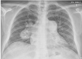
X-ray chest Frontal projection: Enlarged both hila, more on left side and right infra hilar soft tissue opacity. Otherwise lung fields are clear with clear costophrenic angles. Heart size not enlarged .
