Open Access Journal on Medicine and surgery

Impact of Air Pollution on Semen Quality: The Specific Situation of Terni (Central Italy)

Introduction

Infertility is a prevalent condition affecting an estimated 72, 4 million people globally that is well recognized by The World Health Organization (WHO). Although prevalence data are lacking, 9% of couples struggle with fertility issues and male factor contributes to 50% of the issues. Many genetic and lifestyle factors have been implicated in male infertility; however, about 30% of cases are still thought to be idiopathic [1]. The mechanism by which medical conditions affect fertility includes effects on hormonal levels, impairment of sexual function (including ejaculatory function), or impairment of testicular function/spermatogenesis. In the last 70 years, a decrease in sperm fertility and quality has been observed, including sperm count, ejaculate volume, alterations in sperm concentration and morphology [2]. Recent studies suggest that men with abnormal semen parameters have a higher risk of testicular malignancy [3]. Nowadays, environmental and lifestyle factors could be possible contributors to infertility conditions, such as use of smoke sigarettes, increasing of both parents age conception, abuse of alcohol and drugs, physical inactivity, obesity, social stress, exposure to environmental contaminants (polycyclic aromatic hydrocarbons-PAHs, or heavy metals, for examples) and air pollution [4,5]. In particular, epidemiological and experimental studies explained the link between air pollution and alterations of sperm parameters as the main risk factors for male infertility.
Human activities such as transport, industrial and agricultural emission are considered the main causes of air pollution (solid particles, liquid droplets or gases), and people that living near these area, are more exposed to henanced emission source of carbon monoxide (CO), nitrous dioxide (NO2), sulfur dioxide (SO2), ozone and lead [6]. Ambient air pollution is associated with systemic increases in oxidative stress, to which sperm are particularly sensitive. In this contest, reactive oxidative species (ROS) have been related with a broad array of spermatogenensis effects, including the decrease of progressive motile sperm count, viability, abnormal sperm morphology, and fertilization rate and spermatogenic cell numbers [7]. In Italy, 12,482 areas with a high risk of environmental pollution, mainly due to industrial emission, were been identified. Some central provinces, such as Città di Castello, Foligno and Perugia exceeded the limit set for particulate matter with diameter less than 10 microns (PM10) and O3 emissions, in 2019. In particular, Terni is one of the most polluted urban and industrial area in Central Italy [8]. In fact, is situated in an intermountain depression, delimited by the Apennine mountain range. This area is characterized by the presence of typical urban PM10 emission sources such as vehicular traffic, domestic heating and industrial emission sources from a power plant for waste treatment. Peculiar geomorphological and meteorological conditions of Terni basin, limit the dispersion and augment the accumulation of the atmospheric pollutants.
The “Thyssen Krupp AST”, a large steel factory founded at the end of 19th century and two more recent chemical industrial areas, are located close to the city center [9]. As a result of the intensive industrial activities and the geographical location, atmospheric pollution is the major local issue with high PM concentrations occurring throughout the year. According to European Commission Law, the daily maximum PM10 concentration allowed in cities is 50 μg-3 [9]. The threshold has not to exceede more than 35 times per year. In Terni, the atmospheric PM10 concentration exceede that daily limit on more than 70 days in 2012, as recorded by Regional Agency for Environmental Protection (ARPA), Umbria. The aim of the present study is to provide an association between ambient air pollution and sperm quality, analyzing seminal biofluid parameters of man living in the urban area of Terni- Papigno, with a high risk of pollution, comparated with those who live in rural areas with low risk of pollution.

Materials and Methods

Study Participants

Signed written consent was obtained from all 52 participants (age of 20–40 years) enrolled in this study from January 2018 to December 2019. Patients referred to the Seminology Laboratory of the Division of Andrology and Urology Department for an infertility evaluation. Female partners of the infertile men were subjected to general gynecological evaluation and were reported to have normal reproductive health. Residence in the province of Terni was an inclusion criteria while, systemic and cronic disease, genetic abnormalities, alcohol or drug abuse, hormone treatment, varicocele infection microchidism and cryptorchidism, prostatitis and other factors that could affect semen quality (such as fever, medications, exposure to X rays etc.) were exclusion criteria. Men were divided in group A and B that includes 30 patients from the high pollution environmental risk area of Terni-Papigno and 22 subjects that live in neighboring areas, with a low risk of pollution, respectively (Table 1).

biomedres-openaccess-journal-bjstr

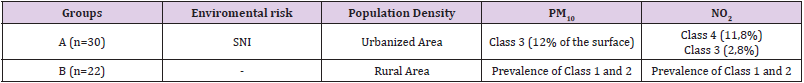
Table 1: Demographic and environmental patient’s classification.

Note: SNI: Sites of National Interest; PM10, Particulate Matter ≤ 10 μm; NO2, Nitrogen dioxide; OCSE Organization for Economic Cooperation and Development, class 1 and 2: rural area with population density <150 inhabitants/ km2 and PM10< 10 μg/m3; class 3 and 4: rural area with population density >150 inhabitants/ km2 and PM10> 10 μg/m3

Semen Analysis and Preparation of Samples

Semen samples were collected at the Andrology and Urology Laboratory by masturbation into a sterile container after 2–7 days of sexual abstinence and were analyzed immediately after liquefaction, according to the WHO guidelines [10]. Each sample was evaluated for seminal volume, pH, total sperm count, progressive motility, and morphology and leukocyte concentration. Semen volume was measured by graduated pipettes. Calibration strips were used to measure the seminal fluid pH. For the evaluation of the sperm concentration, following semen liquefaction, 10 μL of non-diluted, well-mixed semen sample was at first loaded in the middle of a clean Burker counting chamber, maintained at the temperature of 37 °C, gently covered with a cover glass, and examined using 200× or 400× magnification. The sample was diluted before proceeding with the sperm count. 1:2 dilution was used, strictly following the WHO 2010 manual recommendations [10]. The final concentration was calculated as: [(number of spermatozoa counted/the number of lines) x dilution factor] and expressed as 106 spermatozoa/mL. To evaluate the sperm motility, immediately after semen liquefaction, 10 μL of undiluted, well-mixed semen sample was loaded in the middle of a clean Neubauer counting chamber, maintained at the temperature of 37 °C, gently covered with a cover glass, and examined using 200× magnification.
Sperm motility was assessed in 200 random spermatozoa and characterized as progressive and non-progressive motility. The total motility was calculated as the sum of progressive and non-progressive motility. Both progressive and total motility were expressed as percentages. Sperm morphology was evaluated in 200 spermatozoa and the value was expressed as percentages. Finally, the vitality was assessed using eosin staining according to the WHO recommendations [10].

Statistical Analysis

Data were analyzed with GraphPad Prism 6.0. Results were reported as mean ± standard deviation (SD) Analysis of variance and Kruskal-Wallis tests were performed. The significance threshold was set at 0.05.

Results

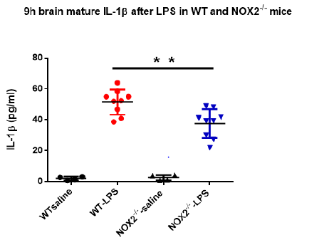
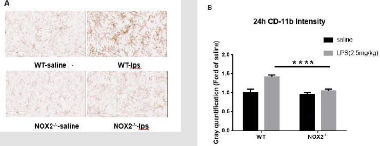
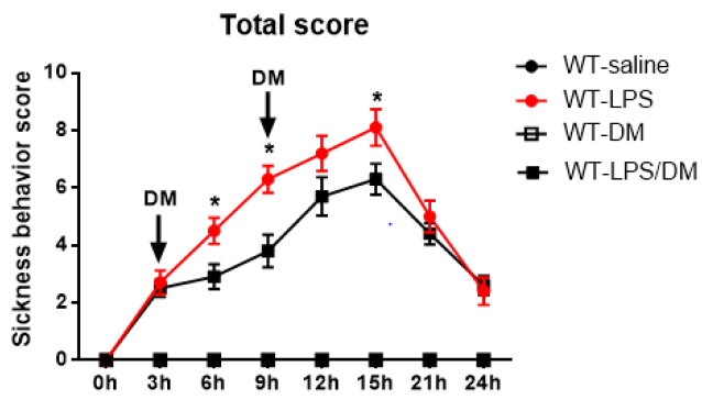
Patients were classified according to Demographic and Environmental characteristics (Table 1) Total sperm count (Figure 1) and sperm concentration (Figure 2) decreased significantly in seminal biofluid of patients living in urbanized area of Terni- Papigno (group A) when compared to subjects who lived in rural areas (group B). Seminal volume (mL) did not reach significant difference (Figure 3) between the groups considered.

biomedres-openaccess-journal-bjstr

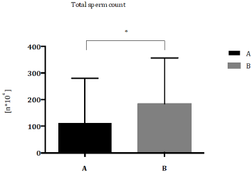
Figure 1: Total sperm count (n*106) expressed as median with range and interquartile range in group A compared to group B. *P<0,05.

biomedres-openaccess-journal-bjstr

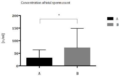
Figure 2: Concentration of total sperm concentration (n/ml) expressed as median with range and interquartile range in group A compared to group B. *P<0,05.

biomedres-openaccess-journal-bjstr

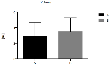
Figure 3: Seminal volume (ml) expressed as median with range and interquartile range in group A compared to group B. *P<0,05.

Discussion

In this study, conventional semen parameters (volume, appearance, pH, motility, and viability, and morphology, presence of aggregations, agglutination and leukocyte concentration) were analyzed for each sample according to the 2010 WHO criteria. Our results showed a significant decrease of total sperm count in group A patients respect to group B subjects (Figure 1). Nowadays, several studies showed how pollution can affect human fertility. Sperm count and sperm motility were reduced in men living in polluted cities. A decrease of sperm count was observed in America and Northern Europe during the last 50 years [2] and the reduction of semen quality and sperm count especially in industrialized countries supports the evidence that adverse environmental factors are key factors for men infertility [11]. Furthermore, different studies reported the enhancement of infertility, urogenital malformation and chronic disease (cancer, diabetes, etc.) in areas with a high environmental pressure [12]. In group A, a significant decrease of sperm concentration was demonstrated, respect to group B patients (Figure 2). In 2019, a longitudinal analysis on 8.945 semen samples, suggested the specific role of O3 pollution in sperm concentration [13]. The effect of air pollutants and ozone on sperm quality, was strongly elucidated by Sokol, et al. [14]; they reported that pollutants have no effect on sperm quality, however, ozone could make changes in sperms, including sperm DNA fragmentation via oxidative stress, resulting in decreased fertility [14].
The toxicity of O3 was largely demonstrated to be the major oxidant of photochemical smog and its exposure produces reactive ROS at respiratory level [15,16]. Extra pulmonary toxicity suggests that O3 or O3 reaction-products can cross blood-gas barrier and be absorbed into the circulating bloodstream, creating an environment caring to an inflammatory reaction [17,18]. It is unclear how O3 can negatively affect sperm quality, but O3-induced oxidative stress could be a possible mechanism, through which testicular and sperm function could be altered [19,20]. Under physiologic conditions, spermatozoa exist in a balanced environment of ROS and antioxidants, where ROS determine the biochemical steps required for normal fertilization (capacitation and the acrosome reaction). However, excessive amounts of ROS produced by leukocytes and immature spermatozoa can damage mature spermatozoa and the integrity of sperm DNA [21,22]. Concerning seminal volume (mL), our results did not reach significant difference between groups considered (Figure 3). Several studies suggested that alterations of semen parameters, including volume, progressive motility, total motility or morphology, may not relate to air pollution [23]. Several works observed a reduction of both sperm motility and morphology, associated with air heavy metals (lead, mercury and cadmium) [24,25], that determine alterations in sperm DNA.
Rubes and colleagues showed that short-term exposure to pollutants caused serious damages in men and women’s reproductive system. Moreover, this study demonstrated that the incidence of sperm DNA alterations due to air pollution is higher in middle-aged men [26]. Also, exposition time of contaminants seem to be determinant, in fact a study of Hammoud showed that 3 months exposure to air pollution decreases motility levels, while removing contamination can restore normal parameters levels [27]. In an extensive cross-sectional study, Xu and co-workers, founded a strong correlation between motility, concentration, and morphology of semen biofluid of men exposed to air pollutants with the use of cigarette and alcohol [28]. On the other hand, Selevan, et al. [29] did not observed any relevant alerations in sperm count, motility and morphology of young men exposed to air pollutants, except for sperm DNA and chromatin [29]. Moreover, several authors specify that the morphological change of spermatozoa after exposure to pollutants is not an indicative diagnostic parameter. Association between air pollution and alteration of specific semen parameters is not clear and different relevant factors, such as geographic areas and lifestyle, should be considered for male infertility diagnosis.

Conclusion

The current study provides evidence of an association between ambient air pollution and sperm quality. Patient residents in areas with high environment exposure had a significantly decrease in sperm quality especially for sperm concentration and count but had no impact on the other sperm parameters of spermogram (motility, morphology, vitality). The individual role of specific pollutants is difficult to identify, since patient in this study are typically exposed to several pollutants simultaneously. The physiopathology leading to altered fertility is poorly understood. In the literature there are forward four mechanisms to explain the negative impact of air pollution on sperm, as hormonal disturbances, oxidative stress induction (ROS), cell DNA and epigenetic alterations, probably working in combination. Clearly, more research is needed to understand the detrimental effect of the pollutants on sperm and to characterize their action in more detail. Our results suggest the important role of human sperm as an early and sensitive biomarker of environmental pollution as it could represent an ideal tool for investigating and promoting health surveillance especially in environmental risk areas. Environmental contaminants and bad lifestyles can impair reproductive health and overall health, encouraging the development of chronic degenerative diseases affecting the adult and, through the sperm epigenome changes, future generations. Thus, identifying risk factors to improve the management of human wellness and health throughout standardized analysis, which correlates the toxic bioaccumulation of the seminal fluid with the multiple semen parameters, might be the main objective to be considered in public prevention policies.

For More Articles: Biomedical Journal Impact Factor: https://biomedres.us

Open Access Journal on  Chemistry and Chemical Engineering

Preparation of 3D-Porous Graphene Aerogel for High- Performance Anode of Lithium-Ion Batteries

Introduction

Energy is recognized as an important material basis as well as a guarantee for human survival and development all the time [1,2]. However, with the excessive use of coal, petroleum and natural gas, the ever-increasing energy shortage and environmental contamination had become two major challenging problems facing all global nations. Thus, to effectively address such critical issues, extensive effort has recently been devoted to exploring efficient and clean energy conversion applications, such as lithium ion, sodium ion and fuel batteries. As an excellent energy storage device, lithium ion battery (LIBs) has attracted worldwide concerned and has achieved commercialization. Due to the prestigious advantages of high power per unit mass, high cycle life and high safety performance, LIBs has been applied in a wide range of fields, such as in mobile phones, electric vehicles and aviation [3,4].

The structural configuration of LIBs mainly consists of positive and negative electrode materials, a diaphragm, electrolyte, and current collector [5]. In a typical discharging process, the Li+ generated by the negative electrode is embedded into the positive electrode through the electrolyte and the electrons are collected by the current collector and move in the opposite direction of the external circuit [6]. While for charging, it is indeed the reverse process as described above. Thus, during the charging and discharging period, the more Li+ ions moving between the positive and negative electrodes, the greater the activity being generated and the higher the charge-discharge specific capacity [7]. As a consequence, such principle of electrochemical conversion has confirmed that suitable anode materials are now playing pivotal role in improving electrochemical performance of LIBs.

As is well known, graphene, consisting of a single layer carbon atoms with sp2 bonding, is a typically two-dimensional carbon material with outstanding excellent electrical conductivity and mechanical strength [8, 9]. Its theoretical capacity can reach up to 744 mAh g-1, which is twice as much as that of graphite. This may be attributed to the fact that both sides of a graphene sheet can accommodate two Li+ ions, in each hexagonal loop of carbon (Li2C6) [10]. Therefore, graphene is regarded as a potential candidate to substitute commercial graphite anodes and has been widely used in LIBs.

However, the powerful π-π stacking interactions and Van Der Waals forces lead to graphene sheets easily self-aggregate, which significantly reduces the available specific surface area and increases Li+ ion transmission resistance [11]. In order to solve the aggregation problem, researchers have devised a great deal of advanced strategies. In particular, self-assembling graphene nanosheets into GA is one of the most effective methods. For example, our previous work demonstrated that PS nanospheres were used as sacrificial templates to fabricated nitrogen-rich graphene aerogel, where PS microsphere intercalation prevented the accumulation of layers in the GO sheet [12]. The obtained GA exhibited excellent electrochemical performance due to the layered porous structure. In addition, Zhang [13] et al. successfully fabricated free-standing nitrogen-doped graphene aerogel as the anode material for sodium ion batteries and it showed a high cycling performance (287.9 mA h g−1 after 200 cycles at a current density of 100 mA g-1). In another similar work, Albarqouni [14] et al. successfully prepared rGO aerogel as supercapacitor electrode materials by utilizing the reduction capability of carbonic acid in soft drinks and enhanced charge storage capacity (121 F g-1 at 0.4 A g-1). Thus, GA exhibit promising applications in electrochemistry field. However, to the best of our knowledge, works related to GA for LIBs are still limited.

In this work, GA used as the anode of LIBs were prepared by a facile hydrothermal strategy without any additives and templates. GA prevented the restacking of graphene nanosheets to enlarge the contact area between the electrolyte and the electrode, which simultaneously reduced ion and electron diffusion resistance [15]. Besides, it was feasible to tailor the oxygen-containing functional group and surface defects of GA through adjusting the hydrothermal synthesis time. Benefiting from their porous structure and large specific surface area, the prepared GA-based LIBs show outstanding cycling stability (664.8 mAh g−1 at 0.1 A g−1 after 100 cycles) and superior rate capability, which demonstrates their potential application for Li+ storage. The current research provides a valuable guidance for developing high-performance anode material for application in LIBs.

Experimental

Materials and Chemicals

All reagents were purchased commercially without further purification. GO was obtained by the method mentioned in our previously reported work [16].

Materials Characterization

To investigate the microstructure and morphology of the asprepared samples, they are examined by advanced characterization tools like X-ray diffraction (XRD, Bruker, Germany), ULTRA 55 scanning electron microscope (SEM, ZEISS, Germany) and Raman spectroscopy (LabRAM HR 800 UV), respectively. The functional groups and elemental analysis of all samples were evaluated by X-ray photoelectron spectra (XPS) on ESCALAB 250Xi (Thermo Scientific, USA).

Electrochemical Measurements

The mixture of active materials, carbon black and polyvinylidene fluoride (PVDF) (weight ratio = 8:1:1) was dissolved in N-methyl- 2-pyrrolidinone (NMP) solvent to prepare working electrodes. The assembling of 2016 coin-type cells was similar to that of reported previously. Cyclic voltammetry (CV) curves were recorded by a Shanghai Chenhua CHI 760e electrochemistry workstation in the range from 3.0 V to 0.01 V at 0.2 mV s-1. Furthermore, galvanostatic charge/discharge cycles were measured using a NEWARE CT-4008 battery testing system at a current density range from 0.05 to 2 A g-1 versus Li/Li+.

Results and Discussion

Structural Characterization

Fig. 1(a) presents the preparation and lithium storage process of the GA. Firstly, 10 mL of GO (3 mg mL-1) was transferred to 25 mL stainless-steel autoclave and then was maintained 180 ℃ for 2, 6, 10, and 14 h. Subsequently, the GA were obtained by freeze-drying and labeled as GA-2, GA-6, GA-10 and GA-14, respectively. The excellent Li+ storage performance of GA-X electrode was due to the porous and defective structure of graphene aerogels, which can provide more Li+ ions pathways and reduce diffusion resistance. In the process of hydrothermal reduction of graphene oxide, oxygen-containing functional groups and hydrogen bonds on the surface of graphene oxide promote cross-linking between the sheets, and the gaps are filled with water molecules [5,17]. As a result, the porous structure is preserved by a sublimation process in freeze drying.

Shown in Figure 1(b~k) are the SEM images of all samples in order to investigate their morphologies and microstructures. Through low magnification (Figure 1(b, d, h and j)) and high magnification (Figure 1(c, e, i, and k)), all samples exhibit the typically hierarchical and interconnected framework structure consisting of twisted and cross-linked graphene nano-sheets, which can provide more channels for Li+ ions transport and reduce electrons transport resistance, thereby enhancing the electrochemical performance of GA-X.

biomedres-openaccess-journal-bjstr

Figure 1: Schematic illustration of procedure to prepare GA anode and SEM images of GA-2(b, c), GA-6(d, e), GA-10(h,i) and GA-14(j,k).

To better clarify the structure variation of synthesized materials after the hydrogenation process, XRD, Raman and XPS measurements are performed. For comparison, Figure 2(a) shows the XRD patterns of graphite, GO, GA-2, GA-6, GA-10 and GA-14, respectively. It could be obviously observed that the characteristic peak of graphite appears at 26.3°, which corresponds to the interlamellar spacing of 0.348 nm [9,18]. After the Hummers method of oxidation process, the sharp peak shifted to 10.6°, which could be assigned to the (002) reflection [19], and the interlamellar spacing was expanded to 0.838 nm, which was caused by the presence of various oxygen-containing functional groups (hydroxyl, carboxyl, epoxy and carbonyl) and the introduction of H2O molecules [20]. More importantly, after hydrothermal treatment, the broad (002) diffraction angle and interlayer spacing of GA-2, GA-6, GA-10 and GA-14 gradually increases to 24.48° (0.372 nm), 24.80° (0.367 nm), 24.94° (0.365 nm) and 25.14° (0.362 nm), respectively, which is attributed to the fact that the surface functional groups and intercalated H2O molecules gradually decrease. This confirms that the hydrothermal time exerts different degrees of reduction impact on graphene oxide.

As can be seen from Raman spectra of all samples in Figure 2(b), two characteristic peaks at about 1348 and 1583 cm−1 appear [21]. G band represents the stretching vibration of sp2-bonded carbon atoms of graphitic layers, and D band corresponds to vibrations of sp3-bonded carbon atoms of the defective graphitic structure or disorders [22]. Generally, The D/G peak intensity ratio (ID/IG) value was used to assess the disorder of graphitic materials [23,24]. ID/IG of the GO (0.97), GA-2 (1.21), GA-6 (1.22), GA-10 (1.52) and GA-14 (1.61) increase with the increment of hydrothermal time, which was due to the partial removal of the oxygen-containing functional groups on the graphene surface, indicating the high degree of reduction in the process of crosslinking graphene into aerogels.

X-ray photoelectron spectroscopy (XPS), a significant characterization tool to give direct information about the elemental composition and surface functional groups of the obtained samples, has been carried out for further analysis. Two obvious peaks at 285 and 532 eV in Figure 2(c), which can be related to C and O elements, respectively [25]. More importantly, the atomic ratio of carbon and oxygen (C/O), is in the following order: GO (2.26) < GA-2 (4.52) < GA-6 (4.76) ~ (GA-10 4.73, GA-14 4.71), which indicated that the oxygen functional groups of GA successfully are decomposed with the hydrothermal reaction time and no significant change occurs after six hours (the detailed data was shown in Table 1). Based on in-depth analysis of the shark peak in high resolution C 1s XPS spectrum (Figure 2d), four types of C species can be detected in those samples. Furthermore, the peaks located at 290.9, 288.3, 285.8 and 284.3 eV correspond to carboxyl groups (-COOR), carbonyl groups (C=O), epoxy groups (C-O), sp3 and sp2 carbon, respectively [26-28]. Shown in Fig. 2(e) is the high resolution O1s spectra, from which it can be seen that GA exhibits four peaks at the binding energy of 536.1, 532.8, 531.3 and 530.1 eV, which are assigned as the H2O, HO-C=O, C-O-C and C=O, respectively [21, 29].

biomedres-openaccess-journal-bjstr

Table 1: Relative atomic percentages according to the XPS spectra.

biomedres-openaccess-journal-bjstr

Figure 2: (a) XRD patterns; (b)Raman spectra; (c) XPS spectra of GA-X. High-resolution XPS spectra of (d) C 1s, and (e) O 1s in GA-6.

To further investigate the electrochemical properties of GA-X in LIBs, the corresponding samples are tested by the standard half-cell configuration. Shown in Figure 3(a) are the cyclic voltammogram curves (CVs) of the GA-6 electrodes with the first three cycles being measured at 0.2 mV s−1. In the first cycle, a sharp reduction peak at 0.25 V appears, accounting for the irreversible decomposition of electrolyte and the subsequent formation of a solid electrolyte interface (SEI) film on the surface of carbonaceous structure [30-32]. In the subsequent cycles, CV curves are closely overlapped, which proves that the GA-6 anode has stable cyclic performance during the insertion and de-insertion process [33].

The charge-discharge curves of GA-6 composite materials in the 1st, 2st, 3st cycles are displayed in Figure 3(b). The GA-6 exhibits an initial charge and discharge capacity of 1744.4/976.8 mAh g-1 and the Coulombic efficiency is 56.0% for LIBs, which far outweighs that of others 1380.4/762.0 (55.2%), 1509.9/809.8 (53.6%), and 1411.8/828.1 (58.6%) mAh g-1. The massive irreversible capacity loss for LIBs is caused by the SEI layer decomposition, irreversible reaction between Li+ and surface functional groups and irreversible desorption of ultra-fine pores [31]. Shown in Figure 3(c) are the testing results about the rate performance of GA-X anode at diversified current densities from 50 mA g-1 to 2 A g-1. In addition, the corresponding specific capacities of GA-6 are 671.2, 453.1, 292.9, 238.4, 201.2, 168.9 and 113.8 mAh g-1, respectively, which can be observed in Figure 3(d). However, another three samples only exhibit specific capacities of 583.2, 384.3, 241.5, 188.4, 159.5, 133.8, and 89.7 mAh g-1 for GA-2; 572.5, 387.6, 240.3, 194.1, 170.9, 143.8 and 91.3 mAh g-1 for GA-10; 562.3, 366.8, 224.9, 187.8, 160.7, 133.8 and 83.8 mAh g-1 for GA-14, respectively. It should be noted that when returning to the current condition of 100 mA g-1, there is still a reversible capacity of 428.4 mAh g-1 in GA-6, higher than 355.8 (GA-2), 380.2 (GA-10) and 353.3 (GA-14) mAh g-1. The dramatically enhanced Li+ storage capacity of GA-6 is mainly because the three-dimensionally porous structure is capable of providing more Li+ ion transport channels and electrical conductivity [34-36].

Shown in Figure 3(e) is the information on the cycling stabilities of the four samples, and the corresponding test was performed at 100 mA g-1. As the hydrothermal reaction time increases, GA-6 (about 664.8 mAh g-1) exhibits a significantly enhanced cycling performance than GA-2 (around 499.9 mAh g-1) after 100 cycles. This may be ascribed to the reason that the hydrothermal time is too short, which leads to insufficient reduction and too many oxygen-containing functional groups. When the hydrothermal time continues to increase, the capacity decays again (around 560.7 and 577.6 mAh g-1) due to the excessive aging of graphene oxide. The results obviously indicates that the hydrothermal synthesis time exerts great impact on the electrochemical performance of the anode. Additionally, as shown in Figure 3(e), all GA anodes show nearly 100% coulombic efficiency, which further confirms that the cycling stability of the obtained GA materials is very high for LIBs (Figure 4).

biomedres-openaccess-journal-bjstr

Figure 3: (a) CV curves of GA-6 at a scan rate of 0.2 mV s-1. (b) Charging/discharging of GA-6 at current density of 50 mA g-1. (c) Rate performances of GA-X electrodes. (d) The rate capabilities of GA-6 electrodes cycled at different current densities. (e) The cycling performance of GA-X electrodes in 100 cycles at 100mA g-1. (f) Nyquist plots of the GA-X.

biomedres-openaccess-journal-bjstr

Figure 4: Nyquist plots of the electrodes.

The electrochemical impedance spectra (EIS) measurements are shown in Figure 3(f), which can further explain and verify the superior electrochemical performance. Nyquist plots of the composite illustrates an arc at the frequent regions corresponding to the interfacial charge-transfer resistance [37,38], and an approximate straight sloping line at the less frequent regions corresponding to Warburg impedance [39]. By contrast, GA-6 has a smaller semicircle and a higher slope, which is attributed to the porous structure preventing the self-aggregation of the GO sheets and providing more Li+ ion diffusion channels. In addition, the GA-X samples have the same equivalent circuit fitting values (Table 2) and the result affirms that the GA-6 composite possesses a smaller charge transfer impedance RSEI (100.3 Ω) and Rct (20.6 Ω) than the GA-2 (150.8 Ω and 38 Ω), GA-10 (225.1 Ω and 2.4 Ω) and GA-14 (135.7 Ω and 176.8 Ω), which demonstrated that porous structure could decrease charge-transfer resistance and improve the electrical conductivity. In addition, to better quantitatively analyze the testing data, an ideal equivalent circuit is utilized for fitting the impedance spectra, as is displayed in Figure 4. Re denotes the resistance caused by the electrolyte, RSEI represents the resistance of the electrode surface film, Rct is the charge-transfer resistance and CPESEI and CPEct are constant phase elements assigned as the surface film and double layer capacitance, respectively, and CPE3 is the constant phase element [40,41].

biomedres-openaccess-journal-bjstr

Table 2: The detailed values of the equivalent circuit components used for fitting the experimental curve.

Conclusions

In summary, a simple yet effective hydrothermal approach was reported to synthesize self-assembled porous carbon framework (GA) with prestigious advantages of large surface area, specific porous structure, superior electronic conductivity, excellent mechanical characteristic and ultrafast electron transport kinetic. Benefiting from their surface defects and abundant porosity, the 3D porous graphene exhibits highly stable cycling performance (664.8 mAh g−1 at 0.1 A g−1 after one hundred cycles). Moreover, it is convinced that by controlling the hydrothermal synthesis time, the surface defects of GA can be easily tailored. Correspondingly, GA-6 obtained by hydrothermal reaction for GA-6 exhibits the highest electrochemical performance. The present work provides a simple strategy to the design of electrodes with high performance for LIBs.

For More Articles: Biomedical Journal Impact Factor: https://biomedres.us

Open Access Journal on Respiratory and Critical Care

Dextromethorphan As A NOX2 Inhibitor Mitigates Endotoxemia-Induced Sickness Behavior and Brain Interleukin-1β Production in Mice

Introduction

Severe systemic infections (sepsis) can cause cytokine storm and lead to acute brain inflammation through the overproduction of proinflammatory factors, such as reactive oxygen species (ROS), nitric oxide (NO) and cytokines release by activated microglia [1]. Acute brain inflammatory responses might result in both short-term behavior alterations (sickness behavior) and chronic neuronal damage via sustained microglial activation [2-5]. Sickness behaviors associated with infections by bacteria, viruses or fungi are common [6]. Sickness or sickness behavior refers to a series of physiological and behavioral changes, which is thought as a malfunction due to systemic inflammatory response during infections or injury, including fever, fatigue, sleepiness, anorexia, cognitive dysfunction, social withdrawal [7,8]. Although it might be beneficial for individuals to fight the infection induced by pathogenic microorganisms [8], immoderate sickness could further aggravate its harm to the health, even death.
For example, neuronal excitotoxicity would be exacerbation following persistence increased temperature in brain tissue during fever, even destroy the blood–brain-barrier permeability [9,10]. Multiple cytokines such as IL-1β, IL-6 and TNFα are associated with sickness behavior [7]. IL-1βseems to be the predominant mediator of sickness behavior in the brain, because blockade of its action by central administration of the IL-1 receptor antagonist significantly attenuates sickness behavior [11]. The propagation of neuroinflammatory cytokines during peripheral immune challenge not only induces sickness behavior but also triggers neuroinflammation characterized by the activation of microglia, the resident macrophage in the brain [12]. Moreover, prolonged neuroinflammation can cause long-term neuronal loss via the selfpropelling vicious cycle between injured neurons and overacted microglia [3,5]. Among various pro-inflammatory cytokines, IL-1β showed a high potent neurotoxicity [13,14].
Our recent study has revealed that the IL-1β signaling is essential for maintaining endotoxemia-associated microglial activation. Reducing brain IL-1β levels prevents chronic neuroinflammation and resultant neurodegeneration [15]. In this study, we investigated the role of NADPH oxidase (NOX2) in LPS-elicited sickness behaviors. NOX2, a major superoxide-producing enzyme, plays a critical role in the development of neuroinflammation [5- 18]. We hypothesized that NOX2 is essential for sickness behavior and brain IL-1β maturation during endotoxemia-mediated sepsis. Besides NOX2-/- mice, we also employed a NOX2 inhibitor dextromethorphan (DM), a widely used non-opioid cough suppressant, which exerts anti-inflammation and neuro-protection through the inhibition of microglial NOX2 [19,20]. In this study, we found that genetic knockout of NOX2 or pharmacological inhibition of NOX2 with dextromethorphan diminished sickness behavior in the LPS peritoneal injection mice model.
Furthermore, the deficiency of NOX2 or the post-treatment of dextromethorphan significantly decreased IL-1β production in the brain and resultant microglial activation. This study demonstrated that NOX2 plays a critical role in modulating the brain level of IL- 1β and associated sickness behavior. Our data also suggests that dextromethorphan could be used in endotoxemia patients for relieving sickness behaviors and neuroinflammation.

Materials and Methods

Animals and Treatment

The C57BL/6J mice and NOX2-/- mice were obtained from the Jackson Laboratory (Bar Harbor, ME). Housing and breeding of animals were performed humanely with regard for alleviation of suffering following the National Institutes of Health’s Guide for the Care and Use of Laboratory Animals (Institute of Laboratory Animal Resources, 2011). LPS (2.5mg/kg) or saline (2.5 mg/kg) were intraperitoneally administrated to 8-week male C57BL/6J mice and NOX2-/- mice initially, and sickness behavior was observed every 3hs after LPS injection until 24h. Dextromethorphan (12.5mg/ kg) or saline (12.5mg/kg) were also intraperitoneally injected to C57BL/6J mice two times (3h and 9h after LPS injection) and sickness behavior was accessed every 3hs after LPS injection until 24h.

Sickness Behavior Assessment

We applied behavior test to evaluate the mice’s sickness behavior after treatment, including the response, the moving distance, ptosis, piloerection and excretion in eyes.
a) Response. The toughing frequency until the mice moved. We have four degree: 0. Animal moved freely without stimulation. 1. The movement of mice under one- or two-times’ toughing stimulation. 2. Three to five toughing stimulation. 3. More than five stimulations.
b) Moving distance. We also set four degree to assess moving distance. 0. The animal moved freely more than 20cm without any stimulation. 1. The moving distance is more than 20cm with stimulation. 2. The moving distance is 10cm to 20cm under toughing stimulation. 3. In case of mice moving a few steps with stimulation, they were regarded as grade 3.
c) Appearance of ptosis, piloerection and excretion in eyes. 0. Negative. 1. Positive.

Immunohistochemistry

Mice were euthanized using fetal plus at the desired time points after injection. Brains were fixed in 4% paraformaldehyde and processed for immune staining as described previously [21,22]. We used the following primary antibodies for immunohistochemistry: antibodies against CD11b (1:400, AbDSerotec, Raleigh, NC). Immuno-staining was visualized by using 3,3’-diaminobenzidine (DAB) and urea-hydrogen peroxide tablets.

Quantification of the immunohistochemistry staining density

The quantification CD11b immunohistological staining in SN was performed by Image J software based on a protocol for quantifying western blots [23]. Briefly, the image was first converted into the grayscale picture, and the background was adjusted before the quantifying area was selected for the measurement of the total pixels. The relative density of the staining was compared based on the density of the total pixels of a certain brain region (total pixels/ area).

Cytokine Analysis

The brain tissues from WT and NOX2-/- mice were measured for mouse IL-1β (R & D Systems Inc.,Minneapolis,MN) according to the manufacturer’s instructions.

Real-time RT-PCR Analysis

Total RNA was extracted from the mouse brain by using a RNeasy Mini Kit from QIAGEN (Valencia, CA) to detect the level NOX2 (gp91) according to the previous description [4]. Total RNA was reversely transcribed with MuLV reverse transcriptase and oligo-dT primers. Then SYBR green PCR master mix was used for realtime PCR analysis. The primer sequences were as follow: GAPDH F (5′ TTC AAC GGC ACA GTC AAG GC 3′), GAPDH R (5′ GAC TCC ACG ACA TAC TCA GCACC 3′), NOX2 F (5′ GAT TCAAGA TGG AGG TGG GAC 3′), NOX2 R (5′ GGT CAG TGT GAA TGG GTG CC 3′). Real-time PCR amplification was performed using SYBR Green PCR Master Mix and QuantStudio 6 Flex Real-Time PCR System (Applied Biosystems, Foster City, CA, USA) according to manufacturer’s protocols. Amplifications were done at 95°C for 10 s, 55°C for 30 s, and 72°C for 30 s for 40 cycles. All samples were tested in duplicate and normalized with GAPDH using the 2-ΔΔCt method. Fold changes for each treatment were normalized to the control.

Statistical Analysis

All data were represented as mean ± SEM. Data were analysed using one-way or two-way ANOVA. Tukey’s post hoc tests were applied when comparing groups to only the control, or Sidak’s post hoc test when comparing between multiple treatment groups. Number of mice used is indicated in each figure legend.

Results

NOX2 Plays a Critical Role in Mediating LPS-Elicited Sickness Behavior

C57BL/6J and NOX2-/-mice received a single injection of LPS (2.5mg/kg; ip) for investigating the role of NOX2 in the development of LPS-induced sickness behavior. Sickness was assessed every 3hs after LPS administration up to 24h. In WT mice, the sickness behavior score showed a time-dependent increase over time, peaked around 9-15h following LPS injection, then declined gradually afterwards, but still remained higher than the base line values at 24h after LPS injection. A similar pattern of change in the sickness behavior score was observed in LPS-injected NOX2-/- mice. However, a significant reduction in sickness behavior score in NOX2-/- mice was observed at 9h,12h,15h, compared to WT mice (p<0.05; Figure 1).

biomedres-openaccess-journal-bjstr

Figure 1: The total scores of sickness behavior in different groups after LPS administration. NOX2-/- mice (N=9) displayed attenuated sickness behavior after LPS challenge compared to WT mice (N=9). Sickness behavior scores were measured every 3hs until 24h after LPS i.p. injection (2.5mg/kg) in WT and NOX2-/- mice.*p<0.05, the mean differences in total score between two groups was observed at 9h (p<0.0001), 12h (p=0.0405) and 15h (p=0.0382) after LPS treatment. Repeated-measure ANOVA and multi¬variate ANOVA to test the differences over time and differences between groups.

The absence of NOX2 Suppresses Brain Levels of IL-1β

As mentioned previously, increase in brain IL-1β plays a critical role in mediating sickness behavior. Our previous study indicated that the production of mature brain IL-1β in brain peaked around 9h after LPS injection [15]. Since the sickness behavior analysis showed high scores at 9h upon the peritoneal LPS stimulation (Figure 1), we measured mature IL-1β concentrations in brain tissues at 9h in both WT and NOX2-deficient mice. Results showed that level of IL-1β in brain tissues of WT mice was higher after LPS injection than that in vehicle-treated mice. Notably, the level of IL- 1β in brain tissues from NOX2-/- mice after LPS administration was significantly lower than that of LPS-treated WT mice (Figure 2). This result indicates that NOX2 is essential in mediating LPSinduced increase in the IL-1β production.

biomedres-openaccess-journal-bjstr

Figure 2: The brain mature IL-1β at 9h after LPS in WT and NOX2-/- mice. Mature IL-1β concentrations in brain tissue were measured by ELISA at 9h after LPS treatment and they were decreased in NOX2-/- mice compared to WT mice after LPS injection (2.5mg/kg) (n=4-9/group). **p<0.05, the differences of IL-1β concentrations between LPS-treated WT and NOX2 groups. Two-way ANOVA followed by Sidak’s multiple comparison test. (treatment×group, F(1,22)=5.152, p=0.0334; effect of groups, F(1,22)=4.434, p=0.0469; effect of treatment, F(1,22)=177.3, p<0.0001).

The Deficiency of NOX2 relieves LPS-Elicited Activation of Microglia in The Substantia Nigra

The reduction of brain mature IL-1β prevents sustained microglial activation induced by endotoxemia [15]. Therefore, we examined microglial activation in the absence of NOX2. Because sepsis-associated neuroinflammatory responses involve multiple brain regions and the substantia nigra (SN) was found a good area in our previous study to observe the persistence of microglial activation [3,4], we chose the SN in this study to evaluate the degree of microglial activation by immune-staining analysis. Typical morphological changes reflecting microglial activation were observed in WT mice 24h after LPS injection. Activated microglia exhibited significant enlargement of cell bodies, irregular shapes. This morphological activation was significantly reduced in NOX2 deficient mice, characterized by a small and round shape compared with WT mice. Analysis by gray quantification showed pattern changes similar to those of morphological observations (Figure 3a & 3b).

biomedres-openaccess-journal-bjstr

Figure 3: Microglia activation in SN was detected with CD11b IHC at 24h after LPS (2.5mg/kg) treatment.
(a) Attenuated microglial activation in NOX2-/- mice compared to WT mice. Following a single injection of LPS (2.5 mg/kg; ip) or saline vehicle, mice were perfused 9 h thereafter for microglial CD-11b immunostaining (n = 3/group). Representative images at 9h were shown (Figure 3A). Scale bar = 300 μm.
(b) CD-11b density was quantified. Results are expressed as folds of vehicle control. Histograms represent degree of microglial activation quantified by measuring the density of CD-11b staining with ImageJ. ****p<0.0001, the differences of gray quantification between LPS-treated WT and NOX2-/- groups. Two-way ANOVA followed by Sidak’s multiple comparison test. (treatment×group, F(1,68)=20.45, p<0.0001; effect of groups, F(1,68)=35.93, p<0.0001; effect of treatment, F(1,68)=56.21, p<0.0001).

Targeting NOX2 as a potential therapy for sepsis-related sickness behaviors

Results from NOX2-deficient mice suggest a potential target for developing drug therapy for sepsis-related sickness behaviors. As proof of principle, we employed dextromethorphan (DM), which displays potent anti-inflammatory property via inhibition of microglial NOX2 activity [20,24]. At 3h and 9h after mice received a LPS injection, DM was intraperitoneally injected and sickness behavior was accessed every 3hs for 24hrs. In LPS treated mice, the sickness behavior score increased gradually up to15h after LPS injection, then it decreased significantly until the end of observation. Analysis of ANOVA showed that DM significantly attenuated sickness behavior at 6h,9h, and 12h compared to mice with LPS treatment alone (p<0.05; Figure 4).

biomedres-openaccess-journal-bjstr

Figure 4: Post-treatment of dextromethorphan (12.5mg/kg) mitigatedLPS-induced sickness behavior in WT mice (N=5-10 for each group). Sickness behavior scores were measured every 3hs until 24h after LPS i.p. injection (2.5mg/kg) and indicated administration of dextromethorphan. *p<0.05, the mean differences in total score between two groups was observed at 6h (p=0.0459), 9h (p=0.0411) and 15h (p=0.0483) after initial LPS treatment. Repeated-measure ANOVA and multi¬variate ANOVA to test the differences over time and differences between groups.

DM reduces LPS-elicited the IL-1β production, NOX2 mRNA level and diminishes the microglial activation

LPS-elicited increase in IL-1β production from brain tissues and microglial activation were determined in WT mice. Similar to the results from NOX2-deficient mice, DM treatments effectively reduced LPS-induced IL-1β production (Figure 5a). NOX2 mRNA levels increased significantly following initial LPS treatment. While the NOX2 mRNA expression in DM post-treatment groups at different timepoints (6h and 12h)were lower than LPS-treated alone (Figure 5b). Pathologically, DM diminished the activation of nigral microglia as well (Figure 6a & 6b). Taken together, these results suggest a potential use of DM in ameliorating the severity of sickness behaviors and neuroinflammation in endotoxemia-related patients.

biomedres-openaccess-journal-bjstr

Figure 5: Post-treatment of dextromethorphan alleviated brain mature IL-1β levels and NOX2 mRNA expression.
a) Post-treatment of dextromethorphan alleviated brain mature IL-1β levels. (LPS 2.5mg/kg) (n=5-10/group). *p<0.05, the differences of IL-1β concentrations between LPS-treated and post-treated dextromethorphan groups. One-way ANOVA followed by Tukey’s multiple comparison test. (F=32.6, p<0.0001).
b) (NOX2 mRNA levels were measured by qPCR at 6h, and 12h after LPS treatment. DM was post-added to mice at 3h and 9h after LPS treatment, respectively. #P<0.05, ##P<0.01 for the differences of NOX2 mRNA level between LPS-treated and post-treated DM groups at 6h and 12h, NS for 9h. One-way ANOVA followed by Tukey’s multiple test (F(3,32)6h=31.42, F(3,32)9h=29.57, F(3,32)12h=32.66). **P<0.001, the differences of NOX2 mRNA level over time in both LPS -treated groups and post-treated DM groups. Repeated-measure ANOVA and multivariate ANOVA test the difference over time and differences between groups.

biomedres-openaccess-journal-bjstr

Figure 6: Post-treatment of dextromethorphan alleviated brain microglia activation in SN.
a) The microglia activation in SN was detected with CD11b IHC at 24h after LPS treatment. (n = 3/group) (LPS 2.5mg/kg, DM 12.5mg/kg). Representative images at 9h were shown (Figure 6A). Scale bar = 300 μm.
b) CD-11b density was quantified. Results are expressed as folds of vehicle control. Histograms represent degree of microglial activation quantified by measuring the density of CD-11b staining with ImageJ. One-way ANOVA followed by Tukey’s multiple comparison test. (F=62.73, p<0.0001). ***p<0.001, the differences of gray quantification between LPS-treated and posttreated dextromethorphan groups. ****p<0.0001, *p<0.05 and NS compared to vehicle group.

Discussion

This study provides the first evidence indicating that microglial NOX2 plays a critical role in endotoxemia-related sickness behaviors. Mechanistically, inhibition of microglial activation and subsequent reduction in IL-1β production through the inhibition of NOX2 underlie the amelioration of LPS-elicited sickness behaviors. This conclusion was based on both genetic knockout of NOX2 gene and pharmacological inhibition by DM. This study also suggests that microglial NOX2 can be a therapeutic target for developing anti sickness behaviors drug. In present study, we found that mice with NOX2 subunit knockout exhibited lower sickness behavior score than WT mice after LPS administration. Furthermore, compared to WT mice following LPS treatment, reduced activation of microglia (24h after LPS treatment) and decreased production of proinflammatory IL-1β (9h after LPS treatment) were both observed in this study. Mechanistically, enhanced production of NOX2- generated superoxide and its related ROS is regarded as the major source of pathological oxidative stress in CNS leading to neuron damage, inflammation, and dysfunction.
In the current study, the NOX2 deficiency reduced acute production of IL-1β and activation of microglia, indicating that the NOX2 deficiency could attenuate the LPS-induced acute symptom via reducing acute neuroinflammation. After elucidating the role of microglial NOX2 in mediating LPS-induced sickness behaviors, as a proof of principle we further provide evidence showing high efficacy of DM in ameliorating sickness behaviors. In addition, post-treatment with DM following LPS also attenuated microglia activation, IL-1β production, as well as NOX2 mRNA in vivo. Over decades, numerous studies demonstrate that DM, a non-opioid cough suppressant, has neuroprotective effects, which is attributed to its antagonistic effect on NMDA receptor [25-27]. However, we previously reported that anti-inflammatory effect of DM is attributed by inhibiting preventing microglia over-activation [28] induced by LPS and MPTP–elicited neurotoxin in neuron-glia culture and in vivo studies [29].
Furthermore, these mechanistic studies revealed that DM reduced the NOX2-generated superoxide free radicals from microglia, which in turn reduced in production of iROS and cytokines. The ill patients always have representative characters in behavior and/or emotion, including fever, fatigue, anorexia, fragmented sleep, losing interesting in physical and social environment, even showing emotion disorders such as depression. Over past two decades, a term of “sickness behavior” is suggested to describe such changes mentioned above in ill individuals or animals. Many studies have clarified the underlying mechanism that pro-inflammatory cytokines, such as IL-1α, IL-1β, TNF-α, IL 6, play a critical role in sickness behavior and CNS dysfunction [30,31]. Despite moderate inflammation or sickness behavior is beneficial for coping with foreign microorganism-elicited infection, the uncontrolled cytokine storm or serious sickness behavior could impair the immunity system, interfere with inflammatory responses and produce further damage to patients.
Therefore, from clinical perspective, it is desirable for ill patients to control or relieve excessive inflammation and sickness behavior after proper anti infection therapy. Our results suggested that DM and NOX2 deficiency not only inhibited activation of microglia, reduced sickness behavior, but also suppressed NOX2 gene expression and production of IL-1β, which is also a critical pro-inflammatory cytokine in neuroinflammation. IL-1β plays a significant role in the initiation of inflammatory response in CNS and transition to chronic neuroinflammation induced by various stimulation [32]. IL-1β could impair the integrity of BBB, leading to infiltration of peripheral immune cells into brain, and aggravating the neuroinflammation [33]. Meanwhile, IL-1β activated microglia and astrocytes, which in turn generate other pro-inflammation cytokines, such as IL-18, TNF-α, IL-6 in CNS. Furthermore, the IL- 1β indirectly recruits leukocytes by augmenting the expression of chemokines.
These mechanistic studies indicated the IL-1β is a critical inflammatory cytokine in both acute and chronic inflammatory response in CNS. Thus, this study not only demonstrates a beneficial effect of inhibiting NOX2 in mitigating the sickness behavior symptoms during acute inflammation, but also can be beneficial in preventing the transition from acute to chronic neuroinflammation. Over the years, a number of studies have reported that DM has neuroprotective effects [25,34-36]. The mechanism of neuroprotective activity has been attributed to DM’s potential antagonistic effect on the NMDA receptor complex. Recently, studies indicated that DM also play neuroprotective roles in cognition deficit following systemic inflammation [37], together with previous reports [19], suggest that DM achieves these extraordinary protective effects through the inhibition of NADPH oxidase. Our study demonstrated DM and NOX2 deficiency attenuate the microglial activation during acute inflammation, meanwhile, the reduced IL-1β production and NOX2 gene expression in brain also was observed. Subsequently, the sickness behavior induced by LPS relieved following acute inflammation.
This finding is similar with the protective effect of DM in chronic neuroinflammation [18,20]. Several studies have investigated the role of a variety of pro-inflammatory cytokines such as TNF-α, IL-1, IL-6, IL-8, IL-10 and IFN-γ and reactive oxygen species like superoxide and iROS in the pathogenesis of sepsis or infection. To relieve inflammation in septic or infected patients via blocking single cytokine (e.g., IL-1β receptor antagonist), while the crosstalk in cytokine network makes this effort uncertain and controversy [38-40]. The present study offers a novel approach by using DM to attenuate infection-related sickness behavior, which selectively targets NOX2. Furthermore, the findings from this study provide a potential therapeutic viewpoint: DM would be an alternative drug for septic or endotoxemia-related sickness behavior besides antibiotics and other life supportive therapy, such as ventilation, oxygen, fluid replacement therapy. In conclusion, DM attenuated sickness behavior in mice induced by LPS via inhibiting NOX2 activation and suppressing the production of IL-1β in brain. The present findings demonstrated that NOX2 is a novel therapeutic target for sickness behavior. Furthermore, this study also suggests that owing to its excellent safety record and high efficacy, DM has a great potential to be tested in clinical settings.

For More Articles: Biomedical Journal Impact Factor: https://biomedres.us

Open Access Journal on Biomedical Engineering

Anti-Radical Activity of Low Frequency and Low Amplitude PEMF

Introduction

If we expose biological tissue to Pulsed Electromagnetic Fields (PEMF) we can induce a measurable biological response and modulate inflammatory processes, promote bone repair, repair of skin wounds, even chronic ones, improve metabolic processes. Electromagnetic fields can interact with biological tissue inducing responses depending on their frequency and intensity. In the therapeutic field, the most promising results are obtained with the use of low intensity and very low frequency pulsed electromagnetic fields, or Extremely Low Frequency, indicated by the acronym PEMF LFLA. In fact, exposing cultures of fibroblasts and endothelial cells to PEMF LFLA, which vary during the stimulation process, we can quantitatively measure a reduction in the free radicals produced. Recent evidence has demonstrated that LPEMFs can prevent inflammation and oxidative stress as a non-invasive therapeutic method for various diseases. As an example, in 2019 a study [1] presented how, in an in vivo test, PEMF LFLA promote functional recovery following spinal cord injury, potentially by modulating inflammation, oxidative stress and HSP70. Furthermore, studies confirmed that PEMF LFLA can reduce ROS levels and enhance antioxidative stress responses in osteoblasts [2]. A polish clinical research [3] in 2018, attested that variable low frequency PEMF therapy meaningfully improved the overall condition of 57 patients through a decrease of oxidative stress markers while significantly affected positively their psychophysical abilities after stroke. In the present study it is presented a biophysical hypothesis of interaction between PEMF and biological working principle regarding the oxidative stress modulation PEMF derived, particularly in sport activities. Finally, we evidenced some results in vitro in fibroblast and endothelial cells and in vivo, in a case report on professional running athletes.

Biophysical Hypotheses Of Interaction between PEMF and Biological Tissues

There are several hypotheses of interaction between electromagnetic fields and cellular structures. Among the various hypotheses proposed in the scientific literature, the use of nonlinear physical descriptive models, seem to be the best tools for describing the induced biological phenomena. In particular, the use of solutions of Schrödinger equation, relating to waves with constant amplitude, called “solitons”, which will be better described below, allows to explain, as a theoretical model, the effects of PEMFs on the modulation of oxidative stress. The magnetic field can, in fact, induce variations in electrical charges through the insulating effect of the plasma membrane, acting on electrons, ions, radicals, molecules and macromolecules.
The hypothesis of interaction of PEMF LFLA with cellular structures includes (1):
1. Action on RNA polymerase and stimulation of cell replication
2. Action on the synthesis and hydrolysis of ATP
3. Effectiveness of oxidation-reduction reactions
Electro-magnetic fields can influence biological redox processes. The electron transport chain, involved in the synthesis of ATP and in respiration, is a process of oxygen reduction (by NADH and FADH2) through electron transfer in the mitochondria (it is the initial part of oxidative phosphorylation). The relatively small mass molecules involved in the chain, such as cytochrome-c, can move quite “easily” out of the mitochondrial membrane, carrying electrons from donor to acceptor [4]. This electron transport can be described using very complex physical theoretical modelling. Trying to simplify, the various models consider electron donor molecules loosely bound to “bridging” molecules, in turn loosely bound to acceptor molecules. But if we consider high molecular weight molecules such as the NADH-ubiquinone oxidoreductase proteins and cytochrome c-oxidase, which are also involved in the electron transport chain, these result to be practically fixed in the inner mitochondrial membrane. Most of these proteins have an alpha-helix conformation. When the electrons are released from the donor molecule, they are transferred into the protein and accompanied by a deformation of the endoplasmic reticulum which forms a potential barrier that attracts the electron itself. A relationship between protein conformation and formation of the electron-lattice deformation bond is hypothesized.
The charges then propagate, accompanied by a deformation of the lattice. Mathematically, through the solutions of non-linear equations, it is possible to describe this propagation which assumes a waveform with particular characteristics, such as the amplitude that remains constant. We will see that this wave has been called “soliton”. To demonstrate this result, the ion-resonance frequency relationship can be inserted into the solution of the non-linear Schrödinger equation, defined in quantum mechanics [5]. If we consider the presence of potential U(x,t) the formula of the equation can be written as follows:

Cyclotron ion resonance is a phenomenon related to moving ions immersed in a magnetic field and is based on the Lorentz force. The magnetic field acts on electronically charged elements such as ions (eg Na +, K +, Ca ++) and electrons, certainly available in biological systems. The magnetic field can act on the movement of these electric charges through a force called Lorentz and defined by

where the contribution of the electric field E which acts on the charge q, and the contribution of the magnetic field of intensity B, which acts on the same charge as a function of its speed v, determines the intensity of the force F. Thanks to the Lorentz force, a free ion immersed in a magnetic field of intensity B, moves on a circular trajectory with an angular velocity ω, dependent not only on the intensity of the magnetic field but also on its own charge and mass. Using the information of the Lorentz force and the circular trajectory, the following formula can be obtained:

If we insert this frequency value into Schrödinger’s equation and solve it, the solution obtained is the wave function of the electron ψ (x,t ) .
More correctly, its square provides information on the probability of finding the electron when trying to measure its position. Applied to the electron transport chain, the wave function provides information on the position of the particle along the protein lattice. Considering an alpha polypeptide chain, the solution of this equation is a particular wave, which indefinitely maintains its amplitude. In the literature this wave has been descripted through the shape of a “soliton”.

The soliton is a solitary wave that keeps its amplitude constant and represents the “module” for transporting energy and information at the cellular level. With the same solution we can calculate the deformation of the lattice (chain) in the “soliton” state, which is proportional to the probability of the presence of the electron (x). From the mathematical demonstration we obtain that the electron and the deformation of the lattice together, propagate along the polypeptide chain, with constant speed, from a donor molecule to an acceptor molecule, as a localized wave. This wave is exceptionally stable, able to propagate along macroscopic distances, thanks to the combination of low energy dispersion and non-linear phenomena (non-linear nature of its formation). The oscillatory character of soliton propagation with frequency given by Equation 1 is accompanied by the propagation of the local deformation of the polypeptide chain.
At a “macroscopic” level, solitons are well known and used in some fields of application. For example, in optical fibre transport networks, the non-monochromatic light, due to the Kerr effect, can propagate without changing over time. Tsunamis are common in seismology, generated by submarine earthquakes, capable of spreading for kilometres without dispersing energy, before impacting the coasts, with all their destructive force. They are therefore waves that do not lose their energy and their intrinsic characteristics by propagating and this is due to the combination of dispersion and non-linear effects, which interact by cancelling each other. The name “soliton” is due to their characteristic of remaining unchanged in the event of reciprocal interaction: two soliton waves that collide do not add up their effects. The hypothesis that some biological phenomena are based on the presence of solitons is due to Alexander Davydov who in 1979 and then in 1985 hypothesized their existence in alpha polypeptide chains [4]. Davidov demonstrated, mathematically, that the energy from ATP hydrolysis could be stored as a vibration of the amide group of the alpha helix protein, through a non-linear interaction that traps, the energy into a soliton [6,7]. The energy comes from the hydrolysis of ATP, as widely known in biology, but this is stored in the form of mechanical vibration of the amide, with constant amplitude. The subsequent propagation of this energy can explain the muscle contraction [8] (Figure 1). Over the years, scientific literature has shown, mathematically, how the solitons mechanism was a good solution to explain biological phenomena. It therefore becomes clear how PEMF LFLA can interact with these mechanisms, promoting biological phenomena.

biomedres-openaccess-journal-bjstr

Figure 1: Alexander Davydov was the first to hypothesize the action of solitons in biological systems.

Biological Working Principle of ROS Regulation with PEMF Exposure

Both in the therapeutic and sports fields, an important effect of PEMF LFLA is the reduction or, more correctly, the regulation of oxidative stress. This plays an important role in numerous pathological conditions on an inflammatory basis, involving the cardiovascular, neurological, metabolic and respiratory compartments. In the respiratory tree, free radicals cause epithelial and vascular necrosis, connective tissue degradation and activation of pro-inflammatory factors [9]. Sports-related oxidative stress slows post-workout recovery, increases fatigue and the risk of myocyte injury. ROS free radicals are produced during physiological chemical reactions that use oxygen. These molecules are particularly reactive and unstable due to at least one unpaired electron in their outer orbital. To reach an electromagnetic equilibrium state, they try to “recover” that electron binding other atoms and leading to new and further unstable molecules. Modulated PEMF LF LA can reduce the average “duration” of the oxidative free radical, limiting its damage action, in particular during inflammatory processes, both acute and chronic, which limit the physiological healing processes or sports recovery. The “spin chemistry”, through the “radical pair mechanism”, can describe the mode of action of the low intensity magnetic field with chemical reactions [10].
During an oxidative reaction, radical pairs are formed, therefore, as known, with “unpaired” electrons. These electrons can be “parallel” or “antiparallel”. In the first case the radical couple will be in the electronic “triplet” state, while in the second case the couple will be in the “singlet” state [11]. During the chemical reaction, with the formation of the radical pair, there is an interconversion between singlet and triplet states. This reaction influences the speed and the course of the chemical process and can be influenced by applied magnetic fields, in particular if they have low energy and variable frequency. The PEMF LFLA fields therefore promote the transition to a more stable state, that is the “triplet” state, reducing the “average life”, that is the duration, of the free radical [12].

In vitro tests

In order to demonstrate the ROS-reducing action, PEMF LF LA was applied to in vitro cultures of fibroblasts and endothelial cells [5]. By means of a solenoid with 54 coils and a generator of sinusoidal electrical signals that allowed the combination through DDS of several sinusoids at different frequencies and above all, to make the frequencies vary during the treatment, impulsive e.m. fields have been generated. at frequencies that can be varied in the range 0-300Hz and with average intensities of 100 μT. In conditions of oxidative stress, free radicals accumulate, but by exposing cell cultures to modulated pulsed electromagnetic fields, it is possible to limit their production, or, more correctly, reduce the duration of activity of ROS by reducing their quantity overall. The following figure (Figure 2) shows the comparison of the quantities of ROS, measured by the Oxyselect kit in cultures of fibroblasts and endothelial cells [5], exposed to the described LF LAPEMF 20 minutes/day for 21 days [13]. The curves show the progressive variations of ROS species on a daily basis: in fibroblasts a reduction is observed, compared to the control, starting from the 8th day, while in endothelial cells the reduction of ROS is evident from the first exposure to PEMF LFLA 0-300Hz, 100uT.

biomedres-openaccess-journal-bjstr

Figure 2: Quantity of ROS species in fibroblast (left) and endothelial (right) cells vs treatment days (20min / d): green line with squares represents the treated cells, while the red line with rhombuses the control cultures.

Clinical Test Report on Athletes

An explorative clinical test report was performed in order to evaluate the ROS influence as a systemic effect after low intensity and low frequency PEMF exposure. The PEMF emitting systems was composed by a mattress with three pairs of solenoids at increasing numbers of coils (18, 36, 50). The coils are connected to a generator which supplies the therapy as per in vitro tests (variable frequencies 0-300 Hz and variable intensity 100 μT mean). We measured systemic ROS levels of n.5 professional athletes with the use of a portable system (FRAS 5, H&D S.r.l. – Italy) before and after exposure with PEMF at different timepoints during their training session. A small amount of capillary blood was taken from the fingertip and analysed through photometer which determines the blood concentration of ROM (Reactive Oxygen Metabolites = Free radicals) in U CARR (10). Patients undergo a 30-minute PEMF exposure lying on the mattress with solenoids once a day. At time 0, 7, 14 and end time (21 days) the analysis of the values of free radicals (d-ROMs) were carried out, before and after a treatment session with PEMF, and then athletes continued the treatment at home. The points in the above figure (Figure 3) showed a decrease of the measurement of systemic ROS after 30 minutes of exposure with PEMF in every session resulting in an overall decrease at the end of the study. This is confirmed by the mean between the measurement before and after PEMF treatment throughout the study in five professional running athletes (Figure 4).

biomedres-openaccess-journal-bjstr

Figure 3: Mean of d-ROMs in U CARR in capillary blood of 5 athletes before (pre-PEMF) and after (post-PEMF) exposure with PEMF treatment at start of treatment (t0) and after 7, 14 and 21 days (tend). In green the standard deviation.

biomedres-openaccess-journal-bjstr

Figure 4: Overall Mean of d-ROMs in U CARR in 5 athletes before (pre-PEMF) and after (post-PEMF) exposure with PEMF treatment of the 4 sessions.

Discussion

LFLA PEMFs are able to interact with biological tissue and modify its responses, for example by reducing free radicals and consequently their action in inflammatory processes. The theoretical hypothesis is based on the modulation of soliton waves capable of acting on cellular elements with an electric charge, in the processes of cellular respiration, realizing the transport of electrons, and promoting the formation of stable states and reducing the duration of action of the free radical. Initial evidence has been found in vitro in fibroblast and endothelial cells, where the exposure to PEMF resulted in a decrease of ROS levels. Clinical exploratory test report on professional athletes showed a decrease of systemic ROS levels after PEMF exposure. LFLA PEMF showed capability to reduce ROS level in vitro and in vivo, but further studies need to be implemented in a wide population in order to confirm this first experimental results.

For More Articles: Biomedical Journal Impact Factor: https://biomedres.us

Open Access Journal on General Practice

Successful Treatment of Extranodal Natural Killer T Cell Lymphoma Nasal Type Complicated by Severe Hemophagocytic Syndrome A Case Report

Introduction

Extranodal Natural Killer/T-Cell Lymphoma, Nasal Type(ENKL), is a rare malignancy of Non-Hodgkin lymphoma characterized by an aggressive clinical course and poor prognosis [1]. It is mostly endemic to East Asia and is associated with Epstein–Barr Virus (EBV) infection [2]. Its lesions are predominantly present in the Upper Aerodigestive Tract (UADT) such as the nasal cavity, nasopharynx, paranasal sinuses or palate. Less commonly, ENKL can manifest at extra nasal locations like the lung, skin, soft tissue, gastrointestinal tract, and testis. Since the neoplasm can destroy the midline facial structures, the disease used to be known as lethal midline granuloma [3,4].The presentation of the disease at extra nasal locations is nonspecific and may mimic many other benign or malignant lesions. Patients with extra nasal presentation often have more adverse clinical features such as an advanced stage, elevated LDH and poor performance status, and the survival rate is inferior compared with the nasal sites [5-7]. Hemophagocytic Syndrome (HPS), also known as Hemophagocytic Lymphohistiocytosis (HLH), is highly heterogeneous and comprises primary and secondary types.
Secondary HPS is associated with a variety of underlying conditions such as infection, malignancy, and autoimmune diseases. ENKL is frequently complicated with HPS, and survival is discouraging in this circumstance [8]. However, no standard treatment has been established based on the results of randomized controlled trials because of the rarity of the disease [9]. Here, we report a case of ENKL complicated by severe Lymphoma- Associated Hemophagocytic Syndrome (LAHS). The patient was treated according to the R-DEP (ruxolitinib, liposomal doxorubicin,VP-16,dexamethasone) chemotherapeutic regimen, and the hemophagocytic lymphohistocytosis gradually improved during chemotherapy. Then P-Gemox (pegaspargase, gemcitabine, oxaliplatin) chemotherapy in combination with the use of anti- PD1 antibody (Sintilimab Injection ) were performed and achieved PR(partial remission). For personal reasons, the patient chose a different hospital to continue the treatment. He achieved CR (Complete Remission) with the therapy of anti-PD1 antibody and Chidamide. Unit now the patient has received the therapy for almost one year and follow up regularly in outpatient department.

Case Report

A 32-year-old non-smoking man was emergently transferred to our hospital on June 30, 2019, because of a month-long highgrade fever, cough, left-sided chest pain and blood in phlegm. Prior to this, the patient had a history of trauma to the right lower limb and the wound healed itself. About ten days later he presented to a local hospital with the above-mentioned chief complaint and a chest computed tomography scan showed multiple nodules, ground-glass opacities and patchy infiltration scattered in both lung fields. Then he had been treated as for pneumonia. However, his clinical condition did not improve. The patient was therefore admitted to our hospital for further evaluation and treatment. There was no relevant personal or family medical history for this patient. The physical examination upon admission revealed a palpable mass(3cm)on the left dorsal side and the lower margin of the right 9th rib, respectively. Fine rales were heard in the lower left lung. Hematologic examination showed a white blood cell count of 2.67×10⁹/L, a hemoglobin concentration of 11.6 g/ dL, and a platelet count of 77×109/L. Serum chemistry showed abnormal results as follows: aspartate aminotransferase (AST)115 IU/L, alanine aminotransferase (ALT)133 IU/L, fibrinogen 1.13g/L, lactate dehydrogenase 966IU/L (normally 114–240 IU/L),Creactive protein 25.53 mg/L (normally less than 10mg/L),the procalcitonin level 0.17ng/ml(normally<0.05ng/ml),triglyceride 2.96mmol/L (normally 0.33-1.7 mmmol/L), serum ferritin8141.3 ㎍/L (normally 20–200 ㎍/L), soluble interleukin (IL)- 2 receptor>7500U/mL (normally 223–710U/mL) and natural killer (NK) cell activity 1.75%.In addition, his plasma EBV DNA level was 4300copies/ml. A bone marrow aspiration did not show lymphoma involvement (Figure 1).

biomedres-openaccess-journal-bjstr

Figure 1:

a) PET/CT revealing heterogeneous hypermetabolic masses in both lung fields, as well as hepatic, splenic, osseous, soft tissue, and multiple lymph node metastases that involved the mediastinum, porta hepatis, bilateral hilum of lung.
b) Chest CT scan showing multiple nodules, ground-glass opacities and patchy infiltration in both lungs.

The patient was HIV negative. Two sets of blood cultures, and tumor markers, including CEA,SCC, CFRA21-1,Pro-GRP, and NSE were all normal. The G test and GM test were both negative. Antinuclear antibody, anti-ENA antibodies, and anti-neutrophil cytoplasmic antibodies were all negative. Administration of broadspectrum antibiotics did not resolve hissymptoms. Bronchoscopic examination did not give a definite diagnosis. Lung nodules increased and grew larger, and hypoxia progressed. A CT-guided transthoracic needle biopsy of the left lower lung was performed (Figure 2). During the course of treatment, FDG PET/CT was conducted and revealed heterogeneous hypermetabolic masses in both lung fields, as well as hepatic, splenic, osseous, soft tissue on the left dorsal side and the lower margin of the right 9th rib, and multiple lymph node metastases that involved the mediastinum, porta hepatis, bilateral hilum of lung. Then we also performed a ultrasound-guided percutaneous puncture biopsy of soft tissue at the lower margin of the right 9th rib. Immunohistochemical staining of these two specimens both yielded positive results for CD56, CD3, CD2,and the Ki-67 proliferation index was 80%;in situ hybridization for EBV-encoded early small RNAs (EBER) was also positive; however, results were negative for CD79a,CD20.

biomedres-openaccess-journal-bjstr

Figure 2: PET-CT on December 24, 2019, showing that the lesions almost disappeared.

Moreover, Hemophagocytic lymphohistiocytosis was confirmed by cytopenia, fever, splenomegaly, hyperserotonemia, hypertriglyceridemia, low natural killer (NK) cell activity and increased circulating soluble IL-2 receptor. A diagnosis of ENKL complicated by LAHS was made. The markedly elevated circulating plasma EBV-DNA level supported the diagnosis. R-DEP(ruxolitinib, liposomal doxorubicin,VP-16,dexamethasone) chemotherapeutic regimen was performed to control the LAHS and it gradually improved during chemotherapy. Then six cycles of P-GEMOX(pegaspargase, gemcitabine, oxaliplatin) chemotherapy in combination with the use of anti-PD1 antibody(Sintilimab Injection) were performed and achieved PR(partial remission) after the treatment. Although the patient suffered from septic shock caused by Klebsiella pneumoniae in the neutropenic period, he was cured by broad-spectrum antibiotics when neutrophil improved. For personal reasons, the patient chose a different hospital to continue the treatment. He achieved CR(complete remission) confirmed by using positron emission tomography-computed tomography (PETCT) with the therapy of anti-PD1 antibody(Tislelizumab Injection) and Chidamide. Until now the patient has received the therapy for more than one year and follow up regularly in outpatient department (Figure 3).

biomedres-openaccess-journal-bjstr

Figure 3: Pathologic findings in CT-guided transthoracic needle biopsy specimens.
a) Histologically, a small number of nuclear hyperchromatic cells presented with large areas of observable necrosis(HEstaining;magnification,×400).
b) Immunohistochemical staining positive for the expression of CD2(magnification,×400).
c) Immunohistochemical staining positive for CD3 expression (magnification,×400).
d) Immunohistochemical staining positive for the expression of CD56 (magnification,×400).
e) Immunohistochemical staining positive for the expression of TIA-1(magnification,×400).
f) In situ hybridization positive for EBV-encoded RNA (magnification,×400).

Discussion

To our knowledge, there have been only a few reports documenting long-term remission in patients with ENKL complicated by severe LAHS. Han et al conducted a study comparing NK/T-LAHS with LAHS associated with other T cell lymphomas, which indicated that both had poor prognosis with a median survival time of 28 and 33 days, respectively [10]. Chang, et al. [11] identified that a long diagnosis time was a poor prognostic factor for patients with LAHS. So the early identification of lymphoma-associated HPS is essential to improve patient, prognosis. However it is challenging since misdiagnosis often occurs as fever and pancytopenia may also be caused by severe infection. The CT findings of pulmonary non-Hodgkin’s lymphoma are varied and nonspecific. Patchy consolidations, nodules, and masses are the most frequent CT findings and infiltrations can also be seen [12]. It is reported that PET-CT may act as a significant tool to assess patients with LAHS, as it is highly sensitive in detecting neoplasms of the majority of histologic subtypes of lymphoma, and also demonstrates extensive 18‑fluorodeoxyglucose (FDG) uptake in tumor tissues [13]. Anyhow there is no non-invasive test specific enough to make a correct diagnosis of ENKL. So lymphoma complicated with HPS should be considered in patients presenting with fever, cough, dyspnea and pancytopenia, associated with unilateral or bilateral pulmonary consolidation and pleural effusion, when combination therapy involving numerous antimicrobial agents has failed. A proper diagnosis may be established by a histopathological examination.
The International Peripheral T-cell Lymphoma Project demonstrated that extranasal NK/T-cell lymphoma (nasal type) has worse clinical features and survival rate, even in cases with apparently localised disease, than nasal NK/T-cell lymphoma in extranodal NK/T cell lymphoma [14].In this report, the tumor involved multiple organs throughout the patient’s body. As with the progressive disease courses and poor prognosis, effective therapeutic strategies are urgently needed. As for HPS, the patient was treated according to the R-DEP(ruxolitinib, liposomal doxorubicin,VP-16,dexamethasone) chemotherapeutic regimen, and the overall condition of the patient gradually improved during chemotherapy. Nevertheless, it was equally important to treat primary diseases as well as treating HPS [11]. Regarding ENKL, no standard treatment has been established based on the results of randomized controlled trials because of the rarity of the disease [9]. ENKL cells are associated with a high expression of P-glycoprotein, leading to multidrug resistance that is likely responsible for the poor response to conventional anthracycline-based chemotherapy [15]. P-GEMOX is a modification of the Gemcitabine, L-Asparaginase, and Oxaliplatin (GELOX) regimen in which L-asparaginase is switched to pegaspargase and was also included as a suggested treatment regimen for ENKL in the NCCN guidelines [16]. In a retrospective analysis of 117 patients with ENKL (96 with newly diagnosed ENKL and 21 with relapsed/refractory(R/R) disease), the P-GEMOX regimen resulted in an ORR of 88% and responses were similar for patients with newly diagnosed and R/R ENKL [17].
Recently, newer agents for ENKL, including immune checkpoint inhibitors and histone deacetylase inhibitor, have been shown to exhibit promising efficacy. Tislelizumab is an anti-human programmed death receptor-1 (PD-1) monoclonal IgG4 antibody that is being developed by Bei Gene as an immunotherapeutic, anti-neoplastic drug. Tislelizumab has been investigated in haematological cancers and advanced solid tumours, leading to its approval in December 2019 in China for patients with relapsed or refractory classical Hodgkin’s lymphoma after at least secondline chemotherapy [18].The registration of tislelizumab for other indications is currently underway. Chidamide (CS055, HBI-8000),a novel oral benzamide class of subtype-selective inhibitor of HDAC 1,2,3 and10,inhibited cell proliferation and interfered withPI3KAkt- mTOR and MAPK signaling. A prospective phase II trial in China illustrated that chidamide monotherapy was effective in 15 patients (detailed disease stages not reported) with relapsed/refractory extranodal NK/T-cell lymphoma [19]. In our case the patient has already received the therapy of anti-PD1 antibody with Chidamide for more than one year, showing that this treatment is effective and safe. As randomized trials comparing different regimens have not yet been conducted and standard therapy has not yet been established for these patients, treatment should be individualized based on patient, tolerance and comorbidities.

Conclusion

At this moment, there is no recommended treatment for ENKL complicated with HPS because of the extreme rarity of this entity. It was equally important to treat primary diseases as well as treating HPS. L-asparaginase-containing regimens are the cornerstone for treating ENKL. In this modern ENKL treatment era, newer agents are being investigated for treating ENKL and prospective multicenter trials need to be performed to establish an optimal treatment for this rare and dismal disease.

For More Articles: Biomedical Journal Impact Factor: https://biomedres.us

Journal on Electrical Engineering and Computer Science

Fast GPU Interpolation for Medical Robotics Using the Conformal Geometric Algebra Framework

Introduction

Generating trajectories for 3D objects is a fundamental problem in areas of robotics, image processing, and others. There are few mathematical algorithms for the interpolation of points that use vector calculus, quaternions, dual-quaternions, and linear algebra. Geometric algebra is a powerful mathematical framework for solving problems using basic geometry entities (circles, points, spheres, planes, and lines), and can represent Euclidean geometry, quaternions, and dual quaternions. For this reason, it can be used for interpolating geometric entities for applications in medical robotics, graphic engineering, robotics, and aeronautics. In this work, a GPU implementation for interpolating geometric entities of conformal geometric algebra is proposed. The design is based on an interpolation algorithm that maps rotations and translations of motors as 8D vectors in the Study manifold.

The paper is organized as follows: section ?? reports on related work, section 2 gives a brief introduction to geometric algebra, with special emphasis on conformal geometric algebra. Section 3 presents the motor interpolation algorithm based on geometric algebra for interpolating entities in different dimensions, while section 4 describes the optimization and implementation of the interpolation algorithm in GPU and section 5 shows the experimental results. Finally, section 7 presents our conclusions and future work. Due to the easy representation of complex problems in robotics and image processing, several works have been proposed to accelerate geometric algebra operations. Most related techniques for accelerating geometric algebra algorithms are based on speeding up basic operations (inner, outer and geometric product, etc) with FPGA Co-processors [1-4] and GPU parallelization [5,6]. More complex architectures have been developed for applications on computational vision. In [7] the authors present an algorithm of Clifford convolution and Clifford Fourier transforms for color edge detection, and an alternative algorithm based on rotor edge detection is proposed in [8].

Gerardo Soria-Garc´ıa et al. introduce an FPGA implementation of Conformal Geometric Algebra Voting Scheme for Geometric Entities Extraction, such as lines, and circles on images of edges [9]. A GPU implementation for conformal geometric algebra interpolation application based on GPU is presented in [10]. The algorithm presented in this work is a modification using motors of the dual-quaternion method to interpolate rotation, translation, and dilation of the geometric entities like points, lines, circles, pair of points, planes, and spheres.

Geometric Algebra

In an n-dimensional real vector space Rn, we can introduce an orthonormal basis of vectors { }, i = 1, …, n. This leads to a basis for the entire geometric algebra.

The Geometric Algebra (GA) of the real n-dimensional quadratic vector space (V, Q) is denoted as Gn. In the case Gp,q,r (n = p + q + r) denotes the GA of (V, Q) where p, q, r correspond the number of the basis vectors which square in GA to 1,-1,and 0, respectively. The geometric product of two vectors a and b is written as ab and can be expressed as a sum of its symmetric and antisymmetric parts:

where the inner product a · b and the outer product a ∧ b is defined by  and 

Gn = Gp,q,r when n = p + q + r with p-unit vectors, q-unit vectors and r-unit vectors which square to 1,-1 and zero respectively. Gn is a graded linear space

where the elements of ∧n are referred to as (homogeneous) multivectors of grad k for k = 0,1, 2,…..,n. In the following, for short, elements of ∧1 V are called vectors. Thus, any M ∈ Gn can be uniquely decomposed into a sum Σk < M >k where < M >k∈ ∧k V. Furthermore, Gn is a Z2-graded algebra in the following sense:

Where

Then,

 is called the even (resp. the odd) part pf Gn. Note that due to equation (4),  is a subalgebra of Gn. Later in this paper, the even subalgebra of Gn will be denoted by .

The dimension of Gn is 2n. The multivector basis of Gn has 2n bases for scalars, bivectors, trivectors and k-vectors. A k-blade is either the identity element 1 of Gn (when k = 0 or, when k > 0), it is defined as the wedge product  A linear combination of k-vectors is called a homogeneous multivector. Consider two homogeneous multivectors Ar and Bs ∈ G n of grades r and s, respectively. The geometric product of Ar and Bs can be written as

Conformal Geometric Algebra

In Conformal Geometric Algebra G4,1 (CGA), the Euclidean vector space R3 is represented in R4,1. The space for G4,1 has an orthonormal vector basis given by {e1, e2, e2, e4, e5} with the properties 

The null basis {e0 ,e∞ } (origin and point at infinity) is defined as

These null vectors satisfy the relations 

Let be E = e∞ ∧ e 0 the Minkowski plane. The unit Euclidean pseudo-scalar is Ie:= e1∧e2∧e3, and the conformal pseudoscalar Ic = Ie E is used for computing the inverse and duals of multivectors. Given a nonsingular k-vector Ak ∈ G4,1 ≡ G4,1,0, the dual and its inverse are respectively.

where A+ stands for the reversion of A and |A| its magnitude. (Table 1) where IPNS and OPNS stand for Inner Product Null Space and Outer Product Null Space respectively.

biomedres-openaccess-journal-bjstr

Table 1: Representation of entities in conformal geometric algebra.

Points, Lines, Planes, and Spheres

The representation of a 3D Euclidean point x = [ x, y, z ]T in R3 in the geometric algebra G4,1 is given by

Given two conformal points xc and yc, their difference in Euclidean space can be defined as

and, consequently, the following equality

is fulfilled as well. The line is formulated as a form in the Inner Product Null Space (IPNS) as follows

where n (bivector) is the orientation and the vector m the moment of the line. The plane is formulated as a form in IPNS

where the n (bivector) for the plane orientation and d is the distance from origin orthogonal to the plane.

The sphere is formulated as a form in IPNS

A poin is a sphere with zero radius. Considering the dual equation for the sphere, we can write the constraint for a point lying on a sphere

The sphere can be directly computed by the wedge of four conformal points in the Outer Product Null Space (OPNS)

Replacing any of these points with the point at infinity, we obtain the OPNS plane equation

Similar to Eq. (17), the OPNS line equation can be formulated as a circle passing through the point at infinity

Rigid Transformations

In CGA many of the transformations can be formulated in terms of successive reflections between planes.

Reflection: The equation of a point x reflected with respect to a plane π is

Translation: The transformation of geometric entity O formulated as two successive reflections w.r.t. the parallel planes π1 and π2 is given by

where a = 2dn, d is the distance of translation, and n is the direction of translation

Rotation: Similarly, we can formulate a rotation as the product of two reflections between non-parallel planes π1 and π2 which cross the origin.

The geometric product of the unit normal vectors of these planes n1 and n2 yields the equation for the rotor

where the unit bivector n = n1 ∧n2, and teh angle θ is twice the angle between π1 and π2

Screw Motion: The operator for screw motion or motor M is a composition of a translator T and a rotor R, both w.r.t. to an arbitrary axis L. The equation for a motor is as follows

where the screw line is n and m stand for the orientation and momentum of the screw line and the dual angle angle . Finally, the motor transformation for any object O ∈ G4,1 reads

Interpolation Algorithm Using the Study Quadric Manifold

where SE (3) is the special Lie group for 3D Euclidean rigid transformations, α represents any Euclidean rigid transformation and the vector X containing homogeneous coordinates X ∈ M6. Note that the motor algebra is a sub-algebra of the 3D conformal geometric algebra G4,1, thus the interpolation uses motors depending upon which algebra is used one can use I for motors of or e∞ for motors of G4,1 which both square to zero and are acting similarly as the isotropic operator of the dual quaternions which use ε which squares to zero as well. Given the set X ∈ M6 containing three homogeneous points X1, X2, X3 ∈ P7, the interpolation curve X ∈ M6 is generated by interpolating the given homogeneous points X1, X2, Xn ∈ P7which satisfy the set X computed as follows

The interpolation polynomials f0(t), f1(t), and f2(t) are formulated as follows

where t, t0, t1, and t2 are interpolation values between 0 and 1. t represents the segment of curve where the point is calculated, t0, t1 and t2 are values that represent the section of curve where interpolated and control points meeting. The Study-quadric bilinear form is given by the following matrix G,

If one wishes to interpolate more points, the above-explained interpolation equation can be extended and adapted as De Casteljou algorithm [12]. This is shown on Algorithm 1. This algorithm calculates a homogeneous point for the given set X that contains the control points (X1,··· , XN) and the interpolation values (t, t0,t1,t2) and the the Study quadric bilinear matrix Q. Here t, t0, t1, t2 are interpolation variables. t is the position interval variable and allows to determine the position within the curve of the interpolation point, the value t goes from 0 to 1. To calculate all the points of the curve, all the values of t are traversed. (t0, t1,t2) are values that indicate at which point the interpolation points, in the functions that came in Klawiter library [12], the values t0 = 0,t1 = 0.5,t2 = 1 are proposed so that the interpolated points correspond to the initial control point, the mid-control point and the endpoint respectively. These are fixed for each curve, only t is what can change when calculating a point within a specific curve.

biomedres-openaccess-journal-bjstr

Algorithm 1: Study quadric interpolation.

First, the algorithm checks the number of elements in X. If there is only one point, this is returned as the solution. Otherwise, X is used to get intermediate control points via equation (26) and saved in M. M is used to call the algorithm recurrently until we get only one interpolation point and returned it. Figure 1 shows the algorithm interpolation for five control points. See that in each interpolation step, for N control points, we get N−2 interpolation points and N −1 repeats are required. Since in the last step, equation (26) required 3 points, an odd number of control points are needed. where X is the set of control homogeneous points (X1, ·, XN) that describe the curve trajectory, G is the quadric matrix, t0, t1, and t2 are global constant values, in this case,0, 0.5, and 1 respectively. where G is the quadric matrix, and X the array of homogeneous points that contains the points X1 to Xn, t0, t1, and t2 are constant values, in this case, 0.5, and 1 respectively. As is seen int the algorithm, is required that the array X have an odd number of homogeneous points to get an interpolation curve. Note that the solution is in fact a rational motion utilizing a interpolating spline which in turn involves rational sub-spline motions, see [13].

biomedres-openaccess-journal-bjstr

Figure 1: Interpolation of five control points with algorithm 1.

In general, the Study quadric interpolation algorithm uses homogeneous points represented by dual-quaternions or by homogenous matrices. As we know, motors are isomorphic to dualquaternions and a motor can be represented as a homogeneous point as well. This motor represented as a vector  
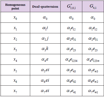
Lies on the Study Manifold, see Table 2. In this regard, we propose a modified interpolation algorithm. For that, the homogeneous points are replaced by motors and substitute matrix operations with GA operations. Since the matrix multiplications in (26) with form , which result is a constant value expressed as:

biomedres-openaccess-journal-bjstr

Table 2: Study quadric homogeneous point represented in dualquaternion and its equivalences as motor in G+3,0,1 and as motor in G4,1

Changing the homogeneous points with their respective motor representation in CGA and analyzing the motor multiplication, is shown that the coefficient of the blade e123∞ (α3) from the motor multiplication is the double value of (29). This coefficient (α3) can be extracted from any motor using the partial derivative:

or via inner product with the Ie and e0:

Using GA, the equation (26) is reformulated for a motor based interpolation algorithm. It uses 3 control motors as follows

In the same manner as Algorithm 1, for more control points, equation (32) can be rewritten using the De Casteljou algorithm as described on Algorithm 2. This algorithm calculates one motor gave the set M of control motors (m1,··· ,mN) and the interpolation constants (t,t0,t1,t2). Similar to Algorithm 1, the interpolation function is called recursively with the calculated motors from the last step as input until we get only one motor. Since the last step needs three motors to calculate the last one, M must contain an odd number of motors. (Algorithm 2)

biomedres-openaccess-journal-bjstr

Algorithm 2: Motor Interpolation on CGA.

Speeding up the Algorithms Using a GPU Accelerator

The algorithmic complexity using the framework Gn ∈ R 2n is: for binary operations O(22N); for unary operations O(2N), and for M interpolated motors using K motors O(MK). To accelerate the motor interpolation algorithm for real-time applications, one can utilize arithmetic accelerators based on FPGA, ASIC, and GPU. We utilize an accelerator based on CPU-GPU co-design, where the GPU is the GTX-970 by Nvidia, which has a computing capability of 5.2 and it can be implemented on any GPU using CUDA. The algorithm was developed to compute in parallel single or multiple interpolation curves. For the computing of a single interpolation curve, the default GPU was implemented, where each motor is computed in one thread launched in a Stream. For the case of multiple curves, the multi-streaming implementation was selected to overlap the streams. GTX-970 GPU does not support recursive algorithms, due to hardware limitations. To implement the motor interpolation, we translated the Algorithm 2 into an equivalent-iterative version given by the Algorithm 3.

biomedres-openaccess-journal-bjstr

Algorithm 3: Motor Interpolation on CGA (iterative).

The versatile Compute Unified Device Architecture (CUDA), is a platform for parallel computing, which was developed by NVIDIA for highly parallel implementations and uses its GPUs. CUDA is based on C/C++ programming languages, however other languages like Python or Fortran can be used as well. Our accelerator was written with CUDA C++ for the GPU acceleration and C++ for the CPU interface. The GTX-970 GPU does not support recursive algorithms; thus the Algorithm 2 has to be translated into an equivalent iterative version as presented in Algorithm 3. In this regard, the algorithm uses one stream for one curve of interpolation and multi-stream for multiple curves. Here, the GPU global memory is the largest in GPU, it has the highest latency, and it can be accessed from any stream for multi-stream applications. This is very helpful, if the number of motors to be computed is large, or if we compute multiple curves, or launch many algorithms employing the same GPU via multistreaming. The GPU and CPU have different architectures.
The CPU has a few sets of cores for sequential applications. In contrast, the GPU has many cores for highly parallel computing. Figure 2 compares the CPU and GPU architectures. Note that GPU cores run asynchronously using threads. Each core is limited to 32, 64, 128, 256, or 512 threads. If a parallel GPU implementation is launched, it is required a certain number of cores. These are figured out as a direct function of the selected threads per block. In the case of the motor interpolation, each thread launches a copy of the algorithm.
All GPU operations run explicitly or implicitly on a stream. If a kernel is launched in GPU, it runs explicitly or implicitly on a stream. A stream is a sequence of asynchronous operations which are executed on a device following an order ruled by the host code. A stream involves these operations to maintain order, to permit operations to be queued in the stream, and to be executed after all preceding operations. It cares for querying the status of queued operations. These operations include data transfer and kernel launches. Recent GPUs allow a parallel technique known as multi-streaming, where multiple streams are launched, and the transference data and kernel launch can be overlapped as shown in Figure 3. For the case of parallel calculation of multiple curves, the multi-streaming implementation is used, and each curve is computed in an independent stream.

biomedres-openaccess-journal-bjstr

Figure 2: Architecture comparison of
a) CPU and
b) GPU.

biomedres-openaccess-journal-bjstr

Figure 3: Different streams used for a concurrent implementa- tion
a) 1-stream,
b) Concurrent approach for device-to-host asynchronous memory copy,
c) Concurrent approach for host-to-device asynchronous memory copy,
d) Concurrent approach for host-to- device and device-to-host asynchronous memory copy.

Experiments

The Study’s quadric and the motor interpolation are compared to analyze which of the algorithms has the best speed performance. Many motors were calculated using 15 test groups of 5 motors considering following amount of points 20, 50, 100, 200, 500, 1000, 2000, 5000, 10000, and 20000. The test was programmed using MATLAB on a Desktop with Intel core I7 microprocessor at 2.4 GHz, 32 GB of RAM, and a GTX-970 GPU by NVIDIA. The results are presented in Figure 4. The execution time of both algorithms grew linearly while the number of points increases, thus the algorithm complexity is O(n). We see that the motor interpolation version is faster than the Study quadric interpolation, namely for 20000 points, it is 28× times faster. Figure 4 shows that the motor interpolation algorithm selected for sequential and parallel implementation utilizes same hardware parameters. The sequential implementation was programmed using C++ language and CUDA C++ for the parallel approach using 32 threads per block in the kernel. The interpolated motors were computed using single and double-precision floating-point representation. All the measures consider only the algorithm execution, but data transference, file writing, and reading are excluded. Results are shown in Figure 5.

biomedres-openaccess-journal-bjstr

Figure 4: Comparison of Study’s quadric and Motor interpola tion speed-up.

biomedres-openaccess-journal-bjstr

Figure 5: CPU and GPU speed-up analysis.

It was confirmed that the GPU has the best time performance for calculating the interpolation algorithm. When the time execution in CPU increases linearly, the time in GPU for 500 or fewer points remains almost constant. For the case of 20000 motors, the GPU implementation runs 79× and 60× times faster than the CPU for single and double floating-point respectively. The different multi-streaming implementations were analyzed; 16 different interpolation curves were calculated using the concurrent approach for host-to-device and device-to-host asynchronous memory copy with 4 configurations (1, 2, 4, and 8 streams) with 32 threads per block. The measure times include data transference. Figure 6 depicts that the use of multi-streaming reduces the execution time considerably, i.e. for 20000 points, 8 streams version is 2.7×, 1.9× and 1.6× times faster than 1, 2 and 4 streams respectively.

biomedres-openaccess-journal-bjstr

Figure 6: Comparison of the Multi-Stream speed-up.

Interpolation of Surgery Motion

This Study’s quadric-based motor interpolation was used to interpolate trajectories in medical robotics for kidney surgery, see Figure 7. The robot manipulator hast to follow a path for suture around a certain region on the kidney phantom. We design to tasks follow an Ultrasound (US)-probe trajectory and the interpolation of a given 3D points sequence. One gives the point coordinates and their desired orientations, these are interpolated by the motor algorithm, see Figures 7b-7d. Afterward, we check the precision error using inverse kinematics. The inverse kinematics was also formulated in conformal geometric algebra, the algorithm was reported in [14 -17].

biomedres-openaccess-journal-bjstr

Figure 7:

a) Robot; views:
b) X-Y,
c) X-Z,
d) Y-Z.

Conclusion

In this work, an optimized version of the Study quadric interpolation algorithm is presented using the conformal geometric algebra framework. A Matlab implementation confirms that conformal geometric algebra interpolation is 28x times faster than a Study quadric interpolation for 20000 motors. The performance of CPU and GPU approaches of motor interpolation algorithm were contrasted for the cases of single and double floating-point precision. The comparison shows that the GPU is 79x and 60 times faster than the CPU implementation for single and double precision respectively. Furthermore, 16 interpolation curves were calculated via multi-streaming. These results confirm that multi streaming reduces the execution time for multiple curves interpolation. We show an application of the motor algorithm for suture by kidney surgery. In near future, we will pursue to implement the interpolation algorithm using multiple GPUs and reduce the data dependency. Also, we will build an OpenCL library of the algorithms using Python.

For More Articles: Biomedical Journal Impact Factor: https://biomedres.us

Open Access Journal on public and environmental health

Awareness and Utilization of Emergency Contraception Among Female High School Students in Southeast Ethiopia

ABSTRACT

Background: Despite the existence of sound contraceptive methods including emergency contraception, there are huge numbers (84 million) of unplanned pregnancies occurring globally. Fifty percent of these pregnancies ended up in induced abortion out of which, 21 million are unsafely manipulated. Teen age girls are at double risk of the situation. The situation is worse in the developing countries like Ethiopia and much worse in rural and sub urban areas. The paucity of information in most rural part limits interventions that fit to the local situation.Objective: To assess awareness and utilization of emergency contraception and associated factors among female high school students in Dodola town, Oromia regional state, Ethiopia.Methods: A cross- sectional quantitative study was conducted using stratified random sampling technique. Data were entered, cleaned and analyzed using SPSS version 20. Bivariate and multivariate logistic regression analyses were conducted to examine the factors associated with the awareness and utilization of emergency contraceptives.Result: From a total of 773 female students 719 completed the questioners correctly; making the response rate of 93.02%. Four hundred fifty-three (63%) of the respondents were aged between 13-15 years with the mean age of 15.46(SD +/-1.76). Majority 578(80.4%) of the study participants were from Oromo ethnic group. About 304 (42.3%) had ever heard the existence of EC and about 9.8% of sexually active respondents had used it. Awareness of EC was associated with age greater than 15 years (AOR= 2.92(95% CI (1.99,4.26)], having radio (AOR= 4.43(95%CI (2.64,7.38)], having TV (AOR= 1.99(95% CI(1.19,3.30)] and chewing chat (AOR=3.08(95%CI (1.39, 6.82)]. Utilization of EC was associated with age, mothers’ educational status and having constant pocket money.Conclusion and Recommendations: The study revealed that awareness and utilization of emergency contraceptives is low. This leads to increased risk of unintended pregnancy and unsafe abortion. Therefore, the study recommends all concerned bodies should take parts in designing programs which increase the awareness and utilization of EC among high school students.

Plain English Summary

The long-established contraceptive methods including emergency contraception have not contributed adequately to stop the occurrence of huge numbers of unplanned pregnancies globally. Close to half of these pregnancies ended up in induced abortion and significant proportions of it are manipulated unsafely. Teen age girls in developing nations including Ethiopia are at increased risks. The situation is worse in the developing countries like Ethiopia and much worse in rural and sub urban areas. However, the gap in information has been affecting the interventions. Therefore, this study is conducted with the aim to assess the awareness and utilization of emergency contraception and associated factors among female high school students’ south-east of the Oromia region, Ethiopia. The study employed a cross- sectional quantitative using stratified random sampling technique. The overall data management and processing used sound statistical software for this purpose. Appropriate statistics were used to reveal statistical associations of related variables to the emergence contraception awareness and utilization. Ninety three percent of the study participants completed the questioners correctly. More than half of the respondent were in the age range between 13-15 years with the mean age of 15.46(SD +/-Majority of the study participants were from Oromo ethnic group. Less than half of the respondents had ever heard the existence of emergency contraceptives. Small proportions of sexually active respondents had used it. The study indicated that awareness and use of emergency contraceptives increase with age and level of education both the students and that of the mothers. The study revealed that awareness and utilization of emergency contraceptives is low. This leads to increased risk of unintended pregnancy and unsafe abortion. Therefore, the study recommends all concerned bodies should take parts in designing programs which increase the awareness and utilization of EC among high school students.

Keywords: Emergency Contraceptive; Awareness; Utilization; Adolescent

Abbreviations: CI: Confidence Interval; EDHS: Ethiopian Demographic Health Survey; EC: Emergency Contraceptive; ECPs: Emergency Contraceptive Pills; IRB: Institutional Reviewing Board; IUCD: Intra Uterine Contraceptive Device; KAP: Knowledge, Attitude and Practice; SPSS: Statistical Package for Social Science

Introduction

Background

Emergency contraception (EC) is contraceptive methods that can be used by women within five days following unprotected intercourse to prevent an unplanned pregnancy.EC provide a unique opportunity for preventing pregnancy after unprotected sex that no other contraceptive method can provide. It is as effective as 75-99% if taken within recommended time frame [1]. The capability of EC addressing the problems of unintended pregnancy in adolescent and young people posits the service one of the most crucial remedies in contraceptive arena for the fact that the sexual activity during adolescence is usually infrequent, unplanned and unprotected [1,2]. Moreover, the steady increase in school attendance rates in developing countries has given the chance for large proportion of young people to become sexually mature while they are at secondary school or earlier. When unintended pregnancy occurred to young girls they forced to dropout from school or goes for unsafe abortion [3]. In connection to the magnitude of the problems, nearly 210 million pregnancies occur each year globally. About 40% of these pregnancies were unintended. Of these unintended pregnancies 50% ended in abortion. About 42 million induced abortions each year, nearly 20 million abortions are unsafe. This represents 13% of maternal death. More than 95% of unsafe abortions occur in developing countries and nearly half of maternal deaths in sub-Saharan Africa were due to unsafe abortion [4,5]. In most developing countries complication of pregnancy and childbirth are a leading cause of death and disability among women of reproductive age. Teenage girls are at double risk of dying from pregnancy and birth complication than women over 20 years. This risk is five times higher for those girls under 15. In sub-Saharan Africa nearly 60 % of women who have unsafe abortion are younger than 25years of age, and 25% are teenagers [5,6].
The magnitude of abortion related morbidity and mortality are highest among young unmarried women in developing nations. For instance, in Uganda among women hospitalized with abortion-related complications, about two-thirds were 15 to 19 years old, two-thirds were students, and 80 % of them had never been married. In Nigeria Hospital-based studies have shown that up to 80% of patients with abortion-related complications are adolescents. In Ethiopia abortion accounts 60% 0f gynecological admission and girls under age 15 are three times more likely to end their pregnancies in abortion as compared to those ages 20-24. According to the 2011 Ethiopian Health and Demographic Survey (EDHS), 12% of young women age 15–19 have already begun childbearing [1,6,7]. Study conducted in Nepal among youths of 15-24 years in higher secondary and undergraduate students, the level of awareness of EC found to be 47%. But, only few mentioned the correct definition (17.02%) and consuming time (9.58%) of emergency contraceptive pills (ECP). Similarly, a study done in Dar Es Salaam, Tanzania, revealed that 57% of the respondents aware of ECP and only 14% had used it. A study conducted in Dessie, Ethiopia, on knowledge, attitude and practice (KAP) of emergency contraception among female college students disclosed that among those who had history of having sexual intercourse 78.3% of them faced unwanted pregnancies and 43.3% of these unwanted pregnancies resulted in abortion.
Of the respondents who had heard about emergency contraception only 15.4% of them made use of it. A study conducted in Jimma town, south west Ethiopia among female high school students revealed that (40.5%) had heard about EC. From those who heard of EC, 27% of the respondents mentioned the correct recommended timing for oral pills of emergency contraception [8- 11]. Currently, there are more adolescent girls in school than ever in our country. It is imperative that these girls have to complete their secondary education and beyond without any risk of unwanted pregnancies. EC is one of the known contraceptive methods to avert such problem. Knowing the level of awareness and utilization of EC by school adolescents is the first step of all the intervention to be implemented to protect adolescents from unintended pregnancy. Though few studies have been conducted on EC, they were focused only on students of higher institutions in big cities of the country. However, there are few or no scientific based research has been conducted so far in a district town like Dodola in which students come from both the town and rural areas around it. Thus, this study was conducted with an intention to determine awareness and utilization of EC and factors affecting the awareness and utilization of EC among Dodola female high school students. The study finding aimed to shape the local intervention by equipping district and zonal health leaders and service providers. Furthermore, the finding of the study will enable the comparison of service utilization within the region and outside; hence area specific interventions can be planned.

Methods and Materials

Study Setting

Dodola is located in southern part of Ethiopia, Oromia national regional state, west Arsi Zone at distance of 320km from the capital city Addis Ababa and 75km from Shashemene, the zonal center. According to 2014\15 woreda base plan, Dodola has total population 30,238 of which 14,917 males and 15,321 females. There are two administrative kebeles. With respect to some social services, the town has one district hospital and two health posts owned by the government. Moreover, it has one higher, four medium, six lower clinics, one pharmacy and eleven drug stores owned privately. There are three elementary, one high school and one preparatory public school.

Study Design

The study employed an institutional based cross-sectional quantitative study design with internal comparison carried out among female high school students in Dodola town, Oromia regional state in January 2016.

Population

Source Population: The source population of the study includes all high school female students in Dodola town who were enrolled in 2015/16 academic year.Study Population: The Study populations were female high school students which were randomly selected for the study from both high schools in the town.

Sample Size

The sample size was determined using the following assumptions: for the first study objective, expected prevalence of level of awareness of EC (64.48%) [12], Level of confidence 95%, and 5% margin of error (d=0.05). Formula for calculating the sample size n= [(Ζ 2) ×P (1 – P)/D2]. Considering design effect of 1.5 and using correction formula for finite population (N=1123female students) by adding nonresponse rate of 10% the final sample size=395. Following similar assumption and formula by taking the prevalence of EC utilization to be (25%) [11] sample size for the second objective was 344. However, when adjusted for sexually active students, the sample size increased to 773. By comparing two sample sizes, the largest 773 was taken as final sample size to ensure the maximum sample size (Figure 1).

biomedres-openaccess-journal-bjstr

Figure 1: Conceptual framework for EC. Note: Berhanu Desta and Nigatu Regassa 2011.

Sampling Technique

The schools were stratified in grade levels (9th& 10th). Respondents from each section were selected by simple random sampling using school enrollment list as sampling frame. Proportional distribution of sample was assigned to the respective schools, grades and sections based on the enrolment data for the academic year (Figure 2).

biomedres-openaccess-journal-bjstr

Figure 2: Schematic Presentation of Sampling Technique.

Data Collection Procedure & Quality Management

The data collection instrument was an anonymous selfadministered structured questionnaire. The questionnaires initially prepared in English and translated to Afan Oromo, and again back to English to ensure consistency and validity by language experts. Five percent of the samples were pretested before the actual data collection and some questions were amended. Training was given for data collection facilitators and supervisors for two days before the pretest and for a day after the pretest. The questionnaires were distributed to randomly selected students. Once the students finished filling the self-administered questionnaires, they had deposited them in a designated box, as informed earlier, to assure their anonymity. The overall data collection process was supervised by Principal investigator.

Operational Definition

Utilization of Emergency Contraceptives: Ever practice of emergency contraceptives.Awareness of Emergency Contraceptive: Ever heard the existence of emergency contraceptive.Unprotected Sexual Intercourse: Sexual intercourse that may result in unintended pregnancy.

Data Processing and Analysis

The collected data ware checked for their completeness and consistency and entered into SPSS version 20 for analysis. Data was summarized and organized using appropriate descriptive measures; frequencies and percentages of the responses were calculated. Associations between variables were assessed by using Odds Ratio with 95% Confidence Interval. First all variables were analyzed with bivariate analysis, then variables with p-value less than 0.25 were considered in Multivariate logistic- regression. Statistical association was declared at p-value less than 0.05 both in Bivariate and Multivariate logistic-regression.

Results

Socio-Demographic Characteristics of the Study Population

Seven hundred nineteen of the total 773 school adolescents were completed the survey questionnaire. Fifty four (6.98%) of the questionnaire were discarded due to incompleteness. Majority of the respondents 452(62.9%) were aged between 13-15 years with the mean age of 15.46 year with standard deviation (SD) of 1.76. About (51.6%) were attending grade nine and most of the respondents (96.2%) were single during the survey. The dominant religion was Islam (73.3%) followed by Orthodox (18.8%) and the majority were from Oromo ethnic group 578(80.4%).

Majority of the respondents 692(96.2%) had ever heard about modern contraceptives but only 304(42.3%) had ever heard the existence of EC before the survey. From those who ever heard of EC, 65.8% of them said oral pill should be used within 72hrs (3days) and 20.4% of them said Intra uterine Contraceptive Device (IUCD) should be used within 120hrs (5days) after unprotected sex to prevent unintended pregnancy. Of those who heard of EC, 207(68.1%) respondents said EC should be used after unprotected sex whereas 90(29.6%) of them said it should be used after unwanted pregnancy (Table1).

biomedres-openaccess-journal-bjstr

Table 1: Socio demographic characteristics of female high schools students of Dodola town, January 2016.

Utilization of Emergency Contraceptive

From the total 275 sexually active students only 27(9.8%) of them had used EC. Oral pill was the only methods utilized as EC. About 21(77.8%) of users took the first dose within 72hours after unprotected sexual intercourse and 17(63%) of them were recommended to use EC by their female friends. More than eighty five percent of EC users had used it two and more times. (Table 2).

biomedres-openaccess-journal-bjstr

Table 2: Awareness and utilization of female high schools students towards emergency contraception in Dodola town, January 2016.

Determinant Factors Related to the Awareness and Practice of Emergency Contraceptives

During bivariate logistic regression, age of respondent, having TV, having radio, having constant pocket money, drinking alcoholic beverage, chewing chat, mothers’ educational status and place of primary education showed significant association with the awareness of EC. Similarly, age of respondent, grade level, having TV, having constant pocket money, drinking alcoholic beverage and mothers’ educational status showed association with utilization of EC when analyzed using bivariate logistic regression. However, multivariate logistic regression analysis indicated that, age >15years (AOR= 2.92(95% CI(1.99,4.26)], having radio(AOR= 4.42 (95% CI(2.64,7.38)], having TV(AOR= 1.99(95% CI(1.19,3.30)] and chewing chat(AOR= 3.08 (95% CI(1.39,6.82)]showed statistically significant association with the awareness of EC (Table 3). Likewise, only age >15years (AOR= 3.52(95% CI(1.04,1.92)], having constant pocket money(AOR= 3.52(95% CI(1.15,10.75)] and mothers’ educational status(AOR= 4(95% CI(1.32,12.09)] were remained the real predictors of EC utilization when adjusted to the confounders with multivariate analysis (Tables 3 & 4).

biomedres-openaccess-journal-bjstr

Table 3: Bivariate and multivariate logistic regressions of selected variables in relation to awareness of EC among female high school students in Dodola town; January 2016.

biomedres-openaccess-journal-bjstr

Table 4: Bivariate and multivariate logistic regressions of selected variables in relation to Utilization of EC among female high school students in Dodola town; January 2016.

Discussion

Unintended pregnancy that usually followed by unsafe abortion is a major reproductive health challenge in developing countries. This RH risk is higher among young females than adult women(4). Nevertheless, unwanted pregnancy resulting from unprotected sex can be prevented by the proper use of EC. The result of this study showed that 38.2% of the respondents were sexually active. Forty percent of sexually active students had history of pregnancy, which 83.6% of those pregnancies were unwanted and 58.7% of unwanted pregnancy resulted in unsafe abortion. Similar studies in Dessie and Mekele cities have showen lower prevalence of unintended pregnancy and unsafe abortion. High rate of unwanted pregnancy and unsafe abortion in this study than the above two studies may be due to high awareness and utilization of EC in those studies than current one which is 70% for Dessie and 90.3% for Mekele [9,13]. When compared to Dodola, the small rural town, the former two cities are with relative better positions both in health service accessibilities and information dissemination. This is more or less similar with studies of Addis Ababa University students 43.5% and high school students of Jimma town 40.5% [11,14]. But it is lower than those studies conducted among tertiary students in Nigeria, college students of Dessie town and Adewa preparatory students 65.3%,69.9%&64.48% respectively [9,13,15]. This discrepancy could be due to socio-demographic and difference in the education status of the study population.
This study revealed that, out of sexually active respondents only 9.8% of them were utilized emergency contraceptive methods. It is more or less similar with research done among youths of Parbat district, Nepal 8.34% [10]. The finding of this study found to be lower than study conducted among Tertiary Students in Osun State, Southwestern Nigeria 32.6%, Mekele preparatory school 53.5% and Debre Markos University 18.4% [13,15,16]. This might be due to the age and knowledge difference between university and high school students. Regarding determinant factors for the awareness and utilization of EC, the present study found that respondents whose age is greater than 15 years were about three times more likely to be aware and utilize EC compared to the age group of fifteen or less years. The finding is consistent with previous studies conducted in Addis Ababa, Mekele, Haramaya and Dessie which reported older age groups were more likely to be aware and practice EC when compared to younger age groups [9,17,13-14]. This might be due to younger youth may be less concerned about unwanted pregnancy and EC as they have not yet started or newly joining to sexual activity. The other possible explanation for this could be as age increases the education level and knowledge of sexual matters may increase.
This study found that respondents who had constant pocket money were more than three times more likely to practice EC than those who had no pocket money. This may be explained by having constant pocket money may help students to decide by themselves to buy and use EC if they faced unprotected sexual intercourse without the consent of others. Moreover, the more they are financial sufficient, the more that they have freedom to practice sex. This is in agreement with the study conducted in Hawassa University about the patterns of risky sexual practice among students [18].

Conclusion and Recommendation

The study finding showed low awareness and utilization of emergency contraceptive among female high school students. This may be among the reasons for high rate of unintended pregnancy and unsafe abortion among the study population. Awareness and utilization of female high school students on EC are affected by a range of circumstances. Given the high rate of unintended pregnancy and unsafe abortion reported by the study participants; the academic institution with the health sector should take measures that can increase awareness of students and ensure easy availability of EC at school level or adolescent and youth friendly clinic in the nearby.

Acknowledgement

We would like to thank school of public and environmental health, College of medicine and health sciences, Hawassa University for technical and financial support. We also would like to extend our gratitude to Dodola town education office, school principals, teachers, data collection facilitators, supervisors and study participants for their invaluable assistance and participation during the study period.

Declaration

Ethical Considerations

Ethical clearance was obtained from Hawassa University, College of Medicine and Health sciences Institutional Review Board (IRB). Official letter from Hawassa University was submitted to Dodola town educational office and high school’s administrators. Informed verbal consent was obtained from the study participants after detail explanation of the purpose and the benefit of the study. Participants were informed that they have full right to discontinue or refuse to participate in the study at any time in the continuum of the data collection. They were also informed the anonymity of the study that by any means their participation in the study and response shall remain confidential.

Consent for the Publication

This manuscript is an extract from the MPH thesis. All ethical issues including communication of the finding to various stakeholders were properly communicated and their consents were obtained verbally.

Data Availability

The data for this study will be available when needed from the SPSS data pool.

Declaration of Funding

As mentioned earlier, this study was part of the fulfillment for the requirement for the master’s degree in public health; small proportion of fund received from Hawassa University and the other portions of expenses were covered by me in support from my family and friend.

For More Articles: Biomedical Journal Impact Factor: https://biomedres.us

Open Access Journal of Maxillofacial Surgery

Oral Manifestations of Behcet’s Syndrome: Case Report

Introduction

Systemic diseases may be preceded or spread, with oral manifestations. The lesions of the oral cavity are attended, for the most part, by Stomatology Specialists, so that knowledge of the oral manifestations of systemic diseases contributes to correct behavior in the face of these conditions that, finally, must be referred to the specialist in the attention of each one of them [1]. Behcet’s syndrome is an autoimmune disease of the seronegative vasculitic type, with a disease of vessels of variable caliber [2]. Although the etiology of Behcet’s Syndrome is unknown, there are numerous investigations that indicate the participation of genetic, infectious (viral and bacterial) and immunological factors. Among the most important are the association with the HLA genotype of the patients, the cross-reactivity with human peptides and the activation of the vascular endothelium [3]. This disease is more common and severe along the old “silk road”, which extended from East Asia to the Mediterranean, Turkey’s current territory. 80 to 370 cases are reported per 100,000 inhabitants in Turkey, while the prevalence ranges from 13.5 to 35 per 100,000 in Japan, Korea, China, Iran, Iraq and Saudi Arabia; being a more severe manifestation in Japan [4]. In relation to sex, a predominance of female sex in countries of America and male sex in Asia is described, mainly between the ages of 20 and 40 [5]. In Cuba there are no prevalence studies of this entity [3]. The first description made of this pathology arises may have been described by Hippocrates in his third book of endemic diseases [4]. In 1930, the Greek ophthalmologist Benediktos Adamantiades reported to a patient with arthritis, oral and genital ulcers, phlebitis, and iritis but it was not until 1936 that the Turkish dermatologist Hulsui Behcet determined the description of this disease of more accurate way [6]. The clinical spectrum of this disease, characterized by a symptomatic triad (recurrent oral and genital ulcers with uveitis and hypotension) in addition to the fact that the histopathological study of the affected organs and the analytical data are nonspecific, makes its diagnosis based on the clinical method of description and grouping of signs and symptoms [7]. In 1990, the criteria for the diagnosis of this entity were established for the first time by the International Study Group for Behçet’s Disease [8].
In 2006, new diagnostic criteria of the International Group for the study of Behcet’s disease were established [9], which declared a major or mandatory criterion: recurrent oral ulcers and minor criteria: recurrent genital ulcers, eye lesions, skin lesions, vascular lesions and positive patergia test [10]. In 2013, the international criteria for the diagnosis of the disease were reviewed, as a result of the analysis they proposed to award 2 points to ocular lesions and oral and genital aphthous lesions and assign 1 point to skin lesions, of the Central Nervous System, vascular manifestations and the positive patergia test. According to these criteria, a patient with a score ≥ 4 points is classified as a Behcet disease [3]. At present it is known that it is not curable, but treatable [4]. The goals of therapy are to: suppress inflammation, reduce the frequency and severity of recurrences. To be effective, treatment must be implemented early [11]. Objective: Describe the oral manifestations and therapeutic behavior to be followed before a patient with Behcet syndrome.

Case Presentation

Patient of 20 years of age, female sex, black race, urban origin, who comes to the emergency services of Maxillofacial Surgery of the body of guard of the Dr. Gustavo Aldereguía Lima Hospital in Cienfuegos, Cuba referred by the specialist in Stomatology General Comprehensive that provided services in the area of health corresponding to the patient for presenting multiple ulcerated lesions throughout the oral cavity and lips Figure 1.

biomedres-openaccess-journal-bjstr

Figure 1: Ulcerated lesions on lips.

Anamnesis

During the interrogation, the patient was referred to as inflammation of the entire oral cavity, pain during food intake and difficulty swallowing (dysphagia), burning sensation and burning of the mouth, halitosis, recurrent episodes (7 to 8 times a year) ulcerated bleeding lesions, which previously healed spontaneously until their total disappearance but during the last episode the lesions persist. It also refers to the presence of genital ulcers, skin rash in the frontal region and persistent joint pain.

Physical Exam

Multiple ulcers were detected in the oral cavity of approximately 1cm and more, covered by a yellowish-green pseudo membrane and with fetidity due to the over-added infection of the lesions, with irregular, undefined, elevated, rounded, hyperemic edges, bleeding to the minimum stimulus, painful on palpation, located in the region of the oropharynx, tongue, palate, gum, floor of mouth, mucosa of cheeks and lips Figure 2a. In addition, irregular and rough surface was detected upon palpation of the frontal cutaneous region Figure 2b and in the left axillary and subaxillary region Figure 2c. The case of the patient was consulted with the Internal Medicine services for a better treatment of genital lesions and joint pain.

biomedres-openaccess-journal-bjstr

Figure 2:(a) yellowish-green pseudo membrane located in the tongue and oropharynx;
(b) Skin rash in the frontal region;
(c) Irregular and rough surface in the left axillary and subaxillary region.

Complementary Exams

The following complements were indicated:
a) Blood count
b) Leukogram
c) Blood chemistry
d) Hepatic Profile
e) HIV serology
f) Rheumatoid Factor
g) C-reactive protein
h) LE cells
i) Immunoglobulins Serum
j) Patergia test
k) Surface antigen
l) Hepatitis C antibody
The results of the complementary examinations were within the normal parameters except the Leukogram in which there was a considerable increase in segmented leukocytes (neutrophils) with a value of 80%, which showed that the over-added infection of the lesions was bacterial. During the patergia test, a small sterile needle was inserted into the skin of the forearm, causing a small red papule to appear at the needle insertion site one or two days after the test was performed, which indicated hyperreactivity of the immune system to minor trauma or damage Figure 3. It is not 100% specific, only a proportion of patients with Behcet syndrome have a positive response.

biomedres-openaccess-journal-bjstr

Figure 3: Red papule on forearm skin two days after the patergia test.

Diagnosis

With these results, some possible diagnoses were ruled out: Human Immunodeficiency Syndrome, Rheumatoid Arthritis, Hepatitis B and C, Herpes Simplex type 1, Recurrent Aphthous Stomatitis, Parvovirus, Discoid Lupus Erythematosus. Finally, the patient was diagnosed with Behcet Syndrome with a scientific basis in the criteria for the diagnosis of this syndrome of 2013, complying with the mandatory or major requirement: recurrent oral ulcers in oral mucosa with a minimum of 3 episodes during a year (2 points ) and with three minor criteria: genital ulcers (2 points), erythema nodosum (1 point) in this case in the left frontal, axillary and subaxillary region and positive patergia test (1 point). According to this current classification criterion it is only necessary to accumulate a score ≥ 4 points, in this case the patient added a total of 6 points.

Treatment

As part of the Stomatological treatment, the patient was indicated a series of hygienic dietary measures such as: eliminating or reducing acidic foods from the diet (lemon, orange, tangerine, pineapple, guava, tomato, natural yogurt); avoid ingesting foods with artificial condiments and at elevated temperatures and instead use natural condiments and ingesting food and drinks that are refreshing but not gaseous; Eliminate local irritants such as coffee, cigar and alcohol. In addition, local topical anesthetics such as 0.2% lidocaine were indicated half an hour before eating food to relieve the pain caused by chewing and swallowing in these cases. In this case, the indication of mouthwash with saline solution or 20% chamomile tincture was very effective, 20 drops in 200 milliliters of boiled water 3 times a day for 7 days, the use of chamomile was preferred as part of the natural medicine treatment and traditional for its properties: anti-inflammatory, analgesic, antibacterial, antiulcer, antiviral and antifungal.
The Internal Medicine services treated the patient with a prednisone in a 20-milligram bulb intravenously every 8 hours, then gradually reduced to a maintenance dose of 10 milligrams orally in tablets, this corticosteroid was very effective in the treatment of inflammatory component of oral, genital and arthropathic lesions of the patient and as an immunomodulator. In addition, colchicine (0.5 mg) was given one tablet every 8 hours; methotrexate (2.5 mg) four tablets in weekly dose; folic acid (5 mg) one tablet daily, except on the day of Metrotexate, all in order to control the rheumatologic component. Currently, the patient is compensated for the disease.

Discussion

Systemic diseases are those morbid processes that affect more than one organic system. The etiopathogenesis of many of these diseases is still not completely clear, but it is well known that, in large part of them, inflammatory processes and immune system disorders that give rise to the various manifestations are involved. The majority of patients with recurrent oral aphthous ulcers have no other involvement, but in others the presence of chronic aphthous stomatitis lesions is associated with systemic processes [1]. Behcet’s syndrome is a very rare autoimmune disease condition in these latitudes [4]. The most accepted concept so far defines it as: in a chronic, multisystemic, recurrent inflammatory process whose main alteration lies in a vasculitis that involves the arteries and veins of any caliber. It is characterized in the clinic by presenting patients with inflammation of the mucous membranes, translated by oral and genital ulcers; in addition to uveitis; digestive symptoms, skin lesions, arthritis and occasional neurological intake.
The etiology of Behcet’s disease is unknown and although the majority of cases are sporadic, some studies support the possible genetic origin with evidence of autosomal recessive inheritance as it is reported that in 1 in 10 families there is another member with the disease or other autoimmune diseases in first-degree relatives such as hypothyroidism, scleroderma, discoid lupus erythematosus and juvenile idiopathic arthritis. Another of the arguments described in defense of this genesis is the increased risk of suffering from Behcet’s disease when it is associated with the presence of the HLA-B51 main histocompatibility system antigen [12]. It is believed as for many autoimmune or auto-inflammatory syndromes, that certain infectious factors (in particular, Streptococcus antigens) and / or environmental factors are capable of triggering symptoms in individuals with certain genetic variants [13]. In some investigations, the herpes simplex virus type 1 and parvovirus B 19, among the triggers of Behcet’s syndrome, other research reports that in this syndrome there is an alteration in the number and activation of lymphocytes, so the CD4 + / CD8 + index inversion has been observed.
The syndrome is usually more serious and frequent in men [3], however the case presented corresponds to a female patient. The white skin color shows a predominance, although there is a great miscegenation in Cuba [12], this ailment is uncommon in the black population but when it appears they present greater complications and worse prognosis [14]. Over the years different criteria have been used to diagnose the disease, the International Study Group for Behçet’s Disease in 1990 considers the existence of recurrent oral ulceration with at least two of the following clinical manifestations: recurrent genital ulceration, ocular involvement, involvement cutaneous or positive patergia test to make the diagnosis [8]. In 2006 the International Group declares it necessary to present: Mandatory Criteria or major criteria: recurrent oral ulcers (minor canker sores, major canker sores or herpetiform ulcers, in oral mucosa with a minimum of 3 episodes for a year). Minor criteria: recurrent genital ulcers (ulcers or aphthous scars in genital areas observed by the doctor or the patient) (2 points). Eye lesions (anterior or posterior uveitis, or the presence of vitreous cells in the examination with slit lamp or vasculitis retinal diagnosis by an ophthalmologist) (1point).
Skin lesions (erythema nodosum, folliculitis, papulopustular lesions, acneiform nodules, observed by the doctor in postadolescent patients not treated with corticosteroids) (2 points). Patergia test: positive (cutaneous hypersensitivity characterized by the appearance of a sterile pustule, 24 to 48 h after needle puncture, observed by a doctor) (1 point). Vascular lesions (arterial, venous thrombosis or aneurysms) (1 point). The diagnosis is made with the mandatory criterion plus 3 points [10]. In 2013, an international group, composed of representatives from 27 countries, reviewed the international criteria, according to these criteria, a patient with a score ≥ 4 points is classified as a Behcet disease [3]. The presence of recurrent, painful, variable-sized canker sores that heal in 1-3 weeks usually without scarring in most cases are the first manifestation, as occurred in this case. The genital canker sores present in 72-94% of cases are morphologically similar to oral ones, usually heal at 2-4 weeks, but they do leave a scar [7]. There are no typical laboratory alterations of Behcet Syndrome. Erythrosedimentation and C-reactive protein levels are often moderately high but do not correlate well with disease activity, in this case these values were kept within normal parameters.
Acute phase reactants may be elevated, mainly in patients with vasculitis of large vessels. Serum immunoglobulins are sometimes elevated and immunocomplex levels may be elevated, but autoantibodies such as rheumatoid factor, antinuclear antibodies, anticardiolipin and neutrophil antithoplasm (ANCA) are negative [3]. The patergia test is not 100% specific, only a proportion of patients with Behcet Syndrome have a positive response and the positivity in patients with the syndrome vary in different geographical areas (60-70% in Turkey and Japan, but rare in America and Europe) [11], however, the case presented is of a patient born in Cuba who is part of the American continent in which the test was positive, not coinciding with world statistics, where positivity is rare in the Americas. The differential diagnosis should be made with: Reiter’s disease, Steven-Johnson syndrome, aphthous stomatitis, recurrent Mollaret meningitis, Whipple’s disease, multiple sclerosis, Harada’s disease, inflammatory bowel diseases, sarcoidosis and ankylosing spondylitis. The diagnosis is based on detailed medical history and long-term clinical observation. There is no way to predict whether a patient with recurrent oral ulcerations will develop the disease even if frequent outbreaks constitute an alarm sign, as confirmed in this case. The choice of treatment depends on the clinical manifestations and their severity.
An early diagnosis followed by a treatment with corticosteroids, medication used in this patient, could prevent the fearful neurological complications in the opinion of several authors, although more recent studies report highly positive results with immunosuppressants [7]. It is described that all patients should use colchicine and steroids, with Metrotexate being the diseasemodifying drug most used in the treatment of this [5]. The majority of patients diagnosed with Behcet syndrome have a productive life. The symptoms are controlled with a healthy diet, rest, physical exercises and drug treatment and the prognosis varies according to the affected organs [7]. Behcet’s disease has a chronic course with exacerbations and unpredictable remissions, the frequency and severity of which may decrease over time. After the first five years, the disease acquires a stable course or towards improvement; after the fourth decade, clinical severity is decreased, with longer intervals of recurrence between exacerbations. The prognosis is good, unless there is commitment of vital organs. The presentation at the age of early onset (before age 25) has been associated with more severe manifestations of the disease and increased mortality [13].

Conclusion

While it is true that multiple systemic diseases present with aphthous oral manifestations, oral ulcers in Behcet’s disease are the first clinical manifestation, which in most cases, lead the patient to seek medical help and constitute for countless authors the cornerstone to reach the diagnosis of this disease, so it is considered vital that the Comprehensive General Stomatologist putting into practice the knowledge acquired about the syndrome, consider it a possible differential diagnosis more before a patient with recurrent oral ulcerations. The timely management of a patient with an unusual and exceptional pathology can have an optimal evolution and reduction of complications in the quality of life. It is recommended to expand coverage and health education to be able to detect these cases in the first instance, thus avoiding underdiagnosis.

For More Articles: Biomedical Journal Impact Factor: https://biomedres.us

Open Access Journals on Surgery

Oral Manifestations of Behcet’s Syndrome: Case Report

Introduction

Systemic diseases may be preceded or spread, with oral manifestations. The lesions of the oral cavity are attended, for the most part, by Stomatology Specialists, so that knowledge of the oral manifestations of systemic diseases contributes to correct behavior in the face of these conditions that, finally, must be referred to the specialist in the attention of each one of them [1]. Behcet’s syndrome is an autoimmune disease of the seronegative vasculitic type, with a disease of vessels of variable caliber [2]. Although the etiology of Behcet’s Syndrome is unknown, there are numerous investigations that indicate the participation of genetic, infectious (viral and bacterial) and immunological factors. Among the most important are the association with the HLA genotype of the patients, the cross-reactivity with human peptides and the activation of the vascular endothelium [3]. This disease is more common and severe along the old “silk road”, which extended from East Asia to the Mediterranean, Turkey’s current territory. 80 to 370 cases are reported per 100,000 inhabitants in Turkey, while the prevalence ranges from 13.5 to 35 per 100,000 in Japan, Korea, China, Iran, Iraq and Saudi Arabia; being a more severe manifestation in Japan [4]. In relation to sex, a predominance of female sex in countries of America and male sex in Asia is described, mainly between the ages of 20 and 40 [5]. In Cuba there are no prevalence studies of this entity [3]. The first description made of this pathology arises may have been described by Hippocrates in his third book of endemic diseases [4]. In 1930, the Greek ophthalmologist Benediktos Adamantiades reported to a patient with arthritis, oral and genital ulcers, phlebitis, and iritis but it was not until 1936 that the Turkish dermatologist Hulsui Behcet determined the description of this disease of more accurate way [6]. The clinical spectrum of this disease, characterized by a symptomatic triad (recurrent oral and genital ulcers with uveitis and hypotension) in addition to the fact that the histopathological study of the affected organs and the analytical data are nonspecific, makes its diagnosis based on the clinical method of description and grouping of signs and symptoms [7]. In 1990, the criteria for the diagnosis of this entity were established for the first time by the International Study Group for Behçet’s Disease [8].
In 2006, new diagnostic criteria of the International Group for the study of Behcet’s disease were established [9], which declared a major or mandatory criterion: recurrent oral ulcers and minor criteria: recurrent genital ulcers, eye lesions, skin lesions, vascular lesions and positive patergia test [10]. In 2013, the international criteria for the diagnosis of the disease were reviewed, as a result of the analysis they proposed to award 2 points to ocular lesions and oral and genital aphthous lesions and assign 1 point to skin lesions, of the Central Nervous System, vascular manifestations and the positive patergia test. According to these criteria, a patient with a score ≥ 4 points is classified as a Behcet disease [3]. At present it is known that it is not curable, but treatable [4]. The goals of therapy are to: suppress inflammation, reduce the frequency and severity of recurrences. To be effective, treatment must be implemented early [11]. Objective: Describe the oral manifestations and therapeutic behavior to be followed before a patient with Behcet syndrome.

Case Presentation

Patient of 20 years of age, female sex, black race, urban origin, who comes to the emergency services of Maxillofacial Surgery of the body of guard of the Dr. Gustavo Aldereguía Lima Hospital in Cienfuegos, Cuba referred by the specialist in Stomatology General Comprehensive that provided services in the area of health corresponding to the patient for presenting multiple ulcerated lesions throughout the oral cavity and lips Figure 1.

Anamnesis

During the interrogation, the patient was referred to as inflammation of the entire oral cavity, pain during food intake and difficulty swallowing (dysphagia), burning sensation and burning of the mouth, halitosis, recurrent episodes (7 to 8 times a year) ulcerated bleeding lesions, which previously healed spontaneously until their total disappearance but during the last episode the lesions persist. It also refers to the presence of genital ulcers, skin rash in the frontal region and persistent joint pain.

Physical Exam

Multiple ulcers were detected in the oral cavity of approximately 1cm and more, covered by a yellowish-green pseudo membrane and with fetidity due to the over-added infection of the lesions, with irregular, undefined, elevated, rounded, hyperemic edges, bleeding to the minimum stimulus, painful on palpation, located in the region of the oropharynx, tongue, palate, gum, floor of mouth, mucosa of cheeks and lips Figure 2a. In addition, irregular and rough surface was detected upon palpation of the frontal cutaneous region Figure 2b and in the left axillary and subaxillary region Figure 2c. The case of the patient was consulted with the Internal Medicine services for a better treatment of genital lesions and joint pain.

Complementary Exams

The following complements were indicated:
a) Blood count
b) Leukogram
c) Blood chemistry
d) Hepatic Profile
e) HIV serology
f) Rheumatoid Factor
g) C-reactive protein
h) LE cells
i) Immunoglobulins Serum
j) Patergia test
k) Surface antigen
l) Hepatitis C antibody
The results of the complementary examinations were within the normal parameters except the Leukogram in which there was a considerable increase in segmented leukocytes (neutrophils) with a value of 80%, which showed that the over-added infection of the lesions was bacterial. During the patergia test, a small sterile needle was inserted into the skin of the forearm, causing a small red papule to appear at the needle insertion site one or two days after the test was performed, which indicated hyperreactivity of the immune system to minor trauma or damage Figure 3. It is not 100% specific, only a proportion of patients with Behcet syndrome have a positive response.

Diagnosis

With these results, some possible diagnoses were ruled out: Human Immunodeficiency Syndrome, Rheumatoid Arthritis, Hepatitis B and C, Herpes Simplex type 1, Recurrent Aphthous Stomatitis, Parvovirus, Discoid Lupus Erythematosus. Finally, the patient was diagnosed with Behcet Syndrome with a scientific basis in the criteria for the diagnosis of this syndrome of 2013, complying with the mandatory or major requirement: recurrent oral ulcers in oral mucosa with a minimum of 3 episodes during a year (2 points ) and with three minor criteria: genital ulcers (2 points), erythema nodosum (1 point) in this case in the left frontal, axillary and subaxillary region and positive patergia test (1 point). According to this current classification criterion it is only necessary to accumulate a score ≥ 4 points, in this case the patient added a total of 6 points.

Treatment

As part of the Stomatological treatment, the patient was indicated a series of hygienic dietary measures such as: eliminating or reducing acidic foods from the diet (lemon, orange, tangerine, pineapple, guava, tomato, natural yogurt); avoid ingesting foods with artificial condiments and at elevated temperatures and instead use natural condiments and ingesting food and drinks that are refreshing but not gaseous; Eliminate local irritants such as coffee, cigar and alcohol. In addition, local topical anesthetics such as 0.2% lidocaine were indicated half an hour before eating food to relieve the pain caused by chewing and swallowing in these cases. In this case, the indication of mouthwash with saline solution or 20% chamomile tincture was very effective, 20 drops in 200 milliliters of boiled water 3 times a day for 7 days, the use of chamomile was preferred as part of the natural medicine treatment and traditional for its properties: anti-inflammatory, analgesic, antibacterial, antiulcer, antiviral and antifungal.
The Internal Medicine services treated the patient with a prednisone in a 20-milligram bulb intravenously every 8 hours, then gradually reduced to a maintenance dose of 10 milligrams orally in tablets, this corticosteroid was very effective in the treatment of inflammatory component of oral, genital and arthropathic lesions of the patient and as an immunomodulator. In addition, colchicine (0.5 mg) was given one tablet every 8 hours; methotrexate (2.5 mg) four tablets in weekly dose; folic acid (5 mg) one tablet daily, except on the day of Metrotexate, all in order to control the rheumatologic component. Currently, the patient is compensated for the disease.

Discussion

Systemic diseases are those morbid processes that affect more than one organic system. The etiopathogenesis of many of these diseases is still not completely clear, but it is well known that, in large part of them, inflammatory processes and immune system disorders that give rise to the various manifestations are involved. The majority of patients with recurrent oral aphthous ulcers have no other involvement, but in others the presence of chronic aphthous stomatitis lesions is associated with systemic processes [1]. Behcet’s syndrome is a very rare autoimmune disease condition in these latitudes [4]. The most accepted concept so far defines it as: in a chronic, multisystemic, recurrent inflammatory process whose main alteration lies in a vasculitis that involves the arteries and veins of any caliber. It is characterized in the clinic by presenting patients with inflammation of the mucous membranes, translated by oral and genital ulcers; in addition to uveitis; digestive symptoms, skin lesions, arthritis and occasional neurological intake.
The etiology of Behcet’s disease is unknown and although the majority of cases are sporadic, some studies support the possible genetic origin with evidence of autosomal recessive inheritance as it is reported that in 1 in 10 families there is another member with the disease or other autoimmune diseases in first-degree relatives such as hypothyroidism, scleroderma, discoid lupus erythematosus and juvenile idiopathic arthritis. Another of the arguments described in defense of this genesis is the increased risk of suffering from Behcet’s disease when it is associated with the presence of the HLA-B51 main histocompatibility system antigen [12]. It is believed as for many autoimmune or auto-inflammatory syndromes, that certain infectious factors (in particular, Streptococcus antigens) and / or environmental factors are capable of triggering symptoms in individuals with certain genetic variants [13]. In some investigations, the herpes simplex virus type 1 and parvovirus B 19, among the triggers of Behcet’s syndrome, other research reports that in this syndrome there is an alteration in the number and activation of lymphocytes, so the CD4 + / CD8 + index inversion has been observed.
The syndrome is usually more serious and frequent in men [3], however the case presented corresponds to a female patient. The white skin color shows a predominance, although there is a great miscegenation in Cuba [12], this ailment is uncommon in the black population but when it appears they present greater complications and worse prognosis [14]. Over the years different criteria have been used to diagnose the disease, the International Study Group for Behçet’s Disease in 1990 considers the existence of recurrent oral ulceration with at least two of the following clinical manifestations: recurrent genital ulceration, ocular involvement, involvement cutaneous or positive patergia test to make the diagnosis [8]. In 2006 the International Group declares it necessary to present: Mandatory Criteria or major criteria: recurrent oral ulcers (minor canker sores, major canker sores or herpetiform ulcers, in oral mucosa with a minimum of 3 episodes for a year). Minor criteria: recurrent genital ulcers (ulcers or aphthous scars in genital areas observed by the doctor or the patient) (2 points). Eye lesions (anterior or posterior uveitis, or the presence of vitreous cells in the examination with slit lamp or vasculitis retinal diagnosis by an ophthalmologist) (1point).
Skin lesions (erythema nodosum, folliculitis, papulopustular lesions, acneiform nodules, observed by the doctor in postadolescent patients not treated with corticosteroids) (2 points). Patergia test: positive (cutaneous hypersensitivity characterized by the appearance of a sterile pustule, 24 to 48 h after needle puncture, observed by a doctor) (1 point). Vascular lesions (arterial, venous thrombosis or aneurysms) (1 point). The diagnosis is made with the mandatory criterion plus 3 points [10]. In 2013, an international group, composed of representatives from 27 countries, reviewed the international criteria, according to these criteria, a patient with a score ≥ 4 points is classified as a Behcet disease [3]. The presence of recurrent, painful, variable-sized canker sores that heal in 1-3 weeks usually without scarring in most cases are the first manifestation, as occurred in this case. The genital canker sores present in 72-94% of cases are morphologically similar to oral ones, usually heal at 2-4 weeks, but they do leave a scar [7]. There are no typical laboratory alterations of Behcet Syndrome. Erythrosedimentation and C-reactive protein levels are often moderately high but do not correlate well with disease activity, in this case these values were kept within normal parameters.
Acute phase reactants may be elevated, mainly in patients with vasculitis of large vessels. Serum immunoglobulins are sometimes elevated and immunocomplex levels may be elevated, but autoantibodies such as rheumatoid factor, antinuclear antibodies, anticardiolipin and neutrophil antithoplasm (ANCA) are negative [3]. The patergia test is not 100% specific, only a proportion of patients with Behcet Syndrome have a positive response and the positivity in patients with the syndrome vary in different geographical areas (60-70% in Turkey and Japan, but rare in America and Europe) [11], however, the case presented is of a patient born in Cuba who is part of the American continent in which the test was positive, not coinciding with world statistics, where positivity is rare in the Americas. The differential diagnosis should be made with: Reiter’s disease, Steven-Johnson syndrome, aphthous stomatitis, recurrent Mollaret meningitis, Whipple’s disease, multiple sclerosis, Harada’s disease, inflammatory bowel diseases, sarcoidosis and ankylosing spondylitis. The diagnosis is based on detailed medical history and long-term clinical observation. There is no way to predict whether a patient with recurrent oral ulcerations will develop the disease even if frequent outbreaks constitute an alarm sign, as confirmed in this case. The choice of treatment depends on the clinical manifestations and their severity.
An early diagnosis followed by a treatment with corticosteroids, medication used in this patient, could prevent the fearful neurological complications in the opinion of several authors, although more recent studies report highly positive results with immunosuppressants [7]. It is described that all patients should use colchicine and steroids, with Metrotexate being the diseasemodifying drug most used in the treatment of this [5]. The majority of patients diagnosed with Behcet syndrome have a productive life. The symptoms are controlled with a healthy diet, rest, physical exercises and drug treatment and the prognosis varies according to the affected organs [7]. Behcet’s disease has a chronic course with exacerbations and unpredictable remissions, the frequency and severity of which may decrease over time. After the first five years, the disease acquires a stable course or towards improvement; after the fourth decade, clinical severity is decreased, with longer intervals of recurrence between exacerbations. The prognosis is good, unless there is commitment of vital organs. The presentation at the age of early onset (before age 25) has been associated with more severe manifestations of the disease and increased mortality [13].

Conclusion

While it is true that multiple systemic diseases present with aphthous oral manifestations, oral ulcers in Behcet’s disease are the first clinical manifestation, which in most cases, lead the patient to seek medical help and constitute for countless authors the cornerstone to reach the diagnosis of this disease, so it is considered vital that the Comprehensive General Stomatologist putting into practice the knowledge acquired about the syndrome, consider it a possible differential diagnosis more before a patient with recurrent oral ulcerations. The timely management of a patient with an unusual and exceptional pathology can have an optimal evolution and reduction of complications in the quality of life. It is recommended to expand coverage and health education to be able to detect these cases in the first instance, thus avoiding underdiagnosis.

For More Articles: Biomedical Journal Impact Factor: https://biomedres.us

Open Access Journal on Chemistry

Mechanistic Role of Egyptian Propolis Against Ehrlich Ascites Carcinoma and Methotrexate Limitations

ABSTRACT

Although several conventional anti-cancer drugs are available, most of them are toxic and intolerable after long-term use. Therefore, there has been a global increased interest to identify novel agents that can possess anti-tumor effects by it or maximize the anti-tumor effects of low doses of conventional anti-cancer drugs. Egyptian propolis is a powerful antioxidant and free radical scavenger produced by bee. The current review was designed to characterize Egyptian propolis, investigate its anticancer effect and a potential protective role against methotrexate (MTX) toxicity.

Keywords: Cancer; Propolis; Methotrexate; Ehrlich Ascites Carcinoma

Abbreviations: EAC: Ehrlich Ascites Carcinoma; TSTA: Tumor Specific Transplantation Antigen; ALL: Acute Lymphatic Leukemia; DHFR: Dihydrofolate Reductase; PMNs: Polymorphonuclear Neutrophils; ROS: Reactive Oxygen Species; CZE: Capillary Zone Electrophoresis; INFs: Induce the Production of Interferon’s; PEE: Propolis Ethanolic Extract; CAPE: Caffeic Acid Phenyl Ester

Cancer and Its Incidence

Cancer is characterized by the uncontrolled growth of abnormal cells which disrupt body tissue, metabolism. It tends to spread locally to various parts of the body. The genetic and environmental factors are crucial for cancer formation and progression. Among the environmental factors are the lifestyle of people that include tobacco, alcohol, and physical activity. Industry, microbial factors (radiation, pollution, infection) and diet are also important reasons for cancer development [1]. About 18 million cancer cases and 10 million cancer deaths were estimated in 2018. One of the most diagnosed cancers is the lung cancer that leads to death. It is followed by breast cancer, prostate cancer, colorectal cancer stomach cancer, and liver cancer for mortality. Lung cancer is the most frequent and leads to death among males, followed by prostate and colorectal cancer. Breast cancer is also the most diagnosed cancer that causes cancer death followed by colorectal and lung cancer. Cervical cancer ranks the fourth for both incidence and mortality.
In Egypt, the commonest sites of cancer in Egyptian males are liver, bladder, non-Hodgkin’s lymphoma, lung and prostate. These sites represent 60.2% of all cancer in males. The commonest sites in Egyptian females are breast, liver, non-Hodgkin’s lymphoma, colorectal and ovary. These represent61% of cancer in females as in Figure 1.

Ehrlich Ascites Carcinoma (EAC)

Experimental tumors are important for modeling, where EAC is one of the commonest. Initially it appears as a spontaneous breast cancer in the female mouse. It is used as an experimental tumor by transplanting tumor tissues subcutaneously from mouse to another [2,3]. The liquid form in the peritoneum of the mouse and called it as “Ehrlich ascites carcinoma” owing to the ascites liquid with the carcinoma cells. EAC is an undifferentiated carcinoma. It is originally hyperdiploid that has high transplantable capability, rapid proliferation and shorter life span. It has 100% malignancy and has no tumor specific transplantation antigen (TSTA). EAC likes human tumors. The tumors are most sensitive to chemotherapy because they are undifferentiated and have rapid growth rate. The rate of proliferating of EAC cells was characterized in four phases.
The phases are
(1) A logarithmic phase for four or five days, after 107 tumor cells transplantation intraperitoneal (i.p.),
(2) A plateau phase where the number of cells stayed constant on the 5th to 13th day,
(3) A transitory proliferating phase on the 13th to15th day,
(4) A second plateau phase on the 15th to 18th day [4].
In general, the EAC model could be applied owing to its assessment of the cancer biology. Also, the cellular and molecular mechanisms of novel anti-proliferation, anti-angiogenic, antimetastatic effects of compounds against cancer could be assessment.

Cancer Treatment

The treatment of cancer is a multidisciplinary therapy that consists of radiotherapy, surgery, chemotherapy and immunotherapy [5]. The treatment depends on the cancer type, its state of advancement and its location. The first treatment is surgery. It is useful in removing of solid tumors. It may be necessary for the early stage of cancers and benign tumors. It may leave residual cancer cells and has undesirable side effect. It changes the rate of growth of the remained cancer cells via triggering a faster metastatic process. The multimodal therapy such as immunotherapy, radiotherapy, chemotherapy and other forms of treatments come after surgery in order to provide a good chance to kill the metastatic cancer cells or keeping them in the remission state [6]. The radiotherapy may cure cancer in different sites of the body. This technique destroys cancer cells by using high-energy X-rays targeted directly to the tumor. During the radiotherapy treatment, the water molecules in the body form what is called free radicals. The free radicals destroy the cell’s DNA by oxidation while preventing its replication.
They kill the cancer cells which rapidly divide. Location, type, stage of cancer, person’s age and general health are all factors affect the radiotherapy. Radiotherapy may be the only treatment with chemotherapy before surgery to shrink the tumor. It is used after surgery to kill off any remaining cancer cells and to ease symptoms such as bleeding. The common side effects of radiotherapy are the killing of dividing healthy cells near by the cancer cells including the fatigue and the skin itchiness and color changes [7].

Chemotherapy

Chemotherapy is considered as one of cancer treatment that involves the use of therapeutic agents which have direct tumorkilling properties. It is the most effective for cancers that divide rapidly and have good blood supply. The treatment by chemotherapy cures maintains long term remission (free of disease) increases the effectiveness of surgery or radiotherapy and helps to control pain and other symptoms. The effective drugs in treating cancer interfere with the cancer cells activity. This takes place by going directly into the sabotage of a specific phase of cell development or by sending messages that confuse the cells to destroy themselves. It is well known that not all drugs are effective against all cancers but various groups of drugs act in different way [8]. Nowadays, the antitumor chemotherapy is very limited although there are many advances in the research and development of different cancer drugs [9]. A major problem of chemotherapy drugs is that the drugs are not specifically selective for neoplastic cells but can eliminate the normal healthy cells too particularly those often divide. If multiple cell lines have destroyed the organ toxicity occurs. Patients with cancer receiving chemotherapy are commonly suffered from anemia, proneness to infections and hair loss. Examples for different chemotherapies are 5-flurouracil, cisplatin and methotrexate (MTX) and their side effects.

Methotrexate

Methotrexate, (2,4-diamino-N10-methyl propyl glutamic acid) is an analog of folic acid in which the groups bonded to the C4 carbon and N10 hydrogen are NH2 and CH3. The structure of the molecule consists of three parts as in Figure 2. MTX has received intensive studies and proved effective therapeutics agents. It treats many solid tumors, autoimmune diseases, and hematologic malignancies [10]. It is a folate antimetabolite with antineoplastic and antirheumatic properties which play a great role in treatment of breast cancer, osteogenic sarcoma, lung cancer, choriocarcinoma, bladder carcinoma, acute lymphatic leukemia (ALL), brain medulloblastoma, primary lymphoma psoriasis, chronic myeloid leukemia and rheumatoid arthritis [11]. The mechanism by which the MTX acts as a cancer chemotherapeutic agent is the inhibition of the dihydrofolate reductase (DHFR) with high affinity giving rise to a depletion of tetrahydrofolates that are needed for the synthesis of both the purines and thymidine. Thus, the synthesis of RNA, DNA and other metabolic reactions is interrupted by MTX. It interferes with mitotic cell division as described in Figure 3 [12].

The dihydrofolate reductase enzyme is the initial cellular target of MTX and other antifolates. It catalyzes the reduction of folate and 7,8 dihydrofolate to 5,6,7,8 tetrahydrofolate. Primary MTX enters the cell by an active carrier transport mechanism and shared by the reduced folates and mediated by the reduced folate carrier (RFC) [13]. MTX is modified inside the cell and retained in it. This is established by the addition of glutamate residues (up to 5) catalyzed by the folylpolyglutamate synthetase enzyme [14,15]. MTX and its polyglutamylated forms are tightly binding inhibitors for DHFR. They interfere with pyrimidine and thymidylate biosynthesis. MTX polyglutamates together with dihydrofolate polyglutamates are inhibitors for enzymes involved in the purine biosynthesis including the aminoimidazole carboxamide ribonucleotide transformylase and phosphoribosylglycinamide transformylase. The enzymatic removal of glutamyl groups from the polyglutamylated forms of folates and MTX is efficiently catalyzed by gamma glutamyl hydrolase [16, 17].
The life threatening may be due to the side effect of high dose of MTX (MTX-HD). However the various doses of oral MTX are variable because of the inter-individual variability of gastrointestinal absorption of MTX. Bone marrow, gastrointestinal mucosa and hair are vulnerable to the effect of MTX. Their high rate of cellular turnover and because the MTX concentration is inversely proportional to renal clearance, the renal toxicity is frequent with MTX-HD [18]. MTX-HD is normally used as a cytotoxic chemotherapeutic agent in many malignancies treatment. This treatment is acute lymphoblastic leukaemia and the treatment of various inflammatory diseases treatment [19]. The efficiency of this agent is limited by its toxicity that causes severe side effects and leads to liver cirrhosis, liver fibrosis, hypertrophy of the hepatocytes, hepatocellular necrosis, hepatitis, and death. In general, MTX toxicity has severe side effects on the haematopoietic system as well as liver enzymes [20]. MTX also increases the amount of H2O2 and free radicals generated by the stimulated polymorphonuclear neutrophils (PMNs). PMNs lead to toxicity and consequently increase the cellular damage rate.
MTX interferes with the homocysteine metabolism via decreasing the levels of 5-methyltetrahydrofolate, homocysteine levels and S-adenosylmethionine (SAM) [21]. In addition, MTX leads to methionine synthesis and reduction in antioxidant enzymes which are catalase, superoxide dismutase, glutathione peroxidase and SAM (SAM acts as an antioxidant) in cerebrospinal fluid of patients on MTX treatment [21]. The deficiency of SAM caused by MTX could be a reason for increasing the reactive oxygen species (ROS). Administration of SAM also caused lipid peroxidation inhibition in a rat model [22]. The undesirable side effects of antitumor drugs could be overcome with compounds able to discriminate the tumor cells from normal proliferative cells. The resistance can be minimized using modality approach combined with different complementary mechanism of action. At this stage, the use of natural sources is belived to have a great value for cancer control and programs destruction [23].
Natural products are rich with chemical compounds having antitumor and cytotoxic activities due to their enormous propensity. This propensity synthesizes a various structural diverse bioactive compounds [24]. Natural products have made significant achievements over 60% of the clinical use of anti-cancer drugs originated from natural products such as plants, marine organisms and bee products [25]. Antioxidant-rich foods are used to assess the human body to reduce the oxidative damage caused by free radicals [26]. There are many synthetic antioxidants used in the pharmaceutical and food industry has toxic and mutagenic effects [27]. The isolation and progressing of natural antioxidants such as polyphenols and flavonoids have attracted the researchers interest everywhere [28].

Propolis

Propolis is a bee product containing resins and other materials that collected by certain species of bees from buds and exudates of plants [29]. This product is also known as bee glue. It is usually sticky in nature and some are dry and gel-like in texture. Bees form the propolis by mixing their own waxes with the resins that previously collected. Certain types of bee use the propolis as glue material for building their hive while some kind of bees like Trigona sp. cover the whole hive with a mass of the propolis [30]. Bees synthesize the propolis in order to protect the bee community. The hive covered with propolis is protected from any possible enemies. It also keeps the inner temperature of the hive at around 35 ℃. Propolis mixed with the bee hive hardens the cell walls and makes it stronger against any physical stress. Some bee’s varieties cover the carcasses of intruders killed in the hive for the attainment of internal aseptic environment. Propolis can defensed the bees against infections, bacteria and fungi [31,32].

Chemical Constituents of Propolis

Propolis is usually creamy to dark brown in color. Some varieties are green and red. The contents of propolis vary according to the environment of the bees and depend initially on the plants around it [32,33]. The compounds present in the propolis are also dependent on the substances that secreted by different plants. Some of these substances are lipophilic materials on the leaves, gums and lattices aside from the resin [33]. Propolis contains esters of phenolic acid (58%), bees wax (24%), flavonoid aglycones (6%), triterpenes (0.5%) and lipids and wax (8%). It also contains minerals and micronutrients such as zinc, manganese, copper and pollen [34]. The buds of black poplar trees in some reigons (Populus nigra) are the main source of propolis that contains high amount of phenolic acids, flavonoid aglycones (flavones and flavonones) and esters [33].

Capillary zone electrophoresis (CZE) technique is normally used for the determination of Twelve different flavonoids as acacetin, pinocembrin, rutin, chrysin, catechin, naringenin, luteolin, kaempferol, galangin, apigenin, myricetin, and quercetin. Two phenolic acids, cinnamic acid and caffeic acid and one stilbene derivative, resveratrol in propolis extracts as shown in Figures 4 & 5 [35]. Biological activity and medicinal use of Propolis

Antimicrobial Properties

Antiviral Activity

Flavonoids of propolis induce the production of interferon’s (INFs). Interferon’s have several antiviral effects. These effects are strengthening of cell membrane and induction of nucleases which destroy the viral genome and the modification of the initiation factor phosphorylation pattern Eukaryotic. This factors affects the transduction of proteins and stops all their biosynthesis including that of viruses [36]. Isopentyl ferulated isolated from propolis was found to inhibit greatly the infectious activity of influenza virus A1 Honey Kong (H3N2) in vitro [37]. Administration of aqueous extract of Propolis decreases the mortality and increases the mean survival length in mice infection with influenza virus A/PR8/ 34 (HONI) [38]. Melliferone, moronic acid, and betulonic acid and four known aromatic compounds were successfully isolated from Brazilian Propolis and tested for anti-HIV activity in H9 lymphocytes. It was found that moronic acid has significant anti-HIV activity [39].

Antibacterial Capacity

The antibacterial activity of propolis against the gram-positive bacteria is due to the presence of aromatic acids, flavonoids, esters, flavone, flavonol, volatile fractions of phenols, caffeic acid and its esters, cinnamic acid, terpenoids, and chrysin that are present in propolis resins. These bioactive compounds can further contribute to antibacterial activity through a mechanism based on two parts, which described in Figure 6. It is known that, propolis represses the bacterial movement and enzyme activity. It exhibits bacteriostatic action towards different bacterial genera. It can also be bactericidal with high concentrations and is able to affect cytoplasmic membrane. The effect of propolis ethanolic extract (PEE) on the physiology of Bacillus subtilis, Escherichia coli, and Rhodobacter sphaeroides, was investigated [40]. H. pylori, is an important factor for gastrointestinal illnesses. This bacterium has the enzyme of peptide deformylase which catalyses the elimination of formyl group from the N-terminus of polypeptide chains. As the activity of this enzyme is fundamental for H. pylori existence, it is viewed as a promising helpful medication target. Caffeic acid phenyl ester (CAPE) is a competitive inhibitor of peptide deformylase.
Propolis combination with synthetic antibiotics allows dose reduction of the selected antibiotics and potentiates their effect. Italian PEE greatly increases the effect of gentamycin, ampicillin and streptomycin. It also moderates the action of vancomycin, chloramphenicol and ceftriaxone. No effect was observed when the PEE was used simultaneously with erythromycin. Furthermore, the PEE which comes from Australian bee suppresses the s. aureus growth although it has a low activity towards p. aeruginosa. The synergism among Bulgarian, Brazilian propolis and the antibiotics (ciprofloxacin and norfloxacin) that dealing with DNA and the metabolism in salmonella typhi was investigated.

Anti-Fungal Activity

The antifungal activity is a function of the chemical variation of propolis [41]. A Poland PEE sample showed high fungicidal activity towards C. albicans, C. krusei and C. glabrata. French Propolis extracts were effective against C. glabrata and C. albicans but less effective towards the A. fumigates. Brazilian PEE proved activity against different Candida strains (C. guilliermondii, C. albicans, C. krusei, and, C. tropicalis [42]. Red and green Brazilian propolis exhibits reasonable activity against various fungal species of Trichophyton. The Argentina propolis microparticles (PMs) and PEE have a good antifungal activity towards clinical yeast isolates, C. albicans and non-C. albicans. The action of different propolis extracts (PEE and propolis water extract (PWE)) as Fungicide was studied against three C. albicans morphotypes. It was shown that the PEE is the most effective.

Antiprotozoal and Antiparastic Activity

The ethanolic and dimethyl-sulphoxide extracts of propolis, were active against Trypanosoma cruzi [43].

Antioxidant and Anti-Inflammatory Activity

The mediators such as vasoactive amines, eicosanoids, platelet aggregation factors, cytokines, kinins and free oxygen radicals are released through injured tissue during the inflammation process. The cytokines which are released from the active macrophages lead to vessel dilatation. During this stage, the phospholiases, cyclooxygenase (COX) and lipoxygenase (LOX) are activated by the phospholipids in cytoplasmic membrane. The activation of these enzymes affects the eicosanoid, arachidonic acid metabolism and generates the main inflammatory mediators. In addition, the reactive oxygen species (ROS) produced during the inflammatory process attacks the cellular components, thus destruct lipids, proteins, and DNA [44]. Also, ROS catalyzes NF-κB translocation in the nucleus and causes transcription of cyclooxygenase- 2 (COX-2), cytokines, nitric oxide synthase (NOS), phospholipase A2, superoxide dismutase. Therefore, it leads to the damage of the tissue. The anti-inflammatory activity of both the flavonoids and phenolic acids that found in the propolis comes from their antioxidative properties.

These bioactive compounds inhibit the activity of lipoxygenase, cyclooxygenase, NADPH-oxidase, tyrosine-protein kinase, ornithine decarboxylase, myeloperoxidase, decrease the levels of prostaglandins (PGE2) and leukotrienes [45]. Propolis overcomes the phosphorylation of inhibitor of κB binding protein (IκBα) and activator protein-1 (AP-1). Propolis is also capable to block the NF- κB activation. The mechanism of propolis protective effect during the oxidative stress is discribed in Figure 7. Flavonoids and phenolic acids can be characterized by their strong antioxidant activity. This activity is related to the chemical structure of these compounds that can inhibit the activity of xanthine oxidase, ascorbic acid oxidase, protein kinase C, cyclooxygenase, cAMP phosphodiesterase, lipoxygenase and, Na+/K+ ATPase. These forbid the generation of ROS through scavenging, disturbing the reactions that result in lipid peroxidation, chelating metal ions, and potentiating the action of other antioxidants.

In Cuban propolis nemorosone is the most abundant compound that shows a good antioxidant capacity as reported by Cuesta-Rubio et al. Kangaroo island propolis ethyl acetate extract is rich with stilbenes that have a powerful antioxidant activity. The propolis of Anhui, China shows powerful scavenging activity due to the presence of caffeic acid and caffeate derivatives. The methanolic extracts of Algerian propolis were found to contain large amounts of flavanones, caffeic acid esters, galangin and kaempferol. They possess powerful scavenging action. The Romanian PEE decreased the concentration of malondialdehyde (MDA) and increased the activity of glutathione peroxidase. Turkish PEE has a good antioxidant activity that increases the catalase (CAT) activity and decreases the MDA levels. The compounds isolated from propolis have also revealed anti-inflammatory activity. Major flavonoids as galangin, chrysin and quercetin have reduced the PGE2 levels. This action occurred by reduction of COX and lipoxygenase expression. The quercetin, kaempferol, genistein, and daidzein inhibits activation of STAT-1 (signal transducer and activator of transcription 1) and NF-κΒ (nuclear factor kappa-light-chain-enhancer of activated B cells) that are important transcription factors for inducible NO synthase [46] (Figure 8). Caffeic acid inhibits the arachidonic acid production as well as COX-1 and -2 activities and many oxidases such as myeloperoxidase, lipoxygenase, ornithine decarboxylase and tyrosine kinase. The artepillin C found in Brazilian propolis inhibits the production of PGE2 [44].

Anti-Proliferative Activity

Propolis possesses an antitumor activity in animal models and cell cultures. This activity result from the propolis ability to inhibit the DNA synthesis in tumor cells, capacity to induce apoptosis and the ability in activating the macrophages to give factors capable of regulating the function of B, T and NK cells. The flavonoids of propolis play a protective role towards the toxicity of the chemotherapeutic agents and radiation in mice. They are promising to have similar protection effect in humans. Combination with an adjuvant antioxidant therapy can increase the effectiveness of chemotherapy via ameliorating the side effect on leukocytes, kidneys and liver and therefore enable the dose escalation. Aqueous extracts from Croatian and Brazilian propolis inhibit the growth of human cervical carcinoma. The chinese hamster lung fibroblast lines, decreased the number of lung tumor nodules in mice [47]. On the other hand, water extract of Japanese propolis inhibited murine S-180 sarcoma cells and the growth of transplanted tumors in mice [48]. Ethanolic extract of Brazilian propolis prevents colon cancer development in rats induced by 1,2-dimethylhydrazine (DMH) [49], reduce human prostate cell proliferation [50]and the growth rate of colon cancer lines as CaCo2, HT-29, HCT116, and SW480. The activity of the anti-colon cancer of Chinese propolis ethanolic extract was reported by Karapetsas et al.
Methanolic extract of Brazilian propolis has cytotoxic effect against human pancreatic cancer line PANC-1 [51].
Chrysin flavonoid constituent of temperate Propolis was found to disturb the cell cycle progression and cancerous cell division. It also decreases in expression of telomerase reverse transcriptase in human [52]. Beside chrysin the caffeic acid and quercetin have exhibited powerful cytotoxic effect on leukaemia cell lines [53]. Chrysin reduces the size and the number of preneoplasmic hepatic nodules that induced by diethylnitrosamine in rats. It also reduced the expression of COX-2, NF-κB [54]. A large number of flavonoids have antineoplastic activity. A flavanol from Mexican propolis showed action towards the A549 lung cancer. The HT- 1080 fibrosarcoma cell line is stronger than 5-fluorouracil. Some terpenes from propolis have proved anticancer potential effect. Two cycloartanes from Burmese Propolis demonstrated strong cytotoxicity against A549, HT-1080, PANC-1 and HeLa. Manool and diterpene of the Greek propolis evidenced specific antiproliferative activity against the colon HT-29 cancer line [55].
Most of studied compounds of propolis against cancer are CAPE. Various studies have illustrated that CAPE has cytostatic and cytotoxic action against several cancer cell lines in both animal and human [56]. CAPE has specific antioxidant activity. It prevents the carcinogenesis by oxidative stress. It has been suggested to be template for the design of anticancer drug within this area [57,58]. Various cinnamic acid derivatives from Brazilian propolis investigated for anticancer activity included artepillin C, drupanin and baccharin [59]. These compounds showed cytostatic effect at low concentration on human gastric, colon cancer and leukemia cell lines [59]. In addition, artepillin C has wide apoptotic antineoplastic activity on human cancer cell lines and in vivo as reported by Kimoto et al.; Bhargava et al; Ferreira and Negri.

Other Important Activities of Propolis

Many investigations revealed useful results in diabetes with propolis. Application on encapsulation of propolis in type-2 diabetes mellitus has been reported to minimize the level of glucose in blood during fasting and also to increase the endogenous activity of insulin [60]. CAPE has also been proposed as an anti-diabetic agent. It greatly stimulates glucose uptake in cultured skeletal muscle cells [61]. Green Propolis of Brazil possesses anti-ulcer action in stomach and diabetic ulcers. It increases the epithelialization rates and reduces the ingress of pro-inflammatory neutrophils and macrophages [44,62]. Propolis of Australia has protective activity against sunburn and skin cancers. It reduces the cutaneous inflammation, immunosuppression and lipid peroxidation induced by UV exposure [63]. Other skin protection activity has been found by CAPE in several melanoma cell lines [64].

biomedres-openaccess-journal-bjstr

Figure 1: Incidence of cancer in Egypt.

biomedres-openaccess-journal-bjstr

Figure 2: Chemical structure of methotrexate.

biomedres-openaccess-journal-bjstr

Figure 3: Mode of action of MTX in cancer.

biomedres-openaccess-journal-bjstr

Figure 4: Major flavonoids found in propolis [35].

biomedres-openaccess-journal-bjstr

Figure 5: Major phenolics and their derivatives found in propolis[35].

biomedres-openaccess-journal-bjstr

Figure 6: The mechanism of propolis as anti-bacterial agent.

biomedres-openaccess-journal-bjstr

Figure 7: Signal transduction pathways.

biomedres-openaccess-journal-bjstr

Figure 8: Major biological activities of Propolis.

For More Articles: Biomedical Journal Impact Factor: https://biomedres.us