Open Access Journal on Pathology

Experience of Indian National Biobank in COVID-19 Pandemic and Future Directions

Introduction

The Coronavirus disease 2019 (COVID-19) is an infectious disease caused by the Severe Acute Respiratory Syndrome Coronavirus-2 (SARS-CoV-2) [1]. SARS-CoV-2 was first reported in China and till date accounted for 169 597 415 confirmed global cases of COVID-19, including 3 530 582 deaths. Worldwide [2]. On March 11th 2020, WHO declared public health emergency [3]. High transmissibility of this virus rapidly surged number of cases and many countries around the globe announced regionalnational lockdowns [4]. The lockdown situations adversely affected businesses, slow down of scientific activities and operational activities of several sectors including biobanks [5]. The National Liver Disease Biobank (NLDB), India is an advanced open resource sharing liver disease biobank for liver and associated disease research established with the joint efforts of the Department of Biotechnology (DBT) and Institute of Liver & Biliary Sciences (ILBS), New Delhi, Government of India [6]. NDLB is India’s first liver disease biobank with a storage capacity of more than 5.4 million biosamples and certified by Tissue Repository Network (CTR. Net) in 2020 [7]. NLDB has been set up in an institute dedicated to patient care and research in liver diseases.
The biobank collects high quality biosamples across the country with clinical data. A total of 73,831 aliquots of serum, plasma, PBMC, urine, tissue, stool, and whole blood from 12,607 patients have been collected and stored at NLDB as of Dec 31st 2020. Biosample and access to the advance analytical facility openly available under one roof for all researchers. In order to deliver cutting edge services for collaborative liver disease research NLDB acquired a non-profitable business and financial model, charging only the cost for utilization of services, NLDB engaged trained and highly competent staff with world class storage and advanced analytical infrastructure, aiming to become a nodal centre for providing the clinical and basic researchers to reliably store biosamples and carry out their research at one platform. The national sudden lockdown was placed on 24 March 2020 in India for 68 days in different phases when the number of confirmed coronavirus cases were approximately 500 [8]. The lockdown restricted people to stay in their homes [9] and all transport services were suspended with exceptions for essential emergency services [10].

Impacts

The sudden lockdown brought both the opportunities and challenges to the biobank. Although, the National Liver Disease Biobank (NLDB) is a liver and related diseases biobank, the government of India designated it as an add-on COVID biobank permitting for collection and storage of COVID-19 biosamples for research, developing diagnostics and vaccines. NLDB faced tri-directional challenges based on financial, operational and sustainability, but were accepted positively with changing in the processes and management.

Crisis Management

The storage facility and associated equipment are one of the key elements in operations of biobank. As per best practices published by International Society for Biological and Environmental Repositories [11], telephone numbers for professional assistance should be clearly posted in the repository and accompanying administrative areas (e.g., engineering or facilities personnel, power companies, fuel supply companies, transportation services). The emergency planning was focused to maintain cryopreservation of biosamples from various possible events that may breakdown the freezers. NLDB has 10 % of the total storage capacity as backup, maintained at operating temperature at all times. Safe guarded by 24×7 CCTV surveillance and a security personal and all mechanical freezers connected with datalogger equipped with SMS alert system. Three biobank personnel are trained and even prepared for 24×7 shifts in case of emergency. Contact numbers of emergency response team (engineering, electricity and security office) are posted on all storage units. Earlier the emergency plan was only focused for natural calamities. Learning from the current situation, an upgraded emergency plan based on management and transportation of sample at satellite center, business strategy, financial planning and operations of biobank is under review. Moreover, NLDB also started to develop contingency plan to keep operating in pandemic positions. There were difficulties in taking consent with COVID infected patients. Leftover diagnostic samples stored at biobank without consent will be utilised for research after approval from ethics board.

Sample Collection

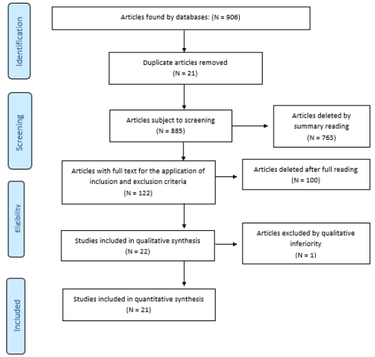
The NLDB follows the “decentralized collection, centralized storage, distribution and informatics” model. (Figure 1). It has collaboration with 18 hospitals for collection of biosamples and supports many research projects by providing biosamples along with associated data. Biosamples are collected with necessary precautions, however, in this pandemic, the need of PPE kit, sanitizer, and establishment of BSL2/BSL3 facility was critical, considering all samples as highly infectious.

biomedres-openaccess-journal-bjstr

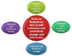
Figure 1: NLDB Model for biosample collection, transportation and distribution.

The challenges confronted while functioning in this pandemic:
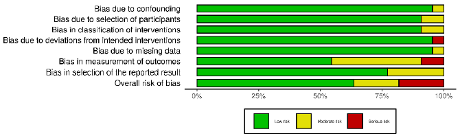
1. There has been a significant decline in the number of samples collected from both host institute and the satellite centres because Outpatient Department and surgeries are only limited for emergency cases (Figure 2). 2. Co-ordination with satellite centers and maintenance of samples became difficult because of limited staff.
2. At initial period, hospitals were not prepared to screen for COVID-19 for all patients, leading to high chances of collecting COVID-19 contaminated samples from asymptomatic patients. Sample processing protocols were revised and precautions were made even for handling samples apparently COVID negative.
3. Biobank was instructed to collect COVID-19 biosamples but processing and storage area was not designed to handle highly infectious samples. To avoid cross contamination, urgent requirements for separate space for processing and storage of COVID and non-COVID samples was flagged.
4. Dedicated routes to transport cryoshippers containing aliquoted COVID biosamples were made from patient ward to BSL2+ facility and then to the storage area.

biomedres-openaccess-journal-bjstr

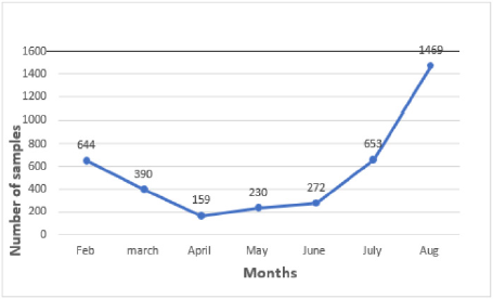
Figure 2: Effect of biosample collection during lockdown.

Logistics and Supply of LN2 Gas

The surging Covid-19 in March, 2020 and the Indian government’s decision to contain the disease outbreak through lockdown adversely affected the domestic logistics sector, especially road transportation, production and supply of essential goods [12]. With increasing number of active cases of Covid-19, the consumption and demand of oxygen was increased throughout the country [13]. Some LN2 industries directed to produce more oxygen in comparison to LN2 gas. The resource management for consumables, refilling of LN2 in cryoshippers, transient storage and transportation of biosamples are managed from main centre established at New Delhi, India. The sudden nationwide lockdown almost got NLDB in a standstill affecting the operational chain such as managing the collection, storage, transportation of biosamples from satellite centres. Biobank has consumption of 100 litres/day to maintain temperature of two LN2 tank. NLDB does not have LN2 plant and dependents only on LN2 supply from outside. Closedown of LN2 factories due to movement of labours, local shortage/limited access to liquid nitrogen, shortage of drivers, made it difficult to get the LN2 tanks refilled. Moreover, the market price of LN2 was hiked up to three times in comparison to the previous routine rate. The pandemic taught that biobank should have inhouse plant for LN2 supply. To avoid such problem in future, NLDB processed to establish an LN2 plant in ILBS premises with capacity to produce approx. 250 litres of LN2/ day (Figure 3).

biomedres-openaccess-journal-bjstr

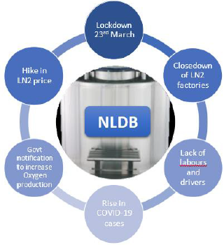
Figure 3: Elements which affected the LN2 supply in the pandemic.

Operations

The ban and restrictions on public transport effected the employees resulting in only 40% attendance of the staff. Biobank staff were seconded in COVID-19 testing lab and the sudden focus and orders to quickly set up procedures to test covid-19 samples, and two-technicians infected with COVID-19 at different time periods and others quarantined for coming in close contact, were big challenges faced. IT experts were not able to resolve the technical issues in the biobank software from home due to nonavailability of remote access for the software. There was temporary interruption of collection and distribution activities as hospitals redirected to treat critical cases and COVID -19 patients only. During lockdown, one of the -80⁰C freezers stopped working, consequently the samples were shifted to the backup freezer. Repair was delayed due to restricted movement and limited supply of spare parts and backup LN2 freezer was being utilised for storage of COVID-19 samples. Biobank samples were on complete risk in case of any failure in storage system as the backup freezers were already in use. Emergency purchase of two -80 freezers was done to accommodate more COVID samples as left over covid-19 samples from hospital diagnostic centers were directed to store in biobank for future research. SOPs were revised as per the knowledge gained in the pandemic. A separate SOP is developed as per guidelines of Indian Council for Medical Research/ Government of India for collection, storage and distribution of COVID-19 samples for research.

Personnel Wellbeing

Commuting for the personnel was big issue in lockdown. However, staff working in COVID lab were provided accommodation in hospital. The safety guidelines issued by Ministry of Health and Family Welfare Government of India to maintain social distancing at work place and transport were followed with necessary compliance [14,15]. Routine test, thermal scanning, sanitizing machine, touch free mechanism installed at all entry and exit points and common areas. Complete ban on non-necessary visit and emergency visits were allowed only after negative rapid antigen test. Two biobank technical staff resigned from their job because their family not allowed them to work on COVID-19 samples.

Management Related Issues

a. Finance: A project for add-on COIVD biobank facility was submitted to the Government of India which was approved and funds released on priority basis in December, 2020.
b. Biobank Information Management System (BIMS): IT related issues were impediment for biobank due to no remote access of clinical databases, biobank systems, slow adaptation and update of software. BIMS was updated with annotation for COVID-19 as per recommendations of ICMR, GOI.
c. HAZARD Management: The primary and basic requirement of biobank is safety of its staff and of the environment against biological and chemical hazards. There were no specific guidelines available for storage, collection, distribution and QMS of highly infectious samples in ISO20387, NCI and ISBER best practices. Sharing of COVID biosamples are not as easy as non-COVID samples thus National Oversight Committee was constituted by ICMR to review the same. NLDB has provision to share the sample after approval of Biosample release committee (BRC). Sample are released after signing MTA and undertaking by recipient to handle COVID-19 and it is informed that any violation or misuse would be dealt with strict action as per laws of Government of India.

Work Culture & Infection

Work culture of biobank has been totally changed due to COVID fright and implementation of new rule and SOPs. Handling the informed consent, annotation forms duly signed by COVID patients was a big issue. WHO and ICMR guidelines are being followed by NLDB to prevention from any infection, Intensive communication and training on good hygiene practices, PPE kit donning and doffing has been provided to biobank personnel. Technicians are equally divided for COVID and non-COVID related work. It is compulsory to wear N95-type masks, use of hand sanitizer, disinfect all documents coming through patents in Ultraviolet (UV) light, and to sanitize work area daily and disinfect the storage area twice a week.

Research Support

The government of India has released huge funds for research focused on Diagnostics, Vaccines, Novel Therapeutics, Repurposing of Drugs or any other intervention for control of COVID-19 as most of the research institutes were closed or had limited access to maintain necessary equipment during lockdown.

Discussion

The sudden lockdown consequent to the COVID-19 pandemic brought both the opportunities and challenges to the biobank. NLDB handled the tri-directional challenges that were operational, financial and sustainability. Sudden changes in operations, supply chain disruptions, manpower presence and remote access of software were major difficulties along with the Handling of Covid-19 biosamples, inaccessibility of donors and challenges in obtaining informed consent. Although, there was neither biobank practices and standards included any plan to run a biobank in a pandemic, NLDB followed the available national [15] and international standards [16] and guidelines [11,17,18] to handle the infectious samples. Though, biobank had an emergency plan for backup storage though there were no thoughts to have an emergency plan for LN2 supply and to work with limited man power. Flexibility in purchase rules, monitoring of efficient utilization, stock management for every one month can be a great help to run biobank in emergency. Biobank must have inhouse LN2 plant along with a rate contract with suppliers to supply LN2 in emergency at equivalent prices. All SOPs revised to treat all sample as infectious Remote monitoring and access of software during emergencies is a must. However, development of remote monitoring software is only possible after the contribution of key stakeholders, such as hospital administration, IT team, privacy legal expert and biobank operations team. In conclusion, NLDB used this pandemic as a learning experience and modifying its operational, emergency and business plans for future crisis and pandemics.

For More Articles: Biomedical Journal Impact Factor: https://biomedres.us

Open Access Journal on Graduate and Professional Studies in Business

Tai Chi, Qigong, and the Treatment of Hypertension

Introduction

Tai chi, also referred to as taiji or taijiquan, is considered both a martial art and a kind of low-impact exercise. Its origins are unclear, but it apparently dates back at least to the thirteenth century. The oldest style is the Chen style, which originated in the Chen village in China [1,2]. The second oldest style, and also the most popular style, practiced by more people than any other style, is the Yang style [3]. The other main styles are the Wu and Wu Hao styles [4], and the Sun style [5], which is the youngest of the five main styles. The various styles of tai chi have much in common, although there are some differences, which we need not discuss in this article. One of the main common features of all styles of tai chi is that they generate healing life energy (qi, pronounced chee), which serves to boost the body’s immune system and prevent the onset of illness and disease. Qi energy also has a beneficial effect on treating existing illness. Many articles and books have been written about the health benefits of tai chi [6-7], so we need not go into the details here. Suffice it to say that many medical studies have found that the regular practice of tai chi can lead to many health benefits, including the treatment of existing diseases and illnesses.
Qigong has been around a lot longer than tai chi, perhaps thousands of years [8]. Many books and articles have been written about this traditional Chinese medicine tool as well [9-74]. It is also a set of gentle exercises that generate qi, which has beneficial healing effects for a wide variety of ailments, including, but not limited to ankylosing spondylitis [75-76], anxiety and stress reduction [77-82], arthritis [83-89], autism [90], back pain [91-92], cancer [93-115], cognitive impairment [116-119], COPD [120-121], COVID-19 [122-123], depression [124-134], elder care [135-138], fibromyalgia [139-141], longevity [142-144], Parkinson’s Disease [145-146], and traumatic brain injury [147], to name a few. The present article focuses on the beneficial effects of tai chi and qigong exercises on hypertension and blood pressure. It reviews a few studies that have found beneficial effects and cites a number of other studies for further reading and research.

Methodology

The PubMed.gov database [148] was searched to find studies that had been done to determine the effectiveness of tai chi and qigong exercises on blood pressure and hypertension.

Findings

The findings reported upon in this article are representative of the numerous studies that have been done examining the effects of tai chi and qigong on blood pressure and hypertension. Additional studies on this topic are cited in the reference section below Liu et al. [149] conducted a meta-analysis to determine the effectiveness of tai chi and qigong exercises in the treatment of essential hypertension (EH). Specifically, they looked at blood pressure (BP), levels of nitric oxide (NO), and endothelin-1 (ET-1). Exercises were performed from 1.5 to 6 months. Nine randomized controlled tests (RCTs) of 516 EH patients in China found that those who did the exercises were able to reduce both systolic and diastolic blood pressure. The exercises also contributed to higher NO blood levels and lower ET-1 blood levels. Although the difference in treatment outcomes using tai chi and qigong exercises versus antihypertensive drugs was statistically insignificant, combining the two therapies resulted in significantly better outcomes than what would occur using only tai chi and qigong or drug therapy. Thus, tai chi and qigong exercises were equally effective as drug therapy in the treatment of hypertension, only without the side-effects that may be present with drug therapy. Liu et al. concluded that tai chi and qigong exercises could be an effective complementary and alternative therapy for EH patients.
The tai chi exercises varied by study, and included the Yang- 24 form, Yang-8, and Chen-style tai chi. The qigong exercises also varied by study, and included Mawangdui Daoyinshu and Baduanjin, among others. Subgroup analyses were performed for the different types of tai chi and qigong, and some were found to be more effective than others. One subgroup analysis of changes in systolic blood pressure ranked the effectiveness of the various exercises as follows, from most to least effective:
a) Chen-style tai chi
b) Mawangdui Daoyinshu Qigong
c) Self-compiled qigong
d) Yang-style tai chi
An examination of different subgroups found that some tai chi and qigong exercises were more effective than others in lowering diastolic blood pressure. The ranking, from most to least effective, was:
a. Chen-style tai chi
b. Self-compiled qigong
c. Mawangdui Daoyinshu Qigong
d. Yang-style tai chi
Liu et al. concluded that Chen-style tai chi might be most effective in reducing blood pressure, while Yang-style tai chi might be the least effective. The authors also compared the effectiveness of the various tai chi and qigong exercises on improving NO levels. The ranking from most to least effective was:
a. Yang-style tai chi
b. Baduanjin Qigong
c. Mawangdui Daoyinshu Qigong
Chen-style tai chi and self-compiled qigong were not statistically significant in improving NO levels. The authors also analyzed subgroup data on the effectiveness of tai chi and qigong in reducing ET-1. The ranking from most to least effective was:
a. Baduanjin Qigong
b. Yang-style tai chi
c. Mawangdui Daoyinshu Qigong
Self-compiled qigong was found not to be statistically significant in lowering ET-1 levels. Thus, it appears that Baduanjin and Yangstyle tai chi may be more effective than other exercises in improving NO and ET-1 scores.
If one were to interpret the findings of this study, one might conclude that choosing qigong and or tai chi therapy might be superior to drug therapy for the treatment of EH for two reasons. Although the study found that qigong/tai chi therapy and drug therapy are equally effective in treating EH, qigong/tai chi therapy has two distinct advantages over drug therapy: qigong/tai chi therapy has no adverse side-effects, and it does not cost anything. Drug therapy, on the other hand, sometimes has adverse sideeffects, and it is not free. The study also found that combining qigong/tai chi therapy with drugs might be superior to choosing just one of the two options.
Pan et al. [150] conducted a systematic review of randomized controlled trials on the effects of tai chi on blood pressure, body mass index (BMI), and quality of life (QOL) on patients suffering from hypertension. Their meta-analysis of 24 studies containing 2,095 patients (1,074 in the treatment group and 1,021 in the control group) found that the intervention group had significantly better outcomes for systolic blood pressure (SBP) [p ≤ 0.001], diastolic blood pressure (DBP) [p ≤ 0.001], physical functioning [ p ≤ 0.001], role-physical [p ≤ 0.001], general health [p = 0.001], bodily pain [p ≤ 0.001], vitality [p ≤ 0.001], social functioning [p = 0.027], role-emotional [p = 0.003], and mental health [p = 0.001] compared to the control group. However, the differences in BMI between the groups were insignificant. Pan et al. concluded that tai chi is an effective therapy to improve SBP and DBP for patients suffering from essential hypertension. Zou et al. [151] found that the practice of baduanjin was beneficial for quality of life (p = 0.004), sleep quality (p = 0.001), balance (p = 0.004), handgrip strength (p = 0.007), trunk flexibility (p = 0.006), systolic (p = 0.0004) and diastolic (p = 0.005) blood pressure, and resting heart rate (p = 0.0005). They examined the results of various studies on each of these topics. In the case of the effect of baduanjin on blood pressure, they examined 9 studies having a total of 743 participants.
Ladawan et al. [152] investigated the effects of qigong exercise on cognitive function, blood pressure and cardiorespiratory fitness in 12 healthy middle-aged subjects who performed qigong exercises in 60-minute sessions, three times a week for eight weeks. They found that the exercises resulted in significant improvements in Trail Making Tests Part A (p = 0.04), systolic blood pressure (p = 0.0001), diastolic blood pressure (p = 0.005), mean arterial pressure (p < 0.001) and maximal workload (p = 0.032). Twelve weeks after cessation of the exercises, they had all returned to the baseline. The authors concluded that it is necessary to perform qigong regularly to maintain the improved health effects.
Ching et al. [153] examined data on 370 subjects from seven randomized controlled trials (RCTs). The following six types of qigong exercises were used:
a) Conventional Qigong
b) Guolin Qigong
c) Shuxinpingxue Gong
d) Dongeui Qigong
e) Ba Duan Jin Qigong
f) Mawangdui Daoyinshu Qigong
They found that the practice of qigong exercises had a significant effect on reducing systolic (p < 0.001) and diastolic (p < 0.001) blood pressure. The above studies are representative of the studies that have been done in recent years on the effectiveness of tai chi and qigong on reducing high blood pressure. Some other recent studies are listed in the reference section at the end of this article [154-188].

For More Articles: Biomedical Journal Impact Factor: https://biomedres.us

Open Access Journal on parkinsons disease

Use of Anti-Inflammatory Drugs in the Treatment of Parkinson’s Disease: A Systematic Review of Perimental Studies

Parkinson’s disease (PD) is a progressive neurodegenerative disease characterized by the loss of dopamine neurons (AD) in the substance nigra pars compacta (CNS) and accumulation of insoluble cytoplasmic protein inclusions called Lewy and Lewy neurites bodies [1]. The precise mechanism underlying the pathogenesis of PD is not yet fully understood. The accumulation of evidence suggests that soluble α-synuclein aggregates, known as oligomers, play a significant role in PD where the neurodegenerative process culminates in impairing several subcellular functions [1]. Thus, clinically, PD presents as muscle stiffness, tremor at rest, bradykinesia (abnormal slowness of voluntary movements), postural instability; some patients also have symptoms related to psychiatric and cognitive disorders. In this context, intraneuronal accumulation and aggregation of alpha-synuclein can start from several sites such as the intestinal tract, where this altered protein (alpha-synuclein) can be transported through the enteric route to the CNS through the parasympathetic pathway [2]. In addition to this hypothesis, there is genetic influence in the functional roles of genes identified as monogenic forms of PD. Mutations in SNCA, LRRK2 and VPS35 genes have been highly penetrating and cause autosomal dominant forms of PD [1]. Thus, showing the existence of multifactorial processes to support the underlying cause of this aberrant protein accumulation. Therefore, what most of these studies show is that when alpha-synuclein is lodged in the CNS itself, it is directly linked to damage triggered by the activation of microglia, which, by releasing inflammatory factors, causes an oxidative burst affecting neuronal cells leading to death [3].
Thus, since there is a pattern of inflammatory characteristics after the beginning of the accumulation of these proteins, this tangle of interleukins, TNF-α, TNF-γ, CCL2, ROS and NO may increase such accumulation and aggregation already in force, thus determining an even more cumulative and oxidative neurodegenerative picture, exponentially affecting the patient’s condition, becoming a real “Parkinson’s snowball”. Thus, this hypothesis suggests a clinical applicability of treatment with anti-parkinsonian drugs of antiinflammatory nature and drugs properly anti-inflammatory drugs (IANES and corticosteroids), where the anti-inflammatory action may provide a therapeutic resource for patients with the purpose of promoting a decrease in levels of dopaminergic cell lesions and lowering of alpha-synuclein accumulation. This study, therefore, aims to correlate the use of these two types of drugs with antiinflammatory attributes to the treatment of PD, observing whether there is an anti-inflammatory or neuroprotective response (via dopaminergic markers) and which group of drugs is better than the other.

Methodology

This study consisted of a systematic review prepared according to the Preferred reporting items for systematic review and metaanalysis protocols (PRISMA-P). The eligibility criteria defined for the inclusion of an article in this review were human and animal studies, contain relevant information regarding the neuroprotective action of the drug in PD, applicability of anti-inflammatory drugs, csf analysis, use of in-silico computational method and clinical results and be indexed in the electronic databases MEDLINE/ Pubmed, LILACS, EMBASE, Scopus and Web of Science. Using the PECOS strategy, the descriptors used in the searches were chosen based on the technical-scientific terms MeSH (Medical Subjective Heading) and DeCS (Descriptors in Health Sciences), combined by the Boolean operator “AND” or “OR” (Table 1). MEDLINE/ PubMed research strategy: “Idiopathic Parkinson’s Disease” OR “Lewy Body Parkinson’s Disease” OR “Parkinson’s Disease, Idiopathic” OR “Parkinson Disease, Idiopathic “ OR “Parkinson’s Disease, Lewy Body” OR “Parkinson’s Disease” OR “Idiopathic Parkinson Disease” OR “Lewy Body Parkinson Disease” OR “Primary Parkinsonism” OR “Parkinsonism, Primary” OR “Paralysis Agitans” AND “Neuroinflammation” OR “Inflammations” OR “Innate Inflammatory Response” OR “Inflammatory Response, Innate” OR “Innate Inflammatory Responses” AND “Anti Inflammatory Agents” OR “Agents, Anti-inflammatory” OR “Anti-inflammatories” OR “Anti-inflammatory Agents” OR “Agents, Anti-Inflammatory” OR “Agents, Anti Inflammatory” OR “Anti-Inflammatories” OR “Anti Inflammatories” OR “Anti-inflammatory Agents, Non-Steroidal” OR “NSAIDs” OR “Non-Steroidal Anti-Inflammatory Agents” OR “Non-Steroidal Anti Inflammatory Agents” OR “Nonsteroidal Anti-Inflammatory Agents” OR “Nonsteroidal Anti Inflammatory Agents” OR “Anti Inflammatory Agents, Nonsteroidal” OR “Antiinflammatory Agents, Nonsteroidal” OR “Nonsteroidal Antiinflammatory Agents” OR “Corticosteroids” OR “Corticoids” OR “Inhibitors, Cyclo-Oxygenase” OR “Inhibitors, Cyclo Oxygenase” OR “Inhibitors, Cyclooxygenase” OR “Prostaglandin Synthesis Antagonists” OR “Antagonists, Prostaglandin Synthesis” OR “Inhibitors, Prostaglandin-Endoperoxide Synthase” OR “Inhibitors, Prostaglandin Endoperoxide Synthase” OR “Prostaglandin Endoperoxide Synthase Inhibitors” OR “Prostaglandin Synthase Inhibitors” OR “Cyclo-Oxygenase Inhibitors” OR “Cyclo Oxygenase Inhibitors” OR “Inhibitors, Prostaglandin Synthase” OR “Inhibitors, Cyclooxygenase 2” OR “Cyclooxygenase-2 Inhibitors” OR “Inhibitors, Cyclooxygenase-2” OR “Coxibs” OR “COX-2 Inhibitors” OR “COX 2 Inhibitors” OR “Inhibitors, COX-2” OR “COX2 Inhibitors” OR “Inhibitors, COX2”.

biomedres-openaccess-journal-bjstr

Table 1: PECOS Strategy.

EMBASE research strategy: (‘parkinson disease’/exp/mj OR ‘parkinson disease’/mj OR ‘parkinson`s disease’/mj OR ‘parkinsons disease’/mj OR ‘paralysis agitans’/mj OR ‘parkinson disease, symptomatic’/mj) AND (‘anti-inflammatory agent’/exp/mj OR ‘antiinflammatory agent’/mj OR ‘anti-inflammatory agents’/mj OR ‘antiinflammatory agents, steroidal’/mj OR ‘anti-inflammatory agents, topical’/mj OR ‘anti-inflammatory drug’/mj OR ‘anti-inflammatory agent’/mj OR ‘anti-inflammatory agents’/mj OR ‘anti-inflammatory agents, steroidal’/mj OR ‘anti-inflammatory agents, topical’/mj OR ‘antiflogistic agent’/mj OR ‘antiinflammation agent’/mj OR ‘anti inflammatory agent’/mj OR ‘anti-inflammatory drug’/mj OR ‘antiinflammatory steroid’/mj OR ‘anti-inflammatory activity’/exp/mj OR ‘anti-inflammatory action’/mj OR ‘anti-inflammatory activity’/ mj OR ‘anti-inflammatory effect’/mj OR ‘anti-inflammatory action’/ mj OR ‘anti-inflammatory activity’/mj OR ‘anti-inflammatory effect’/mj OR ‘antiphlogistic action’/mj OR ‘antiphlogistic activity’/ mj OR ‘antiphlogistic effect’/mj OR ‘nonsteroid anti-inflammatory agent’/exp/mj OR ‘nsaid’/mj OR ‘anti-inflammatory agents, nonsteroidal’/ mj OR ‘anti-inflammatory agents, non-steroidal’/mj OR ‘anti-inflammatory agent, nonsteroid’/mj OR ‘non steroid antiinflammatory agent’/mj OR ‘non steroid anti-inflammatory drug’/ mj OR ‘non-steroidal anti-inflammatory agent’/mj OR ‘non-steroidal anti-inflammatory drug’/mj OR ‘non-steroidal anti-inflammatory agent’/mj OR ‘non-steroidal anti-inflammatory drug’/mj OR ‘nonsteroid anti-inflammatory agent’/mj OR ‘nonsteroid antiinflammatory drug’/mj OR ‘nonsteroid antirheumatic agent’/mj OR ‘nonsteroidal anti-inflammatory drug’/mj OR ‘nonsteroidal anti-inflammatory drugs’/mj OR ‘nonsteroidal anti-inflammatory drugs’/mj OR ‘nonsteroidal anti-inflammatory agent’/mj OR ‘nonsteroidal anti-inflammatory drug’/mj OR ‘prostaglandin synthase inhibitor’/exp/mj OR ‘cyclooxygenase inhibitor’/mj OR ‘cyclooxygenase inhibitors’/mj OR ‘prostaglandin synthase inhibitor’/mj OR ‘prostaglandin synthetase inhibitor’/mj OR ‘cyclooxygenase 2 inhibitor’/exp/mj OR ‘cox 2 inhibitor’/mj OR ‘cox 2 specific inhibitor’/mj OR ‘cox 2 specific inhibitors’/mj OR ‘cox- 2 inhibitor’/mj OR ‘cox-2 specific inhibitor’/mj OR ‘cox-2 specific inhibitors’/mj OR ‘cox2 inhibitor’/mj OR ‘cox2 specific inhibitor’/ mj OR ‘coxib’/mj OR ‘coxibs’/mj OR ‘cyclooxygenase 2 inhibitor’/ mj OR ‘cyclooxygenase 2 inhibitors’/mj) AND (‘modulation’/exp/ mj OR ‘modulation’/mj OR ‘protection’/exp/mj OR ‘protection’/ mj OR ‘protective factors’/mj OR ‘treatment outcome’/exp/mj OR ‘medical futility’/mj OR ‘outcome and process assessment (health care)’/mj OR ‘outcome and process assessment, health care’/ mj OR ‘outcome management’/mj OR ‘patient outcome’/mj OR ‘therapeutic outcome’/mj OR ‘therapy outcome’/mj OR ‘treatment outcome’/mj OR ‘disease management’/exp/mj)
LILACS Research Strategy: “Idiopathic Parkinson’s Disease” OR “Lewy Body Parkinson’s Disease” OR “Parkinson’s Disease, Idiopathic” OR “Parkinson Disease, Idiopathic “ OR “Parkinson’s Disease, Lewy Body” OR “Parkinson’s Disease” OR “Idiopathic Parkinson Disease” OR “Lewy Body Parkinson Disease” OR “Primary Parkinsonism” OR “Parkinsonism, Primary” OR “Paralysis Agitans” AND “Neuroinflammation” OR “Inflammations” OR “Innate Inflammatory Response” OR “Inflammatory Response, Innate” OR “Innate Inflammatory Responses” AND “Anti Inflammatory Agents” OR “Agents, Anti-inflammatory” OR “Anti-inflammatories” OR “Anti-inflammatory Agents” OR “Agents, Anti-Inflammatory” OR “Agents, Anti Inflammatory” OR “Anti-Inflammatories” OR “Anti Inflammatories” OR “Anti-inflammatory Agents, Non-Steroidal” OR “NSAIDs” OR “Non-Steroidal Anti-Inflammatory Agents” OR “Non-Steroidal Anti Inflammatory Agents” OR “Nonsteroidal Anti-Inflammatory Agents” OR “Nonsteroidal Anti Inflammatory Agents” OR “Anti Inflammatory Agents, Nonsteroidal” OR “Antiinflammatory Agents, Nonsteroidal” OR “Nonsteroidal Antiinflammatory Agents” OR “Corticosteroids” OR “Corticoids” OR “Inhibitors, Cyclo-Oxygenase” OR “Inhibitors, Cyclo Oxygenase” OR “Inhibitors, Cyclooxygenase” OR “Prostaglandin Synthesis Antagonists” OR “Antagonists, Prostaglandin Synthesis” OR “Inhibitors, Prostaglandin-Endoperoxide Synthase” OR “Inhibitors, Prostaglandin Endoperoxide Synthase” OR “Prostaglandin Endoperoxide Synthase Inhibitors” OR “Prostaglandin Synthase Inhibitors” OR “Cyclo-Oxygenase Inhibitors” OR “Cyclo Oxygenase Inhibitors” OR “Inhibitors, Prostaglandin Synthase” OR “Inhibitors, Cyclooxygenase 2” OR “Cyclooxygenase-2 Inhibitors” OR “Inhibitors, Cyclooxygenase-2” OR “Coxibs” OR “COX-2 Inhibitors” OR “COX 2 Inhibitors” OR “Inhibitors, COX-2” OR “COX2 Inhibitors” OR “Inhibitors, COX2” .

Web of Science Search Strategy

TÓPICO (Parkinson disease*) AND TÓPICO (inflammation*) AND TÓPICO (anti-inflammatory*).

Scopus Search Strategy

(TITLE-ABS-KEY (Parkinson AND disease) AND TITLE-ABSKEY ( inflammation ) AND TITLE ( anti-inflammatory ) ) .
The selection of articles was performed by two researchers blindly and independently through reading the titles, reading the abstracts and, finally, full reading of the articles. Any disagreement in the selection was resolved in consensus meetings. Articles that fully met the eligibility criteria were included in this study. The selection process is described in Flowchart 1 adapted from PRISMA (Figure 1). In order to analyze the methodological quality of the included studies, each article was evaluated by a researcher based on the items of the ACROBAT-NRSI (A Cochrane Risk of Bias Assessment Tool for Non-Randomized Studies) [4]. Acrobat-NRSI scores were used to exclude articles that did not present hardhitting information to the research, besides serving as a basis for discussing the methodological quality of the articles and the possible viruses in the generalization of their results (Figures 2 & 3). From each article included, data related to the objectives of this review were extracted, such as author, title, type of study, population, PD induction drug, drugs used applied, positive results. These data were computed and compared using the t-Student test for independent samples, with the purpose of comparing the percentage s percentages of the and effects on PD between NCAs and other anti-inflammatory drugs (Table 2).

biomedres-openaccess-journal-bjstr

Table 2: Characteristic of selected experimental clinical trials.

biomedres-openaccess-journal-bjstr

Figure 1: Adapted from PRISMA.

biomedres-openaccess-journal-bjstr

Figure 2.

biomedres-openaccess-journal-bjstr

Figure 3.

Findings

Twenty-one articles were analyzed, separated between two groups according to the drug used for pre-clinical study, antiparkinsonian drugs of anti-inflammatory nature and drugs properly anti-inflammatory drugs (IINES and corticosteroids). Improvement in motor function, decreased movement patriotization, increased levels of striatal dopamine, decreased interleukins and blockage of inflammatory pathways, such as those participating in MPP+ and COX-2, as well as increased and/or decreased loss of neurons armed with tyrosine hydroxylase (TH) enzyme, an important marker of neuroprotection, were identified.

Discussion

In view of these findings, this systematic review demonstrated that there is an effective therapeutic relationship in the use of anti-inflammatory drugs in PD through findings such as, mainly, quantitative increase or decrease in the loss of tyrosine hydroxylase enzyme [5-9]and improvement of motor function or prevention of motor decline [5,10-16]. However, since these are experimental studies in animals where clinical failures are commonly recorded in this methodology, caution should be exercised in the face of these findings, even if it shows clinical relevance. In addition, the importance of the therapeutic look is emphasized, especially in pathophysiological terms elapsed by the articles, observing in most of them that this disease, which affects the nicrostriatal region harboring the substantia nigra and quite rich in microglia, has the cumulative character of alpha synuclein in its altered form, which leads to the formation of a highly fibrillar aggregate by very little known pathways, thus, there is the beginning of a cascade of events that lead to the release of inflammatory toxic factors and a progressive dopaminergic neurodegeneration [17,18]. It is identified, therefore, that within this pathophysiological mechanism there is linked an inflammatory response, so there is a target to be investigated and possibly treated, demonstrating possible therapeutic purposes against PD.
In parallel, this review was able to investigate some other parameters found in experimental animal studies. Some motor tests showed improvement in the face of performance tests, applicability of previous training or open field observation, in addition, motor improvement of the forelimbs and later [5], significant decrease in cataleptic behavior [10], improvement of ambulation and immobilization time [7]and reduction of hypokinesia [15]. These results reinforce the hypothesis of a neuroinflammatory cause of Parkinson’s and once again the application of anti-inflammatory drugs for a possible therapy. It can be observed that characteristics that are found in patients such as muscle stiffness, tremor at rest, bradykinesia and postural instability could be solved or attenuated by a drug with function, absorption and mechanisms similar to what were found in this review. Therefore, there is a vast ness of possibilities for anti-inflammatory pharmacological use, in which, however, there is still a need to weigh the pros and cons, the latter being something of changeable capacity within the pharmaceutical industry, in which with investments in research and advanced technology can be achieved a less deleterious profile to the body, such as raising blood pressure, interaction with anti-hypertensive drugs, reduction of renal perfusion and gastrointestinal symptoms [16].
Within this context, it was also possible to identify an increase, then neuroprotection from levels of dopamine, TH enzyme and dopaminergic neurons in some animals. These results can be explained by the fact that the neuroinflammatory process, in its characteristic of exponential cascading lesion of dopaminergic neurons [8,19], was blocked and there was no more decrease in degenerative character. All this was observed from immunohistochemical analyses of TH (Tyrosine Hydroxylase) levels, an enzyme involved in dopamine synthesis through a series of biochemical reactions that has the amino acid tyrosine as a precursor and a molecular marker of dopaminergic neurons, along with dopamine dosage [5-9,18,19]. Thus, it was demonstrated what can occur in a neural system previously healthy, but with microglia activated by the pathophysiology of PD, in this case by mimetic drugs of PD such as rotenone and 1-methyl-4-phenyl-1,2,3,6 tetrahydropyridine (MPTP). Thus, it is envisaged, once again, the use of these drugs or something more advanced both in patients already diagnosed and living with the disease chronically, as well as in patients at the beginning of diagnosis and mild clinical picture, promoting neuroprotection and, consequently, a greater defense and increased quality of life.
Some drugs in the studies acted directly on microglia and other inflammatory foci, some of them are very common, such as ibuprofen, meloxicam, piroxicam, AAS, Valdecoxib and Parecoxib (NHEMS, which act by inhibiting COX-2, prostaglandin and ultimately reducing cytokines), dimethazone (Corticosteroid that reduces the gene expression of pro-inflammatory cytokines). All of them obtained good results regarding the lowering of glial hyperactivation and intracellular inflammatory, in addition to stimulating the recovery and regeneration phase, avoiding in some cases the toxicity of MPTP [20], which shows that even having extensive knowledge and applicability of these drugs, they can still be key parts for the advancement of neural therapy in PD. Similarly, oxymatrine, an alkaloid compound found at the root of a Chinese herb (Sophora flavescent), promoted relief of motor deficits induced by MPTP and conferred significant neuroprotection, in addition to inhibiting the activation of microglia and exacerbated release of pro-inflammatory as cytokines [13]. This shows that within the vastness of drugs known and disseminated by the pharmaceutical industry, there are still a gigantic number of other substances that can be used in the treatment of this disease [20-27].

Conclusion

Our study has concluded that there is a need for investment in quality, more robust, broad-spectrum preclinical studies, with minimal view to achieve the ideal pharmacological therapeutic for this target. Thus, it is necessary more clinic trials to confirm this relationship between an inflammatory profile and use of antiinflammatory drugs which possible therapeutic agents to treatment of PD.

For More Articles: Biomedical Journal Impact Factor: https://biomedres.us

Open Access Journal of Obesity and Diabetes

Type 2 Diabetes Mellitus and COVID-19 in Mexico. A comprehensive Assessment

Introduction

On February 11, 2020, the International Committee for Taxonomy in Viruses named SARS-CoV-2. Composed of a genome of 30,000 base pairs, belonging to the Coronaviridae family of the order Nidovirales. Phylogenetically coronaviruses are classified into alpha, beta, gamma and delta. Coronaviruses were identified 50 years ago as pathogens responsible for the common cold, mainly HCoV-OC43, HCoV-229E among other variants. At the beginning of 2002, coronaviruses were considered exclusively veterinary pathogens, however, by 2019 they were identified in biological samples from patients diagnosed with pneumonia [1-4]. Showing an age trend initially with geriatric patients, it has been shown that the risk of mortality increases after 75 years [5]. However, today age is no longer a dependent factor for infection. It is important to mention this since it may be due to multiple etiologies in addition to infection, such as: comorbidities, lack of metabolic control, suspension of work in the outpatient clinic due to hospital oversaturation derived from the pandemic, sedentary lifestyle, among others.
Hence it is important to emphasize the lack of metabolic control derived from all those cardiometabolic diseases, such as: obesity, hypertension, dyslipidemias and mainly diabetes mellitus, which turns out to be the first pandemic that has not been adequately controlled since ancient times [6]. All these factors are directly and proportionally related to the risk of severe progression and poor prognosis due to the chronic inflammatory state that generate more the acute systemic inflammatory response derived from COVID-19. In the case of obesity, another factor shared by both pathologies increased even more derived from confinement due to the forced closure of sports centers, favoring a sedentary lifestyle. The anxiety derived from the pandemic favors a greater consumption of foods with low nutritional power, again favoring obesity and lack of metabolic control. Therefore, in the context of a controlled diabetic patient, the measures that had to be implemented as a strategy to reduce the rate of infections are one of the factors to generate lack of control. The percentage of uncontrolled diabetics since the beginning of the pandemic is more and more common and continues to rise, which entails greater spending on health, greater generation of medical supplies and resources. There is an excess of mortality in the Mexican Republic derived from the pandemic, not only due to COVID-19, but also due to other causes [7,8] without forgetting to mention the possibility of under- registration that exists, for example, in marginalized areas or those who could not have hospital access derived from the same scenario. That is why the relevance of this article where a comprehensive scenario is proposed for the knowledge and management of COVID-19 in those patients who already have a chronic damage such as Diabetes Mellitus.

Pathophysiology

The incubation period for SARS-CoV-2 is 5 days with a range of 2 to 14 days [9]. The spectrum of diseases generated by coronavirus infection is mainly acute respiratory, chronic, enteric, hematological, endothelial and of the central nervous system. The mechanism of transmission of the disease by SARS-CoV-2 is from person to person through the airway by the drops of Flügge that are exhaled when coughing, sneezing or speaking and are inhaled or deposited in the mouth and ocular conjunctiva, as well as surfaces, which can function as fomites [10]. The main structural proteins found on the membrane surface of the SARS-CoV-2 viral particles participate within the pathophysiology, which are: Spike (S), membrane (M) and envelope (E). Among other, these are responsible for the anchorage and entry of these microorganisms to the host’s cells. It should be noted the type 2 angiotensin converting enzyme (ACE 2) which is a type I membrane protein that contains receptors in the lung, heart, kidney and intestine, endothelium, nervous system, mainly. The ACE 2 receptors that are located in the lower respiratory tract of humans are the cellular receptors for SARS CoV-2. Since the virion has the S-glycoprotein or Spike protein, which projects through the viral envelope and forms the spicules of the crown, this is glycosylated and is responsible for mediating the binding of the receptor (protein S + ACE 2), as well as its fusion with the host cell [11,12].
This strong bond unites the entire SARS-CoV-2 membrane with the host cell membrane, entering it through endocytosis. Viral particles release their RNA that binds to viral DNA, initiating the viral replication cycle, which leave the host cell through exocytosis. Once the RNA of the SARS-CoV-2 particles begins its translation and transcription, two processes are generated: the first related to the high demand for manufacturing viral proteins causing cellular stress that ends in apoptosis of the target cells; while in the second, the viral RNA acts in a molecular pattern associated with pathogens, which leads it to be recognized by the cells of the immune system, initiating the activation of the cytokine cascade and the migration of neutrophils. Hypercoagulability, venous stasis and endothelial damage is another of the main characteristics mediated by the ACE 2 receptors that SARS-CoV-2 particles possess, being observed in the endothelium of the veins, arteries and arterial smooth muscle cells of the brain; This produces dysfunction and inflammation of the microvasculature that alters vascular flow and initiates platelet activation, increasing risk for macrovascular and microvascular thrombosis, pulmonary thromboembolism, deep vein thrombosis, catheter-related thrombosis, ischemic cerebrovascular disease, acrosyndromes, and capillary leak syndrome. in organs such as lungs, kidneys and heart, increasing mortality, one of the main complications [13] (Figure 1).

biomedres-openaccess-journal-bjstr

Image 1: Physcopathogenesis of COVID-19.

SARS-COV2 as a Diabetogenic Agent

Diabetes is associated with a chronic low-grade inflammatory state that favors the development of an exaggerated and constant inflammatory response. At the molecular level, there is an increase in the levels of IL-6 and C-reactive protein (CRP), so the proinflammatory state typical of diabetes can favor the cytokine storm and the systemic inflammatory response that accompanies the acute respiratory distress syndrome (ARDS) in patients with COVID 19 [14]. This is why diabetics infected with SARS-CoV-2 have a higher rate of hospital admission, severe pneumonia, and higher mortality compared to non-diabetic subjects [15]. SARS-CoV-2 is considered diabetogenic since it is also capable of causing direct damage to the pancreas, due to the expression of ACE 2 (mainly in islet cells) even in a higher proportion than at the lung level, which could worsen hyperglycemia and even induce the onset of diabetes in previously non- diabetic subjects [16]. It should be noted that only 1-2% of patients with mild COVID-19 infection present pancreatic lesions, while 17% of patients with severe cases present with lesions of the pancreas, which can accentuate the systemic inflammatory response and, therefore, Therefore, accelerate the appearance of ARDS [17]. On the other hand, the current scenario of the pandemic even in uninfected subjects may favor the deterioration of metabolic control due to difficulties in accessing the health system, lack of physical activity and increased stress associated with confinement.
Therapeutic strategies should be aimed at facilitating access to the health system through telemedicine to advise the patient on the adaptation of treatment or any other remotely manageable medical situation and guide patients and caregivers in the control of diabetes in order to prevent hospitalization [18]. Clinical symptoms. Different stages of SARS-CoV-2 disease have been described in humans depending on the clinical severity, which can range from mild symptoms such as: fever, myalgia, headache, cough, anosmia. Up to severe symptoms characteristic of pneumonia with severe respiratory impairment [19,20-25]. Table 1 Mild and moderate infections comprise 80.9% of the registered cases; the severe ones, 13.8% and the critical ones, 4.7%. In the adult population it is 1.2%; while in pediatric population it is 15.8% [26]. The prevalence of asymptomatic patients differs according to the age group and can be reported by up to 40% [27]. Due to the high percentage of asymptomatic patients not only in Mexico, but also worldwide, it is vitally important to continue using a facial mask in our daily lives in order to reduce the risk of contagion. Even people with a full vaccination schedule are not exempt from COVID-19 infection.

biomedres-openaccess-journal-bjstr

Table 1: Clinical symptoms of COVID-19 severity.

Prognostic factors for serious and severe disease are considered: cardiovascular disease, diabetes mellitus, hypertension, chronic lung disease, cerebrovascular disease, cancer, chronic kidney disease, obesity and smoking [28,29]. Some alterations in laboratory parameters associated with a pro-inflammatory and procoagulant state are indicative of a poor prognosis, such as multiorgan failure [30]:
• Lymphopenia.
• Elevated liver enzymes.
• Elevated LDH.
• Elevation of acute inflammation markers (CRP, ferritin, procalcitonin).
• D-dimer elevation.
• Prothrombin time lengthening.
• Elevation of troponins.
• CPK elevation.
• Markers of kidney damage (elevated creatinine, anuria). Diagnosis. There are different detection techniques for SARSCoV- 2, each with different sensitivity and specificity. We currently have three types of diagnostic tests [17,18]:
a) Nucleic acid detection tests (PCR). In the case of the gold standard. Being its high cost the main limitation for its application.
b) Antigen (Ag) detection tests.
c) Antibody detection tests (Ab): IgM / A and IgG.

We must emphasize that a negative result does not exclude infection, therefore, if the clinical suspicion is high (clinical data, epidemiological context, radiological findings, sometimes earlier in computed tomography than the positivity of the PCR and analytical studies), it is recommends repeating the same sample in 48-72 hours or trying to obtain it from the lower respiratory tract, especially in severe or progressive disease [16]. Throughout the pandemic, a high percentage of false negatives has been observed in the practice of antigenic tests, the most used in Mexico due to the difference in cost between PCR, which has perpetuated in the patient the uncertainty of being or not with the infection, which means that they do not follow the medical indications and finally contribute to continue perpetuating the contagion. Educating the patient about what a negative result implies despite high clinical suspicion is part of our work in this pandemic and therefore, as health professionals, we should not base our treatment on a laboratory test and the recommended measures should be initiated in the context of isolation, symptomatic treatment and continuous monitoring of associated comorbidities in order to avoid complications as explained in detail.
Treatment of diabetes mellitus in patients with COVID-19. Treatment depends on the clinical characteristics of each patient, risk of complications, age, ease of access to the health area, socioeconomic status, risk of drug interactions especially in patients with polypharmacy, etc. Treatment for COVID-19 infection should be symptomatic, that is, based on the clinical picture presented by each patient, which can be: antihistamines, cough suppressants, thromboprophylaxis, analgesics and anti-inflammatories, educate for self-monitoring of vital signs and provide all the necessary alarm data. As outpatient management in non-serious patients and mild symptoms, the following should be taken into account: prevention of infection, healthy lifestyle, general measures to improve diabetes control, treatment of hyperglycemia, treatment of comorbidities and support doctor (Figure 2). For the treatment of asymptomatic or non-severe patients, the following is recommended: home management, follow usual treatment for diabetes control, goal of fasting glucose 70-130 mg / dL, HbA1c <6.5%, use of telemedicine to clarify doubts and education, indicate alarm and isolation measures, adjust the medication only if there is lack of control. Speaking of telemedicine, Mexico is not fully prepared, since it has a technological development of around 25%, however, thanks to portable technology such as a cell phone that facilitates the use of telemedicine, it can favor the medical attachment of chronic degenerative diseases and likewise surveillance of the clinical evolution of COVID-19 in those patients with a high risk of complications. Up to 70% of the population could benefit from these programs [22,24,30].

biomedres-openaccess-journal-bjstr

Image 2: Measures to be implemented in diabetic patients with COVID-19 taken with modified from M.M. Lima-Martínez et al.

In the case of patients with mild-moderate infection: home management with close monitoring, assess risk of progression and assess the need for in-hospital management, medication adjustments according to glycemic control, fasting blood glucose target of 72-144 mg / dL, HbA1c <7%, close medical contact. For those with severecritical infection: use insulin in continuous intravenous infusion or basal-bolus-correction regimen, fasting glycemic goal of 72-180 mg / dL, HbA1c <8%, strict monitoring of plasma glucose, electrolytes, ketone bodies, renal and cardiovascular function, procoagulant markers among others. Always in-hospital (22,30). (Figure 3). With the above mentioned, the need for extra medication should be taken into account depending on the symptoms of COVID-19 according to the evidence reported so far. It is intended to exemplify the treatment of these two entities together, since if we only dedicate ourselves to treating the patient based exclusively on the diagnosis of COVID-19, forgetting about their underlying pathology, in this case diabetes mellitus, we increase the risk of complications and mortality. Special considerations for drugs for diabetes mellitus in COVID-19 should be taken into account, such as: Metformin, SGLT2-i, GLP-1 analogs, DPP-4 inhibitors, sulfonylureas, and insulin. Each one with specific indications, making the appropriate dose adjustments according to the patient’s needs, to optimize therapeutic goals, but it is important to emphasize that for those who require hospitalization derived from COVID-19, the drug of choice for glycemic control will be insulin [22].

biomedres-openaccess-journal-bjstr

Image 3: Indication in the management of covid 19 according to the clinical severity of diabetic patients. Takane and modified from M.M. Lime Martinez, et al. & Medina – Chavez JH, et al.

In diabetics hospitalized for COVID-19, the use of prophylactic doses of low molecular weight heparin, such as Enoxaparin, is suggested in the absence of contraindications (active bleeding or platelet count <25 × 109 / l, and others), with dose adjustment for patients with frank elevation of D-dimer and those that present severity criteria [15]. It is important to individualize the prothrombotic risk according to the age and associated comorbidities of each patient, even in patients with mild symptoms thromboprophylaxis is indicated, the duration of this measure will also depend on how many associated risk factors present and the clinical severity, which requires a minimum of 2 weeks in those asymptomatic or mild symptoms and up to 6 weeks in severe conditions. Even with the resolution of the symptoms and / or the hospital discharged, this measure must continue for a minimum of 7 days [30].

Conclusions

The union of protein S with ACE 2 is the most important point within the pathophysiology since it culminates in a systemic inflammatory response and endothelial damage, which opens the door for a wide panorama of complications in the organism, even that a patient debut as diabetic from infection. At the beginning of 2020, when the first case of COVID-19 was registered, to date, the Mexican population presents data of exhaustion derived from isolation. Despite this, the vaccination program that was established in Mexico has not been fast enough, placing itself practically in the last place in Latin America for complete coverage of vaccines and reducing the rate of infections to be able to restore daily activities in a greater proportion and better still reduce morbidity and mortality in vulnerable groups. In addition to this, the lack of supplies and medical personnel in the health sector remains constant, which does not favor the scenario of both pandemics since it also worsens the medical adherence required by patients with chronic degenerative diseases, leading to a greater risk of complications, greater risk of contagion and finally higher mortality; thus, generating a vicious circle. Offering a broad panorama as a comprehensive evaluation of what COVID-19 implies in a patient with Diabetes Mellitus offers us new opportunities to reduce complications and serious progression of the disease, emphasizing the need to establish strategies such as telemedicine if necessary for better medical surveillance, promote pharmacological adherence and provide timely help in case of seriousness, always treating together.
We are in a century where two pandemics converge with each other, increasingly diabetic patients with lack of metabolic control, generating catastrophic damage to health, psychosocial and the economy. It is necessary to control both, starting with preventive measures to be able to modify the impact that has been generated so far. The points to follow in the context of DM2 and COVID-19 will be prevention measures where isolation is the most important, educating the patient, surveillance of comorbidities and glucose self-monitoring to be able to adjust the dose or change the medication in case of lack of control, monitor alarm signs and offer symptomatic treatment according to the needs of the patient, without forgetting the necessary use of telemedicine as a support tool.

For More Articles: Biomedical Journal Impact Factor: https://biomedres.us

Open access Journal of Dental Surgery

Dental and Oral Health Care Coverage for Seniors in the United States

Introduction

Oral health is a key component of general health. Estimated prevalence of oral health problems is a staggering 50% worldwide [1]. In addition, diseases of the mouth have been associated with serious chronic diseases, especially among elderly adults [2]. In the US, federal legislators are currently debating proposals to expand Medicare, the public insurance for adults over age 65, to provide dental, vision and hearing benefits. However, these proposals raise both cost and feasibility concerns. Interim steps can be undertaken now to facilitate planning for providing dental benefits to seniors in public insurance schemes.

Health Impact of Oral Disease

Chronic diseases correlated with poor oral health range from diabetes and heart disease to arthritis, and mouth pain interferes with eating which, in turn, causes nutritional deficits that impact overall health [2]. Also, tooth loss is disfiguring, with mental health sequelae, such as shame, isolation and loss of self-esteem. All these problems are more common and more severe among older individuals, especially those with disabilities and among racial/ ethnic minorities or low socioeconomic groups. Assessing the true extent of the problem is hampered by a lack of outcome measure standardization and reliability [3]. This knowledge gap creates an evidence vacuum, likely to be filled by political agendas and shortterm cost considerations.

Current Policy Debate

The Build Back Better Act of 2021 includes vision, hearing and dental benefits for seniors as part of a $3.5 Trillion spending bill for health and other topics. By September 16, the proposal had passed in two committees of the House of Representatives that are on the pathway to a full House vote. Unresolved issues include the fact that many low-income seniors are covered by Medicaid, instead of Medicare, and some states have not extended Medicaid dental coverage to all eligible residents. In addition, the Congressional Budget Office estimated that the cost of providing dental benefits would be higher than the costs for vision and hearing services ($238 Billion over 10 years for oral health for seniors, versus $30 billion for vision care and $89 Billion for hearing benefits). This led to provisions that phase-in coverage for dental treatment beginning in 2028. Additionally, debate between public health advocates for seniors and representatives of private practice dentistry center on whether patients and providers would actually participate in a public system, and about the feasibility of new government regulations [4,5]. One example of a regulatory barrier is that medical practice is reimbursed via diagnostic codes, but dental practices are typically reimbursed via treatment codes.

Interim Policy Options

If it is not possible to provide oral health benefits for all seniors now, then demonstration projects could focus on what works for seniors and private practice dentists. This applied research could be overseen collaboratively by health agencies and the US Small Business Administration. The projects should research the impact of various payment models (e.g., fee-for-service vs. Valuebased care) among small dental businesses in major regions of the country. Primary outcome measures would be cost efficiency, cost effectiveness and participation rates of both seniors and dental providers. Secondary study aims might be reliability of treatment outcome measures for dental function, esthetics, disease, and comfort, especially in high-risk seniors and those with disabilities.

Conclusion

US seniors have an urgent need for dental and oral health care. The minimum policy response would be research conducted now to pave the way for a workable system of dental coverage by 2028. Given the increasingly clear connection between oral health and overall health, some of these projects should be cost-effectiveness studies with both oral and general health outcomes. Investments in oral health today may not only save money on overall health costs in the long run, but improve the quality of life, and may even save the lives of seniors.

For More Articles: Biomedical Journal Impact Factor: https://biomedres.us

Journal of Nursing

Predictors of Mortality Among Children Co-Infected with Tuberculosis and Human Immunodeficiency Virus in Region, North Ethiopia, Retrospective Follow- Up Study

Tuberculosis (TB) and human immunodeficiency virus (HIV) co-infection remain a major global and national health problem that requires substantial action to achieve the Sustainable Development Goals (SDG) and the END-TB strategies [1]. Both TB and HIV are the leading causes of death from infectious diseases worldwide [2]. Mycobacterium tuberculosis and HIV co-infection in the human body, potentiate each other and accelerate to death by deteriorating body immunity causing premature death if untreated [3]. Tuberculosis is a major cause of morbidity and mortality in HIV-infected children [4]. In 2015, the World Health Organization (WHO) report showed that nearly 41,000 children died from TB and HIV co-infection. Of which more than 83% were occurred in Africa [5]. Mortality among children co-infected with TB and HIV varied in different settings and fluctuated widely from 6.2% to 36.5% [5-7]. In Ethiopia, mortality of children co-infected with TB and HIV was 14% [8] and co-infected children had six times greater death than TB disease alone [9]. Furthermore, more than 1 in 5 TB and HIV coinfected individuals were died [10], but this huge problem was not specifically known in children.
The prevalence of TB and HIV co-infection in children was under-assured due to the problem of reaching a definitive diagnosis. However, the WHO report showed that HIV prevalence among children with active TB disease ranges from 10 to 60%, depending on the background rates of HIV infection in countries with moderate to high prevalence of TB [11]. The estimated rates of tuberculosis among HIV positive children also had a wide variation, depending on the TB epidemic and the coverage of highly active antiretroviral treatment (HAART) coverage in the area [4]. Data on the survival of TB and HIV co-infection in children are still lacking and the available information is difficult to interpret due to problems with the diagnosis and selection of study populations [4]. In developing countries, including Ethiopia, the management of TB and HIV co-infection in children is very challenging due to the inaccessibility of appropriate formulations of drugs, drug-drug interactions, pill burdens, drug side effects, and poor drug adherence [12-14]. This may result in high TB incidence and mortality among HIV-positive children. TB is not only the most commonly reported opportunistic infection [15], but also a major cause of hospital admission and death in HIV infected children [16]. The cause of death is also multifactorial and determined by socio demographic, clinical, laboratory, drug and follow-up related factors [8]. Which are poorly understood. Therefore, studies on mortality and its predictors in TB and HIV co-infection in children are very significant to designate appropriate action according to their ages.
Most of the studies on TB-HIV co-infection focused on adult, fewer studies on general co-infected population, little is known in pediatrics sub-age group. Still, the problem in children is masked and actions are taken based on findings from studies in the adult population. However, the problem is very alarming in children due to immature immune system and fast deterioration into death [17,18]. A previous study in the comprehensive specialized hospital of Gondar University in Ethiopia lacks a time specification on the TB and HIV co-infection period, rather they prolonged their follow-up after TB was cured. This makes the study more biased.
To some extent, there is better evidence on the incidence and predictors of tuberculosis in HIV-infected children [19,20], but evidence on survival and mortality after co-infection is limited in Ethiopia. Therefore, survival and predictors of mortality among children co-infected with TB and HIV have not been well documented in Ethiopia. Therefore, this study was to try to fill the above gaps by estimating survival and identifying predictors of mortality among children co-infected with TB / HIV in public general hospitals in Mekelle and the southern zone of Tigray region, northern Ethiopia.

Methods

Study Design, Setting, and Period

A retrospective hospital follow-up study was conducted in two zones of the Tigray Region (Mekelle and Southern), which is located in the northern part of Ethiopia by reviewing 10 years (2008- 2018) medical records of children co-infected with TB and HIV in 2019. About 1,179,687 populations lived in these two zones. Of which 515,524 were children [21]. The study was conducted from October 1,2018 to June 30, 2019 in three selected general hospitals (Mekelle, Alamata, and Maychew).

Population and Sampling

Source Population

All children infected with TB and HIV co-infected under 15 years of age who received follow-up care from January 1 / 2008 to December 30/2018 in the ant-retroviral treatment (ART) clinic at public general hospitals of the Mekelle and southern zone of the Tigray region, North Ethiopia.

Study Population

All children co-infected with TB and HIV, under 15 years of age and those who followed up from January 1 / 2008 to December 30/2018 in the ART care clinic of selected hospitals in the study area.

Inclusion and Exclusion Criteria

Children infected with TB-HIV co-infected younger than 15 years were included in this study and had follow-up care from January 1/2008 – December 30/2018 in a selected hospital. Children who had missed key information on clinical, immunological, drug information and their outcomes had not been recorded on medical charts were excluded.

Sampling Technique

In the Mekelle and Sothern zones of the Tigray region, five general hospitals were found to provide ART services. These are the general hospitals of Mekelle, Quiha, Maychew, Alamata, and Korem. However, this study used cluster sampling by randomly selecting three hospitals (Mekelle, Alamata, and Maychew). Since we used cluster sampling, all children co-infected with TB and HIV who were enrolled in selected hospitals in two zones who met the inclusion criteria were included. The medical charts of children with TB and HIV co-infected from 2008 -2018 were reviewed.

Data Collection and Analysis

Data were collected from medical records (charts) using a data extraction checklist developed from the national HIV intake and follow-up form [22]. The checklist consisted of sociodemographic, clinical, and HIV care/ART/ follow-up related information. Data were collected from April 15/2019 to May 20/2019 from medical records. If the child is co-infected with TB and HIV, the follow-up should continue for the entire life (for HIV care) even if the child was cured from TB. After verifying completeness and consistency, the data were coded and entered into Epi-data manager version 4.4.2.1 and then exported to Stata version 14 for analysis. Kaplan–Meier survival graph and Log-rank test were used to compare the survival difference between intragroups of categorical variables. Mortality rate, person-time observation, and mean survival time were calculated by Stata. The Cox proportional hazard model was used for analysis. The Schoenfeld residual test (estat phtest) or global test was used to check the Cox proportional hazard assumption, it was non-significant (Prob>chi2 = 0.4179) indicates the hazard was proportional over time. Regarding multi- collinearity, the mean VIF was 1.39 indicates, collinearity between variables was within the acceptable range.
Both bivariate and multivariate analysis was computed to determine the association between predictor variables and the outcome variable. These variables that were significantly associated with a p-value of <0.2 in the bivariate analysis were entered into the multivariate analysis. Variables significantly associated with the outcome variable at a p-value <0.05 in the multivariate analysis were considered independent predictors of mortality. Finally, the adjusted hazard ratio with 95% CI and P value was used to measure the significant association between predictors and outcome variable.

Ethical Considerations

The study protocol was evaluated and approved by the Institutional Review Board (IRB) of Mekelle University, a college of health sciences, and then ethical clearance was obtained. A cooperation letter was written to the chief executive managers of each hospital. Since the study was retrospective and document review, it did not cause any risk to the study participants.

Results

Sociodemographic Characteristics

A total of 282 children with co-infected TB and HIV were enrolled in the general hospitals of Mekelle, Alamata, and Maychew. Of which 29 were excluded from the study due to lost cards or incomplete data. The remaining 253 children co-infected with TB and HIV were included in the study. The median age of the study participants was 8 years with IQR (4-13). One hundred and thirtyone (51.8%) of the children were females (Table 1).

biomedres-openaccess-journal-bjstr

Table 1: Sociodemographic characteristics of children co-infected with TB and HIV in general hospitals of two zones of the Tigray region, North Ethiopia, 2019 (n=253).

Clinical and Immunological Related Characteristics

Of a total of 253 children co-infected with TB and HIV, 186 (73.6%) of them developed TB after starting ART. At baseline, 165 (65.2%) of the children co-infected with TB and HIV had WHO stage III, and 129 (51%) had a CD4 count of less than 350 with a median of 330 cells (IQR (176.50-519.50)) cells/μl. During followup, 145 (57.3%) of the children co-infected with TB and HIV had improved their WHO staging to stage I & II. However, 66 (26.2%) of the children had a CD4 count of less than 350 with a median of 540 IQR cells (322.50-840.50) cells/μl. Thirteen (5.2%) of the children had anemia (HGB <10mg/dl) with a median HGB level of 13 (IQR (12-14.4)) mg/dl (Table 2).

biomedres-openaccess-journal-bjstr

Table 2: Clinical and immunological characteristics among children co-infected with TB and HIV in general hospitals of two zones of the Tigray region, North Ethiopia, 2019 (n=253).

Education and Follow-Up Related Characteristics

One hundred and ninety-seven (77.9%) of the respondents had taken co-trimoxazole preventive therapy and 145 (57.3%) had also taken isoniazid preventive therapy before developing TB. The initial ART regimen was changed in 59 (23.3%) of the children due to side effects 35 (13.9%), TB 9 (3.6%), treatment failure 13 (5.1%) and other reasons 4 (1.6%) such as drug toxicity. Firstline ART treatment failure was observed in 13 (5.1%) children. Of these, 10 (76.9%) of them initiated second-line ART regimens. Regarding ART adherence, 211 (88.4%) of the children had good ART adherence (Table 3).

biomedres-openaccess-journal-bjstr

Table 3: Medication and follow-up related characteristics among children co-infected with TB and HIV in general hospitals of two zones of the Tigray region, North Ethiopia, 2019 (n=253).

The Mortality Rate Among Children Co-Infected with TB and HIV

Of a total of 253 children co-infected with TB and HIV included in the study, 38 (15%) deaths and 215 (85%) censored were recorded. Of the censored cases, 186 (73.5%) were alive until the end of the follow-up period, 14 (5.5%) were transferred out, 15 (5.9%) were dropped out of follow-up, and the rest were in TB treatment. Those 253 TB and HIV co-infected children were followed for different periods (1 month to 12 months), which provides 226 child-month observations with a mean survival time of 10.75 (95% CI; 10.37 -11.14) months. In this study, the mortality rate was 0.17 (95% CI 0.12 to 0.23) per 1,000 child-month observations. The majority (73.7%) of the deaths occurred in the first six months of followup period and 15 (40%) occurred during the initial phase of TB treatment. All deaths 38 (15.02%) had occurred during ART. The cumulative probability of survival at the end of 2 months, 6 months, 9 months and 12 months was 94.0 %, 88.0%, 85.0 % and 82.9%, respectively (Figure 1).

biomedres-openaccess-journal-bjstr

Figure 1: Kaplan-Meier cumulative survival estimate of children co-infected with TB and HIV in general hospitals of two zones of the Tigray region, North Ethiopia, 2019.

Predictors of Mortality Among Children Co-Infected with TB and HIV

Bivariate and multivariate analyzes were used to assess the significant association between exposure variables and the outcome variable. Underweight at baseline, moderate / severe wasting at baseline, IPT, CPT, baseline hemoglobin level, level of adherence to ART, type of tuberculosis, WHO staging during follow-up, and hemoglobin level during follow-up were statistically significant at 0.2 level of significance in bivariate analysis. In multivariate analysis; underweight at baseline, IPT user/not/, ART adherence level, type of TB, WHO staging during follow-up, and hemoglobin level during follow-up were statistically significant at 0.05 significance level (Table 4).
The risk of death among children with TB and HIV co-infected with underweight was approximately 8 times higher than children with normal weight at baseline (AHR=7.9 (95% CI 1.26, 49.3)). Children who did not take IPT were approximately 4 times more likely to experience death than children who had taken IPT (AHR=3.69 (95% CI=1.26, 10.8)). The risk of child death with poor adherence to ART was approximately 4 times higher than children with good adherence to ART (AHR = 3.82 (95% CI: 1.38, 10.54)). The risk of death among children infected with extrapulmonary TB was also approximately 3 times higher than infected children with pulmonary TB (AHR = 2.9 (95% CI: 1.1, 7.6)). During follow-up, children with advanced WHO staging (III & IV) were approximately 7 times higher risk of death than children with stage I and II (AHR=6.79 (95% CI= 1.85, 24.9)). Anemic children were approximately four times more likely to experience death compared to nonanemic children during follow-up (AHR=3.76 (95% CI= 1.06, 13.27)).

biomedres-openaccess-journal-bjstr

Table 4: Results of the bivariate and multivariate analysis among children infected with TB and HIV in general hospitals of two zones of the Tigray region, North Ethiopia, 2019(n=253).

Discussion

The study provides information on the overwhelming problem of high mortality and associated predictors among children with TB and HIV coinfected. The mortality rate in this study was 0.17 (95% CI 0.12–0.23) per 1000 child-month observations. The result was lower than the mortality rate reported from a single study conducted in four developing countries (Burkina Faso, Cambodia, Cameroon and Vietnam), which is 0.370 per 1000 child- month observations [23]. The difference may depend on the sample size difference used by the studies.
In this study, mortality was higher in underweight children at baseline. A similar finding was reported from a study conducted in Thailand [24]. This might be the effect of underweight on reducing body metabolic processes resulting in inadequate energy acquisition that increases disease progression, which may end up in death. Furthermore, inadequate weight gain in TB treatment indicates a poor response to treatment [25]. However, stunting and wasting were not significant in this study. This could be due to a higher proportion (90%) of children diagnosed with malnutrition in this study who received treatment for malnutrition. The study also revealed that children who did not take IPT were three times more likely to experience death than children who did take IPT. This was in line with a study conducted in Gondar, Ethiopia [8]. The possible reason might be that IPT reduces the severity and spread of TB disease. However, CPT was not found to be statistically significant in this study, which was reported as a protective factor for death in a study conducted in Gondar, Ethiopia [8]. This may be because a higher proportion (78%) of our respondents had taken CPT and were unable to make a difference. The number of children who didn’t take CPT and died was too few (5.1%). For better survival, HIV positive children should take both CPT and IPT as preventive prophylaxis. In this study, the risk of death among children infected with extrapulmonary TB was three times higher than that of children infected with pulmonary TB. This result was in line with a study conducted in Gondar, Ethiopia [8]. The reason might be that the easy diagnostic technique for EPTB is not available in most of our clinical settings, resulting in delayed initiation of anti-TB treatment leading to rapid disease progression and easy involvement of vital organs.
During follow-up, this study revealed that anemia was associated with higher child death. No previous studies examined anemia during follow-up, but at the beginning of the study, it was identified as a predictor of mortality in studies conducted in Gondar (Ethiopia) and Thailand [8-24]. Higher mortality with anemia may be associated with decreased oxygen and nutrient care capacity of the blood, resulting in inadequate oxygen and nutrient supply to vital organs that become synergistic with TB and HIV [8]. In contrast to other studies in Gondar (Ethiopia) [8], Thailand [24], Nigeria [6], Malawi [26], and a single study in four developing countries [23]; WHO staging, CD4 count, and hemoglobin level at baseline were not significantly associated with mortality in this study. The reason might be that unlike these studies, our study assessed the effect of the variables during follow-up time and at baseline. Most of these variables were significantly associated during follow-up, which shows a better effect on the outcome variable than at baseline. This is one of the strengths of this study. Assessing the effect of these variables during follow-up enables us to overlook the more accurate effects of exposure variables on the outcome variable. The study also considered the time of the event, which enables us to consider the contribution of censored cases.

Limitation of the Study

Since the study was a retrospective review of the chart (secondary data), some variables not documented in the child’s medical records were missed. A further prospective study is needed to address other important issues not addressed by this study.

Conclusion

The mortality rate of children co-infected with TB and HIV in two zones of the Tigray region was high. Most deaths occurred within the first six months of the follow-up period. Underweight at baseline, IPT non-user, poor ART adherence, extrapulmonary TB, advanced WHO staging during follow-up, advanced/severe immunosuppression status during follow-up, and hemoglobin level < 10mg/dl during follow-up were predictors of increased mortality. This study is important for planning and decision making by pointing out gaps to make a successful strategy to combat TB and HIV and related consequences to increase the overall effectiveness of therapy in TB and HIV co-infected infected children.

For More Articles: Biomedical Journal Impact Factor: https://biomedres.us

Journal of Agricultural Sciences

Isolation and Molecular Characterization of Methicillin – Resistant Staphylococcus Aureus (MRSA) In Hospital Patients

Staphylococci are gram positive bacteria belonging to the Staphylococcaceae family. They are catalase positive, spherical in shape arranged in clusters or tetrads, non-spore-forming, and immobile. Many staphylococci can grow under various conditions, in the presence and absence of oxygen, with another market concentration (10% NaCl) and a temperature between 18 °C and 40 °C. Staphylococci are found mainly on the skin and mucous membranes of mammals, some species have a preferential host such as Staphylococcus hominis in humans, while others such as Staphylococcus aureus, find it in more hosts. S. aureus is present on the skin and mucous membranes in 20-30% of healthy people. Adolescents and adults often carry short-term or persistent S. aureus, approximately 15% of healthy adults are persistent carriers. The adult is colonized by S. aureus for a 30-50%, 20% of the population in a persistent way. There are also conditions such as diabetes, drug addiction, immunodeficiency that support colonization and proliferation and transmission [1-3]. S. aureus is one of the most common and important human pathogens, both in the community and in the hospital. The most common S. aureus infections, defined as staphylococcal, are of the supportive type, affect various organs and systems with a high and variable degree of virulence. Infections affect the skin, cutaneous glands, and subcutaneous soft tissues. There may be localizations in the site of abscesses in various organs, therefore infections in surgical wounds and systemic forms.
Other infections are represented by Ritter’s disease or burned skin syndrome, due to the epidermolysin staphylococcus produced. It is a toxin capable of detaching the superficial layers of the skin and by the toxic shock syndrome, TSST-1, also deriving from action of a toxin that involves symptoms such as: fever, hypotension, desquamative erythroderma and organ symptoms [1,4,5]. The main factors that increase susceptibility to infections are the prolonged or inefficient antibiotic or corticosteroid therapies, the use of invasive procedures (vascular and bladder catheterization, tracheal intubation, etc.), prolonged hospitalization and surgical interventions [6,7]. S. aureus is also responsible for food poisoning, due to the multiplication in foods of strains of S. aureus producing toxins resistant to cooking temperatures and the action of digestive proteolytic enzymes [8,9]. S. aureus is provided with a polysaccharide capsule, with phagocytic power, neutralized by specific antibodies. On the cell surface there are proteins that are able to cooperate with those of the host, such as fibronectin and fibrinogen, playing the role of adhesions. Among these, the clumping factor is a protein which, interacting with fibrinogen, forms aggregates that can be highlighted on the slide. Another important surface protein of S. aureus is protein A.
This is involved in complement activation, inhibits the phagocytosis of the bacterium by polymorphonuclear leukocytes, invokes hypersensitization and stimulation of lymphocyte production, contributing significantly to increase the virulence of S. aureus [3,10]. Furthermore, S. aureus has always been an absolute protagonist of acquired antibiotic resistance. Of particular importance and interest was the evolution of the resistance of S. aureus to β-lactam antibiotics, characterized by two distinct periods of hospital infections. A first hospital infection, which developed early (around the early fifties of the last century) and rapidly spread all over the world, was sustained by penicillinresistant strains, which became such having acquired the ability to produce penicillinase [11]. The end after 10 years thanks to the advent of new antibiotics (such as penicillinase-resistant penicillin and the first cephalosporin’s), even if the phenotypic and genotypic characteristic of β-lactamase production remained definitively acquired by most of both hospital community. A second hospital infection, still ongoing today, is that sustained by methicillinresistant strains (internationally known with the acronym MRSA, methicillin-resistant S. aureus), that is, competent of resisting methicillin, the progenitor of penicillinase-resistant penicillins [4]. Methicillin is characterized by an acyl group in 6 ‘which sterically prevents attachment to the β-lactam ring, thus preserving its activity even in the presence of β-lactamase [12,13].
Furthermore, MRSA are resistant not only to penicillinaseresistant penicillins but to all β-lactams, and in addition they are characterized by a demonstrated multi-resistance [9,14]. The onset of MRSA has occurred over time in at least three different areas that have seen changes in those involved in infections: hospitalized people, therefore nosocomial infections, people outside the hospital community and animals. The presence of MRSA was reported for the first time as a nosocomial infection (hospital – acquired MRSA, HA -MRSA), affecting hospitalized patients, so much so that up to the 1970s strains of MRSA represented the major cause of hospital infections. The beginning and spread of HAMRSA has been associated with typical risk factors related to the hospital environment and isolates from patients who were MRSA negative at hospital admission or MRSA isolates are still defined as HA-MRSA. Between 1970 and 1990 several HA-MRSA epidemics occurred in the USA and Japan; pandemics followed by some cases in Europe [15-17]. Since the 1990s, invasive MRSA infections of the skin have occurred in patients who are not hospitalized and who did not possess characteristics to be attributable to HA-MRSA strains [18-20]. The S. aureus that affects such infections are called community-acquired MRSA (CA-MRSA). Described for the first time in the United States, they are potentially dangerous even for the “healthy” population, and are, unfortunately, responsible for most of the children’s deaths. It was possible to discriminate between HA-MRSA and CA-MRSA strains thanks to not only phenotypic but above all genotypic characteristics.
Most infections caused by CA-MRSA involve skin and soft tissue, and some also produce the toxin PVL [21-24]. S. aureus owes its resistance to methicillin to the presence in the SCCmec cassette of the gene encoding a variant of the penicillin binding protein (PBP) referred to as PBP2a. Beta-lactam antibiotics work by binding PBPs to the wall, inhibiting the synthesis of peptidoglycan, the main component of the bacterial wall, thus causing cell death. The PBP2 variant is unable to bind β-lactams, so the synthesis activity can continue, making the action of these ineffective. It is a form of resistance that develops with the production of a protein like the drug’s target, but not susceptible to it. The mecA gene is regulated by the Mecl repressor and the β-lactam sensitive transmembrane signal transducer, MecRI. In the absence of β-lactam antibiotics, MecI represses the transcription of all the genes of the mec complex, therefore not only mecA, but also MecRI and mecI. MecRI with an autocatalytic cut activates the cytoplasmic metalloprotease domain, which splits the link between Mecl and the operator region of the mecA gene, allowing the transcription and production of PBP2a, in the presence of β-lactam. Therefore, the staphylococcal chromosomal cassette mec (SCCmec) is the main genetic determinant able to discriminate between the two groups of HA and CA-MRSA [11,21,25,26]. SCCmec is a mobile genomic island that encodes various resistance determinants. Currently 8 different types of SCCmec have been described. Types I, II, III and VIII are associated with HA-MRSA.
While type IV, V, VI and VII are associated with CA-MRSA, virulent mainly, which mainly affected previously healthy young subjects. Therefore, according to the single clone theory, the cassette would have been introduced only once in S. aureus with horizontal transfer from a species of Staphylococcus, therefore MRSA would have a single precursor, unlike the multiple clone theory which predicts that there have been different events and factors involving different strains of S. aureus [27,28]. Multi-Locus Sequence Typing (MLST) demonstrated that the 5 pandemic clones of MRSA evolved from only two genetically distinct ancestral backgrounds: one dating back to the earliest European MRSA strains and to MSSA strains circulating in Denmark towards the end of the 1950s, and the other, a completely different background, attributable to MRSA strains originally isolated in the USA, Japan and in pediatric patients from different parts of the world [29,30].
The first European MRSA isolates were characterized by belonging to the same phage group, resistance to penicillin, streptomycin, tetracycline (PST) and occasionally to erythromycin (PSTE), by a low MIC (minimum inhibitory concentration) of methicillin (6-25 μg/ml), and a heterogeneous expression of resistance [31,32]. These strains have evolved to the current clone called Iberic, which has acquired additional resistance determinants (some resident on mobile elements, such as plasmid pUB110 and transposon Tn554) and is often resistant to the most common antibiotics except co-trimoxazole. And glycopeptides.
The Brazilian and Hungarian clones would also have derived from the first background. The New York / Japan and Pediatric clones would have derived from the second background. The Iberic, Hungarian and New York / Japan clones is sensitive only to co-trimoxazole and glycopeptides. The Brazilian clone is sensitive only to spectinomycin and glycopeptides. The pediatric clone is resistant only to oxacillin, penicillin, gentamicin, and occasionally erythromycin [13,31]. Epidemiologically, the various reports relating to the isolation of Community MRSA strains outline a European reality characterized by a polyclonal character. In Italy, several clones have been described such as ST88, ST30, ST8, ST72 and ST813. On the contrary in the United States, there is the diffusion of a clone called USA300, belonging to the ST8 and USA400 [16,33,34]. The main HA-MRSA clones circulating in the world belong to the clonal complexes CC5, which includes ST5 SCCmec type II (New York / Japan); ST5-IV pediatric, ST228-I (southern German); The CC8 with ST250-I (Archaic clone), ST8-IV (EMRSA-2, -6), ST8-II (Irish), ST239-III (Brazilian / Portuguese), ST247-I (Iberian); The CC22 with ST22-IV (EMRSA-15); CC30 with ST36-II (EMRSA-16); The CC45 with ST45-IV (Berlin) [35,36]. The aim of this work was to characterize the presence of methicillin resistance in Staphylococcus spp. by phenotypic and genotypic methods isolated from hospitalized patients.
In addition, an epidemiological-molecular study was performed on some MRSA isolates from various departments, applying MLST, to understand the origin and spread of circulating clones.

Materials and Methods

Bacterial Isolates

Eighty-one Staphylococcus spp. strains were isolated and identified. methicillin resistant from patients at the University Hospital of Sassari, Sardinia, Italy. The strains were isolated respectively from 14 blood cultures, 41 samples from the respiratory tract (bronchus aspirate, sputum, nasal, and pharyngeal swabs); 14 from swabs and wound fluids and 12 from other anatomical sites (skin swabs, urine, other). Biochemical identification and antibiogram were performed on all isolates, using the VITEK 2 automated system (Advance Expert System 4.01 software, Biomerieux, Rome, Italy) before being subjected to molecular investigation.

DNA Extraction

Two methods were used for DNA extraction: simple boiling or boiling prep and the use of the DNeasy Blood & Tissue Kit – (QIAGEN GmbH, QIAGEN Strasse 1, D-40724 Hilden). Boiling prep. Some colonies (4 or 5 colonies) were collected and resuspended in 150μl of sterile double-distilled water and boiled at 100°C for 10 min, to lysate the bacterial wall and obtain the escape of the DNA. Next it was centrifuged at 10000 rpm for 3 min, allowing the separation between the pellet (the bacterial lysate) and the supernatant containing the DNA. One μl of supernatant was used in the PCR reactions. The DNA thus extracted are stored at – 20 °C. The instructions of the DNA producers were followed extraction DNeasy Blood & Tissue Kit (QD). Bacterial strains were grown in liquid Luria Broth medium under stirring at 37 °C overnight. Pellet was obtained from 1.5 ml of bacterial culture by centrifugation at 7500 rpm for 10 min. The bacterial pellet was resuspended in 180μl of enzymatic lysis buffer (20 mM Tris HCl at pH 8.0, 2 mM sodium EDTA, 1.2% Triton X-100, lysozyme, 20mg/ml) and incubated for 30 min at 37 °C. Then Buffer AL is added with 25μl of Proteinase K (100mg/ml) and incubated at 56 °C for 30 min for further lysis. The lysate thus obtained was added with 200μl of ethanol is transferred to the columns provided by the kit and centrifuged at 8000 rpm for 1 min. This is followed by 2 washes with 500μl of washing Buffer (AW2).
The DNA was then eluted from the column by adding 100μl of double distilled water and centrifuging at 8000 rpm for 1 min. The DNA thus extracted is stored at -20 °C until use.

Detention of S. aureus using PCR Amplification

Validation of S. aureus species identification was performed by PCR using the species-specific primers [37]. Primers were as follows: Fw, SAU1 5’AGGGTTTGAAGGCGAATGGG 3’; and RV, SAU2 (reverse) 5’CAATTTGTCGGTCGAGTTTGCTG3’. The reaction was carried out in a final volume of 25μl which included 22μl of Platinum® PCR Supermix (Hot start recombinant Taq DNA polymerase, buffer 22 mM Tris-HCl at pH8.4, 55 mM KCl, 1.65 mM MgCl₂, 220μM dNTPs, Invitrogen), 1μl of DNA sample and 1μl of each primer (final 0.5μM concentration). The amplification program consisted of an initial denaturation step at 95 °C for 10 min, 35 cycles of denaturing at 95 °C for 30 sec, annealing at 61 °C for 30 sec and extension at 72°C for 2 min; and a final extension at 72°C for 10 min. PCR products were analysed by electrophoresis on a 1% agarose gel, previously stained with GelRed® Nucleic Acid Gel Stain, 10,000X (Biotium, Inc. Landing Parkway. Fremont, CA), and run at 5 V/cm for 40 min. The molecular marker used was a 100 bp ladder (Invitrogen, Waltham, Massachusetts, USA). The sizes of the PCR products sequenced after PCR were 296 bp amplicon.

Detection of the mecA, mecC (mecALGA251), spa e pvl genes using Multiplex PCR in S. aureus Sample

Was designed a Multiplex PCR for 13 samples identified as S. aureus and 14 invasive CoNS strains, isolated from all blood culture samples, from several departments (intensive care unit, surgery, hematology, pneumology, medical pathology, ENT, nephrology, and dialysis departments) (23,52) to detect the mecA regulatory genes, MecC, spa and pvl genes. Primers: mecA P4, 5´TCCAGATTACAACTTCACCAGG 3´; mecA P7, 5´CCACTTCATATCTTGTAACG 3´; spa- 1113F, 5´ TAAAGACGATCCTTCGGTGAGC 3´; spa-1514R, 5´ CAGCAGTAGTGCCGTTTGCTT 3´, to amplify mecC, mecALGA251 MultiFP, 5´ GAAAAAAAGGCTTAGAACGCCTC 3´; mecALGA251 MultiRP, 5´ GAAGATCTTTTCCGTTTTCAGC 3´; pvl-F, 5´ GCTGGACAAAACTTCTTGGAATAT 3´; pvl-R, 5´ GATAGGACACCAATAAATTCTGGATTG 3´. A 50μl PCR reaction contained final concentration 1 U of Platinum Taq DNA Polymerase (Invitrogen); 0.25 mmol/L of each dNTP (GeneAmp, Applied Biosystems, Warrington, UK); 4 mmol/L of MgCl2; 0.4 μmol/L of each of forward and reverse primers (spa; mecA; mecALGA251; pvl) and 2 μl of DNA template. The amplification program consisted of an initial denaturation step at 94 °C for 5 min, 30 cycles of denaturing at 94 °C for 1 min, annealing at 59°C for 1 min and extension at 72°C for 1 min: and a final extension at 72°C for 10 min.
The sizes of the expected PCR products were 162 bp for mecA, 138 bp for mecC, 85 bp for the gene encoding Panton Valentine Leukocidin (pvl) 180-600 bp for spa fragment (the absence of fragment spa indicates that the isolate is not a S. aureus) [37,38].

Multilocus Sequence Typing

MLST with standard primers introduced by the MLST database was performed on 7 MRSA isolates based on seven housekeeping genes (arcC, aroE, glpF, gmK, pta, tpiA and yqiL) as described by Enright et al. (2000). The following seven housekeeping genes were used in the final MLST scheme, and the fragments were amplified by using the primers shown in (Table 1). PCRs were carried out with 25 μl reaction volumes containing 1 μL of chromosomal DNA (approximately 0.5 mg), 1.25 μL of each primer, 21,5 μl di Platinum® PCR Supermix (Hot start recombinant Taq DNA polymerase, buffer 22 mmol/L Tris-HCl a pH8.4, 55 mmol/L KCl, 1.65 mmol/L MgCl₂, 220 μM dNTP, Invitrogen). The PCR was performed in a PTC-200 DNA engine (MJ Research, Boston, Mass.) with an initial 3 min denaturation at 94°C, followed by 30 cycles of denaturing at 94 °C for 30 sec, annealing at 55 °C for 30 sec and extension at 72°C for 30 sec; and a final extension at 72°C for 5 min. The amplification products were purified with a MinElute 96 UF PCR purification kit (QIAGEN, Venlo, and The Netherlands) and the samples were sent to the sequencing service, Sequencing Service LMU Munich, Germany (http://www.gi.bio.lmu.de/sequencing). Allele numbers and sequence types (STs) were assigned according to the S. aureus MLST website (http://saureus. mlst.net). Trace files of putative novel alleles and the allelic profiles of novel STs were sent to the database for allele or ST number assignment and admission into the database.

biomedres-openaccess-journal-bjstr

Table 1: Sequences of primers used in the Multiplex PCR.

Statistical Analysis

Statistical analysis was performed using Statgraphics Centurion® XV for Windows.

Results

In this study, 81 strains of Staphylococcus spp. were recovered from infected blood samples (17%), respiratory tract samples (51%), wounds (17%) and samples of various kinds (15%). Of the 81 strains, the majority came from inpatients in intensive care (84%). Strains identified included the following Staphylococcus species: 84% Coagulase negative staphylococci (CoNS) of which S. epidermidis, S. haemolyticus, S. hominis, S. warnerii, and S. aureus (16 % n=13) (Figure 1).

biomedres-openaccess-journal-bjstr

Figure 1: Staphylococcus spp. identified by the Vitek2 biochemical system.

Antimicrobial Susceptibility

The following resistance patterns were observed among Staphylococcus spp. isolates: cefoxitin (95%), oxacillin (81%), benzyl penicillin (97%), gentamicin (77%), levofloxacin (85%), erythromycin (86%), clindamycin (48%), and trimethoprim sulfamethoxazole (43%). All isolates were susceptible to vancomycin, teicoplanin, linezolid and tigecycline. On the contrary, all Staphylococcus spp. isolates were sensitive to vancomycin, teicoplanin, linezolid and tigecycline. Of 13 Staphylococcus aureus isolates, 11 (85%) were MRSA and MDR. The predominant resistance profile among MDR isolates included a resistance profile to 7 antibiotics (53.9%) followed by 6 antibiotics (7.7%), 5 antibiotics (15.3%), 3 antibiotic (7.7%) and 2 antibiotics (15.3%) simultaneously.

Distribution of mecA, mecC (mecALGA251), spa and pvl

Multiplex-PCR analysis for detection of different mecA, mecC (mecALGA251), spa and pvl revealed the mecA gene for methicillin resistance in all 14 CoNS (100%) and 11 of 13 of the MRSA (84.6%). The mecC gene was found in 9 MRSA isolates (69.2%). All MRSA samples have showed the presence of spa and the absence of pvl. On the other hand, the previous genes (spa and pvl) were not found in 14 CoNS strains.

MLST

According to the MLST method, isolates were assigned to five different sequence types (STs) (ST5 in 1 strain, ST8 in 1 strain, ST10 in 1 strain, ST22 in 2 strains, and ST228 in 2 strains). Furthermore, the 3 MRSA of care unit were belonged to ST8 (n = 1) and ST228 (n = 2), the strain isolated from the Surgical Clinic showed ST5, from hematology the ST10, while the isolates of Infectious Diseases (n = 1) and of Pneumology (n = 1) were ST22.

Discussion

S. aureus is one of the species most frequently implicated in the etiology of hospital infections in different parts of the world, especially in the intensive care, pneumology, hematology, and surgery departments [39,40]. Although with lower percentages, CoNS are also emerging as important opportunistic pathogens, and are often involved in hospital epidemics [41,42]. This study, in agreement with these studies, highlighted beyond the isolation of S. aureus, a high percentage of CoNS from clinical samples from acutely patients, confirming the growing involvement of these problems in nosocomial infections. The MRSA spread infections is increasing and is achieving worrying levels in several countries, including Italy. Since Staphylococcus spp., in particular MRSA is transmitted through infected people, or vehicles, the first strategy to contain this spread may therefore concern the implementation of prevention, as suggested by the guidelines [43,44]. In this work, all methicillin resistant strains were found to have high resistance to other classes of tested, in accordance with what was reported by the European Center for Disease Prevention and Control (CDC) [45]. The mecA gene was considered the “golden standard” for detecting methicillin resistance in MRSA, however, recently methicillinresistant mecA negative strains have been found, in which the presence is associated with the mecC analogue (mecALGA251).
In this work 97% of methicillin-resistant staphylococci had showed the presence of the mecA gene. Instead, in two isolates, despite being resistant to methicillin from the analysis with Vitek2, they did not possess the mecA and cC genes, highlighting, as reported by other authors, the limits of the phenotypic systems [46,47]. The data confirmed that HA-MRSA showed the virulence gene of Protein A (spa) but not the Leukocidin Panton – Valentine (pvl) gene, usually associated with CA-MRSA a community circulation [48]. Through the MLST profile have been identified 5 different clones of S. aureus, 4 of which ST5, ST8, ST22 and ST228 already circulating in Italy and worldwide, while the ST10 was not yet reported in Italy, was present only at community and veterinary level, confirming the trend of diffusion and exchange between CA-MRSA and HA-MRSA [49]. The ST5 profile strain from surgical clinic, linked to the type of sequence of a HA-MRSA widespread throughout the world and responsible for nosocomial, tract, mucosal and wound complications. Strains of ST8 and ST228 were identified in the intensive care unit isolates, detecting the circulation of at least two different clones in this unit. The presence of strains with characteristics such as to be included in ST8 and ST228, found to be circulating in both hospital and community settings, has been reported throughout the world [3,31,43].
Furthermore, MRSA with ST22 type sequence had been isolated from different types of samples from infectious disease and pneumology department, clone was found mainly in hospital and outpatient clinics, but also in communities and in animals in close contact with humans (dogs and cats) [3,46]. Finally, in this work, a type of ST10 sequence never reported in Italy was found coming from a nasal swab of the hematology department.

Conclusion

In conclusion, this study demonstrated the importance of constant supervision of the clones circulating in the several hospital departments, colonization, and the probable, but already possible, diffusion and exchange of strains found in the hospital and then in the community. This study was conducted on clinical samples that were chosen to represent the reality nosocomial situation. Although conducted on a restricted number of samples, it provides a database for the design of targeted screening and preventive molecular diagnostics.

For More Articles: Biomedical Journal Impact Factor: https://biomedres.us

Open access journal on Physiotherapy

Effects of Different Cryotherapy Techniques on Skin Surface Temperature, Agility and Balance – Comparison Between Cold Water Immersion, Game Ready®, And Ice Pack: A Randomised Clinical Trial

Cryotherapy is widely used for the treatment of acute soft tissue [1] to reduced pain, slow edema formation, decreased tissue temperature and cell permeability, induced superficial vasoconstriction, and prevention of secondary hypoxic injury [2,3]. However, its results are directly associated with the technique used, application time, decrease in skin temperature, and depth of cold penetration [4]. Among the cryotherapy techniques the ice pack is a low-cost device which is easily accessible, causing a reduction in tissue temperature and physiological changes to at least one centimeter [5,6], the cold water immersion (CWI) allows a greater body area to be exposed to cold [7], maintaining tissue cooling for longer and providing increased analgesia [8] and cryotherapy with intermittent compression can deliver similar results [9,10]. The literature shows that cryotherapy with intermittent compression is widely used for recovery of ligament injuries [11], total knee arthroplasty [12] and reduction in post-operative pain [10].

This form of application is effective in reducing pain [10,13,14], improving tissue oxygenation and microcirculation, and providing faster recovery from ligament disorders [15]. In addition, Murgier at. al. Murgier, et al. [11] reported that pneumatic compression and cryotherapy may reduce pain and postoperative blood loss, and lead to higher functional scores [12]. On the other hand, cryotherapy and intermittent compression equipment are more expensive and less accessible for health professionals. Several studies have aimed to evaluate the differences between the application of cryotherapy techniques, with divergent methods and contradictory results [10,16,17]. In addition, Hawkins and Hawkins [18] reported that sports physical therapists applied cryotherapy with great variability for acute or sub-acute ankle sprain and that efforts need to be made to substantiate the evidence of cryotherapy application [19].

Thus, little is known about the differences between the applications of an ice pack, cold water immersion, and cryotherapy associated with intermittent compression for the purpose of reducing skin surface temperature (related to treatment efficacy) and changing agility and balance. Therefore, new research should include controlled and reproducible different techniques. To contribute to discussions on the effects and indications of different techniques of cryotherapy on the ankle joint, the present study aimed to establish differences between applications of cryotherapy techniques on the ankle superficial skin temperature (SST) in different regions of this joint; the effects on agility and dynamic balance were also evaluated. We hypothesized that the three cryotherapy techniques studied would have the same effects on decreasing skin temperature, agility and balance. In addition, we hypothesized that the anterior, posterior, and lateral regions of the ankle would present different values of cooling. Thus, the results may support clinical decision making and aid choices made according to preference among the techniques.

Methods

This was a randomized clinical study, approved by the Research Ethics Committee of the institution (case N. 2.117.378) and registered at http://www.clinicaltrials.gov (ID NCT03659474). All procedures were performed at the physiotherapy outpatient clinic of the University. Twenty young and physically active subjects (ten men and ten women), without injuries or complaints in the ankle participated in the study. The women’s evaluations were carried out outside the menstrual period, since when they are menstruating, worsening in agility and dynamic balance can occur [20]. The sample size was calculated based on the results of the manuscript “The Magnitude of Tissue Cooling during Cryotherapy with Varied Types of Compression” [21], considering the SST over the mid-portion of the right gastrocnemius belly in twenty minutes after application of ice only and the elastic wrap. The values used were the means of each group (15.51 and 11.01) with a standard deviation of 4.39 (p <0.05). The program used was the Power and Sample Size, with a 95% confidence interval, 5% alpha level, and test power of 90%. Thus, 20 participants were recruited.

All participants underwent cryotherapy for the ankle with three different applications: cold water immersion (CWI), cryotherapy+Game Ready® compression (CGRC), or icepack (IP). Entries were randomly performed by a researcher not involved in the study through the platform http://www.ramdom.org and recorded in three different periods, with a 48-hour interval between measurements (Figure 1). All participants performed the three interventions, and there was no sample loss. Initially, a pre-intervention evaluation was performed where the SSTs of the anterior, lateral, and posterior regions of the ankle were collected with a professional Flir C2® thermal camera (FLIR® systems, Inc. Washington, USA). The thermographic camera had automatic ZOOM, it was positioned perpendicularly to the ankle, with a distance of 30 centimeters from the anterior joint line of the ankle, lateral malleolus and midpoint of the calcaneus tendon. In sequence, the Y test and Side Hop Test (SHT) were developed, already used in previous studies for the analysis of dynamic balance (22,23) and agility (24), respectively. Participants were familiarized with the tests prior to conducting the data collection.

After the baseline assessment, the participants performed a warm-up with a 15-minute walk on the treadmill (Movement®) at a constant and comfortable pace. Immediately after walking, subjects were referred to one of three cryotherapy techniques for testing. For the CWI group, the ankle joint was immersed in cold water up to the mid portion of the tibia, at approximately 4°C [22], controlled by the thermal camera. For the CGRC group, the ankle joint was wrapped (cold wrap) using maximum dynamic intermittent compression (established by the equipment) and programmed to maintain a temperature of 1°C, according to the manufacturer’s instructions. For the IP group, the ankle joint was wrapped with three ice packs, each containing 500g of crushed ice. All treatments were performed for 20 minutes. The ambient temperature was always between 25 and 27 degrees Celsius. During cryotherapy applications, the participants sat on a comfortable chair, with their knee and ankle at 90º (Figure 2).

biomedres-openaccess-journal-bjstr

Figure 1: Flow chart depicting methodology for the assessments and interventions using cryotherapy.

biomedres-openaccess-journal-bjstr

Figure 2: Cryotherapy techniques.
A. cryotherapy using cold water immersion
B. cryotherapy + Game Ready® compression; and
C. cryotherapy using an ice pack.

The same position was adopted for the three interventions to reduce the effects of the peripheral blood flow on the cooling and rewarming of the skin. The SST assessment was performed immediately after, 10 minutes, 20 minutes, and 30 minutes after the cryotherapy techniques. The same procedure was carried out for the functional tests. The participants attended data collection on three distinct days and were randomized into one of the three interventional groups each day. Statistical analysis was performed using SPSS software version 2.2 (SPSS Inc. Chicago, Ill, USA). The level of significance was set at 5%. The Shapiro Wilk test was used to establish the normality of the data. The values obtained were compared using a two-way repeated measure ANOVA, one-way ANOVA, and the Bonferroni post-test. To verify the effect size (d), the following formula was used: d= (x1−x2)/averages of the standard deviations (SDs), where x1 is the average of the analyzed variable in the initial assessment, and x2 is the average of the analyzed variable in the final assessment [23]. The average of the SDs was calculated through the arithmetic mean of the standard deviations related to the initial and final assessments: (SD1+ SD2)/2. The effect size was defined as ≤ 0.5 representing a small effect, between >0.5 and ≤ 0.8, a medium effect, and > 0.8, a large effect [23].

Results

Twenty physically active subjects (10 men and 10 women) with a mean age of 21.4±1.9 years, weight of 70.7±12.4kg, and height of 1.7±0.07 meters participated in this study. The subjects engaged in physical activities (gym, soccer, running and other exercises) three to five times a week and were present at the three different data collection moments; there were no sample losses. The three cryotherapy application techniques significantly decreased the SST in the anterior, lateral, and posterior regions of the ankle when the time periods (baseline, immediately post, post10, 20, and 30 minutes) were compared. However, the IP group showed no significant difference for cooling of the anterior region of the ankle 30 minutes after application. In addition, only the CWI group (anterior, lateral, and posterior region of the ankle) and the CGRC group(posterior region of the ankle) exhibited SST sunder 15ºC (Table 1), related to analgesic effects [24]. The comparisons between the three application techniques showed that the CWI induced lower SST values for the anterior and lateral regions of the ankle at up to 10 minutes after application.

biomedres-openaccess-journal-bjstr

Table 1: Assessment of the SST for the anterior, lateral, and posterior regions of the ankle after the application of different cryotherapy techniques.

Note: SSP: superficial skin temperature. CWI: cold water immersion. CGRC: cryotherapy + Game Ready® compression. IP: Icepack. *Significant difference between baseline and rewarming time using the same cryotherapy technique established by repeated measures ANOVA. #Results established by one-way ANOVA for comparison between groups using CWI at different moments.

Both the CWI and CGRC applied to the posterior region of the ankle were equally effective in reducing SST at 10 minutes after the intervention. However, application of CWI produced the lowest temperature in the evaluated regions, except in the posterior region immediately after the ankle was removed from immersion (Table 1). Analysis of the effect size for SST of the anterior ankle demonstrated larger effect sizes for CWI (d = 0.90) and CGRC (d = 0.80), and a smaller effect (d = 0.20) for IP 30-minutes after application. For the lateral and posterior regions of the ankle all the application techniques demonstrated large effects (d > 0.8) for the reduction in SST at up to 30-minutes post-application. Functional performance based on the SHT indicated that the subjects in the CWI group performed worse immediately after application, with a significantly increased time. The subjects in the CGRC group showed improvement 30-minutesafter application, which was not expected. However, no performance differences were identified in the IP group (Table 2).

In addition, subjects in the IP group, even with no performance differences in the SHT at different moments, obtained better results when compared to the CWI group. For the Y test, no significant differences were identified at any moments or between the techniques used (Table 2). For the effect size produced by the different cryotherapy techniques, the CGRC group presented a large effect (d = 0.8) between the initial assessment and after 30 minutes, with improvement in agility performance (SHT). The CWI group demonstrated a large and negative effect (d = 1) between the baseline and immediately after assessments, with worse functional performance in the same test. The results obtained in the present study demonstrated significant differences between the techniques for the reduction in SST as well as changes in agility performance of the participants.

biomedres-openaccess-journal-bjstr

Table 2: Functional performance in the SHT and Y tests after applying different cryotherapy techniques.

Note: SHT: Side Hop Test. CWI: cold water immersion. CGRC: cryotherapy + Game Ready® compression. IP: Ice pack. *Significant difference between application times using the same cryotherapy technique established by repeated measures ANOVA. #Results established by one-way ANOVA for comparison between groups using CWI.

Discussion

The present study showed the cooling of the anterior, lateral and posterior ankle regions, and the posterior region of the ankle showed the lowest skin surface temperature (SST). All the cryotherapy techniques analyzed significantly decreased the SST; however, local cold water immersion (CWI) was better at decreasing SST when compared to cryotherapy + Game Ready® compression (CGRC) and ice pack (IP), up to 10 minutes after the end of the applications, in all regions. The importance of analyzing SST in different regions is justified by the different sites of injury in this joint (direct trauma, ligament injuries, Achilles tendon ruptures, inflammation of burses, etc.), which may respond unequally to the application of cold. The results revealed that the three techniques of cryotherapy applications significantly decreased the SST for up to 30-minutes of rewarming. The CWI group exhibited lower SST values in the anterior and lateral regions of the ankle (approximately 15 degrees immediately after application and better values at up to 10-minutes of rewarming), which is related to a local analgesic effect through inhibiting nerve conduction velocity [25].

However, 20-minutes after application in the anterior and lateral regions of the ankle, the subjects in the CWI and CGRC groups performed better than those in the IP group, which always exhibited higher SST values. These results contradict those by Kennet, Hardaker, Hobbs, & Selfe [26] and Hawkins, Shurtz, & Spears [9] who found lower temperatures after applying a ice pack compared to compression therapy. It is believed that greater rewarming after ice pack application may have occurred due to the lack of compression and worse contact between the skin and ice [21]. The cooling of the posterior region of the ankle demonstrated lower temperatures in all the techniques used, which may be justified by the decreased blood circulation of the Achilles tendon and bursa located in the posterior region of the ankle, since the connective tissue under the skin has less blood flow than the muscles, which does not favor the reheating of this place. It is worth noting that even after 10 minutes the CWI and CGRC groups presented better cooling results.

The best results associated with CWI application can be explained by the greater cooling area of contact [7], the hydrostatic pressure that redirected the skin blood flow to the central circulation [27], and the local vasoconstriction that reduces the fluid flux into the interstitial space. The intervention with CGRC provided satisfactory results that may be related to cooling associated with compression [12]. The results achieved for the effects of cryotherapy on agility performance, evaluated by SHT, confirmed the findings of previous studies, such as those reported by Macedo, et al. [22] and Furmanek, Słomka, & Juras [28] who showed worse functional performance immediately after CWI application. It has commonly been assumed that reduction in nerve conduction velocity may reduce the sensitivity of the afferent mechanoreceptors and the sensitivity of the muscle spindles with less afferent sensory information, causing damage to both the neuromuscular control and functional performance [4,25,29].

Moreover, Kilby, Molenaar, & Newell [30] and Oba, et al. [31] highlighted that the ankle joint capsule is more superficial than the majority of joints, and therefore, the receptors may be more influenced by cryotherapy application. However, after 10-minutes rewarming following CWI and at all intervention moments after CGRC and IP application, the agility performance returned to baseline values, which should be considered for the planning of physical and functional activities, training,and even for the return to sport after cryotherapy. These findings agree with those of Williams, Miller, Sebastianelli, & Vairo Williams, et al. [32] who reported that a 15-minute application of crushed ice on the ankle was not able to change the function of joint receptors. Finally, the Y test did not demonstrate any change after the application of the cryotherapy techniques, which may have occurred because this is a simple and easy test for young and healthy individuals, such as the participants of this study. Thus, we can infer that CWI was the best technique to reduce SST, followed by CGRC, and the IP provided the worst cooling.

This finding is important for clinical practice, since CWI application is widely used, low cost, and produced better results for the variables analyzed. For the posterior region of the ankle, CWI and CGRC were similar in cooling, and even at high cost the CGRC is an appropriate choice and can be used according to the preference of the physiotherapist or the patient. In addition, 10 minutes after the application of CWI, agility is worse and greater care must be taken. As limitations of this study it should be pointed out that healthy individuals were evaluated, intramuscular temperature analysis would be more reliable regarding the application of these techniques, and a force platform would be more appropriate to evaluate balance. Further research should complement the results obtained in this study.

Conclusion

The present study concluded that there is a difference in the application of the three cryotherapy techniques. The applications of CWI and CGRC were more effective in cooling and maintaining lower SST of the ankle. These two modalities should be the preferred treatment options for the anterior, lateral, and posterior regions of the ankle. CWI reduced agility performance at up to 10 minutes after cooling. Dynamic balance was not altered by any of the cryotherapy techniques tested. Ethics Committee of the State University of Londrina (UEL), Londrina, Paraná (Opinion No. 2.117.378). Clinical Trials (NCT03659474).

For More Articles: Biomedical Journal Impact Factor: https://biomedres.us

Journal of Medicine

Joint Aches, Rash, and Fever in A Patient on Mepolizumab

Introduction

Sarcoidosis is a systemic disease characterized by noncaseating granuloma formations in one or more organ systems. Löfgren syndrome is an acute form of sarcoidosis that presents with a classic triad of arthritis or periarthritis, erythema nodosum (EN), and bilateral hilar lymphadenopathy (BHL). The heterogeneous presentations of Löfgren syndrome, especially in the setting of additional rheumatologic diseases, requires high clinical suspicion and acumen for early diagnosis and treatment. We present a case of a 29-year-old male with Löfgren syndrome.

Case Report

29-year-old male with possible eosinophilic granulomatosis with polyangiitis (eGPA)) with severe persistent asthma controlled on mepolizumab presented with arthralgias, rash, and fevers a week after a camping trip. He denied any consumption of unfiltered water, tick bites, trauma, sick contacts, cough, diarrhea, dysuria, or vomiting. His polyarthralgias were asymmetric, present in large joints of all four extremities, and unresponsive to ibuprofen. The patient’s history was notable for a presumptive diagnosis of eGPA dating back to a systemic inflammatory illness at age 16, which manifested with eosinophilia, pericarditis, palpable purpura, pulmonary infiltrates, nephritis and asthma, although the diagnosis was never biopsy confirmed. He had been managed with daily prednisone (20 mg/day) for over a decade due to a lack of health insurance and had been unable to taper due to recurrent asthma, sinusitis, and rashes. Three months prior to presentation, the patient was started on mepolizumab 100mg sq monthly and was able to taper completely off of prednisone.

He was admitted to an outside hospital for severe right ankle swelling and underwent incision and drainage of his right posterior tendon tibial sheath for suspected septic arthritis. The aspirarate was clear, non-purulent and without growth on culture. Laboratory investigations were significant for WBC of 5.08 (ref 4.31-6.4 uL), normal absolute eosinophils, angiotensin converting enzyme (ACE) of 44 U/L (ref 9-67 U/L), negative chlamydia and gonorrhea PCR, and negative ANA and ANCA titers. He also had a negative quantiferon gold test four months prior to presentation. A chest CT demonstrated extensive, bilateral hilar and mediastinal adenopathy (Figure 1). He was started on vancomycin and discharged with cephalexin for right ankle cellulitis. His symptoms transiently improved on antibiotics but he was rehospitalized for progressive arthralgia, fevers, and new onset tender nodules on the left forearm four days later. On readmission, he was febrile to 38.1 C with otherwise normal vital signs.

biomedres-openaccess-journal-bjstr

Figure 1: Chest CT demonstrating bilateral hilar lymphadenopathy.

Physical exam was significant for pain with active and passive range of motion of large joints most pronounced in the right elbow, right wrist, right ankle and left knee. He had ill defined, pink erythematous patches on bilateral anterior lower extremities and 4 subcutaneous, slightly tender nodules on his upper and lower extremities. His laboratory analysis was significant for a WBC 9.52 K/cu mm (ref 3.5 – 10.8 K/cu mm) with 2% of eosinophil (ref 1-3%), creatinine of 1.32 (baseline of 1), ESR 58 mm/hr (ref 0-15 mmg/ hr), CRP 103 mg/L (ref <10 mg/L), and urine analysis without protein, blood, or WBC. Skin biopsy of the subcutaneous nodule on the left shin nodule demonstrated both sarcoidal and tuberculoid (intermittent central necrosis) granulomatous septal panniculitis without evidence of vasculitis (Figure 2). Although the presence of caseation and few eosinophils in his biopsy favored eGPA, his clinical presentation argued more for acute sarcoidosis.

biomedres-openaccess-journal-bjstr

Figure 2: Skin tissue biopsy of subcutaneous nodules demonstrate sarcoidal and tuberculoid granulomatous septal panniculitis.

The differential diagnosis included infection, eGPA nodules, and Löfgren syndrome. Cultures and quantiferon were negative making infectious panniculitis less likely. EGPA nodules were considered, however this patient’s ankle arthritis, erythema nodosum, and hilar lymphadenopathy were most consistent with Löfgren syndrome. He was started on a dexamethasone 16-day taper starting at 6 mg daily and mepolizumab was continued. Post-discharge follow-up with rheumatology demonstrated resolution of arthritis, erythema nodosum, along with normalization of inflammatory markers. Three months post-treatment, his chest x-ray demonstrated decreased appearance of mediastinal and hilar lymphadenopathy when compared to prior CT (Figure 3). One year later, the patient continues to take mepolizumab without corticosteroids, and has had no recurrence of his symptoms.

biomedres-openaccess-journal-bjstr

Figure 3: Chear x-ray three months post steroid taper demonstrating decrease in BHL.

Discussion

Sarcoidosis, first described by dermatologist Jonathan Hutchinson in 1877, is a systemic disease of unknown etiology resulting in noncaseating granuloma formation in any organ [1]. The diagnosis is likely in the presence of clinical symptoms of organ involvement with radiographic correlation, tissue biopsy with histological evidence of granulomas, and exclusion of other diseases with granuloma formation. Exclusion of other granulomatous diseases include granulomatosis with polyangiitis, exposures to particulates (beryllium, dust), and infections such as mycobacterium, coccidiomycosis, and syphilis are important considerations. Löfgren syndrome, first described by pulmonologist Sven Löfgren in 1946, is a subset of sarcoidosis with a distinct phenotype [2,3]. Unlike the chronic often insidious development of sarcoidosis, Löfgren syndrome presents acutely with the classic clinical triad of polyarthritis or periarthritis, BHL, and EN. Fever and uveitis may also be present.

The arthritis or periarthritis is most commonly observed at the ankles but can affect elbows, knees, and metacarpophalangeal joints. Articular symptoms usually precede or present concomitantly with development of tender, erythematous, subcutaneous nodules called EN. Of note, biopsy is not necessary to diagnose Löfgren syndrome. Our case of a 29-year-old male with a history for possible eGPA and severe asthma on mepolizumab who presented with migratory polyarthritis, EN, BHL, and fever is consistent with Löfgren syndrome. Infectious etiologies were considered but less likely given absent history of tick bites, negative sexually transmitted disease and quantiferon gold test, non-purulent right ankle aspirate, and absence of growth on cultures. His history of possible eGPA presented a unique clinical challenge to differentiate from Löfgren syndrome. EGPA, a rare medium size vasculitis, can present with fever, arthralgia, and rash [4]. EGPA can cause subcutaneous nodules on extensor surfaces, which show granuloma on biopsy and may also show eosinophilia and/or vasculitis. Although presence of fibrinoid changes and few eosinophils on skin biopsy made eGPA possible, lack of palpable purpura, necrotic plaques, or retiform purpura on skin exam and overall clinical presentation made a vasculitis etiology less likely.

Given the patient’s recent initiation of mepolizumab, an IL-5 humanized monoclonal antibody, drug-induced sarcoidosis-linked reaction (DISR) was also considered [5]. DISR is a multisystem, granulomatous reaction with an indistinguishable phenotype to sarcoidosis. The four most common classes of drugs to cause DISR include tumor necrosis factor-inhibitors, interferons, antiretroviral therapy, and immune checkpoint inhibitors (Chopra). DISR has a temporal association with initiation of the offending drug and often will self-resolve after the withholding of the suspected drug. Mepolizumab is an anti IL-5 therapy, which is relatively new, but thus far has not been reported to cause DISR. Because this patient remained on mepolizumab even after Löfgren syndrome was diagnosed and had clinical improvement, DISR secondary to mepolizumab is less likely. The diagnosis of Löfgren syndrome in our patient required a multidisciplinary team that recognized the clinical constellation of acute migratory polyarthritis, BHL, and EN. In a young patient with acute arthritis and skin lesions, acute sarcoid should be considered. Work-up should include a chest x-ray to identify BLH and skin biopsy may be helpful in recognizing erythema nodosum. Although Löfgren syndrome is a self-limiting condition, a course of systemic steroids may relieve symptoms and provide quicker recovery.

For More Articles: Biomedical Journal Impact Factor: https://biomedres.us

Journal of Public Health

Background

Prevailing Situation of Tuberculosis in India

Tuberculosis (TB) is the world’s most deadly infectious disease; it claims more than a million lives each year and affects a million more. It is the leading cause of death from a single infectious agent ranking above HIV/AIDS [1]. The global TB situation is dire with TB causing a significant mortality and morbidity [1]. India is the second-most populous country in the world with one fourth of the global incident TB cases occurring in India annually [2]. In 2019, out of the estimated global annual incidence of 10.0 million TB cases, 2.6 million were estimated to have occurred in India [1]. TB is a major public health problem in India, and it has a huge impact on the health and economy of the country [3]. Despite of the fact that TB is a curable disease if there is timely diagnosis and initiation of treatment, around two million people develop TB and 1.5 million die of TB in India every year.1 Poor primary health care and infrastructure in rural areas, irrational use of first- and second-line anti-TB drugs, unregulated private health care, rising prevalence of HIV TB comorbidity, widespread malnutrition fueled by poverty are some of the major challenges to control TB in India [4]. In addition, COVID-19 pandemic threatens to impact the progress made so far in reducing the burden of TB disease posing a considerable challenge for TB control efforts in India [1].

Journey of TB

The journey of TB control in India started with the establishment of sanatoria [5]. This was a maiden attempt in controlling the transmission of infection disease provided a timely diagnosis and initiation of treatment. Around two million people develop TB and 1.5 million die of TB in India every year [1]. The saga of TB control in India spans many decades. It is the most ancient disease with its description available in the ancient Vedas [6]. The evolution of the disease has been need-based, relating to problems of a technical, operational and managerial nature that arose over a period of time in the country [7]. As with most other countries, the initial anti- TB measures implemented in India were unplanned and ad hoc in nature, confined mainly to the establishment of hospitals and sanatoria [8]. This was due partly to lack of resources and partly to a preoccupation by way of isolation. Simultaneously, around the time India gained independence, effective drugs against TB began to be available (Streptomycin 1944, PAS 1946, Thiacetazone 1950, Isoniazid 1952 and Rifampicin1966) [8].

An estimated 4000 clinics and 5,00,000 beds were required for TB control according to western standards of the time in India [9]. Owing to money restrains, attention was directed to prevention of TB by way of BCG vaccination. Along with BCG vaccination, chest radiography, sputum microscopy for case finding, and ambulatory domiciliary chemotherapy for treatment were the other available tools for the control of TB. In order to apply these tools on a large scale, genesis of National TB control Programme (NTP) happened [9]. Now despite of the existence of NTP since 1962, no appreciable change was observed in the epidemiological situation of TB in the country. The situation was further threatened with the emergence of the HIV-AIDS epidemic and the spread of multi-drug resistance TB.7 In view of this, in 1992, came the Revised National TB Control Programme (RNTCP) which was renamed as National TB Elimination Program (NTEP) in 2020 [8,10].

National TB Elimination Program (NTEP)

To revitalize the TB control programme in India, NTEP adopted the internationally recommended Directly Observed Treatment Short-course (DOTS) strategy, as the most systematic and costeffective approach [8]. It started with as a pilot in 1993 and was launched countrywide as a national programme in 1997. Rapid expansion of NTEP began in late 1998. Thirty percent of the country’s population was covered by the end of 2000, and by the end of 2002, 50%of the country’s population was covered under the NTEP. By December 2005, around 97% (about 1080 million) of the population had been covered, and the entire country was covered under DOTS by 24th March 2006 [11]. NTEP was set in motion in Uttar Pradesh (UP) way back in 2006 when in other state it was already attaining maturity [12]. UP being the most populous and vast state in the country contributes to the highest number of TB cases. It is the vastest state in India in terms of demography as well as geography making it challenging to manage the program with 75 districts [13]. UP contributes to 20% of the total notified TB cases in India [2]. Hence, it was decided to review the existing situation of TB in UP and new innovations undertaken to combat TB in UP.

Methodology

Uttar Pradesh is bounded by Nepal on the North, Himachal Pradesh on the north-west, Haryana on the west, Rajasthan on the southwest, Madhya Pradesh on the south and south- west and Bihar on the east. Situated between 23o 52’N and 31o 28 N latitudes and 77o 3’ and 84o 39’E longitudes, this is the fourth largest state in the country [14]. Uttar Pradesh is the densely populous state in the country accounting for 16.4 per cent of the country’s population. It is also the fourth largest state in geographical area covering 9.0 per cent of the country’s geographical area, encompassing 2, 94,411 square kilometers and comprising of 75 districts, 18 divisions, 901 development blocks and 200 million inhabited villages. The density of population in the state is 829 person per square kilometers as against 382 for the country [15]. This case study analyzes the current situation of the NTEP in UP. New initiatives were studied to understand their potential. The case study is based on the analysis of secondary data from the management information systems of the national, state and district levels. Information regarding health infrastructure and human resources was collected from Annual TB report, 2021. Data and information were also obtained from official website of the TBC-India.

Current Scenario

Infrastructure

In NTEP infrastructure, UP is headed by State TB cell (STC) located at Lucknow. The State TB Training and Demonstration Centre (STDC) is situated at Agra. STDC was built to effectively monitor and supervise the program. It is a premier institute in the state to impart quality training and workshops to all the key managers and supervisors in the state. The state has established five Regional TB programmatic Monitoring Units (RTPMUs) for better programmatic monitoring [12]. The state has 75 District Tuberculosis Centers (DTC), 993 Tuberculosis units (TU) and 2063 Designated Microscopy Centers (DMC) [2]. In laboratory infrastructure, there are 11 Culture & Drug Susceptibility Testing (C & DST) laboratories out of which 5 are having LPA facility, including 2 Intermediate Reference Laboratory (IRL) and 148 CBNAAT/ TrueNAT sites are operational in the state [2]. A total of 23 DR-TB centers have been established for the management of DR-TB [2].

Case Finding

Since the programme Implementation in 2006, the state has seen noticeable achievement in improving the prevailing situation of TB and thus contributing to the achievement of Millennium Development Goal [12]. UP being the most populous and vast state in the country, it contributes to the highest number of TB cases, and it has made significant efforts in achieving these targets. The New Smear Positive case detection rate in the year 2007 which was the initial phase of implementation of NTEP in UP was 99606 per lakh population [12]. The Annual Case Notification Rate achieved in the year 2020 against target was 61% [2]. Although the state is struggling to achieve the expected rate of more than 70%, the improvement in terms of number is worth noticing. The treatment success rate of UP of 2019 cohort was 83% as compared to the national average of 82%. In the present study, Presumptive TB examination rate (erstwhile Suspect examination rate) and case notification rate (CNR) were examined for the past 10 years.

Presumptive TB examination refers to a person with any of the symptoms and signs suggestive of TB including cough for more than two weeks, significant weight loss, hemoptysis, any abnormality in chest radiograph [16]. As seen from Table 1, Presumptive TB examination per lakh population has increased which relates to a more robust and strengthened case finding activities. Presumptive TB examination rate is the number of presumptive TB cases who have undergone sputum examination per lakh population per year while case notification rate is the number of tuberculosis cases registered in a specified time period (per year) in unit population (per lakh) in a defined area (e.g., TU/district/state) [16]. This depends on the extent to which patients utilize the health services. CNR is remaining steady/decreasing which denotes that even though case finding efforts are accelerated (which is reflected from Presumptive TB examination over the years), case load remains unaffected which is also one of the achievements of NTEP-UP (Tables 1 & 2).

biomedres-openaccess-journal-bjstr

Table 1: Key indicators of case finding activities of NTEP – UP.

biomedres-openaccess-journal-bjstr

Table 2: Physical infrastructure of NTEP in UP.

NTEP was implemented in Gujarat state in 1998 and it is one of the better performing states as far as control of TB is concerned [17]. There are other states in India who have better indicators, but they are small states. Gujarat is demographically comparable to UP state [18], and its key indicators of case finding are much higher compared to UP [2]. In the year 2015, which is considered as benchmark for TB free certification [2] the presumptive TB examination rate in Gujarat was 884 per lakh population which increased to 1164 in 2020 along with increase in CNR from 128 to 173 per lakh population in 2020 while Presumptive TB examination of UP was 624 per lakh population in 2015 which decrease to 482 in 2020 along with increase in CNR from 115 to 158 per lakh population which suggests that despite a decrease efforts for case detection, case notification has increase as compared to 2015 [2,19]. Hence, more of vigorous active case finding strategies are required.

UP is one of the five states besides Maharashtra, Madhya Pradesh, Gujarat and Rajasthan that contributes half of the total notified TB cases in India [2]. The annual TB case notification is increase during the last 10 years. It is evident from, there is a doubling observed in case notification rate from 2011 to 2019 with a fall in 2020 probably due to COVID-19 pandemic situation in the country. Apart from Ladakh, Lakshadweep, Mizoram and Sikkim, all the states and UTs in India presented a decrease in TB notification rates in the March-April 2020 [2]. The same was observed in UP with the first half of 2020 witnessing a 50% fall in TB notification due to vast hampering of the nationwide health system and restriction of movement due to lockdown situation [2]. But the state took vigorous steps like Active case finding and TB-COVID bi- directional screening which helped in increasing the TB notification by 26% in the last half of 2020 [2]. A well-planned screening for active TB among the high-risk groups is an established effective strategy to improve early case detection [2]. Active case finding activities were being implemented in NTEP since 2017 [20,21]. UP conducted ACF activities with 10121 patients diagnosed of TB among 43 million population mapped in 20202 and 14000 TB patients diagnosed among 56 million population mapped in 2019 [22] with the help of mobile TB diagnostic van enabling early TB diagnosis especially in hard-to-reach areas.

One of the major hindrances in TB detection is poorly established specimen collection and transportation systems in India [23]. Even though UP piloted the use of public postal service for sample transportation in 2019 when nationwide efforts were being made to link the sputum transport with India postal services28, it still needs to strengthen and monitor the engagement of public postal service for sample transportation. Figure 1 reveals that private TB case notification have increased over the past 10 years. Much of this increase in notification is credited to the directly transferred benefit (DBT) scheme of NTEP2 and the fact that the TB was declared as a notifiable disease in May 2012 [24]. UP has shown a considerable achievement in private case notification rate over a period of last 7 years since private sector started notifying TB cases.

biomedres-openaccess-journal-bjstr

Figure 1: Comparison of Private Sector notification of NTEP-UP and India.

Multi-Drug Resistant TB

Drug resistant TB is one of the major impediments to achieving the National strategic plan (NSP) goal of ending TB in India [10]. India bears 27% of the global burden of multi-drug resistant TB (MDR-TB) cases [2] An estimated 1,24,000 people developed MDRTB in India in 2019, i.e., 9.1 cases per one lakh population [2]. The first national anti-TB drug resistance survey reported 28% of TB patients resistant to any drugs and 6.2% having MDR TB. India is one of the countries with highest burden of MDR-TB in the world and UP is the one state which contributes to the maximum load of MDR-TB in India [2]. UP has introduced Programmatic management of drug resistant TB services in all 75 districts in 2013 [12]. It has 23 nodal DR-TB centers operationalized by airborne infection control measures [2]. UP contributes to 25% of total DRTB case notification of India [2] (Figure 2) In 2020, Universal drug susceptibility testing. was offered to 60% of notified TB patients in. DR-TB Case notification rate in UP is 5.3 per lakh population in 2020 Thus, more intensive efforts are required to offered UDST to all the eligible DR-TB patients [2]. (Figure 2) reveals an increasing trend of DR-TB cases in the last 10 years with a fall observed in the year 2020 due to COVID-19 pandemic when the overall case notification was reduced.

biomedres-openaccess-journal-bjstr

Figure 2: Trend of Drug resistant TB in NTEP-UP.

TB-HIV

Human Immunodeficiency Virus (HIV) is a risk factor for TB which not only increases the risk of reactivating latent TB infection but also increases the risk of rapid TB progression soon after the TB infection or reinfection. TB in people living with HIV is very difficult to diagnose and treat owing to challenges related to comorbidity, pill burden, co-toxicity and drug interactions [25]. India accounts for the highest burden of TB-HIV co-infected cases [2] India initiated provider-initiated testing and counselling among presumptive TB cases for early detection of HIV [2]. UP is a low HIV prevalence state. Out of the total TB-HIV burden, UP contributes about 6.95% of the total TB-HIV burden in India.2 In 2020, there were 2356 TB HIV co-infected patients diagnosed with HIV among those tested. Guidelines on prevention and management of TB in people living with HIV at ART centers recommended all TB-HIV coinfected patients should receive cotrimoxazole prophylactic therapy to prevent development of other common opportunistic infections [26].

In 2020, 94% of the total diagnosed HIV patients with TB were initiated on CPT whereas 92% were initiated on ART.2 UP reported high coverage of HIV testing among TB patients notified in 2019 especially in the public sector (85%) [27]. In UP, State TBHIV coordination committee (STCC) and State Technical Working Group (STWG) monitors key policy related to TB-HIV collaborative activities. District TB officers were given the charge of District HIV Nodal officers in 2008 making them the focal point for both TB and HIV related activities and better implementation of policy decisions [12,27-30].

Regional TB Programme Management Units: An Innovative Idea

Over the years India has been trying to initiate and implement new ideas of controlling and eliminating Tuberculosis from the country. Thus, in line with the Country’s view the State of Uttar Pradesh is working on the same path and has taken new initiatives to achieve the goals. Though the complete implementation of NTEP in the state was in the year 2006, in year 2014 an innovation through National Health Mission was conceived which lead to the establishment of four Regional TB Program Management Unit (RTPMU) at Agra, Bareilly, Lucknow and Varanasi. Recently, fifth RTPMU was inaugurated at Gorakhpur, UP [12]. Because of the fact that UP being a large state with 75 districts thus NTEP-UP has always faced a challenge of intensive monitoring, supervision and evaluation of all the districts in the programme from one State unit which is the State TB Cell at Lucknow [12]. Thus, a felt need for decentralization of the State Program Management, Supervision & Monitoring from State Head Quarter to the Regional levels units was seen.

The vision of RTPMU is handholding of districts with supportive supervision Each RTPMU is working as a satellite unit of both STC (State TB Cell) at Lucknow & STDC (States NTEP training and quality assurance establishment located) at Agra and are performing their key roles while supporting the districts linked to them which has led to a more effective Administration, Supervision, Monitoring, Training, External Quality Assurance, Reviews, Logistic management etc. [12]. Functions of RTPMU are to share the responsibilities of STC and STDC to effectively supervise, monitor and provide training as well as feedback to their linked districts for TB control. RTPMU is managed by a regional TB program management officer (RTPMO), 2 Deputy RTPMO, Consultant, Data entry operator and office assistant [12]. Thus, since the establishment of the five RTPMU’s state has achieved a noteworthy improvement in the key indicators and had shown the National Program Managers at Government of India & WHO that the preconceived notion of UP being a nonperforming state is gradually negated [31].

Conclusion

UP contributes to the maximum case load of drug sensitive TB (20%) and DR-TB (25%) in India. Being a vast and populous state with 75 districts, it has a mammoth task lying ahead to eliminate TB by 2025. The usual notion of UP being a nonperforming state is gradually changing. This is reflected in the efforts put by the NTEPUP and the achievements attained so far be it in terms of active case finding of TB cases, increase in private sector notification, using Indian postal service to improved sputum specimen transportation, mobile medical van facilities in hard-to-reach area and better monitoring of the program in the state. The RTPMUs established shows the decentralization efforts of the state linking each district to a particular RTPMU and increasing the accountability for TB. And these efforts are on-going, be it the recent establishment of RTPMU Gorakhpur or the Chief Minister of UP urging ministers and lawmakers to adopt one TB patient each to achieve the target of making UP free of TB which shows the hardcore commitment of the state to eliminate TB. Private sector notification and the proportion of UDST offered to TB patients needs to be further increased in UP. A more focused approach is required for each and every component of the TB program in UP. as a slight change in TB situation in UP will have a huge impact on the nation’s TB status and thus will pave way for eliminating TB from India by 2025.

For More Articles: Biomedical Journal Impact Factor: https://biomedres.us