Open Access Journals on Medicine

Compromised Health and Constrained Human Life in COVID-19 Pandemic, and Concurrent Healthcare Transformation

The SARS-CoV-2 Infection and COVID-19 Pandemic

The current ongoing pandemic of COVID-19 caused by SARSCoV- 2, is associated with high morbidity and mortality in several countries across the globe. A prompt and effective detection of the disease is crucial to identify those infected, to monitor the infection from epidemiological perspective, and to take measures for its containment. On the other hand, the early diagnosis and efficient treatment of COVID-19 including newer therapeutic modalities such as monoclonal antibodies against SARS-CoV-2, may contribute to the individual clinical improvement and limit the morbidity and mortality in the society at large. The likely course of COVID-19 pandemic not certain, and the pandemic being considered a major health hazard, may continue in the foreseeable future or may with low or moderate level of transmission become endemic. The COVID-19 vaccines bear hope to bring COVID-19 pandemic under control, paving a way for its endemicity [1]. In this respect, the WHO in a recent communique indicated that COVID-19 in various countries including India may be entering some kind of stage of endemicity with low or moderate level of transmission [2].
The effects and fallouts of COVID-19 pandemic are striking as it has impacted the social, economic, political, and healthcare aspects of human life. The pandemic is being considered a major health hazard that may continue to afflict human life in the foreseeable future. The transformation of life, thus, at the individual level as well as at the community and collective levels, seems inevitable. Another aspect of the COVID-19 pandemic is the unprecedented levels of misinformation, rumours, and conspiracy theories related to COVID-19 relayed and reproduced by lay and social media, dubbed ‘infodemic’ by the WHO, which are counterproductive to the fight against the pandemic in the short and long term. There are concerns about low to middle income countries (LMICs) related to the COVID-19 preparedness, knowledge sharing, intellectual property rights, and environmental health together with the serious constraints regarding readiness of health care systems to respond to the pandemic. In fact, the spread of COVID-19 presents an extraordinary ethical dilemma for resource constrained nations with poorly developed health and research systems.
In the current crisis, sharing of scientific knowledge and technology has an important role to play. In addition, emergency preparedness is a shared responsibility of all countries with a moral obligation to support each other [3]. The ongoing pandemic has led to a situation in which the scale of emergency is similar to WWII, requiring decisiveness and commitment. In LMICs, the greatest challenge is to design strategy for early response to COVID-19 outbreaks. South Asia holds a quarter of the world’s population with currently COVID-19 affected countries including Afghanistan, Pakistan, India, Nepal, Bangladesh, and Sri Lanka which may have severe constraints in management of the pandemic. In fact, the current low number of reported cases from these areas is likely to be due to less testing with limited resources in these countries. The resource allocation should be rational, transparent, and based on scientific evidence as the current COVID-19 crisis presents challenges that are beyond and above the earlier outbreaks. Efforts for developing and supplying medical devices, diagnostic tools, vaccines, therapeutics, and other medical technologies for COVID-19 pandemic should be tackled judiciously.

Restricted Human Life and Compromised Health

The SARS-CoV-2 Infection control measures are recommended to prevent exposure as well as reduce transmission of the infection include the personal preventive measures at individual level such as mask-wearing, diligent hand washing, particularly after touching surfaces in public, respiratory hygiene (covering the cough or sneeze), avoiding touching the face (in particular eyes, nose, and mouth), cleaning and disinfecting objects and surfaces, and ensure adequate ventilation of indoor spaces. Apart from the mask-wearing decreasing exposure to the infection, has also been hypothesized to reduce the viral load when exposed, and hence to reduce the risk of severe illness [4]. There are other public health measures apart from personal preventive measures for infection transmission reduction focused for source control and containment of infection and include social/physical distancing, stay-at-home orders, school, venue, and nonessential business closure, bans on public gatherings, and travel restrictions with exit and/or entry screening.
The preventive measures are supplemented with aggressive case identification and isolation and contact tracing and quarantine. In the containment areas, the residents are encouraged to stay alert for symptoms and practice appropriate measures to reduce further transmission. The widespread testing and quarantine strategies are imposed to quickly identify secondary infections in an exposed individual and reduce the risk of exposure to others. There are strategies involving self-quarantine at home, with maintenance of at least six feet (two meters) distance from others at all times. There are variations about preventive and quarantine measures for vaccinated and unvaccinated individuals, and those with a recent history of SARS-CoV-2 infection. All these measures restrict human interactions and social and economic activities. The COVID-19 pandemic has thus imposed multiple restrictions on human life, with added risks to unprecedented morbidity and mortality, compromising the global human health, in general [5].
The COVID-19 pandemic has profoundly changed the human life, caused tremendous human suffering, and challenged the basic foundations of socioeconomic well-being, beyond the immediate impacts on health. The short and long-term impacts are likely to be severe for the disadvantaged groups such as older people, children, and women in LMICs. The COVID-19 outbreak poses significant challenges for the elderly, who have high risks for serious complications which can significantly deteriorate their functioning, health status, and social connections. The closure of schools and home confinement during health pandemics has enduring effects on child and adolescent psychological well-being. In today’s increasingly urban world, the cities may be better equipped than the rural areas to respond to the COVID-19 crisis as the latter vastly lack health care facilities. The COVID-19 will, thus, have a negative impact on various dimensions of human life and the potential for deeper effects with GDP and average household income falling by over 10%, unemployment rising by 5 percentage points and life expectancy dropping by half a year.

The Evolving Healthcare Options and Innovations

The COVID-19 pandemic has been a reality check for various provisions of healthcare available in different countries, including the preventive and therapeutic, outdoor consult as well as indoor and intensive care. Whereas in China, the totalitarian regime was able to deal with the pandemic with an iron hand, fully bifurcate COVID-19 healthcare from that for non-COVID-19, and ably carry out preventive measures and vaccination program, in other countries situation has been different. The public health surveillance programs and available infrastructures were shown as not consistently optimal. Additionally, the existing healthcare facilities were unable to cope with the sudden surge and manage intense pressure on their workload especially in the settings of acute care. Even with contingency plans well laid out, healthcare systems were incapable to cope with the abrupt surge in demand and needed to be transformed. The COVID-19 pandemic, thus, has acted as a transformation catalyst, accelerating the implementation and adoption of changes in healthcare. The emerging prototypes of healthcare delivery appear to put more emphasis on preventive measures, remote care, and utilization of innovative digital technologies.
The Hospital-at-Home (HaH) concept was already making inroads in the conventional hospital-based healthcare approach for a large number of diseases, with the hospice service being a surrogate example. In fact, it is being dubbed as the next frontier in the healthcare delivery and our experience with the pandemic has fast accelerated the HaH programs. The emerging HaH programs have advantage of lower costs and readmission rate, while maintaining quality and safety levels, and better patient experience. Build on the HaH concepts, the conditions can be identified and progressively dispensed with home-based primary and secondary care (Figure 1).

biomedres-openaccess-journal-bjstr

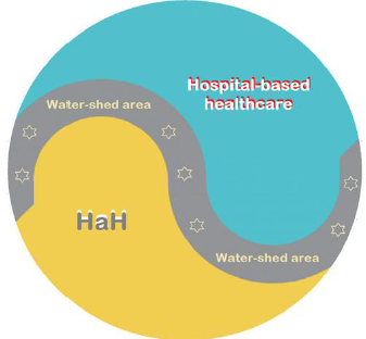
Figure 1: The conventional hospital-based healthcare, the HaH and the water-shed area for intermediate health conditions.

Similar to the scenario in various sectors, the health services and healthcare too have had profound impact owing to COVID-19 pandemic. The COVID-19 pandemic has brought home the realization that a significant proportion of healthcare activities can be tendered remotely equally effectively through technologically empowered approach. As related to the healthcare, there are certain salient aspects likely to emerge in the post-COVID-19 era.
1. There is shifting of large number of patients to remote care. The telehealth services have already been used in emergencies and during crises in the past. With possibility of quality transfer of data, audio and video communications during the COVID-19 pandemic, their utilization has widely accelerated. The pandemic has become a catalyst for swift implementation of online consult and therapy, replacing the clinician/patient face-to-face outdoor consultations.
2. In the hospital setting, the remote care is now being widely used for screening prior to the visit and triage assessment, for the indoor and ICU monitoring and supervising of patients in hospital by off-site experts. This trend is likely to persist to large extent in the post-COVID-19 period, as it provides higher convenience both for clinicians as well as patients.
3. In the mental healthcare, too, the remote consultation is proving helpful. It is likely that once mental healthcare institutions have developed the capabilities of serving their patients through digital technologies, a blended approach in future would emerge, where e-mental-health solutions cover an increasingly greater part of routine services.
4. The remote care system in form of HaH is likely to serve further as an adjunct for the gradual adoption of newer and advanced technologies, such as, the use of drones as delivery vehicles for critical supplies, robotics, the widespread 3D-printing of healthcare-related items, and smartphone-enabled monitoring of patients’ adherence to treatments.

The Healthcare Transformation – Evolution of HaH

As related to the public health, with the availability of the mobile-enabled technologies, there is an improved operation of surveillance systems and data analysis. The mobile-enabled technologies can be deployed en-masse to monitor quarantined individuals and trace exposed individuals with temporal and geographical correlates. The new tools are likely to move further into the public health domain and support the interconnected and hypercomplex global situations in real-time. On the other hand, the healthcare, in general, is needed to be people centred and integrated. The patient centred services include diagnosis and treatment and other supportive aspects of healthcare, whereas integrated healthcare involves adequate provision and efficient delivery of safe and quality health services. The people-oriented approach, on the other hand, implies planning the healthcare services by assessing the needs and expectations of community and applying them in a methodological and efficient way. The integration of modern technologies including telemedicine in healthcare services will improve the quality of healthcare.
The COVID-19 pandemic has led to realization about the limitations of existing healthcare systems and their capacity to respond to healthcare emergencies including infectious disease epidemics. It has underlined the inadequate health literacy among general population to grasp the healthcare recommendations and their outcomes [6]. It has also served as a reminder for proactive planning and preparedness. In addition, it has highlighted the necessity for technologically oriented solutions for healthcare provision and the need for significant healthcare transformation. On the other hand, it has opened the pathways to evolution and expansion of the concept of HaH incorporating communication technology-based approach as a major step to deliver healthcare at home or closer to home with all necessary steps to safeguard the safety and privacy of the participants.
In fact, the healthcare at home (HaH) can be modelled on lines of the hospice care as a multidisciplinary team approach, generally home-based and sometimes providing services through freestanding facilities, in nursing homes, or within hospitals for handling potentially treatable conditions such as pneumonia, heart failure, and alike, with brief hospital stays if necessary (Figure 2).

biomedres-openaccess-journal-bjstr

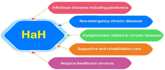
Figure 2: The development of home-based healthcare and potential spectrum of HaH.

The HaH describes a delivery paradigm where the entirety of the hospital-based inpatient care modality is substituted with intensive at-home treatment approach enabled by digital technologies, multidisciplinary teams, and ancillary services [7]. The potential spectrum of HaH can incorporate the hospice care. But as compared to the latter, apart from providing healthcare services for the terminally ill and elderly in form of hospice care, the HaH can be also useful for all those patients who need intense medical care and treatment but can be managed with help of technological monitoring and remote supervision by healthcare professionals at their homes with possible access to a nearby medical facility or hospital. HaH can make possible for people to receive a variety of medical services in their homes and can satisfactorily deal with various health conditions, as it incorporates therapeutic and nursing care, and medical assistance. In fact, the HaH is being envisaged as an alternate attractive model for accommodating increased demand for inpatient health care and as we prepare for the post-COVID-19 pandemic era, there are evolving salient features of HaH potentially promising to maximize the benefits of transformed health care [8].

The Management and Delivery of Healthcare at Home

During the COVID-19 pandemic, there has been a decline in emergency department visits and hospital admission rates in various countries [9]. It seems that in addition to a shift to virtual healthcare, COVID-19 also influenced emergency department visits and hospital admissions unrelated to COVID-19 itself. The studies from both Spain and Italy have shown a reduction in admissions and procedures related to conditions like myocardial infarction and acute coronary syndrome [10,11]. A recent study from Thailand demonstrated that during a national lockdown for COVID-19, there was a significant reduction in daily emergency department visits [12]. Similarly, a study from Melbourne, Australia documented that during times of COVID-19 restrictions there was a significant reduction in ED visits [13]. According to a survey by Canadian Home Care Association, there has been a decline of around 72% in emergency department visits, in turn resulting in the reduction of hospital admission rates [14]. These reductions in outpatient service and admissions underline the need to develop an alternative modality of healthcare for patients still requiring inpatient management for their acute and chronic medical conditions.
The integration of modern technologies like electronic health record (EHR) and telemedicine in healthcare services will save time and resources and provide better healthcare to the users. There are five major technologies which are likely to reform home-based healthcare, and include use of various biosensors, GPS, remote monitoring tools, electronic data and analysis, and telehealth. The e-Prescriptions generated are easy to be transmitted and compatible with the EHR.
In general, the HaH comprises of the following benefits:
1. With the primary focus of HaH, people get medical support at home rather than spending time in a medical facility. Further, it allows people to stay comfortably at their residential facility rather than at hospitals, having lower cost and various psychological advantages.
2. Activities of daily living are not altered and supported in-home in usual ways while maintaining a good quality of life for them in the known and perceptive atmosphere.
3. With the home care provided to patients with chronic health issues such as diabetes and respiratory disease, clinical trials have shown fewer complications and better health outcomes. The personalized and skilled care improves the overall response to the treatment.
4. With the real-time monitoring with technological equipment, the patients are seen and followed in real-time. Along with the AI and automation, the HaH aims to streamline the processes such as scheduling appointment, data collection, maintaining EHR, e-prescriptions, and scheduling and providing other health-related services as and when needed to improve the overall patient care at home.
The Covid-19 pandemic has amplified interest in HaH in the United States, European countries, and elsewhere as an alternative care model for both COVID-19 and non-COVID-19 patients, who can be remotely managed aided by current regulatory flexibilities (15). In fact, the HaH is being envisaged as an attractive model for accommodating unprecedented demand for inpatient capacity created by Covid-19. As we prepare for the health care for the postpandemic era, there are salient issues to be solved to maximize the benefits of HaH –
1. The HaH models must encompass the provision of healthcare of analogous intensity to hospital inpatient standards, and have a specified geographic catchment area, with properly defined correlates.
2. As the HaH is supposed to create the acute hospital care at home and to enable health systems to provide intensive care at home for patients with various acute and chronic conditions, this may lead to a remarkable expansion of HaH.
3. There is a unique opportunity to extend and expand HaH in current times, which can become a new vehicle for integrating non-medical services into healthcare as the patients may require further support due to complexity of their illness.
4. With the advances in digital technologies and their increased utilization by patients and healthcare providers, there is taking place transformation of the home environment into a preferred healthcare delivery site.
5. As the health awareness and rising cost of healthcare services may lead to increase in demand of HaH, managing and delivering HaH with technological backup should be affordable and providing quality service.
6. Further, a regulatory and policy implementation roadmap is required for provision of HaH, which should be accompanied by monitoring tools, such as, public reporting, patient registries, and maintenance of reliable database.

Conclusion – The Healthcare Solutions for the Future

With the COVID-19 pandemic having impact on almost every aspect of human life, the lessons have been learned relating to provision of healthcare. The telemedicine and virtual online consultations have been helpful in dealing with sudden surge and demand for healthcare both outdoor consult as well as emergency visits, and indoor and ICU care. During the COVID-19 and now in post- COVID-19 phase the alterations in provision of healthcare and its transformation have been enormous. The conventional healthcare encompassing outdoor consult and hospital-based care is being increasingly replaced by tele- and video- consultations, remote technologically assisted indoor care, and HaH. While the hospitalbased care cannot be fully dispensed with, a large proportion of it being increasingly assigned to HaH. The technologically assisted remote healthcare, outdoor as well as indoor, with its availability and acceptability, and associated challenges and benefits, is the new reality of current times.

For More Articles: Biomedical Journal Impact Factor: https://biomedres.us

Open Access Journals on Neonatology

Circulating HMGB-1, HistoneH3, and Syndecan-1 in a Newborn with Neonatal Cerebral Infarction

Introduction

Neonatal cerebral infarction is a relatively rare central nervous system disorder that occurs in about 1 in 5000 neonates. The most common cause is ischemic cerebral injury resulting from neonatal asphyxia but can also be idiopathic. Half of the infarctions develop by the day 1, and few occur over the 3rd day of life. Most occur in the middle cerebral artery region, most often on the left side [1- 3]. Convulsions are the most common initial symptom, but there are many non-specific symptoms such as decreased feeding ability and apneic attacks. Treatment is mainly systemic management and symptomatic treatment for convulsions. Currently, there are only a few reported cases of the use of thrombomodulin, tissue plasminogen activator, and Edaravone® (free radical scavenger), of which treatment method has been useful in adults [4], probably because of the many side effects for neonates. Neonatal cerebral infarction is currently classified into six categories from the viewpoint of pathogenesis, and most of the idiopathic cerebral infarctions belong to the category of ischemic cerebral infarction [1,2]. Similar cerebral ischemia and subsequent inflammation are pathological conditions of cerebral infarction in both newborns and adults, and the indication for treatment depends on how many hours have passed since the onset of cerebral infarction. In newborns, infarct lesions may appear shortly after birth or may have already occurred before birth and may be diagnosed by the detection of postnatal symptomsin the infant. Therefore, it is very difficult to determine the onset of neonatal cerebral infarction compared to that of adults.
We have confirmed and reported in a multicenter cohort study that the effectiveness of brain hypothermia in neonatal asphyxia can be judged by changes in the serum high mobility group box- 1 (HMGB-1) concentration [5]. Furthermore, we observed the serial changes of HMGB-1 in the blood of infants who had already developed fetal asphyxia and suffered severe sequelae even though brain hypothermia was started within 6 hours after birth. We found that a long time had passed following the onset of ischemic lesions and reported that postnatal brain hypothermia may be ineffective for such hypoxic ischemic encephalopathy within utero onset [6]. This time we experienced a case of cerebral infarction in one of two twins. We had the opportunity to simultaneously measure three biomarkers, HMGB-1, histone H3, which is a nuclear protein similar to HMGB-1, and syndecan-1, which is present on the surface of vascular endothelial cells and is thought to be released in the blood during angiopathy. As a result, we report a case in which the onset of cerebral infarction was suspected to have occurred before birth.

Case Presentation

The mother was 35 years old and had one pregnancy and zero deliveries. She had preeclampsia and was indicated for an emergency caesarean section due to exacerbation of her hypertension on the 36th week, 3rd day of her pregnancy. A female with a birth weight of 2420 g was born as the second baby of a diamniotic dichorionic twin pregnancy with an Apgar score of 8-9 and umbilical arterial pH of 7.273. At 2 hours and 54 minutes after her birth, she was admitted to the NICU due to an apneic attack. The infant was given intravenous phenobarbital for pedaling-like and muffled mouth movements on day 1 after birth, but it was ineffective and was changed to continuous administration of midazolam on day 2. Head echo showed no obvious lesions, but computed tomography and magnetic resonance imaging showed extensive cerebral infarction in the left middle cerebral artery region (Figure 1). The patient was diagnosed as having idiopathic cerebral infarction because a coagulation system test, amino acid fraction, and ophthalmologic examination were all negative. Midazolam was used from 2 to 8 days of age. No particular abnormalities such as in oral feeding and muscle tone were observed. An electroencephalogram was performed on day 22, and a decrease in activity on the left side was observed, but no obvious seizures were observed, so the patient was discharged from the hospital on day 31 after birth.

biomedres-openaccess-journal-bjstr

Figure 1: Magnetic resonance imaging at the 2nd day of age above: T2-weighted image, below: Diffusion weighted image; T2-weighted images revealed extensive cerebral infarction in the left middle cerebral arteryregion.

Material and Methods

Measurement of HMGB-1 Levels

The HMGB-1 measurements were performed by technicians with no knowledge of the personal data of the patient providing the samples using a commercially available ELISA kit (Shino-Test Corporation, Sagamihara, Japan). The detection sensitivity of this assay system was 0.2 ng/mL [7].

Measurement of HistoneH3 Levels

Because there is no commercially available ELISA kit, the measurement of histone H3 was performed by the ELISA prepared in our laboratory. The following is the method which had been presented in our laboratory was adopted this time as well [8]. Polystyrene microtiter plates (Nunc, Roskilde, Denmark) were coated with 100μL/well of 1 mg/L anti-histone H3 peptide polyclonal antibody (Shino-Test Corporation) in phosphatebuffered saline (PBS), and incubated overnight at 2–8°C. After three washes with PBS containing 0.05% Tween-20 (washing buffer), the remaining binding sites were blocked by incubation with 400 μL/well of PBS containing 1% bovine serum albumin (BSA) for 2 h. The plates were washed again and incubated with 100 μL/ well of diluted calibrator and serum samples (1:10 dilution in 0.2 mol/L Tris pH 8.5, 0.15 mol/L NaCl, and 1% BSA) for 24 h at room temperature. After washing, the plates were incubated with 100 μL/ well of anti-histoneH3 peroxidase-conjugated peptide polyclonal antibody (Shino-Test Corporation) for 2 hours at room temperature. The plates were washed again, and the chromogenic substrate 3,3′,5,5′-tetra-methylbenzidine (TMBZ; Dojindo Laboratories, Kumamoto, Japan) was added to each well. The reaction was terminated with 0.35 mol/L Na2SO4, and the absorbance at 450 nm was measured with a microplate reader (Model 680; Bio-Rad, Hercules, CA, USA). A standard curve was obtained with purified calf thymus histoneH3 (Roche, Stockholm, Sweden). The amino acid sequence of histone H3 is highly conserved throughout species, and that of the antibody recognition in humans, calves, mice, and rats. This ELISA specifically detects histone H3 and does not react with other histone family proteins, including histone H2A, H2B, and H4, even if 104 times excess proteins are loaded. The detection sensitivity of this assay system was 2.0 ng/mL.

Measurement of Syndecan-1 Levels

The following is the ELISA method which was presented originally in our laboratory. Polystyrene microtiter plates (Nunc, Roskilde, Denmark) were coated with 100 μL anti-syndecan-1 monoclonal antibody (R&D Systems) in PBS, and the plates were sealed with a thin adhesive-coated plastic sheet and incubated overnight at 37°C. The unbound antibodies were removed by washing the plate 3 times with PBS containing 0.05% Tween 20, and the remaining binding sites in the wells were blocked by incubating the plates for 2 h with 400 μL/well of PBS containing 1% BSA. After washing, 100 μL of each dilution of the standard and samples in 0.2 mol/L Tris pH 7.4 and 0.15 mol/L NaCl2 containing 1% BSA was added to the wells. The samples and recombinant syndecan-1 standard were diluted 1:10. The microtiter plates were incubated for 20–24 hours at room temperature. After washing, 100 μL per well of anti-human syndecan-1 peroxidase-conjugated polyclonal antibody (R&D Systems) was added, and the plates were incubated at room temperature for 2 h. After washing, TMBZ (Dojindo Laboratories, Kumamoto, Japan) was added to each well. The enzyme reaction was allowed to proceed for 30 min at room temperature. The chromogenic substrate reaction was stopped by addition of stop solution (0.35 mol/L Na2SO4), and the absorbance was read at 450 nm.

Ethical Approval

This study was approved by the ethics committees of the Japanese Red Cross Musashino Hospital (#28060). Parents of the twins were informed of the study design, and their written informed consent was obtained

Results

HMGB-1 and histone H3, which are common substances as nuclear proteins, showed a fairly strong positive correlation with a correlation coefficient of r = 0.965 (Figure 2). Syndecan-1 was low in both twins at each measurement, and no significant correlation was observed between HMGB-1 and histone H3 as previously reported [14]. HMGB-1 and histone H3 showed no significant variation in their levels in the specimens obtained before and after the onset of the first apneic attack (Figure 3).

biomedres-openaccess-journal-bjstr

Figure 2: Correlations between serum HMGB-1 and histone H3 levels. A significant correlation was observed between HMGB-1 and histone H3 (r = 0.965, p < 0.0001). However, no significant correlations were found between syndecan-1 and HMGB-1 (p = 0.431) or histone H3 (p = 0.373).

biomedres-openaccess-journal-bjstr

Figure 3: Serial changes of serum concentrations of HMGB-
1, histone H3, and syndecan-1 in this case twins.
🔴:this case twin, 〇:this case co-twin.

Discussion

Treatment for ischemic lesions of the brain is more effective the sooner it can be initiated after ischemia onset. Within 6 hours after the onset of ischemic lesions, HMGB-1 released from injured cells disrupts the blood-brain barrier (BBB) [9]. Then, within 24 hours after lesion onset, peroxiredoxin released from the cells acts on macrophages migrating from the injured part of the BBB to release inflammatory cytokines such as IL-1β and TNFα. Then, 24 hours after the onset, IL-17 and IFNγ are released from T cells, exacerbating damage to the BBB and brain cells [4,10]. The indication for brain hypothermia to treat neonatal hypoxic-ischemic encephalopathy is “the treatment can be started within 6 hours after birth”, and that for Edaravone®, a free radical scavenger, as a treatment for cerebral infarction in adults is “the treatment should start within 72 hours of onset”. These indications make sense in terms of protecting against this inflammatory response after temporal ischemia. In adults, the origin of onset is often clear, but in newborns, onset does not always occur at birth. In our previous case, severe ischemic encephalopathy had already occurred before birth, and the ischemic encephalopathy became severe even though brain hypothermia was performed within 6 hours after birth [6]. The serum cytokine profile and HMGB-1 level in this case was measured over time to develop a theoretical diagnosis and report. We consider that asphyxia is a condition of systemic ischemiareperfusion and cerebral infarction is a condition of local ischemiareperfusion.
Both HMGB-1 and histoneH3 are nuclear proteins. It was expected that the blood concentrations of these two substances would correlate, as they would be released into the blood when the cells were injured. Although the number of samples obtained was small, the values showed a very strong positive correlation (r = 0.965). In contrast, the serum HMGB-1 levels in this twin remained within reference values we have already reported for all cord blood and early postnatal specimens [7]. HistoneH3, which showed a strong correlation, was also about the same value. As this baby was born by a caesarean section before labor, it was inferred that the so-called “physiological ischemia-reperfusion stress” associated with birth would be minimal [11]. In addition, the changes in the HMGB-1 level suggested that the probability of developing cerebral infarction from immediately after birth to the onset of the apnea was extremely low. Considering that the apneic attack occurred early after birth, it is highly possible that the onset of cerebral infarction in this baby occurred prenatally and that most of the injury process was already completed by the time of birth. This suggested a reason why the HMGB-1 and histoneH3 levels in the cord blood at birth and as measured after birth did not increase [12,13].
Syndecan-1 is a representative proteoglycan expressed on the surface of vascular endothelial cells. So far, it has been reported that syndecan-1 is released into the bloodstream due to vascular endothelial cell damage in the early stage of sepsis [14]. Furthermore, it has been confirmed to increase in an ischemic perfusion injury model used in the animal experiment of Gayosso et al. [15]. Syndecan-1 did not significantly correlate with HMGB-1 and histone H3. This result was consistent with the report in adults by Ikeda et al. To the best of our knowledge, the Ikeda et al. [14] report, which focused on sepsis rather than cerebral infarction, is the only report of the simultaneous measurement of the three biomarkers HMGB-1, histone H3, and syndecan-1. The presentcase may be the second report and especially the first report in newborns. When released extracellularly, HMGB-1 not only directly damages the BBB but also acts on monocytes to increase the expression level of tissue factor and promote fibrin production by thrombin, thereby promoting thrombus formation [4]. Histone H3, a structure of NETs (neutrophil extracellular traps) released from neutrophils, is also involved in thrombus formation when released extracellularly [8]. Furthermore, syndecan-1 is also exfoliated and released into the bloodstream when vascular endothelial cells are damaged, and a thrombus is formed on the surface of the vascular endothelial cells [14]. If it can be confirmed that the levels of these three substances, which are released in common into the blood stream due to cells damaged by ischemia-reperfusion injury, are elevated in the acute phase, the use of drugs such as thrombomodulin and Edaravone® may be effective for neonatal cerebral infarction. Future accumulation of additional cases and simultaneous measurement of these three biomarkers must be useful for determining the onset of cerebral infarction.

For More Articles: Biomedical Journal Impact Factor: https://biomedres.us

Open Access Journals on Department of Pediatrics

The Dilemma of Choosing a Vaccine Against SARSCoV2 in Children? /SARSCoV2 Vaccine in Children/

Introduction

Vaccines for children are the basis for the prevention of serious, infectious diseases, which is why, for the last 6-7 decades, health workers have tried to keep the coverage of the population with vaccines over 90%, at least in developed countries. The rapid spread of the covid19 pandemic has posed a dilemma for us – should we vaccinate children against SARSCoV2 infection, with which vaccine (prepared by known technology or new technology), or should children be exposed to natural infection and stay unvaccinated? Pediatricians are daily exposed to pressure from certain pharmaceutical companies to vaccinate children older than 12 years with a certain vaccine, despite published and positive research on a vaccine that is applicable for children older than 3 years. Is this a simple match of pharmaceutical companies or is it a match between “new” and “old” vaccine technology or is it a fair match of scientific facts? Does the scientific and professional public, worldwide, agree that children should be vaccinated against SARSCoV2 infection, is there a safe and protective vaccine, and what age children should be included in the vaccination? Does vaccination of children against SARSCoV2 have a scientific justification after 18 months from the beginning of the COVID19 pandemic and after the arrival of new strains of SARASCoV2 against which the effectiveness of previous vaccines is partly because they don’t protect against infection but protect against a severe clinical picture? Here we consider the achievements so far on the vaccine against SARSCoV2 infection in children.

Children are often asymptomatic COVID19 i.e. Children are significant carriers of SARSCoV2 in the community. Children suffer mainly from mild to moderate clinical pictures of COVID19. According to the American Academy of Pediatrics, so far an extremely small number of children have suffered from a severe clinical picture of COVID19 (2.4% of total patients) or died of COVID19 (0.08% of total patients), and these are children with comorbidities (obesity, diabetes, neurological progressive diseases) [1]. However, children often show the long-COVID19 or post-COVID19, and these are predominantly children who were carriers of SARSCoV2 or suffered a mild clinical picture of COVID19. Long-COVID19 or post- COVID19 in children is mainly presented as a severe clinical picture in the form of the multisystem inflammatory syndrome (MISC) or similar-MISC which includes myocardial dysfunction, shock, and severe respiratory failure whose treatment is carried out in the intensive care unit.

Certainly, the prevention of COVID19 is more effective than the treatment of a child with COVID19 or long-COVID19, which is a kind of recommendation for vaccination of children against SARSCoV2. Indeed, there is an indication that children must be vaccinated against SARSCoV2 infection. We have been waiting for the results of research on adults for 18 months and accordingly, it is necessary to check the effectiveness of COVID19 vaccines in the child population, of course with the implementation of ethical principles of clinical research. A new circumstance is the poor efficacy of previous vaccines, in adults, against new strains of SARSCoV2 (delta, mu) and the fact that, in the September wave of the COVID19 pandemic, a worrying number of children became ill (25.7% of the total number of patients) compared to previous waves [1].

The basic two groups of vaccines against SARSCoV2 infection are known and apply according to the technology of vaccine preparation. A total of 13 different vaccines are used worldwide. One group of vaccines was made by the known technology of vaccine production with whole, purified, inactivated SARSCoV2 (manufacturers: Sinopharm, Sinovac Biotech, Bharat Biotech) [2,3]. The second group of vaccines was made with a new vaccine production technology using:
1. mRNA against spike protein proteins (manufacturer: Pfizer BioNTech, Modern), or
2. Recombinant adenovirus as a vector against spike protein viruses (AstraZeneca, Institute of India, Janssen/ Johnson&Johnson, Gamaleya National Center of Epidemiology and Microbiology, CanSinoBiologics), or
3. Recombinant spike protein with a new adjuvant (manufacturer: Novavax) or DNA plasmid [4,5].

The first three mentioned vaccine platforms have passed phase 3 and their effectiveness in the prevention of SARSCoV2 infection in adults has been confirmed, while research in the pediatric population is in the initial stages. Application of the fourth platform, i.e., the DNA vaccine began to be used in September 2021, in India, in adults and children older than 12 years [5], so we do not have data on its real effectiveness. We evaluate each vaccine according to its effectiveness, immunogenicity, and safety. Table 1 shows the basic characteristics of individual vaccines which are recommended for children. The efficacy of the inactivated vaccine against SARSCoV2 ranges from 50 to 83.5% [6]. The efficacy of a vaccine containing mRNA against the spike protein SARSCoV2 ranges from 94.1 to 95% [6]. The efficiency of the so-called “vector” vaccines against SARSCoV2 ranges from 65.7 to 91.6% [6]. The efficacy of a vaccine containing a recombinant spike protein with a new adjuvant is 89.7% [6]. The efficacy of the DNA vaccine, estimated in the laboratory, is 67% but is aimed at suppressing the delta strain of the SARSCoV2 virus [5,6].

To achieve high efficiency and immunogenicity of the vaccine, it is necessary to establish an efficient and known mechanism of immunization with phagocytosis using antigen-presenting or dendritic cells that activate T-lymphocytes, which will consequently activate B-lymphocytes, thus achieving cellular and humoral immune response. The second dose of the vaccine enhances and prolongs immunity against SARSCoV2 in terms of an increase in IgG antibody titer to the spike protein SARSCoV2 (S1-RBD) with neutralizing capacity, to a lesser extent to the N-protein SARSCoV2, as well as an increase in INF-gamma secretion after recognition of SARSCoV2 antigen, primarily CD4 lymphocytes and a lesser extent CD8 lymphocytes. Since the time of Pasteur, we have been considering the effectiveness of an inactivated (“dead”) vaccine, that contains the entire infectious agent and conjugate vaccine that contains parts instead of the whole live virus. For example, pertussis vaccination coverage is 86% after 3 doses of vaccine (primarily whole-cell vaccine), which provided a low rate of pertussis in children [7]. It is this efficacy of the pertussis vaccine that can be compared with the efficacy of the inactivated SARSCoV2 vaccine [2,3]. Vaccines that use mRNA or DNA provide human cells with genetic information for an important part of SARSCoV2 against which the immune response is elicited. Vector vaccines transmit genetic information, through another virus for part of SARSCoV2, to human cells that produce a viral protein and elicit an immune response. Protein subunit vaccines produce proteins from viruses so that the human immune system learns to attack them.

Immunogenicity in previous studies was estimated as the percentage of seroconversion, i.e. Increase in the titer of neutral antibodies to SARS-CoV-2 after 28 days of vaccine administration. It is still not specified which antibody titer prevents infection or why there is a quantitative but not qualitative increase in anti-SARS-CoV antibodies after vaccination nor which CD4/CD8 lymphocyte ratio protects against SARSCoV2 infection nor how long post vaccination immunity lasts? The seroconversion achieved by the inactivated SARSCoV2 vaccine found in two independent studies in children was approximately the same: 96.8-100% according to the vaccine dose (1.5 and 3.0 mcg, respectively) compared to 100% after 56 days of the first dose, regardless of whether the dose was 4 mcg or 8 mcg and regardless of age group (3-5 years, 6-12 years, 13- 17 years) [2, 3]. In school-age children, seroconversion with the inactivated vaccine against SARSCoV2 is achieved after 28 days from the first dose [2]. The mRNA produced by Pfizer is 100% effective and contributes to a robust response by producing antibodies to SARSCoV2 in children aged 12-15 years after 7 days of the second dose of the vaccine [4,8]. The vaccine safety data in terms of the number of adverse local and systemic reactions in children are shown in Table 1 [6,9,10]. Data on the efficacy of other vaccines for adults are discussed in the English Covid Vaccination Program [6]. To achieve herd immunity, it is crucial to achieving coverage of the population by vaccination of approximately 80%, which has been achieved by several countries in the world (Portugal, Spain, and Denmark). Until the achievement of collective immunity, it is necessary to implement epidemiological protection measures against the SARSCoV2 infection.

biomedres-openaccess-journal-bjstr

Table 1: Vaccines against SARSCoV2 applicable in children.

For More Articles: Biomedical Journal Impact Factor: https://biomedres.us

Open Access Journals on environmental Science

Agronomic and Ecosystem Services Potentialities of Green Manure Utilization

Introduction

The global impact of climate change caused by the excessive use of natural resources and the over growth of world population harms biodiversity species and disturbs ecosystem functions. It was identified that land degradation, one of many consequences of poor management. (Holden, S., Shiferaw, B., & Pender, J., 2005) [1] of natural capital (soil, water, and biodiversity such as vegetal and animal organisms) is the problem of worldwide caused anthropological manipulation of land, which altered chemical, physical and biological soil properties (Lal, R, 2001). The Global Assessment of Human-Induced Soil Degradation project (GLASOD) in 1988 estimated 1,964 million (nearly 2 billion) hectares degraded worldwide and more than 22% of combined agricultural land pasture and woodland were destroyed by human-induced soil degradation (Chen H. J. et al. 2014). This problem of land degradation evolved the climate change, biodiversity, food security, quality of water and air. The resource land is characterized by a complex structure of two interlocking systems: a system of natural resources ecosystems and human society. The interaction between them determines how natural resources are managed (Temengsgen G et al. 2014). For example, the findings of Lingling Hou (2012) [2] estimated that 50% of the land in China was degraded and more than 466 million hectares have been affected. That situation caused environmental and ecological damages [2].
However, overuses of agrochemical products [3] were fundamental causes of soil degradation in the worldwide. It would like to cite chemical fertilizers and pesticides. The agricultural industry is ranked first for the consumption of pesticides with 1.84 Mt in 2014 and non-biological pesticides account for 91.78% in China [4]. Many researchers stated that green manure is useful to limit those negative effects of using of artificial products in agriculture. The world community would undertake maintaining soil fertility and biodiversity in order to assume the equilibrium between food supplies, the population growth and safe environment [5]. To reach this objective, many governments have undertaken different measures for conservation and sustainable use of biological diversity [6]. Operationally, many farmers must use green manure to limit the excessive application of chemical products in agriculture. The objective of this review is to give the answers to this big question: How green manure practices could perform farming production and environment damage?.
The environment scientists tried to complete the commitment of the States in implementing the convention on Biological Diversity universally acknowledged in 1992 by discovering that the use of green manure could be one of the solutions of land degradation, agricultural products quality and environment damage. This is why this review paper tried to give different explanations and details about various agricultural performances and ecosystems services provided by green manure. By definition, green manure is produced by ploughing leaves and roots of green plants at maturity into the soil. After a while, they become compost or green fertilizer [7]. According to some writers, green manures are the material of plant-incorporated into the field [7,8] and for others green manure is plants used to produce compost or green fertilizer [5]. That big question of research has been answered through reviewing numerous publications based on the benefits of using green manure. Methodologically, this review summarizes and discusses the finding from the following principal publications reviewed below: (Table 1).

biomedres-openaccess-journal-bjstr

Table 1.

Use of Green Manure

According to Costanza Robert (1997) [12], the natural ecosystem services were defined as the benefits that the nature provided to humans through natural resources transformations into a low of essential goods and services [12]. In this way, green manure is used to fertilize degraded agricultural land and save biological diversity in the soil. To optimize the ecological and economic services value from green manure, farmers used diverse models of planting such as rotation fallowing, inter-cropping and double cropping. Those technologies improve ecosystem services, support agricultural production performance and safe environment.
The use of green manure contributes to maintain ecosystem systems and to give ecosystem services value to human interventions which aim to promote sustainable development. Green manuring increased the CO2 concentrations and providing biological-Nitrogen in the soil [13,14], soil shade, soil organic matter (SOM) and soil organic carbon (SOC) when turned into the soil [14,15] as economic benefits [15], ecosystem services [7], and mitigation of pollution and beautify landscapes.

Green Manure Species

In the world, it is observed many kinds of green manure and cover crops those farmers use as green fertilizer in the fields for different reasons. However, the ryegrass (Lolium perenne), winter oilseeds (Brassica napus ssp. oleifera var. biennis), and winter rye (Secale cereale L.) were used in mixture to control the biomass and weeds and to increase crops yields in rotation crops [15]. In South Africa, according to the evaluation of green manure legumes have the potentiality to increase the crops yields in smallholder farms (Jude, J.O.O., 2011) [16]. It can be used in tropical region to increase the yield and agronomic performance of common beans [17]. Then, green manure plants play a significant role in farming systems management through its functions, such as the financial services, ecosystem/ecological services, and cultural landscape services functions [18] (Rovanovskaya A.A 2008).

Economic Services Functions

The practice of green manuring reduces the biomass, the density of the weeds, and increases at maximum the crop yields and the green manure crops could improve the health of soil when it was turning into the ground after maturity [7,8]. Green manure practices bring economic benefits [9] through reducing the costs of inputs of the farmers, increase yields. It is not easy to capture all of these gaps of costs during the process of the market transaction [19] without adapted scientific methods. It is observed the indirect effects of green manure practices on the cost of disease damages management (McGuire, A., 2016). With the incorporation of different leaves of green manure (Abelmoschus esculentus) in Bhendi cropping systems, the crops growth parameters performed well and yield quality, as well as high net profit and benefit-cost ratio were observed [20]. The evaluation of economic services of green manure is very important when the research needs to understand the contribution of green manure in ecosystem system. It is used different potential applications to evaluate the ecosystems services values namely the Life Cycle Assessment (LCA), Contingent Valuation Method (CVM), Willingness -To -Pay (WTP) and Willingness-To-Accept (WTA), bio-economic model (Wang T. et al., 2018), and economic benefits analysis [21]. Currently, those techniques of assessment were applied in environmental impacts assessment (EIA) [20]. Referencing to the capacity of fixing nitrogen, the low-cost nitrogen, a bio-fertilizer, is provided by green manure. Those plants are good patterns of wetland rice cultivation [22].

Ecological Services

Green manure crops have the potentiality to maintain ecosystem systems [9]. Ecosystem systems provide a variety of essential services, including water, air, and health, livelihood, and well-being (Barvanera Patricia Sandras Quijas, Karp Daniel S. et al., 2016). In France, near Toulouse and Orleans, crucifer’s species, grown in the form of green manure (catch crops) released a large amount of mineral nitrogen (N) for later commercial crops and legumes cropped as green manure plants decreased the leaching of nitrates in extraordinary ways. The same authors indicated that mixing crucifers and legumes in farming systems was an operational solution to obtain multiples ecosystem services from both catch crops for providing nitrate and legumes for providing nitrogen as green manure services [23]. Specifically, Azolla, a tiny aquatic fern, was used as green manure in flooded rice planting [24]. Then, green manure crops can regulate multiples environmental problems such as pest and diseases control [25], carbon sequestration [18,26]. It contributes to water filtration mitigation, climate control [26], and beautification of the living environment. However, green manure improves air purification and quality of agricultural products [27,28] because conventional farming systems are the sources of Green Houses Gases (GHG) emissions [26].

Plant Health Improvement

The weeds are unwanted plants that hosted pathogens (Rodgers V.L, Stinson K.A and Finzi A.C. 2008) and play a principal role in various ecosystems. Many of those weeds led to direct and indirect damages in farming ecosystems such as the losses of fertile agricultural lands, biodiversity, areas for grazing animals, and production of livestock, chocking of navigation and canals of irrigation and diminution of the availability of water in the rivers. However, green manuring, by adding carbon into the soil (Blumenthel, D. M. Jordan, N.R. and Russelle, M.P 2003), is one of the different sustainable farming systems which can successfully bring sustainable weed control for environmental, social, and economic benefits and wellbeing (Garnavel L and Natarajan S K 2014). Green manure /covers crops destroy weeds, which could theoretically act as causes of pathogen inoculum for crops and make returning accumulated nitrogen to the soil, reduced nitrogen leaching, avoid erosion, and improve soil structure [23]. In Pacific North- West of United States of America, the green manure (Cover crops, Mustard, Sudan grass, Lupine, and Marigold used as biological control and canola, Crambe, meadowfoam, Milkweed, seed meals organic amendment) reduced the nematode impact (Meloidogyne chitwoodi) on potatoes by 50-80%, provide nematode control comparable to fumigation and improve soil physical characteristics especially water infiltration and penetration of resources [29]. Then, green manure crops and cover crops, used as green manure, played a significant role in controlling diseases and nematodes, which harm cropping patterns.

Carbon Sequestration

Farming systems can be a source of dioxide of carbon (CO2), and when it surpasses plant carbon fixation by photosynthesis, CO2 contributes to climate variability. In 2002, Reicosky estimated that tillage of the soil led to carbon losses between 30% and 50 %. However, when farmers incorporated green manure crops into the soil, they captured CO2 through the humification of soil organic matter (SOM) fractions after the mineralization process and the content of soil organic carbon (SOC) increased [11]. However, green manure displayed a significantly greater soil organic carbon (SOC) than the crops taken as a reference [30]. Thus, green manure crops and cover crops contain the potential capacity to sequestrate carbon and improve smallholder farmers resilience with minimum trade-offs [31]. Allowing a fallow period between two seasons of cropping can increase the soil organic carbon (SOC). Then, the SOC is an effective measurement to compensate for anthropogenic GHG emissions [11,32]. In this context, Yang (2014) found that green manuring is a management strategy for mitigating soil degradation, increasing nutrient levels (nitrogen, carbon, and other micro-elements [33]. The same study indicated that green manure legumes increased significantly in the long term, the total carbon (C) and nitrogen (N), and the formation of the stable aggregate portion of the water measuring from 2 to 5 mm in the soil. Specifically, the nitrogen is the element of nutrients supplied chiefly by green manuring, since nearly all the soil, turning under the crop-legumes increased the nitrogen associated with organic matter [5].

Quality and Quantity of Yield Improvement

Reducing-cost-technology (RCT) consisted of the soil nontillage and decreasing nitrogen over-fertilizing doses with green manures before crop patterns. Those plants have high potential for restoration of soil fertility and enhancement of terrestrial crop production [34] and provided best profits compared with the other plants (Whitmore A. P. et al, 2000) [35], also contributed significantly to the nutritional demand of green manure legumes, thereby providing an agro-ecological and sustainable production [36]. The grazing of green manure, especially oat (Avena sativa), pea/oat (A. Sativa/Pisum sativum) contributed to improving the available nitrogen production in integrated crop-livestock systems [37]. This system of production of agriculture consisted of capturing ecological interactions among different systems of landuse, making agricultural ecosystems more proficient at cycling nourishment, preserved the natural resources and environment and improved the quality of the soil and enhanced biodiversity (Franzluebbers, A. J., 2007;) [26,38]. The green manure vetch (Vicia villosa) more affected the quality of biological maize (Zea mays) compared to that of fallow lands associated with organic fertilizers (phosphorus supplement) on in a field experiment of two years in central Italy [39]. The rotation of crops (Bullock, D.G., 1992) is one of the different modes of planting green manure that brings high yield of crops.

Mitigation of Soil Water Losses, Air Pollution, and Environmental Degradation

Green manuring is one of the ways of moisture conservation in the soil. Thus, the adoption of moisture conservation techniques, in situ, was increasing the moisture availability. However, the growth of green manure crops after cereal harvesting had reduced infiltration from rainfall during the autumn season in Lituany of an average between 19.4 % and 21.7 % (2003) and between 7.0 % and 8.3% (2004) such as clover produced more biomass (0.407 g/m2) with more nitrogen (7.35 g/m2) when clover incorporated into the soil and it increased nitrogen concentration.

Improvement Of Soil and Biodiversity’s Health

The soil organic matter (SOM) plays a central role in the function of farms and particularly in biologic farming [40] (Morton A. C., 2008). Green manure contributes, transformed by ploughing into the soil, to improve the physical and chemical properties and plant growth (Hrishan Chandra, 2005), [41,42]. Then, depending on its potential to fixe biological nitrogen, green manure legumes are providing nutrients to crops in cropping systems [43], The soil organic matter content is the home of millions of microorganisms which brooked down by bacteria and makes nitrogen available to plants (Pieters A.J 1927) [44]. The retention of plant nutrients (carbon, nitrogen, zinc, etc.) from organic inputs depends on the microbial community under environmental conditions [45,46] and bio-chars [47]. By using green manure, nitrogenous compounds and carbons are transformed by soil microorganisms into elements absorbed by crops [5]. In the plants, roots absorbed more nourishment concentration than shoots [48].

Why it is Necessary to Plant Green Manure in Agricultural Land?

The green manure practices safeguarded biodiversity and provided ecosystem services to agricultural systems by transforming nitrogenous compounds and carbons into elements absorbed by the next crops (Thomas Oladeji Fabunmi et al 2012; Pieters A, J., 1927). In the study conducted by Zandvakili showed that the roots had higher concentrations of nourishments than shoots [36,48]. found that the use of native species of the Caatinga Biome could affect the nutritional demand of the Market of Garden Crops significantly, thus providing a form of agro-ecological and sustainable production. Consequently, soil fertility in organic matter is managed mainly by planting green manure. For example, China milk vetch (Astragalus sinicus L.) planted and mixed with chemical nitrogen fertilizer reduced the application of chemical fertilizer (Ma Yanqin and Huang Guiqin, 2019) and increased yield of rice by 28.7% in southern China (Qin, W. et al.2012) [49] and decreased seasonal methane (CH4) flux in the mono-rice cultivation system [49]. When the application rate of milk vetch is increased, also, the yield and production of rice increased (Chang H. L., et al, 2010). The combined utilization of vegetable-green manure and phosphorus-enriched compost (P) can then be considered a reliable option for managing N and P fertility in the short and long term and maintaining plant needs [39]. Green manure and cover crops are well recognized in many systems of agriculture. The application of green manure in smallholdings provides multiples profits. Those benefits are nitrogen fixation, soil organic matter content, and weeds control, the management of disease and pests, and soil erosion control. It is a significant low-cost added-value to technological options that integrate the consideration of nature conservation and productivity of agriculture (Pratt, O.J., 2016).

Ecological Compensation is Needed to Support Planting Green Manure

Although green manuring is one of good practices for sustainable development of agriculture, the increased cost of production of green manure reduces the willingness of farmers to plant green manure. Another method to promote farmers to adopt green manure is ecological compensation [9]. The eco-compensation is an approach like a trade-off in which compensatory laws attribute different values of the benefits of ecosystem services or the damage of the loss of the natural environment. That compensatory act corresponding to those goods or services lost or gains by the environment. The compensatory law can be a mechanism to ensure the ecosystem services flow (restoration of resources, recreation, or conservation of nature) and maintaining the flow of natural capital on which depending on the economy. The eco-compensation policy aims to encourage people (He, K. et al. 2016) to participate in sustainable agriculture.
A study conducted in Spain showed that the eco-compensation practices in environmental impact assessment (EIA) is much fewer because of only 407 of 1302 records of decisions (RoDs) reviewed (31%) mention eco-compensation and only 117 of 1302 RoDs (9%) described the measures of compensation mechanism (Villaroya, A. and Puig, J., 2013. The Willingness-To-Accept (WTA) the eco-compensation standards for farmers of fallow winter wheat in Hengshui, Hebei province, was 0.00095$/hm2 [50]. Many factors affected significantly and positively the willingness of farmers to reduce pesticides, namely the farmers’ environmental concern, cognition of pesticide residues, the quality of agroproducts interest, and controls of inputs. The study also noted that regulations and countermeasures and enhancing farmers’ selfcontrol were essential to guide farmers toward environmentallyfriendly measures in agricultural production (Zhang L et al 2018). On the contrary, to initiate a Pigouvian-tax, tax paid by economic actors when their activities generate negative externalities implies to proceed of actors’ Willingness-To-Pay (WTP) for the negative externalities from agriculture. A study carried out using a model of dynamic equilibrium to assess the effects of the welfare of subsidy estimated that the impact of Pigouvian-Tax on the intensity of financial support was negative (Yang, L. et al., 2018). The ecocompensation based on financial support could give added-value on the well-being of human, maintaining dynamic effects of ecosystems and nature conservation because the Pigouvian-tax alone cannot play a significant role to correct the most considerable externalities in the long-run (Dennis W. C. and Glenn C. L., 1980; Kohn, R. E., 1986) [51-55].

Conclusion

The use of green manure in agricultural fields brings various profits in terms of economic benefits and environmental safeguarding. However, green manure practices must be adopted as a new sustainable development approach of agriculture. The study reviewed various papers related to green manure benefits. It was observed that green manure technologies help farmers to various advantages namely economic benefits, carbon sequestration, nitrogen fixation, SOC content improvement, biodiversity safeguard, etc. Because of high ecosystems services values and few economic profits of farmers, an ecological compensation system could be adopted widely as a new sustainable development approach in farm systems.

For More Articles: Biomedical Journal Impact Factor: https://biomedres.us

Open Access Journals on Biochemistry and Molecular Biology

A New Method for in vivo Targeted Gene Transfer into Oligodendrocytes using Adenoviral and HIV Vectors

Introduction

Viral vectors such as retrovirus, adenovirus (Ad), lentivirus, and adeno-associated virus (AAV) are widely used in clinical gene therapy protocols as vehicles for the delivery of genes into mammalian cells [1-5]. One advantage of these vectors is their ability to transduce a wide range of cell types, but this lack of specificity is also a distinct disadvantage, especially for in vivo gene transfer. This is because transduction of both target and nontarget cells results in massive wastage of the vector, and stable gene transfer into non-target cells by retroviral vectors may increase the likelihood of insertional mutagenesis in the transduced cells [6,7]. An efficient technique for targeted gene transfer would thus be a highly desirable.

Several strategies for targeting cells for gene transfer have been reported. One approach is to achieve transcriptional targeting using a tissue-specific promoter [8,9]. With this strategy, however, non-target cells are also transduced, even though the promoter is silent, resulting in massive wastage of the vectors. Another strategy is receptor-mediated targeting. For example, many investigators have been able to achieve targeted retroviral gene transfer through modification of the vector particles using single chain antibody fragments [10,11] and ligand molecules [12, 13], and by using pseudo type viruses [14,15]. The low efficiency of gene transfer is a serious disadvantage of this approach, however. To overcome these problems, we developed a novel strategy for cell targeting based on tissue-specific expression of an ecotropic retroviral receptor gene using Ad and ecotropic retroviral vectors [16]. With this approach, we achieved efficient targeted retroviral transduction through Ad-mediated, tissue-specific expression of a retrovirus receptor. Unfortunately, non-dividing cells could not be transduced using this method because cell division is required for the retrovirally mediated gene transfer [17].

Human immunodeficiency virus (HIV)-based retroviral vectors have several interesting features that make it potentially useful for targeted gene transfer. Because CD4 antigen is a major receptor for HIV entry, HIV vectors transduce only human CD4-expressing cells [18]. In addition, the HIV vector, itself, provides receptormediated targeting based on the natural tropism of viruses, and it can transduce non-dividing cells [19]. To make full use of these features of the HIV vector, we developed a new method that expands the host range of the HIV vector through a two-step gene transfer protocol [20]. Using this protocol, we were able to stably transduce such non-dividing cells as neurons and terminally differentiated macrophages [21], suggesting a combination of Ad and HIV vectors is potentially useful for transduction of a variety of non-dividing cell types. To further develop this strategy for targeted gene transfer into non-dividing cells, in the present study we constructed an Ad vector expressing the CD4 gene under the control of the oligodendrocyte (OL)-specific myelin basic protein (MBP) promoter [22,23]. We then tested whether OL-specific gene transfer could be achieved using Ad and HIV vectors, and whether this new method could be used for targeting gene transfer into nondividing cells in vivo.

Materials and Methods

Cells

Cos, 3T3, HEK293 and CD4H (CD4+ HeLa) cells [20] were grown in Dulbecco modified Eagle medium (DMEM) supplemented with 10% heat-inactivated fetal bovine serum (FBS) and 100 units/ml penicillin and 100 μg/ml streptomycin (GIBCO-BRL, Gaithersburg, MD) at 37 °C in 5% carbon dioxide. The CG4 OL cell line was maintained and differentiated as described previously in growth medium or differentiation medium [24]. Primary cultures of mixed rat brain cells were prepared as previously described [25] with some modification. Briefly, embryonic brains were minced and dissociated by pipetting after treatment with 0.25% trypsin. After low speed centrifugation (500rpm, 3min) to remove the debris and filtration through a 70mm filter, the cells were grown in MEM/F12 medium (GIBCO-BRL, Gaithersburg, MD) supplemented with 10% FBS to obtain a mixed glial culture.

Production of Ad and HIV vectors

A replication-defective Ad vector was generated using the Saito method [20]. An Ad vector containing the CD4 gene under the control of the MBP promoter (kindly provided by Dr. Ikenaka, National Institute for Physiological Sciences, Okazaki, Japan) was constructed by inserting the expression unit containing the MBP promoter (1.3-kb HindIII fragment) [26], the coding sequence of CD4, and the rabbit ß-globin polyadenylation signal into pAdex1w [27]. Another Ad vector containing the CXCR4 gene [28,29] was constructed by inserting the coding sequence of CXCR4 (kindly provided by Dr. Matsushima, University of Tokyo, Tokyo, Japan) into pAdex1wCA [27]. These constructs and EcoT221 digested Ad DNA terminal protein complex were introduced together into HEK293 cells. The recombinant Ad, Ad/MBPCD4 and Ad/ CAGCXCR4 were isolated, purified and concentrated through cesium chloride gradient centrifugation. The titers of Ad/MBPCD4 and Ad/CAGCXCR4 were 3×10¹⁰ and 1×10¹⁰ PFU/ml, respectively. An HIV vector carrying the enhanced green fluorescent protein (EGFP) gene (HXGFP) was generated by transient transfection of Cos cells with packaging (pCGPE) and vector (pHXGFP) plasmids as described previously [30]. Two days after transfection, the viruscontaining supernatant was concentrated by ultrafiltration using a CENTRIPREP 50 (Millipore Corporation, Bedford, MA) [30]. The biological titer of the concentrated HIV vector was approximately 108 transducing units (TU)/ml when CD4H cells were used as the target cells.

Targeted Gene Transfer into OLs

To achieve OL-specific gene transfer, we applied the two-step gene transfer method using Ad and HIV vectors as described previously [20]. In brief, rat brain primary mixed cultures were incubated with Ad/MBPCD4 and Ad/CAGCXCR4 for 60min at an MOI of 10. After two days of culture in complete medium, the cells were incubated with HXGFP for 48 h at an MOI of 100. For in vivo targeted gene transfer, 6- to 8-week-old Fischer 334 female rats (Japan Clea Co. LTD., Tokyo, Japan) were anesthetized with ketamine (100mg/kg) and nembutal (50mg/kg) and placed into a stereotaxic frame. Thereafter, the skull was exposed, a hole was drilled over the injection site (0.7mm anterior to bregma, 2.0mm lateral, 4.0mm vertical) [31], and 1μl of Ad/MBPCD4 and Ad/CAGCXCR4 was infused over period of 10min using a Hamilton syringe with a 26 gage needle. Three days later, 2μl of HXGFP were infused into the same site. As a control, Ad/CAGLacZ (kindly provided by Dr. Saito, University of Tokyo, Tokyo, Japan) plus Ad/CAGCXCR4 and HXGFP or only HXGFP were injected. All experiments involving animals were conducted according to the institutional guidelines of the Nippon Medical School.

Flow Cytometric Analysis

Expression of CD4 or CXCR4 was analyzed by flow cytometry (FACS Calibur, Becton Dickinson, Franklin Lakes, NJ) after staining with fluorescein isothiocyanate (FITC)-conjugated anti-human CD4 or phycoerythrin (PE)-conjugated anti-human CXCR4 (BD Pharmingen, San Diego, CA).

Immunocytochemistry and Immunohistochemistry

To identify OLs, we used anti-carbonic anhydrase II (CAII) and anti-MBP antibodies (Dako, Hamburg, Germany) as described previously [32,33]. The transduced mixed rat brain cells were fixed in 4% paraformaldehyde for 15min at room temperature. After washing three times with PBS, the cells were incubated with anti-CAII antibody and normal rabbit serum (Dako, Hamburg, Germany) for 2h at room temperature. The cells were then washed three times in PBS containing 0.03% Triton X and exposed to Texas Red-conjugated secondary antibody with normal rat serum (Dako, Hamburg, Germany). The stained cells were examined under a IX/70 inverted fluorescence microscope (Olympus, Tokyo, Japan) or analyzed by FACS Calibur. To analyze in vivo transduction of rat brain, 5 days or 3 months after injection of HXGFP, the rats were anesthetized and perfused with 4% paraformaldehyde. The brains were then removed and transferred to PBS solution containing 20% sucrose and stored overnight at 4 °C. The next day, 40-μm-thick tissue sections were cut using a cryostat, after which the sections were blocked for 1.5h in PBS containing 10% normal rabbit serum with 0.03% Triton X and incubated with anti-CAII or anti- MBP antibody overnight at 4 °C. The sections were then washed three times in PBS containing 0.03%. Triton X and incubated with rhodamine (TRITC)-conjugated secondary antibody (Dako, Hamburg, Germany). After immunostaining, the tissue sections were mounted on slides and visualized and photographed using a confocal laser-scanning microscope (Leica TCSSP, Heidelberg, Germany) as described previously [32].

Results

biomedres-openaccess-journal-bjstr

Figure 1: OL-specific CD4 expression by Ad/MBPCD4.
A. FACS analysis of Ad/MBPCD4 transduced cells.
• The upper panel shows a FACS analysis of Ad/MBPCD4-transduced 3T3 (a) and CG4 (b) cells
• As a control, the lower panel shows Ad/CAGCXCR4-transduced 3T3 (c) and CG4 (d)cells.
B. Immunocytochemical analysis of mixed primary rat brain cells transduced with Ad/MBPCD4.
• (a) Bright-field image.
• (b and c) Fluorescence microscopic images of cells double immuno stained with anti-CD4 (FITC) (b) and anti-CAII (Texas-Red) (c)
• (d) Merged image combining b with c.

Oligodendrocytes, which are known to be myelin-forming cells, are an important target cell for gene therapy aimed at treating such demyelinating diseases as multiple sclerosis and metachromatic leukodystrophy. Because OLs are postmitotic, they cannot be transduced using moloney murine leukemia virus-based retroviral vectors. On the other hand, both Ad and HIV vectors are able to transduce non-dividing cells. Furthermore, Ad vectors with the MBP promoter have proven useful for OL-specific gene expression both in vitro and in vivo [33]. To assess OL-specific expression, we first examined CD4 and CXCR4 expression in 3T3 and CG4 cells transduced with Ad/MBPCD4 or Ad/CAGCXCR4. Expression of CXCR4 was detected in both cell types transduced with Ad/CAGCXCR4, while expression of CD4 was detected only in Ad/ MBPCD4-transduced CG4 cells (Figure 1A). We then confirmed the OL-specific expression of CD4 using primary mixed rat brain cell cultures. Among the different cell types transduced with Ad/ MBPCD4 only CAII-positive cells expressed CD4 (Figure 1B), indicating that Ad/MBPCD4 selectively mediated expression in OLs. We next evaluated the utility of our two-step gene transfer system using mixed rat brain cells first incubated with Ad/MBPCD4. And because non-human cells do not express CXCR4, which is a coreceptor for HIV, these primary cells were also incubated with Ad/ CAGCXCR4. Two days later, the cells were incubated with HXGFP and, after an additional 2 days, they were stained with anti-CAII or anti-MBP antibody. We found that all EGFP-positive cells were also CAII-positive (Figure 2A). In addition, FACS analysis showed that only MBP-positive cells could be transduced with HXGFP (Figure 2B), indicating that OLs first transduced with Ad vectors were then selectively transduced with the HIV vector. Thus transient selective expression of CD4 molecules using Ad/MBPCD4 is apparently sufficient to render OLs susceptible to HIV-mediated gene transfer. To then determine whether this new method could be used in vivo to target gene transfer into OLs, we injected the Ad and HIV vectors into the brains of adult rats. Five days after injection of the HIV vector, some of the rats were fixed and their brains were examined using confocal laser-scanning microscopy.

biomedres-openaccess-journal-bjstr

Figure 2: OL-specific transduction using a two-step gene transfer method.
A. Immunocytochemical analysis of primary rat brain cells transduced using a two-step gene transfer method.
• (a) Bright-field image.
• (b and c) Fluorescence microscopic images of GFP (b) and CAII (Texas-Red) (c)
• (d) Merged image combining (b) with (c).
B. FACS analysis of primary rat brain cells transduced by two-step gene transfer method.
• FACS analysis of primary rat brain cells transduced with Ad/CAGLacZ (a) or Ad/MBPCD4 (b) plus Ad/CAGCXCR4 and HXGFP. After immunostaining with anti-MBP, the cells were analyzed by flow cytometry.

EGFP-positive cells were not be detected in rats injected with the control vector Ad/CAGLacZ plus Ad/CAGCXCR4 and HXGFP or with HXGFP alone (data not shown). On the other hand, we were able to detect EGFP-positive cells in rats injected with Ad/ MBPCD4 plus Ad/CAGCXCR4 and HXGFP (Figure 3A). Moreover, immunohistochemical staining using anti-CAII and anti-MBP (not shown) antibodies revealed that all of the EGFP-positive cells were also CAII-positive (Figures 3B & 3C) and MBP-positive, indicating that only OLs were transduced using this two-step gene transfer method. In some cases, the rats were not sacrificed and analyzed until 3 months after vector injection. Notably, the results obtained 3 months after vector injection were nearly the same as those obtained 5 days after vector injection (Figures 3D-3F). This strongly suggests we were able to integrate the transgene into the host genome and obtain sustained transgene expression

biomedres-openaccess-journal-bjstr

Figure 3: Immunohistochemical analysis of rat brain stained with anti-CAII 5 days
• 5 days (A, B and C) or 3 months (D, E and F) after transduction using the two-step gene transfer method.
• Confocal microscopic images of GFP-positive cells (A and B), CAII-positive cells (C, and D), and merged images (C and F) of the transduced brain tissue sections.

Discussion

Cell targeting is particularly important for in vivo gene transfer into brain, as stable genetic modification of some neurons or neuronal networks could cause serious psychological changes. Therefore, if the targeted cells are glia, undesirable gene transfer into neurons must be avoided. Our findings show that by using a two-step gene transfer system with Ad and HIV vectors we could selectively transduce OLs both in vitro and in vivo. Moreover, these findings imply that with the appropriate combination of vectors and promoters, one could also selectively transfer genes into neuronal cells.

The transduction efficiency for mixed primary rat brain cells was only 4% to 5% (Figure 2B). One likely reason for the low transduction efficiency is that the HIV vector cannot transduce non-human cells, which do not express CXCR4. We therefore had to use two Ad vectors to target rat OLs. On the other hand, only one Ad vector, Ad/MBPCD4, is needed for HIV vector-mediated gene transfer in human brain, which we would expect to increase transduction efficiency. Another possible reason for the low transduction efficiency is the toxicity of Ad vectors [34]. Using Ad vectors it is difficult to achieve highly efficient transgene expression without toxicity [35]. To overcome this problem, to used gutless Ad vectors, which retain no viral genes and have proven to be highly efficient with little toxicity or immunogenicity [36]. In addition, group D Ad reportedly infects primary central nervous system cells more efficiently than group C [37]. Thus, by using recombinant gutless Ad vectors generated from type 17 (group D) Ad, the efficiency of transduction into central nervous system cells using the two-step gene transfer method may be increased.

In summary, we have developed a new method of targeted gene transfer into OLs using Ad vectors with a tissue-specific promoter and an HIV vector. This new method can be used with nondividing cells both in vitro and in vivo. Furthermore, by choosing the appropriate promoters, this method may be useful for in vivo targeted gene transfer into any type of non-dividing cells.

For More Articles: Biomedical Journal Impact Factor: https://biomedres.us

Open Access Journals on Food Systems and Health-Saving Technologies

Technological Properties of Wheat-Triticale-Rye Flour

Introduction

In the diet of the population of this country a large proportion (up to 40 %) is taken by breadstuffs, the main part of which are products on the basis of various kinds of baking and macaroni flour. So, it looks quite promising and much in demand to make flour composite mixtures at flour mills with the specified contents of the main nutritious and biologically active substances: protein, carbohydrates, carbs, fats, vitamins, micro- and macro elements from the products of the main processed crops including wheat, triticale and rye. Such grain mixtures will become basic for particular manufactures which make bakery products, confectionary, pasta and extruded products, dairy and meat products for specific diet: dietary, preventive and curative nutrition [1-11,12-26]. The current trends in the development of one of the most important processing industries – flour grinding – are developing technologies of processing traditional crops (wheat and rye) as well as new technologies of processing non-traditional crops such as triticale. The ultimate goal of the branch development is to introduce new and to improve traditional technologies and to create products of procession of various kinds of crops with a specified composition and qualities. In addition, a co- processing of grain of various crops, including wheat, triticale and rye is most promising [1- 6,17,22,23]. Breadstuffs with products of processed triticale grain are characterized by higher nutritional value since there is more protein and indispensable amino acids, the main limiting acid – lysine. The combination of positive properties of rye – abundance of biologically active aromatic substances and wheat – rheological properties of dough makes it possible to make diet products.

Wheat protein has little lysine, so in the course of breadstuffs production of improved composition products the lack of lysine is compensated by the increase in digestibility and nutritional value of the product [14].

Materials and Methods

Wheat grain line 5170, triticale grain Alexander and winter rye grain Moscovskaya 12 of 2017 were used as the objects of study. The baseline of wheat and triticale grain quality was determined by the infra-red analyzer of grain SpectraStar 2500 XL and is presented on Table 1. The quality evaluation of the obtained samples of wheattriticale- rye flour was carried out in accordance with standards of GOST (State Standardization System) 26574 -2017 “Wheat bread flour. Technical specifications”, GOST 34142-2017 “Triticale flour. Technical specifications” and GOST 7045-2017 “Rye bread flour. Technical specifications“. The grain is grinded at a roller mill “Melnic 100 Lux”. The capacity of the mill is up to 100 kg/hr. and it grinds the grain of various crops into the flour of the higher-grade flour, class 1 and class 2. Cold air conditioning as the most common method was used as hydrothermal treatment. The original grain of wheat and wheat-triticale-rye grain mixture was moisturized up to 15.0-15.5% and was swelling up for 10 hours. In the course of grinding various sorts of triticale grain the mechanical-kinematic parameters of rollers (spacing between rollers, gradient of riffles, number of riffles per 1 cm. location of riffles, balance between rapidly rotating and slowly rotating rollers , the speed of the rapidly rotating roller) and the set of strainers was unchanged.

biomedres-openaccess-journal-bjstr

Table 1: Indicators of the original wheat and triticale grain quality.

Results and Discussion

At the first stage of research the grinding of the original grain of wheat and various wheat-triticale –rye grain milling mixtures was made on a grinding unit “Melnic 100 lux”. To determine and compare the milling properties we processed the original grain of wheat and wheat – triticale – rye mixtures in proportions 50:40:10, 50:35:15 and 50:30:20. The obtained results of experimental grinding are shown on Table 2. According to the results of the tests, it can be concluded that the original grinding mixture of wheat, triticale and rye in proportions 50:40:10 is the most optimal since the yield of higher-grade flour and the total yield of flour from this grain mixture is the highest and exceeds not only the yield of all other grinding wheat-triticale-rye grain mixtures but also the control sample of the wheat. Besides, the flour obtained from that mixture has the highest whiteness typical of the higher grade flour. By analyzing the obtained data, it can be concluded that the quantity of gluten in the flour made from all mixtures, except the flour from the original wheat, does not meet the standards of GOST (State Standardization System) 26574-2017 “Wheat bread flour”, since according to the standard the gluten content is 28% for the higher grade flour. However, the obtained data on the quantity of gluten in all mixtures meet the standards of GOST 34142-2017 “Triticale flour. Technical specifications”.

biomedres-openaccess-journal-bjstr

Table 2: Yield of wheat and wheat-triticale-rye bread flour.

At the second stage of studies the baking properties of the original wheat and wheat- triticale- rye flour were specified. To study the baking properties of wheat and wheat-triticale–rye flour laboratory tests baking were carried out without dough. That method was developed by The Russian Center of assessment the quality of agricultural products [13]. The samples of the baked bread were tested on the total yield, shape stability, organoleptic indicators – appearance, dread pulp condition, taste, smell and physiochemical indicators – acidity and moisture of the bread crumb. The obtained results of the test laboratory baking are shown on Tables 3-6. The bread from wheat and wheat-triticale-rye flour had the total yield from 395 to 585 cm.3/100gr. of flour. Table 6 demonstrates that all bread samples appearance and sourdough were rather highly rated. The surface of the bread from wheat and wheat-triticale-rye flour in proportion 50:40:10 is even, while from other types of wheat-triticale-rye flour (50:35:15 and 50:30:20) is a bit rough and lumpy. The color of wheat bread crust is brown with golden hue, and the other types are light brown (Figures 1-4). The crumb of all breads is light colored and elastic enough. All samples have fine porosity, thin-walled, uneven, except for the sample of bread from wheat-triticale-rye flour in the proportion of 50:40:10 – it has even surface. All breads has taste and smells specific to wheat bread.

biomedres-openaccess-journal-bjstr

Table 3: Quality indicators of the obtained samples of higher-grade wheat and wheat-triticale-rye bread flour.

biomedres-openaccess-journal-bjstr

Table 4: Indicators of quality of the obtained samples of first-grade bread flour.

biomedres-openaccess-journal-bjstr

Table 5: Indicators of bread quality made from various samples of wheat and wheat–triticale-rye flour.

biomedres-openaccess-journal-bjstr

Table 6: Quality indicators from the main flow of wheat and wheat-triticale-rye flour.

biomedres-openaccess-journal-bjstr

Figure 1: Bread from the original wheat flour.

biomedres-openaccess-journal-bjstr

Figure 2: Bread from wheat-triticale-rye flour in proportions 50-40-10.

biomedres-openaccess-journal-bjstr

Figure 3: Bread from wheat–triticale-rye flour in proportions 50:35:15.

biomedres-openaccess-journal-bjstr

Figure 4: Bread from wheat–triticale-rye flour in proportions 50:30:20.

The maximum organoleptic assessment was 30 points of 35 possible the wheat bread sample can have. The wheat – triticale-rye bread from wheat flour in proportion 50:40:10 was a little behind with 29 points. According to the results of the test laboratory baking it can be concluded that the bread from wheat- triticalerye flour in proportion 50:40:10 is the most optimal in all tested quality indicators. It is a lot closer to the control variant in terms of moisture, porosity and acidity indicators in accordance with GOST 27842-88 “Wheat bread. Technical specifications”. In organoleptic indicators it exceeds the samples from wheat-triticale-rye flour in proportion 50:35:15 and 50:30:20.

Conclusion

According to the results of the conducted tests the milling properties of the grinding grain mixture from wheat and wheattriticale- rye grain in various proportions were determined. The tests were carried out at the “Melnic 100 lux” industrial mill. According to the results of the complex study it can be concluded that the grinding mixture of wheat and wheat-triticale-rye grain in proportion 50:40:10 is the most optimal since the yield and quality of the higher-grade flour and the total yield of flour from this grain mixture exceeds all other grinding mixtures and the control sample of the original wheat. In addition, the flour obtained from that mixture has the highest whiteness. The laboratory baking tests were carried out to determine the baking properties of the obtained samples of wheat and wheat-triticale-rye flour. As a result, the bread from wheat-triticale-rye flour in proportions 50:40:10, got the top rating in terms of organoleptic and physiochemical indicators. The obtained new bread from that flour will be healthier compared to the one made from wheat since it will differ in terms of digestibility, higher protein content with the limiting indispensable amino acid–lysine.

For More Articles: Biomedical Journal Impact Factor: https://biomedres.us

Open access Journals on Medical Oncology

Jogging, Physical Exercise and Nutrition Over Time of COVID-19

Introduction

Exercise extends life expectancy and is recommended as a tool of prevention and even of treatment of different pathologies, including cancer. It is therefore not to be considered only a “good habit” for a healthy lifestyle, but it is even counted among the support therapies for cancer patients and several studies have shown its effectiveness both in terms of mortality and risk reduction of recidivism [1]. The current COVID-19 pandemic severely limits the individual’s ability to maintain constant physical activity due to forced quarantine at home. It also seems to contradict the initial assumption; Mattia, the first Italian patient who was diagnosed with a Covid-19 infection, was hospitalized in intensive care for pneumonia. He was 38 years old and was an endurance athlete, in particular a marathon runner. How could a young, healthy and trained man has manifested the symptoms of the infection so heavily? May have exercise influenced his ability to respond to the virus? Some days before the hospitalization, while the infection was incubating, unaware of what was happening in his body, Mattia intensified physical activity by participating in two half-marathons (21km) and in a soccer match, within 12 days [2]. These events, in addition to having potentially caused the infection of family members, teammates and colleagues, may have affected Mattia’s immune status, causing the infection to take root with so much virulence.
Excessive physical stress can temporarily alter the athlete’s immune status increasing the risk of infections and their clinical manifestations, in particular affecting the upper respiratory tract and gastrointestinal tract. The aim of this work is to show the physiological pathways through which stress, induced by physical exercise, causes transient immunodepression and the behavior that can strengthen individual defenses and improve the quality of life (QoL). Adequate physical activity and a balanced diet could promote a better outcome in case of infection, in particular from COVID-19 and even in cancer patient. We hope to sensitize the population to maintain a correct lifestyle in order to strengthen their immune system.

Immunity and Microbiota

Immunity is the ability to defend against infections and diseases. The immune system is made up of several cells, tissues, molecules and systems [3]; among them human intestine plays a role of primary importance in the maintenance and development of the immune system. The human intestine contains about one trillion microbes, bacteria, fungi and viruses, the amount of these microorganisms is called intestinal microbiota [4,5]. The microbiota offers many benefits to the host through the maintenance of the integrity of the intestinal barrier, the production of nutrients such as vitamins, the remodeling of the epithelium and the protection against pathogens [4]. However, infections, antibiotic treatments and diet changes can change the microbiota’s composition [6] with direct effects on the individual health. Diet plays a major role on the microbiota: the bacteria present in the colon have the ability to ferment complex carbohydrates, generating different metabolites, including short chain fat acid (SCFA). SCFAs such as propionate, N-butyrate and acetate are rapidly absorbed by enteral cells and are involved in the regulation of cellular processes such as gene expression, chemotaxis, differentiation, cell proliferation and apoptosis [7]. Butyrate has been shown to have anti-inflammatory and anti-tumor properties and it is an important energy source for intestinal colon cells. Butyrate reduces bacterial translocation and enhances the barrier function of the intestinal mucosa, promoting the assemblage of tight-junctions and the synthesis of mucin. SCFAs also regulate lipid and glucidic homeostasis in the liver [7,8] and influence the regulation of appetite through a mediated receptor mechanism; in fact, propionate acts on beta cells, enhancing the sense of satiety [7]. SCAFAs regulate immune system and inflammatory response, influencing the production of cytokines; for example, they stimulate the production of interleukin 18 (IL-18), involved in the epithelium maintenance and repair. Furthermore, the presence of the microbiota in the intestine influences the colonization by pathogenic microorganisms, competing for adhesion sites and for nutritional resources and producing anti-microbial substances such as catelicidines, lectins C and prodefensins, as well as stimulating the production of IgA [4]. The intestinal microbiota is involved in the synthesis of de novo essential vitamins (vitamin B 12, folate, vitamin K, riboflavin, biotin, nicotinic acid, pantothenoic acid, pyridoxine and thiamine). All these factors can influence the guest health. For example, an alteration in the levels of bile acids, branched fatty acids, choline, vitamins and purine and phenolic compounds is associated with the development of obesity and type 2 diabetes [9,10].

Microbiota Response to Stress Induced by Physical Activity

Physical activity stimulates several neurohormonal systems common to stress answer. A recent review showed two different systems of correlation between stress and exercise: the sympatheticadrenal- medullary system (SAM) and the hypothalamic-pituitaryadrenal axis [7]. The activation of these axes leads to the release of catecholamines and glucocorticoids in the circulatory stream and the autonomic nervous system activation, with the release of neurotransmitters directed to the peripheral tissues, the gastrointestinal tract and the cardiovascular system. The brain-intestine axis through the activity of the vagus nerve connect the autonomic and the enteric nervous system which releases gamma amino butyric acid (GABA), neuropeptide (NPY), dopamine and SCFA and Tryptophan, molecules produced by the intestinal microbiota [7]. The physical and emotional stress, after hours of training, causes an alteration of the physiological homeostasis of the SAM and of the hypothalamic-pituitary-adrenal axis [11]. Recent studies have shown how the brain-intestine axis is linked to the development of the microbiota. Studies with germ-free mice show that minimal stress can induce an over-production of corticosteroids and ACTH (Figure 1); therefore, the composition and maintenance of the microbiota is of primary importance in the development of an appropriate response to stress [12].
During strenuous exercise, the body temperature rises and blood flows from the gastrointestinal tract to muscles and peripheral organs such as the heart and lungs. A redistribution of blood flow and thermal damage can cause a lack of integrity in the gastrointestinal barrier and the activation of an inflammatory response. Furthermore, prolonged intense exercise increases stress hormones and the translocation of lipopolysaccharides (LPS) in the gastrointestinal tract, with an increase in the production of proinflammatory cytokines and intestinal permeability. Hypoxia can also increase intestinal permeability due to reactive oxygen species (ROS) production and the alteration to the microbiota composition [13]. The gastrointestinal tract responds by releasing GABA, NPY and dopamine, which cause gastrointestinal (GI) disorders, anxiety, depression and reduced appetite. The production of butyrate and propionate can increase transepithelial resistance and can improve the function of the gastrointestinal barrier, reducing inflammation [7]. Approximately 20-50% of athletes suffer from gastro intestinal symptoms [13] linked to the type of exercise, intensity and age. In a study the incidence of GI disorders during a triathlon competition was of 93% [14] and 2 out of 29 athletes stopped the competition due to vomiting and diarrhea. Another study [15] showed that physical exercise conducted at 70% of Vo2Max leads to a 60-70% reduction in splanchnic blood flow. This hypoperfusion, together with the increase in the gastro intestinal barrier permeability, leads to ischemic intestinal damage. Stress also induces an increase in the translocation of LPS with a recall of pro-inflammatory cytokines. In fact, very high blood LPS values were recorded in marathon runners, triathletes and ultra endurance athletes and 90% of them developed digestive disorders [16]. It has also been observed that glucocorticoids, released during intense exercise, cause a reduction in the expression of the Toll Like Receptors (TLR), therefore the ability to produce anti-inflammatory cytokines and defense against unwanted bacteria is reduced [17] (Figure 2).

biomedres-openaccess-journal-bjstr

Figure 1: SAM releases epinephrine from the adrenal medullary which facilitates rapid mobilization of metabolic resources and regulation of stress response. Epinephrine increases circulating adrenaline and norepinephrine levels, heart rate, strength, peripheral vasoconstriction and energy mobilization. Stress activates the paraventricular nucleus of the hypothalamus that produces corticotropin (CRH) and vasopressin. CRH stimulate, at the level of the adenohypophysis, the release of adenocorticotropin (ACTH) into the circulatory system. ACTH binds to the receptors of the adrenal cortex and enhances the production of glucocorticoids, they, for a negative feedback system, bind to their brain receptors by inhibiting further secretion of CRH [82]. Intense physical exercise, above 60% of the maximum volume of oxygen consumed per minute (Vo2Max), stimulates the hypothalamic-pituitary-adrenal axis and the release of catabolic hormones; the release of cortisol is not stimulated below this threshold, while above 80% of Vo2Max a significant increase in ACTH is obtained [83]. Some studies have shown high levels of CRH in 60-80% of endurance athletes in the early stages of chronic stress, demonstrating the correlation between exercise-induced stress and hormone stress levels in athletes [84].

biomedres-openaccess-journal-bjstr

Figure 2: Correlation between exercise and immunity.

Exercise and Immunity Function

Moderate regular physical activity is more beneficial in terms of preventing infections than the sedentary lifestyle or intense training of elite athletes. This has been demonstrated in both observational and experimental studies conducted on animals and humans; in particular, many studies have focused on the prevention of upper respiratory tract infections (URTI) [13]. Mice which run 20-30 minutes per day compared to sedentary mice showed less mortality and reduced morbidity after pathogen inoculation [18]. In a study in elderly or obese individuals, it was shown that 30- 45min of physical activity at 60-70% of heart rate (HR), 5 times a week, for 12-15 weeks, led to a lower incidence of URTI and a shorter duration of symptoms compared to sedentary individuals [19,20]. The immunosurveillance is linked to physical activity in relation to the duration, intensity and type of physical activity. For example, an hour of cycling seems to enhance the dependent and independent receptor recognition of neutrophils [13,21]. A moderate and constant aerobic activity guarantees the homeostasis of IgA production (Figure 3), an important factor in URTI prevention [22].

biomedres-openaccess-journal-bjstr

Figure 3: Exercise stimulates the innate immune response and enhances immunosurveillance. Moderate physical activity enhances the type 1 immune response, mainly mediated by helper and cytotoxic T lymphocytes (for example during a viral infection), enhances the switch to TH2 lymphocytes and stimulates the anti-inflammatory response.

Endurance sports increase the concentration of neutrophils and monocytes in the blood during the activity and last about two hours, this process allows the infiltration, repair and regeneration of muscle tissue [22]. However, excessive workload, such as during a competition, and associated stress are linked to immune system dysfunction, increased oxidative stress and muscle damage. The concentration of NK lymphocytes, neutrophils, T and B lymphocytes, salivary IgA is modified after an intense training for more than two hours and the expression of class 2 histocompatibility molecules (MHC2) in macrophages [23], increases the serum content of various lipid compounds (including oxidolipids) and triggers an inflammatory process [7,18]. Oxidolipids are involved in promoting, regulating and turning off this inflammatory process. This inflammatory process induces an infiltration of inflammatory cells and cytokines and impairs the inflammation circulating pool with a weakening systemic defenses [7].
Increased risk of URTI has been shown in marathon runners and ultra-endurance athletes [13]. From 2311 runners of the Los Angeles Marathon, about 13% of the runners reported respiratory infection during the week following the competition, compared to 2.2% of the similarly experienced runners who did not participate (Odds Ratio = 5.9) [24]. In an under analysis, 40% of runners had experienced at least one infectious episode during the two winter months preceding the marathon; this percentage represented athletes who had run more than 96 km per week [24]. In a retrospective study endurance athletes who had a high load of stress and sleep deprivation showed a higher incidence of URTI [25]. Epidemiological data recorded during international competitions [26] have shown that 2 to 18% of athletes experience an episode of infection, with a higher percentage in women and athletes in endurance sports. About half of the infections involve the respiratory tract, while the other half involves the gastrointestinal tract, the skin and the genitourinary tract [26]. Regular physical activity has an anti-inflammatory effect, different pathways control the inflammatory signals (such as the interaction with toll like receptors) and induce the release of muscle myokines, the production of interleukins, the decrement of dysfunctional fat, and stimulate the tissue oxygenation. In this way the function of innate immunity and the homeostasis of the oxidolipins are stimulated [27,28].
Chronic high levels of inflammation biomarkers are linked to multiple pathologies, such as obesity, arthritis, atherosclerosis, renal failure, metabolic syndrome, insulin resistance, type 2 diabetes mellitus, sarcopenia, osteoporosis, dementia, depression and different types of cancer [26]. For example, obesity induces a constant state of inflammation, characterized by a proinflammatory infiltration with macrophages and granulocytes, an altered production of acute phase proteins, reactive oxygen species, metal-proteases, oxidolipins, adipokines and cytokines proinflammatory. After intense and prolonged exercise, the biomarkers of inflammation transiently increase, while they are chronically expressed at low levels in obese individuals [26]. In a study on the incidence of influenza A, Warren, et al. [29] have shown that exercise can restore the normal protective capacity of the immune system in obese patients. In fact, in the obese patient the immune response is often delayed or completely inhibited. Exercise balances energy expenditure, leptin response, INF production and increases specific IgG2c levels in response to influenza A infection and enhances the percentage of circulating CD8 T lymphocytes [29]. Epidemiological studies have shown reduced levels of leukocytes, C-reactive protein, interleukin 6 (IL-6), interleukin 18 (IL-18), TNFα, in fit adults (i.e. who do constant physical activity 3 times per week); however, many trials have failed to demonstrate wane of inflammatory processes due to physical activity, in the absence of weight loss [30]. In fact, the evidence shows a reduction in chronic inflammation only in subjects who train more than 300 minutes a week only when physical activity is associated with a weight loss [30].

How Nutrition Influence Exercise Immune Response

Diet can have a direct and indirect effect on the immune system. The availability of some nutrients in athletes under stress can affect energy metabolism, protein synthesis and endocrine-nervous and immune systems. In general, many athletes consume a high load of carbohydrates and proteins and a low load of fibers and fats to have fast energy resources, but this nutritional behavior can cause an alteration of the intestinal function due to the low fiber load in the diet [7]. Furthermore, the athletes’ diet contains some metals, calcium, amino acids and essential fatty acids and antioxidants. We are aware that the microbiota composition may affect the stress and performance (Figure 4). Carbohydrates supply the hepatic and muscle reserves of glycogen, during prolonged periods of intense exercise, reducing stress hormones such as cortisol and the immunosuppression associated with high physical activity [7,31]. However, post-workout is characterized by the so-called “Open Window”, that is, a temporary drop in the immune system due to the energy and metabolites consumption during exercise [31]. A carbohydrate intake of about 8-10g/kg of body weight per day is indicated to restore the pre-workout glycogen values in 24 hours [31]. At the same time, it is also important to restore body fluids, in fact during exercise there is a consumption of about 150% of fluids based on body weight [31]. Adequate availability of all amino acids is necessary to maintain immunocompetence. In the last decade, particular attention has been paid to some categories of amino acids. In particular, glutamine is the most abundant circulating amino acid, and it alone represents about 20% [32]. More than 70% of the circulating glutamine derives from skeletal muscles [33] where it is released by proteolysis or synthesized de novo [34]. Therefore, after prolonged exercise, there was a drop in the plasma glutamine concentration of about 20%, the so-called “the glutamine hypothesis” [31,35] seemed to explain the transient immunodepression after the exercise and the increased risk of infections. However, despite the essential role of glutamine in the synthesis of cytokines, and in the macrophage and leukocyte function, no study has confirmed that supplementation in the immediate post workout balanced this immune defense dysfunction [36].
Recent epidemiological data have shown how exercise reduces the risk of all types of cancer [37]. A very recent study investigated the role of glutamine on the development of tumor cells on mouse models with triple negative lung and breast tumors [38]. The hypothesis of this study is that physical activity, by reducing the share of circulating glutamine, reduced cancer uptake of glutamine inhibiting the tumor growth. Moreover, the study demonstrated that a decrease in the tumor bioavailability of glutamine, induced by pharmacological means or by physical activity (running on the wheel), caused a decrease in tumor growth. In addition, physical activity reduced the mRNA gene expression of muscle atrophy determining the state of sarcopenia and abolished weight loss [38]. Although the athletes follow a low-fat diet (15-30% of total daily calories) [31], the fat metabolism allows the protection of glycogen reserves during prolonged exercise, improving performance. A highfat diet reduces intestinal inflammation, bacterial translocation and intestinal damage following intestinal hypoperfusion during exercise, consequently reducing digestive disorders [7]. A dose of about 1-2 g / day of omega 3 seems to reduce the production of cytokines and reactive oxygen species (ROS) during exercise [7]. On the other hand, Pedersen, et al. [39] monitored the serum concentration of NK and cytokines in 10 sedentary individuals, who consumed a high carbohydrate diet (approximately 65% of the daily calorie intake), and 10 athletes, who consumed a high fat diet (approximately 62% of the daily calorie intake), during a training session 3-4 times a week for 7 weeks. Immune function was enhanced in the group that consumed the carbohydrate-rich diet. Furthermore, a high-fat diet can have a harmful effect on the composition of the microbiota, increasing the permeability of the GI barrier and the translocation of LPS [7]. A lack of micronutrients such as metals, zinc and vitamin A can have negative effects on the immune function; however, an excessive introduction compared to the real need does not seem to enhance its function. Although high doses of antioxidants can reduce cortisol and lL-6 levels, recent evidence has shown that healthy individuals do not benefit from preventing URTI [13]. Indeed, high doses of a single antioxidant, such as vitamin E, can be pro-oxidant and pro-inflammatory. Instead, suboptimal serum vitamin D levels lead to impaired immune defenses and an increased risk of infections [40,41]. In particular, vitamin D helps keep tight-junctions intact, stimulates the production of antimicrobial peptides, reduces the proinflammatory cytokine cascade, stimulates anti-inflammatory cytokines and promotes the formation of regulatory T lymphocytes [42]. Several observational studies and clinical trials have reported that a vitamin D supplement determines a lower risk of developing flu. However, the benefit of vitamin D supplementation is evident in individuals who lack it [43-45]. Vitamin D has recently been used for the prevention of Covid-19 infection thanks to its anti-inflammatory action and low side effects [45]. It would be interesting to evaluate its use for preventive purposes, we await clinical studies confirm this hypothesis. As for vitamin C, it keeps the alveolar barrier intact and promotes the transcription of protein channels, regulating the clearance of alveolar fluids. During an infection serum vitamin C levels decrease proportionally to the severity of the infection; an intravenous infusion of high dose vitamin C seems to have a protective effect in subjects whose sepsis is related to a respiratory distress syndrome [46]. A study has recently been recorded to evaluate the efficacy of vitamin C infusions in patients with SARSCovid 19 [47].
An insufficiency of a single nutrient is rare, the use of multivitamin complexes or mineral salt supplements can be a valid prevention choice. This method prevents the excessive introduction of a single nutrient, avoiding side effects. However, the benefits are obtained by correcting a slight nutrient insufficiency because of consumption during exercise. It should be considered that the association of some foods can naturally provide these micronutrients, increasing their bioavailability and having a synergistic effect [48]. In a previous study, there has been shown a reduction in the incidence of URTI in individuals who consume 3 servings of fruit per day, compared to those who consume less than two [49]. Many substances are considered potentially beneficial to the immune system, but often these benefits are only evident in in vitro studies which use them at high concentrations. Only a few supplements have shown some solid evidence in humans, such as bovine colostrum, echinacea, probiotics and some polyphenols. Bovine colostrum has beneficial effects on the integrity of the intestinal barrier, on the activity of phagocytes and on the production of salivary IgA [50-52]; however, these benefits are more evident in people who have a compromised immune system. Supplementation with selected families of probiotics is capable of modulating the intestinal flora and improving its homeostasis. These benefits are more evident in individuals who have a compromised immune status as a result of increased risk of gastrointestinal infections or, for example, to prevent antibiotic-induced diarrhea. Polyphenols are powerful antioxidants and in vitro have shown anti-microbial and anti-viral activity [53]. Quercetin is widely used in athletes since its consumption (1000mg / day for two weeks before and two weeks after the competition) has been shown to reduce the incidence of URTI (5% of incidence compared to 45% in the placebo arm) [21]. However, this flavonoid is present in many foods such as onions, fruit and berries, green leafy vegetables and tea; therefore a correct consumption of these foods can provide a protective effect against infections.

biomedres-openaccess-journal-bjstr

Figure 4: Effects of inadequate nutrition on the immune system.

Effect of Exercise on Immunosenescence

Aging is associated with a decreased functionality of organs and physiological systems, including the immune system. Aging causes immune dysfunction that it is called immunosenescence, it is related to a greater susceptibility to infections, autoimmune diseases, neoplasms, metabolic disorders, osteoporosis and neurological disorders [26]. Immunosenescence leads to a reduced response to vaccines, a reducted proliferative capacity and activity of T and natural killer (NK) lymphocytes, a reducted pool of circulating cytokines, a reducted phagocytic activity and an exaggerated inflammatory response to bacterial infections [54]. However, exercise and nutritional habits are important modifiable factors that can have a significant impact on strengthening the immune system in all stages of life. Recent studies have compared the immune function of elderly trained with sedentary population. In one study, 30 sedentary older women were compared to 12 women who had participated in senior running national teams [19]; in trained women there was a higher concentration of NK and an increase in the activity of T lymphocytes compared to sedentary women. In another study, the immune function of 17 senior runners who had been training for about 17 years and 19 sedentary control cases was compared. Also in this study, an increase in T lymphocyte activity was seen in runners [49]. These data show that constant physical activity can modulate the immune system, slowing down aging.

Exercise and COVID-19

The current SARS-COVID-19 pandemic began in China and since January 2020 has become the world’s largest calamity both in terms of contagiousness and death and in economic terms. No drug or vaccine is currently available for the treatment and prevention of the infection [55], and if on the one hand the few sources, constantly updated, are rapidly shared by the whole world scientific community, there is still no certainty on the mechanisms of spread, transmission, incubation, contagiousness and lethality of the virus [56]. Latest news agreed that the most symptomatic and serious patients would develop tissue damage, due to a tropism of the virus for cells expressing angiotensin 2 (ACE2) receptors at high levels (present both at the alveolar and at the level macrophage and endothelial), and an out-of-control inflammatory process, with the consequent formation of clots and thrombi that would cause cardiac, renal, encephalic and lung complications with possible patient is death [57]. The primary goal to date is to try to contain the infection by limiting the movement and social contacts of people. Quarantine, however, has negative consequences, such as, an increase in sedentary activities line watching television or playing video games. Low daily physical activity and reduced energy expenditure, if not balanced with a correct reduction in caloric intake, promote weight gain and worsening of pre-existing pathological conditions such as diabetes, hypertension, respiratory disorders, obesity, and the typical frailty of the elderly patient such as sarcopenia and dementia [58-60]. In obese patients, hormonal status, depression of the innate and specific immune system and sedentary lifestyle are determining factors in the manifestation of the severity of the infection. The UK’s National Intensive Care Audit and Research Center (ICNARC) on COVID-19 published a report (July 17, 2020) in which 73.7% of the 10492 hospitalized COVID-19 patients were observed to be overweight or obese and that among patients with BMI> 30 who had undergone intensive care, 71.9% died [61]. While the data from Istituto Superiore di Sanità (ISS) (July 9, 2020) show that, among 3857 patients who died from COVID-19, the 61.8% presented more than 3 comorbidities, and the overall prevalence of patients who presented obesity as the only comorbidity was of 10.9% [62]. In the obese patient there is a constant inflammatory state determined by a condition of hypoxia and dysfunction of the adipocytes, which results in an exuberant secretion of pro-inflammatory cytokines (TNF-α, IL-1β and IL-6) and the recruitment of macrophages, T and B lymphocytes, creating a self-regenerating inflammatory circuit [63]. Obesity alters the immune response by memory CD8 T lymphocytes in response to influenza virus infection, resulting in increased mortality, viral titer and worsening pulmonary clinic [64]. These adverse effects have been associated with obesity-induced failure to maintain influenza-specific CD8 + memory T cells, which are essential for ensuring vaccine efficacy [64]. Obesity is not only an individual risk but the increase in the number of obese individuals could allow the development of a more virulent viral strain and increase the mortality rate [65]. Chronic inflammation and impaired fibrinolysis contribute to increasing the risk of developing thrombosis, an event that worsens lung damage and death in patients with COVID-19 infection, which justifies the use of heparin for prophylactic and therapeutic purposes [66]. As illustrated above, physical exercise leads to significant benefits for both the healthy and the pathological individual [67,68]. Maintaining constant physical activity such as 30 minutes of moderate physical activity and 20 minutes of intense physical activity [67] per day is a fundamental way of prevention of sedentary lifestyle. Physical activity is of primary importance especially in the elderly individual to maintain physiological functions and reserves in order to fight the symptoms related to covid-19 infection [69].

Exercise and Cancer

Exercise in cancer patients improves the QoL and reduces the symptoms and side effects of treatments [1]. Physical activity and exercise can relieve side effects of antiblastic treatments such as tiredness, nausea, vomiting and significantly improve the patient’s QoL [70-73]. They can also implement strength, muscle elasticity and improve body composition with an increase in lean mass [73,74]. In fact, even in cancer patients, body composition is a determining QoL factor [75], and cancer patients should have an active lifestyle, an intense-moderate aerobic activity of at least 90 minutes per week and strength activities twice a week [76]. Several studies and reviews of the literature have shown that physical activity is safe and even beneficial in metastatic patients, for example with breast cancer [77] or with lung cancer [70,78,79]. The prospective DELCap study showed a linear correlation between intensity of physical activity and reduction of the risk of recurrence and mortality in patients treated for high-risk breast cancer [80]. Exercise lessens the endurance of symptoms, enhances the expectations and hopes, and allows the continuation of treatments. In essence, physical activity with specific and individualized exercises improves physical status and psycho-spiritual representations of the disease on daily life [56]. Other studies have focused attention on the psychosocial impact of exercise such as running for women treated for breast cancer [81], exercise is an important part to recover personal esteem, the challenge for life, the fight against the disease, the recovery of physical and aesthetic condition [82-84].

Conclusion

Moderate physical activity strengthens the immune system and improves health. However, the excessive physical effort associated with other stressful conditions can compromise the immune status, increasing the infections risk, in particular airways infections, because of dysbiosis, alteration of the permeability of the gastro intestinal barrier and immunodeficiency. Playing a moderate and constant physical activity, taking care of nutritional intake and filling any deficiencies is an essential way to maintain an efficient immune system. Sensitize the population to adopt healthier lifestyles, avoids the worsening of clinical conditions or the onset of new pathologies in the event of a COVID-19 infection is a priority. In conclusion, an excessive physical activity can worse the individual health. It is important to modulate it, and to take care of the food intake in order to develop an efficient immune system that can fight infections, such as COVID-19 infection, and the development of tumor pathologies, as well as enhancing the response to therapies and avoiding the onset of sarcopenia.

For More Articles: Biomedical Journal Impact Factor: https://biomedres.us

Open Access Journals on Medical Sciences

Contributions of the Integral Hypothesis to the Confrontation of the Severity of Dengue: A Brief Opinion

Opinion

Dengue is currently considered the most important arbovirus in the world in terms of human morbidity and mortality [1]. The number of cases notified by the World Health Organization (WHO) has increased eightfold in the last two decades, from 505.430 in 2000 to more than 2.4 million in 2019 [2]. The consideration of dengue as an immunopathogenic disease, where the immune response and not the virus per se is responsible for the clinical picture, emerges like a valuable perspective that offers helpful information for its successful confrontation [1]. As it is known, Dengue Hemorrhagic Fever (DHF) and Dengue Shock Syndrome (DSS) are the severe or fatal manifestations of Dengue Fever (DF), however, in numerous occasions some young students of Medical career expose as a personal concern their ignorance about how to act to prevent death [3]. In patients with DHF/DSS, the prognosis depends on the early and effective recognition of shock that is why it is necessary to have an adequate surveillance of them, especially during the critical period, framed in the transition from the febrile to the afebrile phase, which generally occurs after the third day of the onset of the clinical presentation [4]. In this sense, it is recognized that timely and adequate medical assistance reduces mortality rates below 1% [3]. The above makes us reflect on how much still needs to be learned and how much remains to be taught regarding this subject [4].
The relevant role of the immune response in the pathophysiology of severe forms of this illness dates from the late 1980s, but despite having tried to explain exactly, it still remains without a clear explanation. In fact, some valuable hypotheses have been raised to elucidate the serious condition [1], nevertheless, the author agree with other researchers [5] that the Integral Hypothesis proposed by Kouri P, et al. [6], is the most complete when pointing out like no one other, dengue like a multifactorial immunopathological phenomenon. It was in 1987 that Kouri P carried out a research in Cuban territory and came to the conclusion that a better understanding of the worsening of dengue would be possible through the analysis of various factors, to say: factors of individual risks (presence of anti-dengue antibodies, age, sex, race, chronic disease carrier), epidemiological (vector and interval between infections) and factors related to the virus (serotype and virulence of the strain) that, as a consequence of its interaction lead the development of severity [6]. In this way, it is considered paradoxical that, despite the usefulness of these postulates, the researches related to its verification have been so limited. In addition, not only the scientific value of its particular content is distinguished in this hypothesis, but also the advocacy of the ethical, the environmental and the sociocultural with a more dynamic vision of the still enigmatic health-disease process.
Hence, the author commit with this viewpoint, since it exclusively conceives key elements that contribute to solving the problem raised, which from other prospects are often undervalued. It would be regrettable not to exploit the knowledge that this hypothesis contains and also the wide range of representational elements that it considers. Additionally, it must be emphasized that considerations emanated from this view contribute with the most complete and holistic professional training of sanitary personnel, by providing a conception in which, not only is the health sector positioned like the protagonist of the pertinent confrontation of the problem, but also conceives the population involved like a key element or active subjects with the right to make decisions regarding their reality. The need to address the shortcomings revealed in higher medical education regarding to the teaching learning of contents concerning to confronting the evolution of dengue towards its fatal forms, is imminent, and guides this author to discover in the mentioned hypothesis a particular form of the application of the scientific method in the medical profession. It is offering an invitation to reflecting about the possible solutions that may arise as result of a deep analysis of this point of view in order to diminish the uncertainty of students about the knowledge required in this area and thus to enrich the quality of health services. It is known that the problem associated with the way in which optimal decisions are made and should be made, has been treated in numerous studies.
Among the elements of the analysis of said investigations, it is highlighted that: individuals have a limited capacity for processing and analyzing information, as well as knowledge, which allow making the most rational decisions [7]. This process is not only permeated by each individual’s own mental model and social relationships, the quality of the professional teaching educational process pursued by the subject is also considered a determining factor for the choice of the final decision. Based in previous, actually is evidenced a general acceptance to introduce activities that propitiate the clinical reasoning from the first stages of the formation of the doctors [8]. Finally, it is considered that the Integral Hypothesis offers a procedure that develops the incipient capabilities of clinical reasoning and illustrates the applicability of the contents referred in this paper.

For More Articles: Biomedical Journal Impact Factor: https://biomedres.us

Open Access Journals On Medical Science

Full Validation of a New Formula Estimating the Smoking Economic Burden by Morbidity

Introduction

Smoking is a significant health market weakness close related to tobacco consumption intensity. The smoking impact over the health finance management carries to big fiscal spends agree to the tobacco consumption intensity too [1]. In the Health Economy context is usually used the illness burden attributable to smoking (morbidity close related to smoking probability´s) as similar to the smoking economic burden by morbidity (probability to health spend because of smoking) [2]. This limitation is given because there isn´t a single generalized form estimating the smoking economic burden by morbidity because this risk factor is researched as other risk factor without take account the self-particular characteristic from smoking. Then, the precise measurement from this rate is a significant present necessity for epidemiologic researches and health Budget administration too [3]. Smoking is given by the tobacco consumption [4]. Smoking impact over the Public Health mean san opportunity cost because of financial pressure to the health financial administration attributable to tobacco consumption [5]. People agree to tobacco consumption support that smoke is a personal and single decision. However, this argument don´t take account the smoker dependence to tobacco consumption because of nicotine. Also make reference to the effective employment because of the tobacco economy. Nevertheless, this argument don´t consider the labor productivity lose attributable to smoking given by labor time lose smoking and smoker earlier death before retire age [6]. Also is supported that earlier smoker death carries to fiscal save because of reduction in retirement payments. However, this argument don´t take account all social costs attributable to smoking before the smoker death [7].
Acknowledge the whole smoking economic impact over the health financial administration is very important. As consequence is important a precise measurement from this economic impact to design and apply effective policies or the smoking control [8,9]. WHO is agree increasing tobacco price by tax. This economic policy must provide a context where fiscal income would increase and tobacco consumption would reduce because of the general behavior from these economic goods as ordinaries and necessaries goods [10]. Thus is evidenced the real necessity from the precise estimation from the smoking economic burden for the Public Health and the whole society too [11]. In Cuba, fiscal income related to tobacco consumption, the economic heavy from the tobacco industry over the external trade and the social and cultural conditions close related to tobacco consumption don´t make easy the effective smoking control [12]. Cuba had developed several researches describing the relation between tobacco consumption, Price and other economic variables. However, Cuba hasn´t an actualized cost – benefit relation quantified to support an economic policy for an effective reduction from the tobacco consumption. This situation is given by the unknowledge from the smoking economic burden measurement´s [13]. Also had been researched the use from the tributary policy for the smoking control, showing the fesiability from this policy type to reduce the tobacco consumption. Nevertheless, anyone from these researches shows the whole smoking economic burden over the Public Health [14,15]. From the ten main death causes in Cuba seven are close related to tobacco consumption. All of them are no – communicable illnesses [16]. Patients attention because of these morbidity and mortality causes are mainly present in health institutions with middle or high especialization, carring to more expensive health services because of smoking [17].
Cuban´s goverment organizations had established several measures for the smoking control. However, the empiric evidence shows that those measures aren´t applied wholy [18].
By other side, the salarial measures adopted since had contributed to raise the net salary. For example, in 2010 the middle salary in Cuba was $448.00. In 2018 was $777.00. This increasing represent more than 9% growing annualy. However, since 1990´s until 2019 the trade minor price from tobacco was the same, only $7.00 the box of 20 units. This price represent less than $0.30 USD per box. This position had done more available the tobacco products carring to increase the effective demand of health services attributables to smoking. However, the unknow from the smoking economic burden by morbidity don´t may make a detailled valuation from the smoking impact over the health services administration [19]. Estimating the smoking economic impact are used mainly two ways. One is using the standart cost agree to the illness protocol. This method is used in limited context as health institution, for example, because of the unavailable generalization use for big populational research, for example, esstimating the smoking economc impact over the health services in a whole economy. At same time, this situation conditiones the effective capability from fiscal authorities for the smoking control [20]. The second way is agree to the economic burden rate. This method looks for determine the expected relative value from the Public Health spend attributable to smoking. This method is largely used in populational researches and is the main supporting the designing and application of public policies for the smoking control. This is the main method analyzed in this research.

Smoking Economic Burden by Morbidity Agree to the Illness Burden

The illness burden is an epidemiologic concept to measure the morbidity impacto ver the researched population. In relative terms is equivaent to the probability from the selected morbidity in the reearched population. In the analysis from the illness burden is included the analysis from the economic burden from the morbidity cause or risk factor researched. This ter mis equivalent to the probability from the cost attributable to the morbidity cause or risk factor researched [21]. Respect to smoking researching as risk factor, the morbidity attributable is a necessary condition but not sufficient determining the smoking economic burden by morbidity. Since the economic point of view, the illness burden attributable to smoking determines the potential demand of health services because of smoking while the economic burden by morbidity determines the effective demand of health services attributable to smoking [22]. Much researches uses the rate for illness burden attributable to smoking as the rate for the economic burden attributable to smoking by morbidity. This practice carries to a methodologic mistake and may make less trustable the researches results. Especifically this position is assumed when researches uses the economic burden because of active smokers as similar of the smoking economic burden by morbidity. Nevertheless, not all health spend because of active smokers is attributable to smoking and from no smokers health spend a portion is attributable to smoking because of passive smokers [22]. Then, the smoking economic burden by morbidity because of active smokers is always less than the illness burden attributable to smoking. Using both rate as similar carries to over value the true smoking economic impact over the health services.
As example of that is research from 2016 in Latin America. In this case authors used the rate from the economic burden by morbidity in a way wich interpretation suggests that illness burden attributable to smoking because of active smokers is equal to the whole smoking economic burden by morbidity [23]. Other authors as Ariel Barchad [24], Isaranuwatchai [25], Lightwood y Glantz [26], Sung Max [27] had developed similar researches with similar procedure too. This condition shows the necessity to remake the rate estimating the smoking economic burden by morbidity because of active smokers. In Cuba had been developed several researches related to the smoking economic impact. Much from these researches compare the health spends because of active smokers and no active smokers assuming that the difference between then is attributable to smoking. By this way is assumed the same position previosly analyzed where the health spend by active smokers is used as equal to the health spends attributable to smoking [28]. These researches are suppousing that smoking dimension depend only from the active smoker existence witout consider the relation between the tobacco consumption intensity and the added affect from smoking in the morbidity attributable and the economic burden by morbidity too [28].
By other side, in the analyze from the smoking economic burden by morbidity is usually excluded the passive smokers. This condition makes that the smoking economic burden by morbidity isn´t showed wholy. In this case is important appoint that passive smoking is determined by active smoking because the tobacco active consumption determines the active smoking which determines the passive smoking. Then, the tobacco active consumption and the smoker’s number are main variables explaining the smoking economic burden by morbidity and not only the smoker’s number [22]. Passive smoking plays an important role estimating the smoking economic burden by morbidity and particularly in the cost – benefit relation. Passive smokers induce to health spend attributable to smoking agree to the economic burden attributable to smoking because of active smokers. Also, passive smokers don´t carry to fiscal income because they don´t by tobacco. However, passive smokers spend attributable to smoking may be very significant too [29]. Abstracting, since the economic point of view the main limitation identified estimating the smoking economic burden by morbidity is given by attributating the whole smoking economic burden by morbidity to actie smokers. That´s why a precise estimation from the smoking economic burden by morbidity demand two main modification:
1. Remake the rate for the smoking economic burden by morbidity because of active smokers.
2. Include in the analyze the passive smokers.
To answer these limitations Fé Fernández Hernández and Efraín Sánchez González introduced new conceptions that carried to propouse significant transformations estimating the smoking economic burden by morbidity agree to the Medical Journal of Pinar del Río [29]. Nevertheless, it is necessary to valorate the designed rate to measure the smoking economic burden by morbidity. This is the main objective from this research.

Methods

Was made a bibliographic research to describe the estimation process from the smoking economic burden by morbidity. Were utilized as theoretical method the analysis and synthesis, the comparative and the systematization. As empiric methods were used the Principle of Multiplication and the bibliographic research

Results

The economic burden attributable to someone risk factor is determined by the happening of three independient successes at same time: the existence of some person affected by the researched risk factor, the existence of the morbidity related to the researched risk factor and the effective demand of health services attributable to the researched risk factor [22]. Each one from these successes has a probability associated. Agree to the Multiplication Principle, if someone success A is determined by the happening of several independient successes at same time, the probability associated to A is equal to the product of the probabilities from all successes determining the A happening [30,31].

The Smoking Economic Burden Over the Public Health

All tobacco consumption damages the human health. Then, the illness burden by smoking born since the minimun tobacco consumption. In the smoking economic burden by morbidity case´s isn´t thus because this rate is also determined by the effective demand of health services attributable to smoking. Given that the potential demand is higher than the effective demand, then, the effective demand of health services attributable to smoking is less than the potential demand of health services attributable to the same risk factor. In epidemiologic terms means that the illness burden attributable to smoking is higher than the economic burden attributable to smoking by morbidity all rate measured in reative terms as Fé Fernández Hernández and Efraín Sánchez González suggested. However, there is a direct proportional relation between the effective demand of health servics attributable to smoking and the tobacco consumption over the minimum tobacco consumption determining the existence of the effective demand of health services attributable to smoking. This means that higher tobacco consumption over that minimun consumption means higher effective demand of health services attributable to smoking. Also means that with the growing of tobacco consumption the difference between the illness burdens attributable to smoking and the smoking economic burden by morbidity will be closing.
The cited authors introduce two main concepts showed in following.
1. Minimun economic consumption (mec): minimun tobacco consumption determining the effective demand of health services attributable to smoking.
2. Gross economic risk (ger): part from the illness burden by morbidity determined by the effective demand of health services attributable to smoking. It is equivalent to the probability of the effective demand of health services attributable to smoking.
Agree to authors, the mathematic fuction ger determined by the tobacco consumption (tc) has two parts
Ger(tc) = 0; tc ≤ mec and
Ger(tc) = 1 – mec/tc; tc>mec [29]

Analizing Ger

The tobacco consumption intensity and the morbidity attributable to smoking save a proportional relation with a dynamic in the same sense. As result, the effective demand of health services too because the close relaton between these variables [30].
In analytic terms should be equivalent to support that:

By other side the several impact from smoking over the morbidity at same time determine a desacelerated growing because the same patient only recieve a single health service at same time including when must afford several morbidity consequences attributable to smoking at same time. This phenomenon is more evident while tobacco consumption increases because of the growing in the morbidity attributable [30]. In analytic terms should be equivalent to support that:

By other side, it is evident that the smoking economic burden by morbidity is determined by the illness burden attributable to smoking and that´s the main reason of why those rates is close related. However, the accumulative efect from smoking over morbidity suggest a faster growing from the smoking economic burden by morbidity agree to tobacco consumption intensity. This analysis close to the previous may support that:

Abstracting, the rate suggested estimating the effective demand of health services attributable to smoking is trustable and contexted. Also is agree to the main economic and epidemiologic characteristics from smoking related to the estimation of the smoking economic burden by morbidity. As result, the smoking economic burden by morbidity because of active smoker must be calculated as the multiplication of the probability of the morbidity close related to smoking, the probability to has an active smoker and the probability of the effective demand of health services attributable to smoking given by ger. It is very important take account that exposition to tobacco smoke by smoking or passive exposition is a main variable to understand the smoking economic burden by morbidity behavior´s [22]. That´s why passive smoking needs be explicated by tobacco consumption and tobacco passive exposition at same time [20].
Null tobacco consumption means null smoking economic burden by morbidity because of passive smokers. This is because the smoking economic burden by morbidity because of passive smokers is determined by the smoking economic burden by morbidity because of active smokers agree to passive exposition rate. Then, assuming that
1) Morbidity causes attributable to smoking are the same for active and passive smokers, each one agree to self exposition to tobacco consumption,
2) The successes to have an active smoker, a passive smoker and a no smoker person are mutually excluding and complementaries at same time in the researched population the whole suggested rate estimating the smoking economic burden by morbidity is aproppiate [20].
The new formula suggested estimating the smoking economic burden by morbidity also:
a. Acknowledges that only a part from the illness burden attributable to smoking has economic consequences agree to the effective demand of health services attributable to smoking;
b. Acknowledges the main role from the tobacco consumption intensity and the tobacco passive exposition estimating the smoking economic burden by morbidity, and;
c. Acknowledges the self responsibilities from active and passive smokers in the formation of the smoking economic burden by morbidity.
However, the most important from the new formula is given by identify the difference between the potential and the effective demand of health services both attributable to smoking. This suggestion solve the limitation identified previosly in this research. Nevertheless, ger rate isn´t perfect because other rate with similar characteristic may obtain similar results but it easy useful suggest their utilization estimating the smoking economic burden by morbidity. The new formulation was partially utilized in the Cuban context limited to active smokers for 2019. The result showed that the 11.1% from the health spend in 2019 was attributable to active smoking. If in the cited research should be used the illness burden attributable to smoking as similar to the smoking economic burden by morbidity because of active smoker the rate value would be 16.4%. This difference means an over valuation from the smoking economic burden by morbidity because of active smoker in 5.3%, agree to the journal ECronicon Pulmonology and Respiratory Medicine [20]. Moreover the number result, the new formula solve the necessity to acknowledge and include the effective demand of health services attributable to smoking estimating the whole smoking economic burden by morbidity.

Conclusion

The new rate will provide a better estimation from the smoking economic burden by morbidity. Its application will may identify the role of active smokers and passive smokers in the formation of the smoking economic burden. However, its application must be agreed to the supposes identified.

For More Articles: Biomedical Journal Impact Factor: https://biomedres.us

Open Access Journals On Medicine

Diagnostic Significance of Fine Needle Biopsy and Cell Block in Skin and Subcutaneous Nodules

Introduction

Skin and subcutaneous lesions are a common reason for patients to visit family doctors. Ultrasound is increasingly being used to confirm the diagnosis, but deep lesions must be evaluated with magnetic resonance imaging (MRI) or computed tomography (CT) to exclude invasion of the underlying structures and/or malignancy [1]. People can experience a wide range of growths and skin changes over the course of their lives. Family physicians must be able to distinguish between different skin tumors specially to rule out malignancy [2]. Skin malignancy represents one of the most dangerous types of cancers. It spreads gradually in its early stages, so it is necessary to detect it as soon as possible. It is suggested that skin cancer results from the presence of unpaired strands of deoxyribonucleic acid (DNA) in skin cells, resulting in skin genetic defects or mutations [3]. Evaluation of patients with skin or subcutaneous nodules, especially if they are multiple, is a major problem for clinicians. But with full investigations, the problem becomes easier. However, the definitive diagnosis depends on the pathological reports of tissue biopsy [4]. Easy access to skin nodules provides a good platform for performing fine needle cytology and sampling by pathologists or clinicians. Smear cytology gives several default settings [5]. Depth is more important than width. The skin nodule may be mobile or fixed in the underlying tissue. Proper management of skin or subcutaneous nodules needs to know their exact nature, benign or malignant. These nodules can be accessed through needle aspiration, to make smears and cell blocks; this action is relatively non-invasive outpatient procedure and does not require an operating room or anesthesia. It is also a cheap and fast process. Furthermore, it has been suggested that FNAC breast exam is very useful, relatively quick, inexpensive and less invasive test due to the size of the fine needle, and is easier and safer in some lesions, such as very small lesions or those directly under the skin [6]. The question was how is his sensitivity and specificity, and to what extent can he be relied upon in making a treatment decision? Therefore, this study aimed to verify the reliability of examination of smears and cell masses in the diagnosis of skin and subcutaneous nodules and to compare them with examination of tissue biopsies.

Material and Methods

Study Design and Samples’ Preparation

Samples of cutaneous or subcutaneous nodules from two hundred and twenty-five patients who decided and gave written consent to participate in the study were selected for this study. These patients were referred from various related clinics in University Hospitals to the Department of Pathology between January 2018 and December 2020. The patients underwent full history taking, general examination for pulse, temperature, respiration rate, body weight, and height. All malignant nodules were primary lesions. The nodules under study were examined clinically for location, size, plurality, shape, consistency, color, if hair was present or not, and whether there was change in color, size, shape, mobility or fixation. Then, a sterile 10 cc plastic syringe was opened, approximately 1 cc of air was taken, the cover was placed on the needle and it was ready for use. The lesion and the surrounding skin underwent a gentle massage with cotton gauze filled with 95% ethanol, then the needle was inserted close to the periphery of the node (avoiding the center where there may be necrotic material that hinders proper smear taking). The needle was moved in multiple directions with suction; then it was gently removed. Then, the suction site was under pressure with clean, sterile cotton gauze for at least 10 minutes to stop possible bleeding. The needle was gently removed from the syringe, and then 1 drop of the syringe’s content was placed on approximately 4 clean glass slides. The smear was prepared by gently moving the blunt end of the syringe.

The remainder of the fluid underwent addition of a mixture of10% neutral buffered formalin, and absolute ethanol, 1:1 solution by gentle suction of the previously prepared formalin/ ethanol mixture by the syringe. The smear was allowed to dry on the glass slides, then the slides are placed vertically in the staining container, with the addition of 95% ethanol, and left for 20 minutes to fix. Fixed smear slides were gently placed in water for 1 min, then in filtered hematoxylin for 5 min, then in tap water for 30 min, then in eosin for 1 min in tap water for 20 min. The slide smear then, underwent placing in ascending grades of ethanol till absolute alcohol. A cap was placed on the smear, which was then examined with an Olympus XL 30 binuclear microscope. The pellets of cells in the used syringe, were left for one day for fixation, then underwent processing to form paraffin blocks, according to Hegazy Method of tissue processing [7]. Unstained slides were prepared from the paraffin blocks then staining with Hematoxylin & eosin (H&E), Giemsa stain and Papaneacolou stain (PAP). Then, the slides were examined with an Olympus XL30 binuclear microscope and photographed with the digital camera on the microscope. Patients in this study underwent FNAC smears, performing cell masses if possible. Then, histological examinations of tissue biopsies were performed after surgical removal of the lesions.

Statistical Analysis

The correlation between results of FNAC smear and that of cell block study was examined by chi-square test and student T-test. Sensitivity test, & Specificity test for both FNAC smear and cell blocks were examined in relation to the tissue biopsy, considering the results of tissue biopsy examination as the reference positive cases [8]. All statistical tests were done with considering the significance value of chi-square test and T-test (P value significance =0.05 or less) using SPSS 16.0 for Windows (SPSS Inc. Chicago, Illinois, USA)

Results

There was a wide range of ages in the patients who underwent this study. Their ages ranged between 2 and 76 years but most of them (80%) were between 40-45 years old. The genders of patients were predominantly female. The female to male ratio was 3 to 2, respectively. The results of the FNAC smear, cell block examination, and tissue biopsy examination are presented in Tables 1-3. The most relevant pictures of microscopic examination are placed in (Figures 1-10). Sensitivity and specificity test results of both FNAC smear and Cell block examination are summarized in the following data:
For the FNAC smear examination:
True positive cases = 220/225.
True negative cases = 5 /225.
False positive cases =25/225.
False negative cases = 45/225.
Sensitivity test = 220/220+45= 220/265=83.01%.
Specificity test = 5/5+25=5/30= 16.66%.
For the cell blocks examination:
True positive cases = 220/225.
True negative cases = 5 /225.
False positive cases = 0/225.
False negative cases =5/225.
Sensitivity test = 220/220+5 =220/225 =97.77%.
Specificity test = 5/5+0=5/5 = 100%.

biomedres-openaccess-journal-bjstr

Table 1: Results of FNAC smear, cell block examination, tissue biopsy in the benign nodules.

biomedres-openaccess-journal-bjstr

Table 2: Results of FNAC smear, cell block examination, tissue biopsy in the benign nodules.

biomedres-openaccess-journal-bjstr

Table 3: Results of FNAC smear, cell blocks, and tissue biopsy in malignant nodules.

Discussion

In this study, we focused on the performance of cell mass and its importance in diagnosing and differentiating lesions due to its simplicity and reproducibility. We performed FNAC smear first, as it is a rapid test to detect the presence and types of cells in general and also to compare the features found in FNAC smear and those in cell blocks. FNAC is an easy to perform and inexpensive technique. It does not disturb the architecture of the lesion and has nothing to do with the promotion of metastasis of malignant lesions, because it is a non-invasive technique and does not harm the patient. It looks similar to intramuscular or subcutaneous injection of any medication using a fine needle. We utilized the clot method for preparation of cell blocks, other methods described by [9] and [10]. We preferred the clotting method because the structural forms of cells and tissues were more preserved. We utilized the same clotting method in preparation of cell blocks in two previous studies and gave excellent results [11]. Patients in this work ranged in age from 2 to 76 years with a greater transition to senescence (>40 years was about 70%), and the female to male ratio was 3 to 2, respectively with no significant outcome. This means that the appearance or complaint of the skin or subcutaneous nodules occurs more often in females and at older ages. For FNAC smear results, the test processed a sensitivity about 83% that is a significant percentage but lower than that of cell block (97.77%) This meant that cell block is more sensitive test than FNAC smear. Furthermore, cell block technique is more specific than FNAC smear, (100%) for cell block, and 16.66% for FNAC smear).

These results alluded to the role of cell block in appearance of cell orientations which give a picture resembling the tissue biopsy. Similarly, another study reported high sensitivity and specificity of FNAC in diagnosing skin lesions with sensitivity of up to 100% in epidermal cyst and inflammatory lesions, but showed only 67% for adnexal tumors [12]. The authors also reported specificity of 50% for adnexal tumors.In case of benign cyst, the cyst was filled of fluid, so the smear contains scanty cells, which may not appear, and the smear gave an insufficient result. Cell block provided more accurate result, but the performance of cell block may fail because of low cells and need gentle handling. In case of seborrehic keratosis, the smear was insignificant, because the cells were degenerated, with more brown pigmented cells; the features of cells were vague. On contrast in cell block, the benign features of cells were more obvious. This picture gives a confidence in diagnosis as a benign lesion. In cases of lipoma and fibrolipoma, the smear showed fat globules and/or mature fat cells, fibrous elements a picture found by other authors [13]. These features give a confidence of benign lesion, but the specific diagnosis cannot be achieved. The specific diagnosis of lipoma, fibrolipoma can be given easily in cell block. In cases of fibroma; the smear gave a suggestion of benign fibrotic lesion (spindle-shaped fibroblasts and some collagen fibers), but the cell block showed well-formed fibroma. In cases of juvenile fibromas, the smear may be negative because of the dense contents; however, the cell block is specific for diagnosis, moreover, it takes another importance to exclude juvenile fibrosarcoma and fibromatosis (the absence of mitotic activity).

On contrary of the previous lesions, FNAC smear provided satisfactory results that distinguished a benign lesion, and also specific for neurofibroma; because it gave us a picture of twisted nuclei, angulated and a hair-like background of neurofilaments a feature also found by [14]. In cases of pilomatrexioma, the smear gave a picture of benign lesion (ghost cells, mature epithelial cells, giant cells) the same picture found by[15], but the cell block failed in most cases because of the presence of admixed amounts of degenerated, or shadow cells. In cases of dermoid cysts or implantation cysts, the smear showed a mixture degenerated epithelial cells and keratinous material, a picture found also by [16], but the cell block also usually failed due to the keratinous material.In cases of atypical lipomas, FNAC smear showed a very useful picture; pleomorphic, hyperchromatic lipoblasts, mucoid background; the cell block did not add a more information. In cases of basal cell carcinoma; FNAC smear showed malignant cells with basophilic cytoplasm; the same results were found by [17], but the orientation of cells appeared in the cell block with peripheral palisading. In cases of squamous cell carcinoma, the malignant epithelial cells and the individual cell keratinization appeared in the smear. The FNAC smear showed a good picture but the specific feature of cell nests and keratin pearls appeared in the cell block. In cases of FNAC smear of basosqaumous cell carcinoma; the smear gave a picture of malignant epithelial cells, but the specific orientation of basosqaumous cell carcinoma appeared in the cell block,In cases of Madura foot, the smear showed mixed inflammatory cell infiltrate which was non-specific, but cell block was very useful to clarify the mycetoma colonies with appearance of peripheral esinophilic clubs; however, [18] could found this feature in the FNAC smear. The flowchart followed in the study and the main results are shown in Figure 11.

biomedres-openaccess-journal-bjstr

Figure 11: Flowchart showing main steps followed in the study and main findings.

Conclusion

Cell block study gives a more specific and more orientation of the cells similar to that of tissue biopsy. FNAC smear may give a useful advantage. Some lesions give negative results in smears, others give negative results in cell block. Therefore, we recommend performing FNAC smear as well as cell blocks whenever possible.

For More Articles: Biomedical Journal Impact Factor: https://biomedres.us