Journals of Public Health

Huntington Disease Genetic Test Taking: A Challenge between Modern Medical Technology and Humanity

Opinion

Many previous studies investigated the ethical issues and psychological effect behind on the disclosure of Huntington Disease (HD) [1]. Huntington Disease is a devastating and degenerative brain disorder, where it slowly diminishes the affected individual’s ability to physical movements, cognitive, and behavior disturbance [2]. The cognitive dysfunctions may include disorganized thoughts, planning, searching for alternatives, and delays the acquisition of new motor skills [1]. HD affects the physical movements by the inability to maintain a constant voluntary muscle contraction at a constant level [1]. HD can occur to people ages from 2 to 80 but symptoms typically strike at midlife [3]. The disease is characterized by chorea, a progressive dementia [4]. The disease can eventually lead to death, which normally occurs ten to seventeen years after initial onset [5].

Huntington’s disease is the most common inherited neurological disorder, with prevalence ranging from 4.1 to 7.5 cases per 100,000 in Caucasians average 5.6 cases per 100,000 Caucasians, however, with wide variation [6]. Prevalence is less common in Asia and Africa, where approximately 1 in a million are born with the gene [7]. HD is a genetic disorder that develops in people who have inherited a larger than normal huntington gene on chromosome 4 [1]; the expansion in the gene is due to the repetition of CAG [7]. The larger huntington gene produces an abnormal protein that initiates death of brain cell in the middle age [8].

It has no confirmative pharmacological therapy for HD; therefore other means of management therapies may be used such as cooperating with patient’s families and health care professionals, and managing behavior anomalies [2]. Eventually, the patients become dependent upon others for their care; thus, HD profoundly affects the lives of entire families emotionally, socially, and economically causing extreme burden [9,10]. Reported that families of a Huntington Disease patients go through different stages such as adjusting the impact, searching for information that helps them cope with the situation, gathering support from different sources, bolstering spirit, designing individual care, and facing the uncertainty in the future.

Genetic testing has been available for patients to predict the HD and is almost 100% acute by detecting the gene. There is a high inheritance rate of HD of 50% chance as the disease acquires an autosomal dominant pattern if parents were positive with HD [11]. A positive result of the genetic testing signifies that they will develop HD [10]. Genetic information solves the uncertainty of illness, reveals correct information, and consequently sometimes diminishes people’s anxiety and fear of the unknown. However, this new knowledge of genetic information maintains openly to be investigated of ethical issues of informed consent, shared decision making and types of truth telling. Uncovering the participants’ voices and lived experiences and the professional’s own potential values and actions through various socio-cultures and medical institutions or communities may provide deeper understanding the relationship and meaning between the modern medical technology and humanity.

For More Articles: Biomedical Journal Impact Factor : https://biomedres.us

Journals of Physical Sciences

Acute Effects of Aerobic Physical Activities on Attention and Concentration in School-aged Children

Abstract

Purpose: It is unknown whether or not a PA intervention can produce positive effects on students’ attention and concentration late in the school day (i.e., afternoon). Thus, the purpose of this study was to examine the acute effect of 30-min PE lessons on students’ attention and concentration late in the school day.

Methods: A total of 115 fourth- and fifth-grade students participated in this study with the mean age of 9.41 years old for fourth grade students (n=58) and 10.41 years old for fifth-grade students (n=57). One fourth-grade class and one fifth-grade class were randomly assigned to the intervention group, while the other one fourth-grade class and the other fifth-grade class were randomly assigned to the comparison group. The intervention took place after all classes attended a 30-min regular late afternoon academic lesson. The intervention students took the d2 Test of Attention before and after attending a 30-min aerobic PA-focused PE lesson, while the comparison students took the d2 Test of Attention before and after attending a 30-min lecture-typed PE lesson. The d2 Test is standardized paper and pencil letter-cancellation test that measures neuropsychology performance of the students in the areas of sustained and selective attention as well as concentration.

Results: 2 (pre-test vs. post-test) x 2 (Experimental Group vs. Comparison Group) ANOVA revealed a significant effect of time, but no significant effect of group for processing speed (TN), accuracy (E%), and concentration (CP). Further, the repeated measures ANOVA indicated that there was no significant interaction between time × group in TN and E%, but there was a significant level of the time x group intervention in CP, close to p< .05.

Conclusion: It was concluded that after participating in both the 30-min aerobic PA-focused PE lesson and the 30-min interactive lecturetyped PE lesson late in the school day, the students had greater improvement in attention and concentration, compared to after attending the 30-min regular academic lesson.

Keywords : Focused attention; Sustained attention; Concentration; Aerobic physical activity

Introduction

A growing body of studies has shown positive effects of physical activity (PA) on attention and concentration in school-aged children [1-9]. Attention is defined as the ability to resist distractions and concentration is referred to the ability to stay focused [5,10,11]. Attention and concentration are key to cognitive processes such as encoding, recalling, information processing, and problem solving. Therefore, attention and concentration play essential roles in successful academic performance [11-17]. However; children are sitting in the classroom for prolonged periods of academic instructions from morning to afternoon during a school day. Especially, the prolonged sedentary lifestyle reduces students’ attention to academic instructions and concentration on task engagement [11,15,18]. To address this critical issue, investigating the acute effects of PA on increasing attention and concentration in school-aged children has been of great interest in school settings. Empirical studies have examined the acute effects of a single bout of different types, durations, and intensities of PA on attention and concentration performance in school-aged children [1-10]. In a systematic review of 12 studies in acute effects of a single bout of PA on children’s attention in the laboratory and the school settings, four out of seven studies showed that aerobic PA was positively associated with attention and concentration [11]. Supporting that, a study by Tine and Butler [8] examined the acute effects of a 12- min single bout of aerobic exercises on attention and concentration in 164 students aged 10-13. The results indicated that both lowerincome and higher-income students in the intervention group showed significant higher performance in selective attention than the control group who were sitting while watching 12-min film clip [8].

Also, the other study investigated the acute effects of a 50-min aerobic exercise PE lesson with moderate-to-vigorous intensity and a 50-min coordinated ball skills PE lesson with the same intensity on 3-5 grade students’ attention and concentration performance in d2 test, compared to a 50-min regular academic lesson [1]. The results indicated that the students who took aerobic exercise PE lesson and an academic lesson showed a significant higher attention performance from pre-test to post-test than the students who took the coordinated ball skills PE lesson, which had less improvement.1 However, it was unknown when the varying types of the exertions took place across a school time [1].

Further, a study examined the acute effects of 15-min “no break”, passive break, moderate intensity PA break, and vigorous PA break on 123 fifth-grade students’ attention performance [5]. The results revealed that a passive break, moderate intensity aerobic PA, and vigorous intensity aerobic PA yielded a significant better selective attention performance than “no break”. Of the four conditions, participating in moderate intensity aerobic PA resulted in the most pronounced acute effect on attention performance. In addition, a study compared the acute effect of 10-min coordinated exercises with 10-min a regular PE lesson on adolescent students’ attention and concentration in a school setting.2 The results showed that both coordinated exercises group and regular PE group with the same moderate intensity monitored by heart rates significantly increased their attention and concentration from pre-test to posttest. However, coordinated exercises group showed a significantly higher degree of performance in d2 test compared to the regular PE group [2].

Furthermore, a study examined the immediate and 40-min delayed effects of a 20-min cognitively engaging aerobic type of physical games on 2nd-grade students’ updating, inhibition, and shifting performance [3]. The results revealed that the students in the experimental group showed a significant immediate improvement in inhibition performance compared to the control group who listened to age-appropriate story. However, no significant acute effects of the intervention on updating and shifting performance were found. The results showed no significant intervention effect on inhibition after 40 min of cognitively engaging physical activity [3]. In contrast, Schmidt et al. [6] examined 90 fifth-grade students’ attention and concentration in revised d2 test before, immediately after, and 90 min after students’ participating in 45-min coordinated, aerobic PA (experimental condition) compared to the control students who were taking a regular 45- min academic lesson. They found that the experimental students did not show significant improvement in d2 test immediately after the intervention. In contrast, after 90 min of the intervention, the experimental group showed greater improvement in attentional performance [6].

To date, a handful of studies showed empirical evidence that the single bout of aerobic PA with moderate intensity and varying durations of 10-min, 12-min, 15-min, 20-min, and 50-min yielded beneficial acute effects on attention and concentration in schoolaged children [1-11]. A meta-analysis review indicates that the students aged 11-13 showed the largest effect of the positive association between PA and cognition.19 However, most of the studies did not report when the varying durations, intensities, and types of a single bout of PA intervention took place across the school time. Due to a lack of studies examining the timing of the intervention effect on attention and concentration, it is unknown whether or not the PA intervention can produce positive effects on students’ attention and concentration late in the school day (i.e., afternoon). Given the fact that students start their school day from early morning to afternoon, students are engaging in several instructional academic lessons for roughly 6-7 hours of the school time. It was evidenced that students’ attention and concentration were lower late in the school day compared to early in the school day [18].

School teachers often claim that the last two class periods of the school day were the most challenging for them to gain students’ attention to their instructions and to motivate them stay focused on the task engagement and performance [18]. To solve this imperative issue, there is a critical need to develop feasibly implemented intervention strategies that increase students’ attention and concentration late in the school day. Physical education is a school-required curriculum for all students to take and provides all students with quality and variety of PA experiences. Therefore, physical education is used as a feasible and effective intervention strategy for generating a positive acute effect on attention and concentration in school-aged children [1-3,6].

The purpose of this study was to examine the acute effect of 30- min PE lessons on students’ attention and concentration late in the school day. This study will test two research hypotheses:

a) students will show a higher level of attention and concentration performance immediately after the 30-min PE lessons late in the school day than immediately after the 30- min regular academic lesson (before the PE lessons);

b) students in the 30-min aerobic PA-focused PE lesson will show higher positive changes in attention and concentration performance than students in the 30-min lecture-typed PE lesson from pre- to post-test late in the school day.

The significance of this study lies in using a regular school PE lesson as the intervention strategy for improving students’ attention and concentration, especially in the afternoon, the late school day. Positive findings will support the effectiveness of the PE intervention in improving students’ attention and concentration, leading to successful academic performance. The cost-effective, feasible, and scalable intervention wills benefit students in developing academic-enhanced attentional behaviors.

Methods

Study designTwo fourth-grade and two fifth-grade classes at one elementary school were recruited for this study. One fourth-grade class and one fifth-grade class were randomly assigned to the intervention group, while the other one fourth-grade class and the other one fifthgrade class were randomly assigned to the comparison group. The intervention took place after all classes attended a 30-min regular late afternoon academic lesson. The intervention students took the d2 Test of Attention before and after attending a 30-min aerobic PA-focused PE lesson, while the comparison students took the d2 Test of Attention before and after attending a 30-min lecture-typed PE lesson.

ParticipantsAll students enrolled in the two fourth-grade classes and the two fifth-grade classes were invited to participate in this study. A total of 115 fourth- and fifth-grade students participated in this study with the mean age of 9.41 years old for fourth grade students (n=58) and 10.41 years old for fifth-grade students (n=57). An approval for conducting this study was obtained from the University Institutional Review Board (IRB)-Health Sciences and Behavioral Sciences (HSBS) (HUM00122551). The signed consent forms were obtained from the parent/guardian of 115 students. Also, written assent forms were gathered from the students prior to pre- and post-testing. At the end of the study, 22 children who were absent in either session and/or did not complete the d2 Test either at pre-test or at post-test was deleted from data analysis. A final data analysis consisted of 93 students who completed the pre- and post-tests and participated in their respective intervention group (n=39) and comparison group (n=54) (Flow chart 1).Flow chart 1: Timelines for the D2-test after the academic lessons (before the PE lessons) and after the PE lessons.

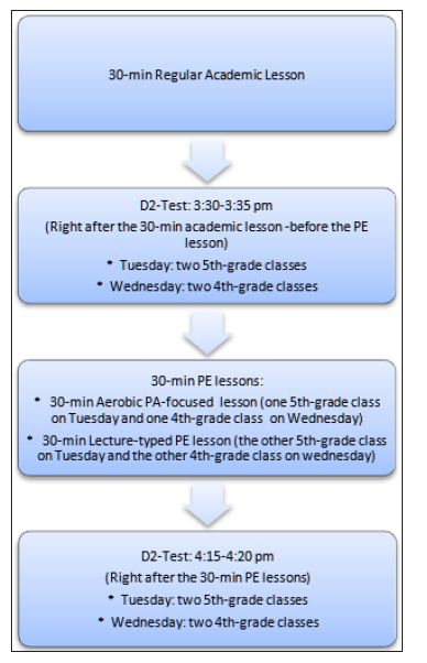
Data CollectionPre-test: Figure 1 shows the timelines for the students in the fourth- and fifth-grade classes to take pre-test (after the 30- min regular academic lessons), 30-min aerobic PA or 30-min PAbenefits lecture, and post-test. One week before the pre-test, each teacher who was trained in the protocols of the test explained the directions for taking the test to the students. The students were then asked to practice the two lines of the test according to the standardized directions for taking the test in order to ensure all students understand the testing procedures. On each of the test days in the afternoon during a school day, the participating students took the d2 Test of Attention in their respective classroom. After completing the pre-test, the students turned in their testing sheets to their PE teachers.Figure 1: Changes in TN for the two groups.
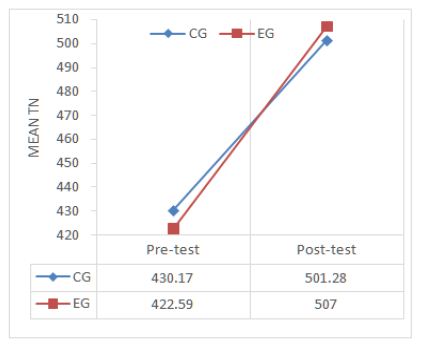
Intervention: Right after the pre-test, the intervention students started the 30-minute aerobic PA-focused PE lesson. The lesson consisted of 5-min warm-ups (jogging around the track, followed by stretching exercises), 16-min relay race running and running through obstacle courses on a regular track and field court (i.e., 400-meter lanes), and 4-min cool down stretching exercises, in addition to 5-min class organization and instructions. In contrast, the comparison students participated in the 30-min lecture on benefits of PA and appropriate methods of PA in a regular PE lesson. During the lecture, the PE teacher showed the pictures in relation to the benefits of varying exercises, asked students to conduct mock interviews with their peers about their PA participation and methods they used, and summarized the benefits of PA and appropriate methods of engaging in PA.Post-test: After finishing the lecture-typed PE lesson, the control students performed on the d2 Test while following the same testing procedures as the pre-test in their classroom. For the intervention students, right after the aerobic PA-focused PE lesson, they returned to their classroom and took the d2 Test of Attention while following the same testing procedures as the pre-test as well. The PE teachers collected the testing sheets from their students.

Outcome Measure- d2 Test of AttentionThe d2 Test is standardized paper and pencil letter-cancellation test that measures neuropsychology performance of the students in the areas of sustained and selective attention as well as concentration [20]. It consists of 14 lines of 47 randomly mixed letters “d” or “p” with 1-4 dashes arranged individually or in pairs above or below the character. The students are instructed to scan the characters and mark only the letter “d” with 2 dashes either above or below, or one dash on top and one on the bottom within 20 s per line for a total of 4 min and 40 s to complete the test. Distractors come in two forms, more or less dashes above or below the “d”, and the letter “p”… .d d d.. .Three parameters of the d2 Test for sustained and selective attention and concentration were used for data analysis in this study. They area. TN: the total number of items processed within the d2 test. TN is a quantitative measure of the processing speed;b. E%: the sum of omission and commission errors divided by the total number of items processed. E% is a measure of accuracy and thoroughness; andc. CP: the total number of correct responses minus commission errors. CP is an objective measure of attention span and concentration ability.Values of both TN and E% are subject to learning effects, while CP is viewed as independent from manipulative [20,21]. The d2 Test had high test-retest reliability coefficients for all parameters, ranging from .95 to .98 [21]. The d2 Test has been proven to be an internally valid measure of scanning accuracy, speed, discriminant validity and fluctuation across trials.21The test-retest reliability of the d2 Test has been shown to be very high (.95-.98) for all parameters [20,21]. Test values for criterion, construct, and predictive validity have been stable over the course of 23 months after the initial testing [20,21].

Data Analysis

Descriptive statistics of TN, E%, and CP at pre- and posttest were computed for each group. Percentage improvement in TN, E%, and CP (percentage improvement= (Mt2-Mt1)/Mt1 x 100)) from pre- to post-test for the experimental group and the comparison group were computed. In addition, independent t-tests were performed to compare the pre-test in the three dependent variables between the two groups. Also, descriptive statistics of the percentage improvement in the three dependent variables were conducted for each group. A 2 x 2 mixed factor analysis of variance with repeated measures was used to examine differences between pre- and post-test (within subjects) and differences between the experimental group and the comparison group (between subjects). ANOVAs with repeated measures were conducted separately for the TN, E%, and CP. When the assumption of sphere city was violated, the Greenhouse-Geyser correction was reported. Post hoc contrast (Bonferroni adjustment) was used to test effects between the two groups. The η2 was calculated as the effect size of ANOVAs. We did not include the gender into the analysis because previous validation studies showed no gender differences in each parameter of the d2 Test. Statistically significant level for all analyses was set at p < 0.05. All data were analyzed using SPSS version 24.

Results

Descriptive Statistics

Table 1 presents the descriptive statistics of pre-and post-test in d2 Test of Attention between the experimental group (EG) and the comparison group (CG) as well as the percentage improvement in the three dependent variables from pre- to post-test between the two groups. At pre-test, the CG’s mean scores in TN (processing speed) and CP (focused attention) were slightly higher than the EG’s mean scores. The higher numbers of TN and CP indicate the better performance in attention and concentration. The CG’s mean score in E% (accuracy and thoroughness) was similar to the EG’s mean score. The lower scores in E% represent the better accuracy. Further, independent sample t-tests revealed no significant mean differences between the two groups in TN, E%, and CP (t=.360, df=78.40, p=.720; t=.389, df=90.94, p= .682; t=.919, df=80.44, p =.895) at p>.05.

Table 1: Means and standard deviations for three parameters of d2 Test at pre- and post-tests and for improvements between the experimental and the control group.

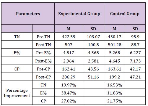
At post-test, the EG’s mean scores in TN and CP were higher than the CG’s mean scores. Also the mean score in E% of the EG was much lower than that of the CG. Regarding the percentage improvement in the three dependent variables from pre- to posttest for each group, both groups showed increased mean scores in TN and CP and decreased mean score in E%. The results indicated the two groups’ performance in TN, E%, and CP was improved from the pre- to the post-test. However, the EG’s percentage improvement in E%, CP, and TN were greatly higher, moderately higher, and higher than the CG’s percentage improvement in the three dependent variables.

ANOVA with Repeated Measures

Table 2 illustrates the results of 2 (pre-test vs. post-test) x 2 (CG vs. EG) ANOVA repeated measures. The results showed a significant effect of time, but no significant effect of group for the three dependent variables (TN, E%, and CP). The results indicated that all participants in both groups significantly improved their d2 test performance and decreased the number of errors from the pre-test to post-test with a relatively large effect sizes (Table 2), and (Figure 1-3). Further, as presented in (Table 2), the repeated measure ANOVA revealed no significant interaction between time × group in TN and E %. However, (Figure 2) shows a relatively large degree of changes from pre- to post-test for the EG compared to the CG. Also, (Figure 2) displays relatively large different mean scores in E% between the two groups at the post test. Echoing the results, (Table 2) shows EG had a larger percentage improvement in E% compared to the CG. In addition, the significant level of the time x group intervention in CP was close to p<.05. The results indicated that the EG showed a higher change in performance of CP (focused attention) from pre-to post-test which compared to the CG (Figure 3). Furthermore, at the post-test, independent t-tests indicated that there was no significant performance improvement in TN, E%, and CP between the two groups (t=.015, df=75.181, p>.05; t=-.003, df=53.545, p>.05; t=.480, df=87.731, p>.05).

Figure 2: Changes in E% for the two groups.

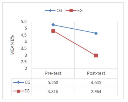
Figure 3: Changes in E% for the two groups.
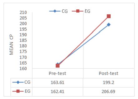
Discussion

This study investigated whether the afternoon PE lessons could improve attention and concentration in school-aged children. Participants took d2 Test before (after taking the 30-min regular academic lesson) and after participating in either 30-min aerobic PA-focused PE lesson (experimental group) or 30-min lecture on benefits and methods of PE lesson (comparison group) late in the school day. Confirming the first hypothesis, the students scored significantly higher in TN, E%, and CP immediately after the 30-min PE lessons (30-min aerobic PA-focused PE lesson and in 30-min lecture-typed PE lesson), compared to the pre-test (immediately after the 30-min regular academic lessons). Our results supported previous consistent findings showing that acute bouts of aerobic PA (e.g.,10-min, 12-min, 15-min, 20-min, and 50-min) immediately enhanced attention and concentration performance in school-aged children [1,2,5,7,8].

Furthermore, the results partially supported the second hypothesis that students in the 30-min aerobic PA-focused PE lesson will show higher positive changes in attention and concentration performance than students in the 30-min lecture-typed PE lesson from pre- to post-test. In this study, the students in the 30-min aerobic PA-focused PE lesson showed a higher degree of better processing speed, accuracy, and concentration performance and a higher level of percentage improvement in the three parameters compared to the comparison group, although did not reach a significant level of p<.05. Partially supporting the present result, Gallotta et al. [1] found that students in the two groups: the 50- min aerobic PA-focused PE lesson and 50-min academic lessons about humanistic subject matter showed a higher level of attention before and after the classes compared to the students in the 50-min coordinated exercises-focused PE lesson.

Previous studies have explored potential physiological mechanisms for generating beneficial acute effects of aerobic PA on attention and concentration in school-aged children (20-30). Studies found that acute bouts of aerobic PA increased cerebral oxygenated blood flow, alpha activity of the precuneus [23,24]. Also, acute bouts of aerobic PA activated connections of the neuro networks between cerebellum and frontal cortex and elevated levels of Brain-Derived Neurotrophin Factor (BDNF) in the blood [5,25,26]. These increased levels play important roles in improving brain functions such as attention span, concentration, and working memories, and visual-spatial abilities [24-29]. However, the previous studies have not examined whether the enhanced acute effects of the aerobic PA on students’ attention and concentration are implied to late in the school day.

The unique finding of this study showed that the fourth- and fifth- grade students demonstrated faster working speed, better accuracy, and higher concentration scores immediately after participating in 30-min aerobic PA-focused PE lesson late in the school day, compared to immediately after taking the regular academic lesson in the afternoon. According to the lesson content described in the methods section, the students were engaged in vigorous intensity level of aerobic PA in the 30-min aerobic PAfocused PE lesson. Our finding was contrary to previous studies reporting a positive effect of moderate intensity level of acute PA on attention in school-aged children [2,5,19].

The inconsistent finding might be related to the intervention conditions which occurred at different timings of the school day. In the previous studies, students taking the d2 Test before and after acute bouts of PA with varying intensity levels and durations took place in the morning of the school day. Previous findings showed that a short bout of moderate intensity level of PA produced positive beneficial effects of attention and concentration for school-aged students in the morning of the school day [2,5,19]. However, our unique finding suggests that participating in 30-min aerobic PAfocused PE lesson has pronounced, facilitating effects on attention and concentration in school-aged children in the afternoon, especially late in the school day.

Another unique finding of this study indicated that the students showed significantly higher performance in attention and concentration immediately after the 30-min lecture-typed PE lesson compared to immediately after the 30-min regular academic lesson. This finding might be related to the lesson content and instructional methods. As described above, during the 30-min lecture-typed PE lesson, the students focused on learning a variety of PA methods and the benefits of PA participation. In order to make the lesson content meaningful and authentic to students, the PE teachers encouraged students to take active responsibility for their learning process through engaging them in conducting peer interviews, sharing their findings of the interviews about benefits of PA and methods for PA from students’ perspectives and experiences, and highlighting the shared findings related to the lesson focus.

These inquiry-based, students-engaged active learning strategies might spark students’ interests in, enhance their motivation for, and increase their enjoyment of learning the lesson content. Motivation and positive mental state play roles in boosting the level of arousal, which facilitates cognitive responses such as attention and concentration performance [30,31]. Similarly, Janssen et al [5]. Found that the students showed a significant higher attentional performance after participating in the 15-min story-telling lecture, compared to the students after attending the regular academic tasks (mathematical problems). Also, in line with the present finding, previous studies evidenced that the interactiveand active-engagement instructional methods are more effective than traditional lectures for gaining students’ attention to the learning process, focusing on the task engagement, and staying focused on the instructional process [32-35]. This study suggests that the lecture-typed lesson in which the students are engaged in active, responsible, and cooperative learning processes is critical to facilitating students’ attentional performance even late in the school day.

In general, the strength of this study is to use the schoolscheduled PE lessons as the treatment conditions to examine their acute impacts on students’ attention and concentration late in the school day compared to the regular school-scheduled academic lesson. Further, the study suggests that in order for students to pay attention to instructions and maintain task-engagement, school administrators may intentionally schedule a regular PE lesson in the afternoon whenever possible, or may alternate a morning PE lesson with an afternoon PE lesson per semester for students. Also, PE teachers may incorporate more aerobic-typed PA that is developmentally appropriate for their students’ fitness and skill levels into their afternoon PE lessons. In addition, school administrators may use professional development activities to provide their teachers with opportunities for learning and implementing innovative instructional strategies such as active and interactive learning, cooperative learning, collaborative learning, and problem-based learning. Teachers may deliberately and appropriately incorporate these instructional strategies into learning and teaching processes to ensure students to be active learners instead of passive learners. The suggestions resulted from this study are potentially feasible for schools to implement in school settings.

It is worth to note that this study has three limitations. First, this study did not use passive rest as the control group. The main reason is that this study took place in a school setting. If we asked students to simply rest for an entire academic lesson merely for the sake of this study, this would interrupt the school’s regular academic schedules, so that the school administrators and teachers as well as parents would be hesitant to allow their students to participate in this study. Second, this study did not objectively measure students’ intensity level of aerobic PA during the 30-min aerobic PA-focused PE lesson, although shuttle running and running through the obstacle courses continuously are good example of vigorous intensity aerobic-typed PA. Due to different levels of students’ aerobic fitness which is an important moderator impacting PA and cognitive functions [25,36,37],future studies may objectively measure students’ intensity levels in different experimental conditions using accelerometer to examine acute effects of aerobictyped PA with varying intensity levels on students’ attentional performance late in the school day.

Third, this study did not examine any delayed effects of the 30- min aerobic PA-focused PE lesson and the 30-min lecture-typed PE lesson on students’ attention and concentration in the afternoon of the school day. Chang et al. [38] found significant acute PA effects on attentional performance 11 to 20-minute after the PA intervention. Further, Schmidt et al. [6] reported students’ showing pronounced improvement in attention and concentration 90-min after the 45- min coordinated motor skills PE lesson in the morning of the school day. As the acute PA produces positive delayed effects on attentional performance, future studies may examine varying delayed effects of the 30-min aerobic PA-focused PE lesson on students’ attentional performance in the afternoon of the school day. The positive findings would further evidence that implementing afternoon aerobic PA-focused PE lessons is instrumental to keeping students stay-focused in the next regular academic lesson.

It was concluded that after participating in both the 30-min aerobic PA-focused PE lesson and the 30-min interactive lecturetyped PE lesson late in the school day, the students had a large size of improvement in attention and concentration, compared to after attending the 30-min regular academic lesson. However, there was no significant different degrees of improvement in the processing speed, accuracy, and concentration between the students in the aerobic PA-focused PE group and the students in the interactive lecture-typed PE group from before to after the PE lessons, although the experimental students showed a higher percentage of improvement in the three parameters compared to the comparison students. This study suggests that a regular school-scheduled PE lesson that focuses on aerobic-typed PA is instrumental to improving students’ attention and concentration, especially in the late school day.

For More Articles: Biomedical Journal Impact Factor : https://biomedres.us

Journals of Biotechnology

Clinical Relevance of Type Specific Clays

Abstract

Today’s life style and food habits are aligned to readymade fast food with challenges in human health safety causing ailments and disorders such as blood pressure, blood sugar, constipation, gastric, weight gain, mental strain etc. They are all responsible to imbalance the life styles. As a result, humans are suffering from varying types of health hazards. Type specific clays are tested at different levels for recovery from such ailments. However, many of such tests are not validated medically, but evidences, being the witnesses of truth, necessitate systematic investigations of type specific clays in soils for their clinical relevance for curing of human ailments and disorders. Clay eating followed by detoxifying potential of clays deserves attention for clinical validation. Ethiopian soils are rich resources for type specific clays that could be tested for clinical uses.

Keywords : Clays; Clinical relevance; Human health; Clay eating; Detoxifying potential

Introduction

Clinical approach refers often to the techniques being applied to the ailment, sickness, disorder and problem in human bodies and requires medical interventions for the purpose of elimination of symptoms in order to restore human health and promote body functioning. Clays have been used for cure of skin infections since long past. Recently, Lynda and Shelley (2010) [16] presented a review report in which the clinical use of French green clay (rich in Fe-smectite) for healing Buruli ulcer, a necrotizing fasciitis (‘flesh-eating’ infection) caused by Mycobacterium ulcerans is highlighted. The clays are interesting as they may reveal an antibacterial mechanism that could provide an inexpensive treatment for such skin infections (Lynda and Shelley 2010) [16] . A review on Soil Science vs Science for Medicine by Mishra and Richa [1] revealed that clay eating is associated historically with treatment for cholera and bacterial infections. Clay tablets were used widely across the Mediterranean as well as European territories in certain religious cause besides curing the poisoning and the plague. However, the clay tablet was used by Roman Catholic Church and was listed in pharmacopeia as late as 1848. The use of eating clay has been studied in America, Sweden, Africa, Indonesia and Australia. In India and many other countries, however, knowledge on soil and clay eating is scanty. The rates of pregnant women eating soil or clay in African countries range approximately from 28% in Tanzania to 65% in Kenya, where clay is selectively identified and sold in markets. They collect it from termite mounds being rich in minerals and eat at an average of 30g daily. Important contributions as recorded herein include the reports of Lynda and Shelly (2010) [16] , Wiley and Solomon [2] and Wilson [3]. Present paper is an attempt to understand the clinical relevance of type specific clays in different soil groups across the world including India as well as Ethiopia and other African countries.

Background

Lynda and Shelley (2010) [16] had arranged type specific clays for clinical uses as follows:

1) Bentonite-type clay has been used to treat infections, indigestion, and other medical problems by both applying clay paste externally to the skin and by ingesting as solution. Bentonite has been prescribed for many dermatologic formulas. Montmorillonite is the main constituent of bentonite.

2) Attapulgite or palygorskite is very absorbent clay, somewhat similar to bentonite. When used in medicine, it physically binds to acids and toxic substances in the stomach and digestive tract. For this reason, it has been used in several anti-diarrheal medications and also as detoxificant.

3) Kaolinite being low activity clay is not as absorbent as most clays used medicinally. It has a low shrink-swell capacity as well as low cation exc hange capacity. It is often called ‘white cosmetic clay’. However, it is used mainly for oily skins.

Mishra and Richa Roy [1] reviewed the role of soils and clays in protective medical treatments to restore human health. Type specificclays are often used on the skin to heal eczema, dermatitis and psoriasis, during bath as a soaking liquid to remove toxins, enrich cells to receive more oxygen, facilitates to alkalize the body and gives relief against digestive problems like constipation, promotes immunity by killing harmful bacteria and viruses, improves teeth, purifies water and is useful as a baby powder alternative as well as for cleaning hair and face. Multani Mitti in India is commonly used for cleaning hair. Advancement in transmission electron microscopes (TEM), field emission scanning electron microscopes (FESEM), atomic force microscopy (AFM), and secondary ion mass spectrometers (SIMS)] have facilitated to undertake investigation on surfaces of clays and similar nano-scale minerals. Efforts are being made to make a clay antibacterial that has not only the potential applications in medicine, but can also contribute to the general understanding of antibacterial mechanisms for permanent cure. Besides, there is emerging interest in geophagy [3,4] to elicit a curing response in humans through ingesting the easily available materials that may physically soothe an infected and inflamed gastrointestinal lining (Lynda and Shetty 2010) [16]. Also, clays are used externally to adsorb toxins from skin and provide heat to stimulate circulation for rheumatism treatment (Lynda and Shetty 2010[16], Gomes et al. [5]. Historical evidences of using clays are available with Aristotle (384-322 BC) Mahaney et al. [6]. The cure of intestinal ailments by ingestion of volcanic muds is also noticed (Lynda and Shetty 2010[16]). However, evidence indicating antibacterial properties of natural and synthetic clay minerals Herrera et al. [7]; Lynda and Shetty 2010 [16]; Wilson [3] lacks the mechanisms responsible for chemical interaction occurring at the clay mineral–bacterial interface, and that inhibit bacterial growth.

Clay for Detoxification

Soil and clay eating may be dangerous if anthrax bacteria and others resist for years in the soil. Medical science believes that eating soil or clay would reduce their hunger and sometimes causes infection. Soil is considered to pose hazard because of

a) chemical contamination, especially heavy metals

b) harmful bacteria, mostly from sewage or manure

c) Parasites, especially roundworms from pet or wildlife faeces.

But, taking a pinch of well-tested clay or soil may be a benefit to the immune system. Some reports indicate that normal children of one and three years of age often eat soil, while aged children may continue to eat soil if there is delay in their growth. A dose of 500 mg a day of soil or clay consumption is considered normal in children up to 3 years old as reviewed by Mishra and Richa Roy [1].

Clay may be used externally as well as internally. It is antiseptic to prevent decay or putrefaction, promotes wound healing, relieves and prevents inflammation, cleans cancer cells (anti-carcinogenic), softens and soothens the skin (emollient), refrigerant cools and reduces body temperature (refrigerant) besides improving skin texture (cosmetic). When used internally, the clay such as bentonite acts as a detoxifier, which can absorb heavy metals like mercury, arsenic, lead, and tin. Besides, it provides minerals and trace elements. Being colloidal in nature, it reduces or even eliminates toxins and harmful ingredients from body. Metallic ions of silver, copper, and zinc have inhibitory and bactericidal effects.

The zeolites with their immense power of absorption as well as adsorption indicate strong affinity for oxidized silver ions and thus form silver exchanged zeolites, which have shown antibacterial potential against aerobic and anaerobic Gram-negative and Grampositive bacteria pathogens including Pseudomonas aeruginosa, Porphyromonas gingivalis, Prevotella intermedia, Staphylococcus aureus, Streptococcus mutans, and Streptococcus sanguis and could be used in dental applications Hotta et al. [8], Kawahara et al. [9], Matsuura et al. [10].

Copper-loaded vermiculite is reported to have better antifungal activity besides inhibiting the growth of E. coli Li et al. [11] Clay or soil eating by and large has revealed some potential for digestive and nutritional benefits. However, the most remarkable evidence is its relevance in detoxification. It is known that ingestion of clays by animal species like rats, birds, parrots etc enables a wide variety of foods free of suffering from any toxic effect. Today, human beings are suffering from variety of ailments caused mainly by some types of toxicity/contamination Mishra and Richa [1].

Clays belong to a crystalline shape with hexagonal networks of silicon-oxygen tetrahedron that provides a large surface area with charged sites that cause bonds to capture charged ions and certain toxins. The well known colloidal properties following the existence of hydroxyl ions within the clay structure may promote its ability to bind and exchange other metals, adsorb water and organic compounds. In low concentrations (4 μg/ml), silver ions produced inhibitory and bactericidal effects with no obvious toxic effect on human blood cells Berger et al. [12]. Elevated levels of copper can inhibit the growth of some microorganisms and exhibit bactericidal activity Gordon et al [13]. The use of copper-coated products or copper alloys has been proposed for surfaces exposed to human contact to reduce the transmission of infectious microbial agents. Other metallic oxides, including zinc oxide, magnesium oxide, and calcium oxide, have antibacterial activity with demonstrated effectiveness against E. coli and S. aureus Sawai [14], Mishra and Richa Roy [1,15].

Clay crystals carry a negative electrical charge as hydroxyl and oxide ions, while impurities or toxins or even bacteria carry a positive electrical charge and during exchange with clay, the positively charged ions are attracted to the negatively charged colloidal surfaces of the clays. The clay colloids thus get electrically satisfied and hold the positive ions until human body could remove both through excreta in toilet and accordingly, clay could maintain its colloidal integrity within the human body without any assimilation or break down. The clay may expand in case of montmorillonite or bentonite and the substance could be absorbed by filling the space between the stacked silicate layers. Thus, clay minerals possess an inner layer charge that behaves and acts like an absorbent and may absorb and bond even with elements showing toxic nature.

Evidence that witnesses the truth of soil/clay eating

As a mark of the first celebration of the World Soil Day on 5th December, 2014, the Department of Soil Science at Bihar Agricultural University, Sabour in India discovered Karu Paswan of more than 100 years of age at a village of Babupur (Bakharpur) in Pirpainti Block of Bhagapur District in Bihar (India), who has been daily eating a type specific soil of Ganga flood plain for the last 60 years Mishra and Richa [15]. He is non-vegetarian with normal food diet, but daily eats almost 200 g of soil additionally. He has two daughters and two sons. At this old age, he has black hairs and walks on foot for 10 to 12 km daily. However, soil sample collected was analysed (Table 1), though further analytical and medical reports are awaited Mishra and Richa Roy [1,15].

Table 1: Comparison of power densities of different ambient energy sources.

Rays of New Hopes in Ethiopia

Ethiopia is a land of soil museum covering appreciable areas under dominant clays and clay minerals of volcanic origin namely montmorillonite, attapulgite, zeolite and vermiculite. In patches, kaolin deposits are also observed. Their clinical relevance needs to be established in near future in order to authenticate their medical uses in days to come Mishra and Richa Roy [1,15].

Conclusion

Soil and clay as the protective medical applications for restoring human health have their long history, which needs to be refined in terms of clinical relevance. Necessity is the mother of invention and challenges often open the door of opportunities. Let’s not forget that the soils across the globe suffer from crucial management risks, even though they possess in their type specific forms like clays certain unique potential for medical uses. Soil as a natural resource helps in getting food, water and even air. However, the truth of evidence as recorded is enough to trust on a bare fact that soil or clay may be uniquely suitable medically to cure a number of ailments, which are becoming common to everyone now-a-days across the world.

This is now time for researchers to come forward to establish the truth in a big way in close association with soil science professionals, who could specify the suitability of soil and clay for medical uses. Soil is thus not only meant for survival and nourishment of human beings, but for protective medical treatment also.

If “Yoga” has been accepted as a symbol to sustain the human health, the clay may be used in clinical application for restoring the human health. However, protocol for clinical uses of type specific clays and even soils is priority and the soil science must address all key issues in course curriculum that suit medical as well as clinical applications.

For More Articles: Biomedical Journal Impact Factor : https://biomedres.us

Journals of Toxicology

Energy Harvesting: A Toxic Free and Reliable Power Source for Implantable Microsystems

Editorial

The implantable medical devices require constant power source and for this requirement, batteries have been developed that has enabled the successful deployment of the devices and their treatment of human disease. While the functional requirements for the batteries used to power these devices vary with the type of device and therapy, there are some characteristics that are demanded by all applications. The batteries must have high life span, be safe during installation and use, have predictable performance that can be interrogated to provide state of discharge information and be highly reliable [1]. Additionally, the batteries must have high volumetric energy density to enable the design of small devices that minimize discomfort for the patient. Thus, long term stability during use, predictable performance, high volumetric energy density and outstanding reliability are key characteristics that define successful systems for biomedical implants [1]. Since the introduction of first cardiac implant in 1972, a variety of battery systems have been developed for implantable medical devices that utilize lithium metal anodes with cathode systems including iodine (Li/I2) [2-4], manganese oxide (Li/MnO2) [5,6], carbon monofluoride (Li/CFx) [7,8], silver vanadium oxide (Li/SVO) [9,10] and hybrid cathodes using both carbon monofluoride and silver vanadium oxide (Li-CFx-SVO) [11,12]. The specification of these batteries is presented in Table 1.

Table 1: Comparison of Different Batteries Used in Implantable Medical Devices.

This range of batteries provides the appropriate power levels as demanded by a specific medical device varying from microampere to ampere level currents. Successful development and implementation of these battery types has helped enable implanted biomedical devices and their treatment of human disease. Among these, Li/I2 has proved to be safer and more reliable than others for use in pacemakers and hence used widely in the last 4 decades [13]. However, these batteries suffer from limited life span and hence require replacement by surgery which is inconvenient to the patient. Hence an alternate energy source is required to continuously power these implants. One such approach which can address the abovementioned problem is Energy Harvesting [14,15]. It is the process of scavenging out energy from different sources in the ambient environment. Various harvesters are being proposed to harvest energy for implantable devices. Some of the recently reported harvesters can be studied in [15-21]. The form of energy used by the harvester to scavenge the power, defines the type of energy harvesting. There are four main ambient energy sources available viz., mechanical energy due to vibrations and deformations (electrostatic, piezoelectric and electromagnetic), thermal energy (temperature variations and gradients), radiant energy (sun, IR, RF) and biochemical (bio-fuel cells). A comparison of the power densities of these different energy sources is presented in Figure 1 [15,22].

Figure 1: Comparison of power densities of different ambient energy sources.

Although research is being carried out on designing different structures of energy harvesters to generate as much power as possible, there are still some aspects that need to be investigated before finally implanting the harvester inside the body. One such area is the stability of the harvester. For example in electret-based electrostatic harvesters, the electret stability degraded at high temperature and humidity and at higher surface potentials which can reduce the life-span of the harvester. Another aspect that has to be investigated is the toxicological effect of the harvester, if any. But given the trend of the energy harvested by different structures being proposed, the practical application of the harvesters in implantable microsystems as a reliable power source will certainly be a reality.

For More Articles: Biomedical Journal Impact Factor : https://biomedres.us

Journals of Orthopaedics

Current Review; Biomarkers in Diagnosing Periprosthetic Joint Infection

Mini Review

Periprosthetic joint infection (PJI) is one of the most dreaded complications after total joint arthroplasty [1]. There is no gold standard for diagnosing PJI, hence, a clinician who encounters a suspected PJI case, ought to use a combination of tests. All of which, besides their expense can be invasive and even this can’t provide 100% accuracy [2]. Several biomarkers have been introduced that are potentially reliable tools for diagnosing PJI [3]. In this article, we aimed to review the current diagnostic measures of PJI with a special focus on molecular biomarkers.

Synovial Fluid Biomarkers

Synovial fluid biomarkers play an imperative role in the diagnosing PJI. Leukocyte esterase (LE), human α-defensin, human β-defensin, synovial CRP, and cathelicidin LL-37 are namely the biomarkers that have shown promising results. LE is an enzyme that is secreted by the activated neutrophils. It can be detected using colorimetric strip tests via reactions that result in a color change [4]. LE is a readily available and simple test and is now part of the minor criteria of the Musculoskeletal Infection Societydiagnostic criteria for PJI [5]. Tischler et al. [6] demonstrated that the LE strip test has a high specificity, positive, and negative predictive value for diagnosing PJI. Wetters et al. [7] investigated the accuracy of the LE test and reported a sensitivity of 92.9% to 100% and a specificity of 77.0% to 88.8%. The important point is to note that bloody samples cannot be evaluated for the LE test without being centrifuged as the presence of blood can potentially interfere with the colorimetric changes of the test strip [6].

Synovial fluid α-defensin test has shown great sensitivityand specificity for diagnosing PJI, 97% and 96% consequetively [8]. Defensins are 2-6 kDa cationic microbicidal peptides that are active against many Gram-negative and Gram-positive bacteria, fungi, and enveloped viruses [9]. Defensins in mammalians are classified into alpha, beta, and theta categories, based on their size and pattern of disulfide bonding. Alpha-defensins are particularly found in neutrophils, certain macrophage populations, and Paneth cells. Defensins are produced in response to microbial products or pro-inflammatory cytokines. The α-defensin mechanism by which microorganisms are killed and inactivated is not yet fully understood. Nevertheless, it is thought that it causes membrane disruption in microorganisms [10]. The spatially separated, charged, and hydrophobic regions, along with the polar topology of α-defensin, allows it to insert itself into the membranes; therefore, the hydrophobic regions are buried within the interior phospholipid membrane and the cationic sites interact with anionic phospholipid head groups and water. The disruption of membrane integrity and function leads to lysis of the microorganisms [11,12]. Several studies have endorsed the role of the α-defens in test in diagnosing PJI. The α-defensin test provides consistent results regardless of the organism type, Gram staining, species, or virulence of the organism [13].

CRP, which elevates in both the serum and synovial fluid of PJI cases, is a protein that is synthesized by the liver in response to acute inflammation [14]. Parvizi et al. [15] reported a statistically significant difference in the mean of synovial fluid CRP comparing septic and aseptic patients; 40 mg/L vs. 2 mg/L, respectively (p<.0001). The study found a sensitivity of 85% and a specificity of 95% when 9.5 mg/L was considered as the threshold.

Human host defense peptide LL-37 is an antimicrobial peptide that induces mediators such as IL-8 and regulates the inflammatory response [16,17]. Gollwitzer et al. [18] reported that LL-37 has a sensitivity of 80% and specificity of 85%, with an area under the curve of 0.875 for diagnosing PJI.

Serum Biomarkers

Serum markers are favorable diagnostic tools due to their accessibility and low-risk nature. The American Academy of Orthopaedic Surgeons and the International Consensus meeting on PJI currently recommend using serum erythrocyte sedimentation rate (ESR) and C-reactive protein (CRP) as the first line diagnostic work up for patients with suspected PJI. Reports have shown asensitivity of 91% and specificity of 72% for ESR and a sensitivity of 94% and specificity of 74% for CRP [19-21]. Serum ESR and CRP are well-known biomarkers that indicate systemic inflammatory response [22]. However, these markers are elevated with any type of inflammation and/or infection, compromising their specificity for diagnosis of PJI. Recent evidence suggests that PJI with slowgrowing organisms may not result in a fully-fledged physiological response and hence may not result in elevation of ESR and CRP in the serum, raising a concern for the sensitivity of these tests. Furthermore, a study by our group showed that the administration of systemic antibiotics can significantly compromise the results of these laboratory values [2]. It is imperative for clinicians to consider the timing of infection prior to assessing patients’ ESR and CRP results, as these markers are frequently elevated in the early postoperative period. Studies have shown that ESR can be elevated up to 6 weeks after surgery and CRP can be elevated for up to 2 weeks [23].

Procalcitonin (PCT) is a serum biomarker that elevates in the presence of bacteria. Bottner et al. [24] measured serum levels of several biomarkers including: PCT, interleukin (IL)-6, tumor necrosis factor (TNF)-α, ESR, and CRP in 78 patients undergoing revision arthroplasty for PJI. The sensitivity of CRP and IL-6 were the highest (95%) when the levels were greater than 3.2 mg/ dL and 12 pg/mL, respectively. The authors recommended that combination of CRP and IL-6 could be used as a screening test for PJI. PCT levels (>0.3 ng/mL) were very specific (98%) but had a low sensitivity (33%) for diagnosing PJI. In another study by Hügle et al. [25] authors showed that PCT with a threshold of 0.25 ng/ mL has a higher sensitivity and specificity for diagnosing septic arthritis than CRP, with a sensitivity of 93% and specificity of 75%. This could possibly be rationalized by the fact that PCT is secreted by the mononuclear phagocyte system only when stimulated by lipopolysaccharide. Therefore, PCT can be a useful tool to differentiate between bacterial infections of the joint and other causes of inflammation. Nevertheless, more recent studies claimed that PCT is not a very accurate tool for diagnosing PJI [26,27].

IL-6 is another serum marker that has gained attention for diagnosing PJI. IL-6 is secreted by different immune cells and triggers the excretion of CRP; therefore, it is believed that the IL-6 levels rise much faster than CRP and has been reported to be a sensitive marker for diagnosing PJI, however it also lacks specificity [24,28]. (p-6) Wirtz et al. [29] advocated the role of IL-6, and in their study authors showed that IL-6 is a better indicator for postoperative inflammatory response than CRP in patients undergoing TJA.

There is a dire need for a sensitive and specific serum biomarker for diagnosing PJI and numerous efforts have been made to pursue this goal. Serum D-dimer is another biomarker that has shown very promising results for diagnosing PJI. Shahi et al. [30] in their prospective study showed that serum D-dimer outperforms both ESR and CRP for diagnosing PJI with a sensitivity of 89% and a specificity of 93%.

Modern medicine has entered a new era where molecular biomarkers play an increasingly important role for diagnosis of various conditions. PJI is no exception and new biomarkers hold great promise for it. Efforts should continue to hopefully find a gold standard test for a timely diagnosis of this serious complication.

For More Articles: Biomedical Journal Impact Factor : https://biomedres.us

Journals of Nursing

A Form of Decision –Making Failure in Arabian Nursing Education

Opinion

Late in the twentieth century and, exactly, in 1983, Yarmouk University/ Jordan started its Bachelor of nursing programme. At that time, when the Faculty of Nursing (FoN) had no nursing PhD holders, other than the Dean, nursing students had the opportunity to study a course titled “Bed Nursing Diagnosis”, which has, overtime, changed to become “Health Assessment” course. This is an essential course that is taught all over the world since decades. The nursing curriculum was never based on the medical model.

Thirty years later when, in October 2012, I joined the University of Benghazi, Libya, to teach nursing, I had the opportunity to view the nursing curriculum that was developed by PhD holders who obtained their qualifications from a developing country. The curriculum was introduced in 2007 after approval by the University higher authorities. The curriculum has no “Health Assessment” course; English language was not considered as a unit that students need to learn before they start the nursing courses offered in this language. Therefore, English language, courses were scattered over the four year duration of study. In addition the curriculum suffered fatal errors as it contained various courses that were purely based on the medical model and were taught by medical doctors. Such courses were “General Internal Medicine”, “Special Internal Medicine”, “Forensic Medicine”, “Obstetrics and Gynecology Medicine & Surgery”, “Geriatric Medicine”, “Pediatric Medicine and Surgery”, “Psychiatric Medicine & Mental Health”, and “Community Health Medicine”. The FoN had a “Quality and Performance Appraisal Unit” headed by an academic member of staff who had a Bachelor of Botany degree and who was also the Faculty research representative at the University Council! The Dean of the FoN who is a medical doctor appointed himself as the Head of Scientific Affairs!

The question that poses itself is: What is going on in the twenty first century with regards to nursing education in Libya? To answer this question in such a way that would help the reader understand why nursing in Libya is substantially lagging behind the world, the facts reported above would be discussed in some detail.

University administrators and decision makers might not be aware that there are huge variations in the quality of nursing education programmes across institutions of higher education in the world; many programmes in the developed countries are much more advanced than those offered in the developing countries. Therefore, the said University and may be others take inappropriate decisions when recruiting teaching staff with low academic capabilities and giving them responsibilities that they cannot fulfill. Evidently, the price of this is, in part, a poor nursing curriculum confirming that teaching faculty might be unaware of what is going on in the world of nursing education, and may not be willing to learn from the experiences of others. The visible landmark of administrative corruption which is evident in the appointment of individuals in positions they are unqualified for complicates the picture further. Nevertheless, such administrative corruption is not unique to the Libyan context; it does affect other institutions of higher education in the middle east region.

With this faulty system in place, the consequences are awesome. Concerned Libyan universities waste massive resources to produce poor outcomes. Nursing students learn little nursing if any; they waste a considerable amount of time and effort on irrelevant medical courses. During my work at the said University, I conducted in February 2014 a medication calculation test for six out of nine nursing graduates who were appointed as teaching assistants because they were the “best”. All of them failed the test which is an essential component of nursing practice! Simply speaking, they did not learn nursing. Therefore, graduates of such programmes would not be in position to practice true nursing and would not, successfully, carry out their roles and responsibilities like their counterparts across the world. Meanwhile, patient safety will be threatened.

To rectify this situation, the said Libyan university and all concerned others need to re-consider their recruitment policies. Like many universities in the region, they need to recruit graduates who obtained their qualifications from developed countries, andbe selective when recruiting applicants who obtained their higher qualifications from developing countries.

Administrative corruption and responsible decision making never meet. This corruption has to, sooner or later, come to an end. Academic and administrative positions must be occupied by qualified and competent staff if progress is to be made. If existing laws do limit the universities ability to achieve this important goal, they need to be amended.

Education is a national security issue and must be viewed as such. Personal relationships should have no place when it comes to education. Ensuring that the right person, with the right skills and experience, is in the right place remains a vital issue that decision makers should always think of and never sacrifice.

For More Articles: Biomedical Journal Impact Factor : https://biomedres.us

Journals on Dentistry

Creating State or Regional Dental Registries to Identify Pediatric Dental Abuse or Neglect

Public Policy

In 1967, the American Dental Association published an editorial noting dentists’ infrequent reporting of cases involving child abuse. Subsequently, the ADA adopted the Principles of Ethics and Code of Professional Conduct along with an official policy addressing child abuse [1,2]. Our current child welfare services, including services for dental neglect and abuse, has a genesis dating back to the 1960s following the publication of “The Battered Child Syndrome” by Dr. Henry Kempe and colleagues and which focused primarily on physical abuse, and subsequently on sexual abuse [3]. Because the evolutionary development of legal responses to pediatric dental abuse and neglect has never been a primary focus of legislatures [4], the recognition by dental professionals of their role as mandatory reporters has been somewhat slower than recognition by other health care professionals serving the medical needs of children [5]. Today, dental professionals are clearly recognized as mandatory reporters of oral injuries indicating possible child abuse or child neglect [6].

Dental neglect may occur when a parent or child’s main adult care provider has been properly alerted by a health care professional regarding the nature and extent of the child’s dental condition, the specific treatment needed, and the mechanism of accessing that treatment [7]. Dental neglect may manifest as cavities in baby teeth, “rotting teeth,” gum disease, gingivitis, failure to follow through with agreed-on treatment regimens, communication deficiencies or inability to speak, and lack of functionality due to complications from tooth decay, with tooth decay being the single most common— and preventable—chronic childhood disease [8]. According to the Child Abuse Prevention and Treatment Act of 1974, child abuse and neglect is defined as “at a minimum, any recent act or failure to act on the part of a parent or caretaker which results in death, serious physical or emotional harm, sexual abuse or exploitation; or an act or failure to act which presents an imminent risk of serious harm [9].”

Michael Wald of Stanford Law School has noted that “[n]eglect and poverty have always been closely linked,” and although neglect has not substantially decreased in twenty years and served as a basis for child protection service interventions, researchers and practitioners have asserted for over 40 years that there is a “deep failure of policymakers to acknowledge the problems in ‘neglectful’ families, and take the steps necessary to meet the needs of their children—the ‘neglect of neglect [10]’”

The issue to address is how to respond and attempt to prevent pediatric dental abuse and neglect? Pediatric dental abuse is often the easier condition for dental professionals to identify, but dental neglect is perhaps more problematic in many instances [11]. If a parent or caregiver obtains a dental treatment plan, but fails to follow through with the child’s treatment, how would the dentist be made aware in the event that the parent or caregiver simply does not bring the child back to the same dentist? Currently, there is no mechanism by which dentists might otherwise be made aware that the patient they are treating has previously been diagnosed and provided with a treatment plan that has been ignored or not complied with by the parent or caregiver. Noncompliance with children’s dental treatment plans may constitute acts of legal child neglect. Such a case triggers the mandatory reporting requirement imposed on dentists and dental care providers.

One possible solution to this recurring scenario would be to establish either statewide or regional digital encrypted registries of dental protected health information (“PHI”). Encryption [12] processes for data should be consistent with the guidelines adopted by the National Institute of Standards and Technology (NIST) [13]. The registry would allow dental providers to post the patient’s treatment plan in a manner whereby other dental professionals might be able to access the information without compromising the medical privacy rights of the patient as [14] defined in the Health Insurance Portability and Accountability Act of 1996 [15] (“HIPPA”). HIPPA provisions do not “inhibit reporting of child abuse and neglect [16].”

A digital data treatment registry based upon the Medicaid billing procedure in which teeth requiring treatment are registered would provide a method for dental care providers to ensure that those patients with pre-existing treatment plans have been followed. In the event that the patient has such a treatment plan recorded in the registry filed under the dental license number of the dentist who crafted the treatment plan and the patient’s contact information, determining whether a child dental patient has had his or her dental needs neglected would be a matter of cross checking the information contained the regional or state registry [17]. Currently, no such registration system is utilized, and those dentists who provide treatment to children who may have already visited a dental office, had a treatment plan proposed, but where parents or caregivers have failed to attend to the child’s dental needs, the registry system would help protect the interests of child patients who may be unable to fend for themselves [18].

followed. In the event that the patient has such a treatment plan recorded in the registry filed under the dental license number of the dentist who crafted the treatment plan and the patient’s contact information, determining whether a child dental patient has had his or her dental needs neglected would be a matter of cross checking the information contained the regional or state registry [17]. Currently, no such registration system is utilized, and those dentists who provide treatment to children who may have already visited a dental office, had a treatment plan proposed, but where parents or caregivers have failed to attend to the child’s dental needs, the registry system would help protect the interests of child patients who may be unable to fend for themselves [18].

For More Articles: Biomedical Journal Impact Factor : https://biomedres.us

Journals on Surgery

Bilateral Lumbar Hernias – A Case Report and An Overview of the Current Literature

Introduction

Primary lumbar hernia is a rare clinical entity. So far, around 300 cases have been described in the literature and almost all of them describe repair for unilateral hernias. We present a case of bilateral primary lumbar hernia repair at our institution.

Case Report

A 79 year old lady, who had enjoyed unremarkable health and was fully independent on her activity of daily living, was presented to us with self-detected bilateral loin swellings for one year. The swellings did not produce any symptoms of pain or discomfort and she denied any history of trauma to the region or symptoms of cough, constipation or urinary outflow obstruction. Physical examination revealed her Body Mass Index (BMI) to be 19.1kg/ m2. Abdominal examination found a non-distended abdomen and there was absence of surgical scars or ventral hernias. Bilateral lumbar protrusions were detected on upright (Figure 1), but they were completely reduced in prone position, which was compatible with lumbar hernias. Computer tomography (CT) scan demonstrated bilateral lumbar hernias just below the twelfth ribs. Retroperitoneal fat was seen inside the right hernia sac and a segment of descending colon was seen inside the left hernia sac (Figure 2). She later consented to an elective repair of her bilateral lumbar hernias under elective settings.

Figure 1 :

Figure 2 :

Perioperative Management

She was admitted one day before the operation for assessment by the list anesthetist as well as for bowel preparation. Klean-Prep polyethylene glycol solution (Helsinn Healthcare S.A., Switzerland) was given in case bowel resection was indicated. She started fasting eight hours before the operation. Amoxicillin and clavulanic acid were given by intravenous route during induction of anesthasia. Repair of the bilateral lumbar hernias was performed under general anesthesia with endotracheal intubation. The patient was first positioned in right decubitus for the repair of the larger left side lumbar hernia. Although prone position had obvious advantage of allowing both sides repair to be performed without position change, we decided to carry out the lumbar hernias’ repair one side at a time in their respective decubitus positions in order to avoid the unnecessary risks associated with prone anesthesia [1,2].

Operative Findings

Both lumbar hernias were from the superior lumbar triangle. The fascial defects measured 3x3cm in size over the left side, and 1x1cm over the right side. A segment of descending colon was found in the hernia sac over the left side, and perinephric fat was noted in that over the right side.

Method Of Repair

Due to the rarity of bilateral primary lumbar hernias, as well as the limited experience available in the current literature on laparoscopic approaches in this clinical entity, we decided for open repair as it remained to be simple, safe and effective [3,4]. An oblique incision was made over the left lumbar hernia. The Latissimus dorsi muscle was retracted cranially and the hernia content was reduced. A 7x7cm Prolene polypropylene mesh (Ethicon, USA) was placed in ‘sublay’ manner beneath the lumbar fascia defect. The mesh was fixed by non-absorbable monofilament stitches applied superiorly to the periosteum of the twelfth rib, anteriorly to the internal oblique muscle, and posteriorly to the quadratus lumborum (Figure 3). The wound was then closed in layers after hemostasis had been attained. Following the completion of the left lumbar hernia repair, the patient was turned to left decubitus position and the same procedure was repeated on the right lumbar hernia. The operative blood loss was 30ml and the operation took 138 minutes. Postoperatively the patient recovered uneventfully and diet was resumed immediately after the patient recovered from the effect of anesthesia on the same day. The patient was prescribed ondemand oral paracetamol 500mg every 6 hours for the first three days after the operation. She was fully mobilized on the next day after the operation and was discharged on post op day 2. There was no recurrence detected on follow-up at 6 months.

Figure 3 :

Discussion

Queen Elizabeth Hospital is a tertiary referral surgical center with a catchment population of one million. Our institution carry out approximately 1200 cases of hernia repair yearly, but the incidence of primary lumbar hernia is extremely low accounting for just one case per year (0.09%). Lumbar hernia is a rare condition. It was estimated that only around 300 cases have been reported in the literature [5,6]. However, the incidence of bilateral lumbar hernias was mostly described in the pediatric population, in which case they were classified as congenital and usually associated with other inborn abnormalities [7-11]. Only a few reports have described experience on the acquired-type bilateral primary lumbar hernias, which are found in adults [3,9]. Despite its rarity, acquired lumbar hernias possess specific clinical features. Acquired lumbar hernias account for around 80% of cases [12]. They are classified as primary (spontaneous) or secondary, depending on the existence of a causal factor (previous incision, trauma or infection). Approximately 55% of acquired lumbar hernias belong to the primary type [13]. Primary lumbar hernias are more often found on the left side and in the upper lumbar triangle [14]. They are precipitated by conditions associated with increased intra-abdominal pressure, such as strenuous physical activity or chronic bronchitis.

The commonest contents of a lumbar hernia include retroperitoneal fat, small and large bowel, omentum, caecum, appendix, stomach, ovary, spleen and kidney [15,16]. Nonetheless, these reports include cases of post traumatic or incisional lumbar hernias where the hernial sac may be of substantial size. The diagnosis is obtained by clinical examination, and can be supplemented by CT imaging, where the sensitivity is approximately 98%. Some have also found MRI useful in confirming lumbar herniadefects [15,17,18]. In contrary to the aforementioned, albeit our patient belongs to the acquired primary type of lumbar hernia, she did not have the typical risk factors for lumbar hernias, hence the development of her lumbar hernias was likely a result of anatomical alterations of the posterior abdominal wall caused by aging [6].

Most lumbar hernias increase in size with time, and overall, there is a 25% risk of incarceration and 8% rate of strangulation. It is therefore generally agreed that lumbar hernias should be repaired early [19]. Primary repair is challenging due to inadequate fascia around the defect making tensionless repair difficult [20]. Various primary repair [21], tissue flaps [9,17,22,23] and mesh repairs (including laparoscopic trans abdominal [3,24-27] and retroperitoneoscopic approaches [19,28-30] had been described. In view of the high failure rates observed in primary repair with facial closure, the principle of tensionless repair by the use of prosthesis became more popular in Lumbar hernia repairs. This has led to more recent publications on the use of pros¬thetic mesh to bridge the defect and minimize tension, which was associated with a lower recurrence rate [31,32]. As tension-free methods became more popular, reports on the use of different prosthesis design and modification followed.

Losanoff and Kjossev reported on the use of a cylindrical shape polypropylene mesh plug. It was proven to promote powerful fibroblastic response to repair the wall defect [17]. On the other hand, Armstrong and co-workers reported the use of the Ventralex patch, which combines a layer of PTFE with a smaller polypropylene mesh. This mesh allowed the surgeon to perform with a small incision and to place the mesh within the peritoneum with polypropylene straps abutting the patch to the fascia, and was associated with a quick recovery as well as good long-term outcome [18]. Solaini and co-workers reported the use of a dart mesh (Bard Mesh Dart, small monofilament knitted polypropylene), which was sutured medially to the quadratus lumborum, externally to the internal oblique, and superiorly to the periosteum of the twelfth rib and the serratus posterior-inferior. Like other similar studies, the author observed a quick recovery and no recurrence upon followup at 11 months [33]. These reports have revolutionized the use of a mesh in achieving tensionless repair in lumbar hernias. It was based on this and our centre experience that open repair using prosthesis was selected for our patient as it is simple, safe and effective. We anchored a Prolene polypropylene mesh (Ethicon, USA) in sublay manner with Nylon stitches superiorly to the periosteum of the twelfth rib, anteriorly to the internal oblique muscle and posteriorly to the quadratus lumborum. The sublay or underlay placement of a mesh is scientifically sound as buttressing the mesh posteriorly takes full advantage of the hydrostatic principle of Pascal, where the mesh repair will be reinforced by the intra-abdominal pressure [34]. Advocates of this approach have found success in the repair of different abdominal wall hernias including parastomal [35-37] and other ventral hernias [38-40].

This technique was also recently reported on the repair of lumbar hernias [32,41,42]. Sublay mesh repair is therefore advantageous as it provides tension-free repair and prevents recurrence behind the mesh, especially in primary hernias. This concurs with the post-operative findings in our patient where a quick postoperative recovery, short hospital stay, minimal analgesic demand and no recurrence at 12 months was observed. Placing the mesh in the sublay manner may be equivalent to extraperitoneal hernioplasty as advocated by enthusiasts of the minimally invasive approaches. Minimally invasive surgery has recently become a popular option for repair of lumbar hernias. One nonrandomized study demonstrated that laparoscopic lumbar hernia repairs resulted in lower operative times, hospital stay, pain medication use, as well as recovery time [43]. However, most of the laparoscopic repair was performed on patients with secondary lumbar hernias following either history of trauma or nephrectomies.

The laparoscopic approach can repair the defect at the deepest layer of the posterior abdominal wall, which allows for complete identification and coverage of the entire defect without a large incision. Nevertheless, the acquisition of expertise, the difficulty in fixing the mesh (due to the lack of strong fascia around the hernia defect), and the higher operation cost due to necessity of special instruments for mesh anchorage (e.g. tack and staple devises) may preclude laparoscopic approach becoming the prevalent option in lumbar hernia repairs. Although some authors may define laparoscopic approach as the best option [6], it is the authors’ opinion that it should be reserved for centres where expertise and resources are readily available; after all, the open approach is as safe and effective.

Conclusion

This report describes a successful repair of bilateral primary lumbar hernias using open tension-free sublay prosthesis. Due to its rarity, lumbar hernias especially bilateral hernias should be treated following the principle of hernia repair by a method that has been proven safe and effective in other more commonly performed hernia repairs.

For More Articles: Biomedical Journal Impact Factor : https://biomedres.us

Journals on Respiratory

Oral Medicine: The Awareness of Patients Using Steroid Inhalers on the Increased Risk of Oral Candidosis: A Service Evaluation

Abstract

Objective: To perform an evaluation of the current practice of informing patients on steroid inhalers of the increased risk of oral candidosis (thrush) as a side effect of using the inhaler.

Method: This is a service evaluation in the form of a survey distributed to patients attending the respiratory medicine outpatient clinic at the Manchester Royal Infirmary (MRI).

Results: The evaluation shows that the majority of patients are not informed of the risk associated with commonly prescribed steroid inhalers, and they feel that it is the responsibility of the doctors to warn them about this side effect. Very few patients received advice from their dentist on this matter.

Conclusion: All healthcare professionals are responsible for the well-being of their patients. Dentist should play active role in advising patients on the oral side effects associated with the use of steroid inhalers.

Clinical Relevance

To perform an evaluation of the current practice of informing patients on steroid inhalers of the increased risk of oral candidosis as a side effect of using the inhaler.

Introduction

Asthma and Chronic Obstructive Pulmonary Disease (COPD) are common respiratory diseases affecting 5.4 and 3.2 million people of the UK population, respectively [1,2]. One of the treatments used for these conditions is steroid inhalers [3]. Steroid inhalers suppress the inflammatory reaction in the lung, which reduces the airway hyper responsiveness to allergens and irritants [3,4]. In 2014, 19.4 million steroid inhalers were dispensed in the community [5]. However, a prominent side effect of this intervention is the local suppression of the immune system, a phenomenon which could result in fungal infection [6-8].

A clinical trial comparing patients on steroid inhalers, nonsteroid inhalers and healthy subjects found that using steroid inhalers has a statistically significant effect on the count of oral Candida albican [9]. This was greater and dose-dependent in patients on fluticasone in contrast to those using beclomethasone [9,10]. However, a previous study found no correlation between the type of inhaler or the dose [7]. The risk increases with the concomitant use of broad-spectrum antibiotics, oral steroids and in diabetics [10]. A high C.albicans count increases the risk of oral candidosis, and once candidosis is established, antifungal treatment should be commenced.

The incidence of oral candidosis in patients on steroid inhalers could be as high as 61 % with clinical symptoms and 66 % with subclinical candidosis [11]. Unfortunately, most of those studies were of inferior design with short follow-up time and lack of control group. Few measures have been proposed to lower the incidence of steroid inhaler-induced oral candidosis. These include: the use of spacer [12], dose reduction [13,14] and the use of amphotericin B mouth rinse [9]. However, simple measures such as a mouth rinse with water after using steroid inhalers have a significant effect on reducing oral candida counts in patients on steroid inhalers [13-15]. As healthcare professionals, it is our duty to ensure the well-being of our patients. This includes the prevention and management of potential side effects induced by the treatment we provide.

Therefore, advising patients on steroid inhalers to rinse after using the inhaler and emphasising the importance of regular visits to the dentist should be an integral part of our treatment plan for such patients. Between 2011 and 2013, the British Lung Foundation (BLF) produced a leaflet titled “Steroids and your lungs”. It included information about the risk of oral candidosis and voice hoarseness as side effects of using steroid inhalers. It advised patients to use a spacer, mouth rinsing after use, oral antifungals and drinking fresh pineapple juice as measures to prevent such a complication [16]. Unfortunately, this leaflet was only circulated for a brief period of time before being discontinued. The BLF explained that they review their leaflets regularly, and that due to limited resources, some of the leaflets had to be discontinued. Fortunately, reliable websites have the same information and they are still accessible to everybody [17,18].

However, not all patients access those webpages to read about the condition and its prevention and management strategies. This is a service evaluation aiming to explore the awareness of patients of steroid inhalers and the increased risk or oral candidosis (thrush) as a side effect of using the inhaler. To the best of the authors’ knowledge, no previous evaluations explored this area from the patients’ point of view.

Materials and Methods

The student attended outpatient clinics at the department of respiratory medicine during the project period (January, 2016 to February, 2016). All attending patients were given the invitation letter and offered to participate in the questionnaire. Those patients who agreed to participate were given the questionnaire to complete anonymously and then drop it in the box. To avoid multiple entries from patients attending a follow-up appointment, they were asked if they had participated in the study. If they had or could not remember, they were not included in the evaluation. Statistical analysis was performed using IBM SPSS Statistics (Version 20.0; IBM Corp., Armonk, NY, USA). “Free text” comments from participants were reviewed by the researcher without statistical analysis. Descriptive statistics such as frequency and % were used for the nominal/ordinal data and the mean and standard deviation for the interval/ratio data. As this is a service evaluation, a hypothesis testing was not carried out.

Results

During a period of four weeks, the student attended six outpatient clinics. A total of 234 patients attended those clinics (average of 39 patients per session) both as new referrals or followups. Every patient was offered the questionnaire to complete and there were 203 returned questionnaires (86.7% return rate) and all of them were completed correctly and included in the analysis. Out of the 203 patients, 148 patients (72.91%) used inhalers as part of their treatment. Only patients who were using steroid-based inhalers (n=142) were included in the analysis of warning of side effects of using such inhalers. Most of these patients, amounting to 57.4% (n = 82), were not warned about possible side effects of steroid inhalers, while 6% (n=8) were not sure or cannot remember if they have been warned (Figure 1).

Figure 1 : Have you been warned about possible mouth problem effects as a result of using your inhaler?

Table 1 : What side effect were you warned about?

Table 2 : Advice given to patients to avoid oral candidosis as a result of using steroid inhalers.

The warnings received by the remaining 36.49.6% (n = 52) are summarised in Table 1. Some patients were warned about more than one side effect associated with steroid inhalers. Therefore, the total number of frequencies exceeds that of patients being warned, and the sum percentages exceeds 100%. Some of the patients who were warned about possible side effects of using steroid inhalers did not receive any advice to avoid this side effect, while other patients received different forms of advice. The advice given to patients toavoid oral candidosis is summarised in Table 2. All patients who mentioned that they were advised to use antifungal medication had at least one episode of oral candidosis, which might suggest that they were warned about this side effect after they had the infection.

As patients could have received more than one form of advice, the total number of frequencies exceeds that of patients being advised and the sum percentage exceeds 100%. All of the patients thought that it is the responsibility doctors to warn them about the side effects of the drugs they prescribe. However, some thought that it is the pharmacist’s and possibly dentist’s responsibility in addition to the prescriber’s. Notably, this evaluation shows that only 55.7 % (n = 29) of the patients, who were warned about the side effects of their inhalers were advised by their doctor regarding the side effects of steroid inhalers.

All patients who chose “other” (28 %, n = 16) as a response to the question (who warned you about the side effects?) specified that it was the specialist nurse who warned them. Table 3 shows that some of the patients were treated for oral candidosis since they started using the inhalers (average of two episodes per patient) and the majority of them were not warned about this side effect beforehand and as explained earlier,some of those patients might have been warned about this side effect after they had the infection. No association was found between the condition patients were treated for and suffering from an oral candidosis episode.

Table 3 : A comparison between patients who suffered from oral candidosis since they started using steroi inhalers in terms of warnings they received about the side effect of the inhalers.

Discussion

This evaluation shows that there is a deficit in the doctors’approach to advise patients on the side effects of steroid inhalers they prescribe. It also illustrates the important role played by other healthcare professionals (e.g. pharmacists and nurses) in warning patients about the side effects of the drug they are using. Those patients who had an episode of oral candidosis and indicated that they had been warned may have been only been warned after contracting an infection. This is because some of those patients indicated that they were advised to use antifungal medication to avoid this side effect. The other interesting finding is the fact that the spacer was rarely advised to patients on steroid inhalers as a measure to reduce the risk of oral candidosis.

Some patients mentioned that they were warned about tooth decay and gum disease as a side effect of using their inhaler. In terms of gum disease, patients could be referring to oral candidosis and they are confusing mucosa and gum. With regards to dental caries, no association between steroid inhalers and caries has been mentioned in the literature and it is not clear why those patients were warned about this side effect. Again, it could be that those patients were warned about oral side effects and they forgot what those side effects are. A few patients were advised by their dentist regarding the oral side effects of the inhalers they use. This could be due to the fact that some patients are not registered with a dentist or do not see their dentist regularly.

On reflection, this evaluation could be improved by conducting a multi-centre evaluation involving both primary and secondary care settings. Furthermore, due to different number of inhalers available, it was difficult to list them all in the questionnaire. Therefore, patients might not recognise the inhaler they use. It might be more appropriate to include a picture of the inhaler in the questionnaire. Some of the patients ticked “other” as the type of inhaler they use. However, they did not specify which inhaler they were using, and were therefore not considered as users of steroid inhalers. This could have skewed the data, as some of those patients were not warned about oral side effects of the inhalers, which could have been steroid-based.

Conclusion

In conclusion, advising patients on the side effects of prescribed drugs should be the responsibility of the prescriber as patients perceive it. However, it should also be the responsibility of all healthcare professionals to ensure the well-being of all their patients and not assume that advice has already been given by prescribers, therein ruling out any further role for them to play. This evaluation shows that only small proportion of patients hasreceived advice on oral side effects of steroid inhalers from their dentists.

Acknowledgment

We are grateful to Miss Jackie Sheppard, specialist asthma nurse, for her assistance in the design of the questionnaire. We also appreciate the co-operation of Miss Sarah Leo, divisional research manager, for her help in obtaining the required approval to carry out this evaluation.

Conflict of Interest

Authors declare no conflict of interest.

Ethical Implications

The project was approved by the Research and Innovation division at the MRI. No ethical approval was required.

For More Articles: Biomedical Journal Impact Factor : https://biomedres.us

Journals on Natural Sciences

Actinomycetes as Tools for Biotransformations of Lignans

Opinion

The current demands for novel and sustainable biotechnological processes, including new microbial enzymes with industrial potential are constantly required. As a form to address the growing need for industrially relevant enzymes, functional screenings of microorganisms and/or (meta)genome mining techniques have emerged as powerful strategies for the identification of promising enzymes to novel or improved industrial processes [1]. Once enzymes have been well characterized, they can be produced, studied, and engineered about their biocatalytic, including possible synergistic activities in multiple protein cocktails. Currently, there is an increased interest in exploring and exploiting microbial enzymes for selective degradation of plant biomass. The efforts are concentrated on the bioconversion of lingo-cellulosic material. While ample enzymes are nowadays available for the degradation and modification of the polysaccharide content of plant biomass, there is a need for effective lignin degrading enzymes.

Focus on novel ligninolytic enzymes from microbial lignindegrading systems can be valuable biocatalytic tools for the valorisation lignocellulosic material, as energetic substrates for bio-products obtaining. Biocatalysis refers to the use of whole cells or enzymes to catalyze reactions or transformations can promote the generation of numerous human needs [2].

One of the most recently microbial groups explored in this area is actinobacteria, known about their special abilities to produce diverse bioactive compounds, including enzymes with multiple biotechnological applications. Due to its ability to produce secondary metabolites with widespread industrial applications, actinomycetes have attracted the attention of many research groups in the world [3].

Adapted to many environmental conditions, actinobacteria are particularly promising, since this group is a potential producer of antimicrobial compounds, enzyme inhibitors, immuno-modifiers, enzymes and growth promoting substances for plants and animals [4]. Additionally these bacteria can also participate in the degradation processes of recalcitrant organic matter, contributing to the ecological balance do carbon in the planet.

Lignin, as molecular compound, comes from the oxidative polymerization of hydroxycinnamic alcohol derivatives [5]. The term lignocellulose is related to the bound of cellulosic material and the phenolic polymer by ferrul oil bonds [6]. Davin et al. (2008) [7] conducted extensive reviews of various aspects of lignin and lignan formation, including a detailed biochemical evaluation of reactions by random or controlled coupling of 4-hydroxyphenyl (H), guaiacyl (G) and siringuil (S), as well monolignol derivatives. In this sense, due to the economic value added in the production of wood and biofuels, lignin biosynthesis, as well as its manipulation, has been described as the target of some research works [8]. Enzymes that lead to the formation of monomers with different compositions, resulting in different proportions of guaiacyl and siringuil units, involving gene and mutation studies [9-11]. In addition, informations about the enzymatic reactions involved in the formation of lignans from coniferous alcohols is still limited [5].

In this context, lignocellulolytic enzymes from actinobacteria are one of the most explored for their application in industries that use lingo-cellulose as raw material. In addition to cellulose, lignin is also considered as one of the most abundant polymers at Earth. Lignin, suber in and condensed tannins are polymers composed by phenyl-propanoids that can contribute significantly to the stability and robustness of gymnosperms and angiosperms, from mechanical or environmental damage, such as pruning or drying [7].

While in the past, it was thought that only (white-rot) fungi were responsible for the degradation of lignin, it is becoming clear that also bacteria can play an important role in lignin degradation [12]. The main bacterial enzyme actors in lignin degradation seem to be laccases and a recently identified family of heme-containing peroxidases known as dye-decolorizing peroxidases (DyPs, EC 1.11.1.19) [13,14]. These enzymes form a distinct group of hemecontaining peroxidases and seem to offer attractive catalytic properties for biotechnological purposes, including ligninolytic abilities [15]. Except for these bacterial peroxidases, laccases also can be potent lignin-modifying enzymes. These oxidoreductases act on various polyphenols which form the core of lignocellulosic material [16,17]. Laccases are classified as multicopper oxidases, with low substrate specificity, which allows their activity under alarge scope of organic compounds. Laccases typically contain four copper atoms to support catalysis. Laccases from actinomycetes were also already described, including Streptomyces griseus, S. cyaneus, S. coelicolor, S. ipomoea, S. Sviceus and Thermobifida fusca. These proteins were found to represent so-called small laccases, containing two domains linked to copper atoms [18,19]. Genes from diverse lignocellulolytic actinomycete strains have been described, cloned and expressed in Escherichia coli [20-22]. Saini et al. (2015) [23] reported about a thermo-alkali stable laccase from Thermobifida fusca, which could promote oxidation of 2,6-dimethyl phenylalanine and p-aminophenol [24]. While some reports show that several bacterial peroxidases and laccases have been reported in literature, the number of available and well-characterized laccases and Dyp-type peroxidases is very limited.

In this sense, studies about lignans and their pharmacological properties have been gaining increased relevance, involving investigations on their cytostatic, antitumor, as well as antiparasitary activities. In this way, enzymatically directed reactions, promoting improvement of regal and diastereo-selectivity, for the desired products, have become elegant tools and extremely relevant in the study of lignans. In order to evaluate the effect of the microorganisms on the biotransformation of lignans, some studies have been conducted [5,25-28].

All of these allied to emerging issues, non-profit, over-thecounter potential efficient medicines, treatment of neglected diseases, sustainable production of fuels, making the use of viable technologies, low cost and reuse of plant biomass. In this way, more investigations are needed in order to converge with the evaluation of the same substrates to the microbial metabolism, like some phenylpropanoids isolated from Necthandra neucantha, belonging to the family Lauraceae [29]. Such a multidisciplinary characteristic of a project that is being conducted by our research group, we are aiming the pharmacological evaluation of the biotransformation of phenylpropanoids with historical parasitic activities, by actinobacteria isolated from distinct Brazilian environmental habitats. Conventional methods using plants to produce compounds are still considered as effective, but they imply in low concentration of the desired compounds and high dependence on agricultural productivity, which involves classical risk factors, including climatic conditions and plant pathogens [30].

Previous results from our group and partnerships, demonstrated that some actinomycetes can be able to promote the bioconversion of lignin compounds, motivating deep investigations about the enzymatic systems that are involved, as well as the characterization of reaction products. In terms of bioenergy view, the conducted study is innovating in the use of non described compounds (lignans) [29], as model substrate to evaluate ligninolytic abilities of microbial strains. At the clinic view, it is possible to verify new possibilities to biotransformed phenylpropanoids with improved activities.

For More Articles: Biomedical Journal Impact Factor : https://biomedres.us