Open Access Journals on Neurodegenerative Medicine

Neuroprotective Effects of a Cameroonian Drink, Tenghõ, on Memory Impairment and Neuroinflammation Induced by Scopolamine on Ovariectomized Wistar Rat

Introduction

Neurodegenerative diseases are defined as hereditary and sporadic conditions, which are characterized by progressive nervous system dysfunction. They include diseases such as Alzheimer’s Disease (AD) and other dementias, Brain Cancer, Epilepsy, Stroke, Parkinson’s Disease, Multiple Sclerosis, Huntington’s Disease, and others. Alzheimer’s disease is the most common form of dementia and is characterized by a progressive decline in cognitive function Farooqui [1]. Alzheimer’s Disease damages the cerebral cortex, a vulnerable brain region implicated in memory, emotion, cognition, and decision-making behavior and is characterized by progressive neuronal loss, but the mechanisms of cell death at different stages of the disease remain unknown Darya, et al. [2]. Care and support of patients with dementia has wide-ranging consequences for families, health-care systems, and society as a whole Etters, et al. [3]. The vast majority of women experience spontaneous cessation of menstruation between the ages of 47 and 55 and the physiological changes associated with menopause cause uncomfortable symptoms in some women. Most importantly, estrogen deprivation resulting from menopause, in combination with age-related factors, disproportionately increases the risk of many neurodegenerative diseases, such as AD Yaffe, et al. [4].

In women, circulating ovarian hormones not only play a main role in reproductive behavior and sexual differentiation, they also contribute to emotion, memory, neuronal survival and the perception of somatosensory stimuli Gasbarri, et al. [5,6]. Ovarian hormones have also been suggested to regulate affective disorders and learning memory beyond their role in pain modulation Gasbarri, et al. [5]. In experimental animals, ovariectomized is a common method to deplete animals of their gonadal hormones. In females, the absence of the ovaries induces a drastic decrease of circulating estrogens Zhao, et al. [7]. Inflammation is increasingly recognized as a causal factor in the pathology and chronic nature of central nervous system (CNS) disease Block, et al. [8]; while diverse environmental factors have been implicated in neuroinflammation leading to CNS pathology Craig, et al. [9]. For that, a combinatorial cocktail approach is suggested as a rationale intervention to attenuate chronic inflammation and confer neuroprotection in Alzheimer’s disease (AD) Mc Larnon [10]. The requirement for an assemblage of pharmacological compounds follows from the host of pro-inflammatory pathways and mechanisms present in activated microglia in the disease process Mc Larnon [10]. In this study, we used the Tenghõ’s drink as a combinatorial approach to relieve memory disorders.

Tenghõ is a drink made by several Cameroonian spices including Zingiber officinale, Allium sativum, Cymbopogon citratus, Ocimum basilicum, Petroselinum crispum, and Bee propolis. In Cameroon, the drink is empirically used for the treatment of inflammation, pain and anxiety. In our previous studies (not yet published), Tenghõ shows an anti-inflammatory and analgesic activities. Knowing the link between inflammation, oxidative stress and neurodegenerative disorders, the present study aimed to assess neuroprotective effects of Tenghõ on memory impairment and neuroinflammation induce by scopolamine on ovariectomized Wistar rat.

Material and Methods

Plant Collection and Preparation of Tenghõ’s Drink

The ingredients used for the preparation of Tenghõ were collected in Dschang (West Region of Cameroon), and consist of garlic (Allium sarivum), basil (Ocimum basilicum), parsley (Petroselinum crispum), lemon grass (Cymbopogon citratus) and propolis. The mixture of these ingredients was macerated in 40% ethanol. The filtrate obtained after filtration with a Wattman paper No4 was lyophilized to obtain a crude dry extract. The therapeutic dose of 400 mg/kg was extrapolated from the traditional used in humans and surrounded by the doses of 200 and 600 mg/kg.

Experimental Animals

Adult female Wistar rats (7-8 weeks old, 115-150 g) were used for the test. Healthy animals were bred in the animal house of the Department of Animal Biology and Physiology, University of Yaounde I. They were maintained under normal laboratory condition of temperature (25 ± 2 °C) with a natural ~12 h light/ dark cycle. Animals had free access to tap water and soy free rat chow ad libitum. Animal handling and experiments were carried out in conformity with the European Union on Animal Care (CEE Council 86/609; Reg.no.FWA-IRD0001954) guidelines adopted by the Institutional Ethics Committee of the Cameroon Ministry of Scientific Research and Technology Innovation.

Chemicals

Scopolamin (SCOPO) were obtained from Jiangsu Huayang pharmaceutical Co. Ltd Zhiongxing (China) and was used to induce neuroinflammation. Piracetam (Nootropyl ®, PIR) was purchased from UCB SA, Braine-l’Alleud-(Belgium) and was used as reference drug.

Behavioral Assessments

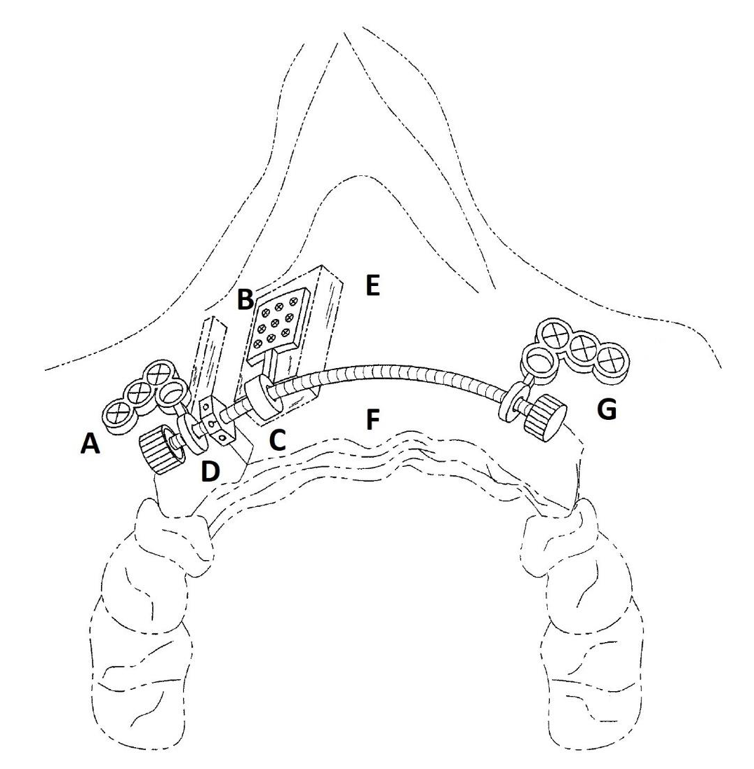
Y Maze Task Test: The Y-maze test is a recognition memory test used to assess short-term spatial working memory. It is a test sensitive to hippocampal damage, genetic manipulation and amnesic drugs. The Y-maze test is particularly useful as an initial test of memory function in rodents Kraeuter, et al. [11]. The device used was made of wood, and consisted of three arms (11 x 50 x 32 cm each) separated between them by an angle of 120°. The block allowing to close one arm of the device, was placed at 5 cm from the center of the device. In this study, the protocol described by Kraeuter, et al. [11] was used. All arm entries were noted sequentially so that the total number of arm entries, as well as the sequence of entries, was recorded. The time spend in the novel arm was recorded and percentage of good First arm entered was calculated according to de formula:

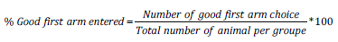
Morris Water Maze Test: The Morris Water Maze (MWM) test is widely used to study spatial and long-term memory Morris [12,13]. The water maze used in this study was consists of a black round tank, which has a diameter of 150 cm and height of 54 cm and is filled with water to a depth of 38 cm. The submerged platform (9.0 cm in diameter, 1.0 cm below the water surface) is invisible in the target quadrant. Fixed, extra-maze visual cues were present at various locations around the maze (posters) in the room with constant brightness (25 lux). In this study, the protocol described by Vorhees and Williams was used Vorhees [14]. In the training session days, the latency time of entry in the target quadrant was recorded and at the day of test session (day 5), the latency time of entry, the number entry and the time spent in the target quadrant were recorded. All parameters were recorded by a video tracking system connected to a computer directly above the water tank. The percentage of Time spent in the target quadrant was calculated according to the formula:

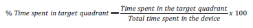
Experimental Design: Twenty-five females Wistar rat were ovariectomized and divided in 5 groups of 5 animal each: Negative control (OVX) and Positive control (PIRA) receiving distilled water, three tests groups (TEN 200, TEN 400 and TEN 600) receiving the Tengho’s drink at the doses of 200, 400 and 600 mg/kg respectively. Five others animals were used as Sham (Control group) and treated with distilled water. After 28 days of estrogen decline, all animal received an injection of Scopolamine (0.6 mg/kg, i.p.) during 14 days, excepted the sham group. The drink was administered orally from day 28 post ovariectomy to day 51. At the days 38 and 46 post ovariectomized, animals were suggested to the Y maze and Morris water maze tests respectively. Animals were sacrificed after 52 days post ovariectomy and brains were harvested. The right cerebral hemisphere was ground in Tris buffer for evaluation of oxidative stress parameters (MDA, SOD and nitrites levels and Catalase activity) in homogenate, while the right cerebral hemisphere was fixed in 10% formalin for H&E and cresyl violet staining as well as immunohistochemistry (Iba-1 and GFAP for microglia and astrocytes expression respectively).

Biochemical Assay

The levels of malondialdehyde (MDA), superoxide dismutase (SOD) and Nitrites as well as Catalase activity were evaluated according to the protocols described by Wilbur, et al. [15-17] respectively

Sectioning for Histology and Immuno-Histochemistry

Sections of 5-μm thickness were obtained from brain tissues embedded in paraffin wax using a standard microtome.

Haematoxylin & Eosin and Cresyl Violet Staining: Brain tissue sections were processed for and Hematoxylin & Eosin and cresyl violet staining based on the work of Dawson, et al. [18,19]. Sections were deparaffinized in 2 changes of xylene and thoroughly dehydrated in solutions of graded alcohol concentration (i.e. 100%, 90%, 80% and 70%). Sections were thoroughly rinsed in 2 changes of distilled water and stained. Immediately after staining, sections were dehydrated through descending alcohol grades (80%, 90%, and 100%), cleared in 2 changes of xylene, and subsequently mounted wet in dibutyl phthalate xylene (DPX) a synthetic mountant, with the use of coverslips. The slides were allowed to dry and prepared ready for microscopy.

Immunohistochemically Labeling with Antibodies: Immunostaining was done in accordance with the standard protocol described by Folarin, et al. [19]. Paraffin sections were dewaxed, rehydrated and immersed in distilled water. Antigen retrieval was done in 10 mM citrate buffer (pH = 6.0) for 25 min, with subsequent peroxidase quenching in 3% H2O2/methanol. All sections were blocked in 2% milk for 1 h to avoid non-specific background staining. Brain sections were immuno-labelled with the following antibodies: anti-Iba-1 (1:1000; Abcam, Cambridge, MA, USA) and anti-GFAP (1:1000; Dako, Denmark). Each antibody was diluted in 1% PBS milk and 0.1% Triton X detergent (to facilitate quick penetration of antibody) and incubated over night at 4oC. HRP-conjugated secondary antibodies in VECTA-STAIN kit (Vector Labs, Burlingame, USA) was subsequently used to detect the bound antibody based on the manufacturer’s protocol. The end product of reaction was improved using 3, 3′-diaminobenzidine as a chromogen (DAB) (1:25 dilution) for 5 min. Sections were thoroughly dehydrated in solutions of graded alcohol concentrations, then passed through xylene, and mounted wet in DPX, cover slipped and allowed to dry. With identical light intensity and exposure settings, images were taken with a bright fild microscope (Biomicroscope, YJ-2005 series) equipped with × 4 × 10 and × 40 dry and × 100 oil objectives and a cameral Inspiration Marvotech (MYCH-10 L) and AmScope ToupView 3.2 software’s connected to a monitor screen of a laptop computer.

Statistical Analysis

All data were expressed as the mean ± standard error of the mean (S.E.M) and analysed with one-way ANOVA followed by the Dunnett post-test (GraphPad Prism® Software, version 5.03, San Diego, CA, USA). The p values < 0.05 were considered significant.

Results

Neuroprotective Effects of Tenghõ’s Drink on Memory Impairment Induced by Scopolamin on Ovariectomized Wistar Rat

Effects of Tenghõ’s Drink on Spatial and Short-Term Memory Evaluate by the Y-Maze Test: The analysis of results presented on (Figure 1) showed that compared to the Sham group, Scopolamine injection induced a non-significant decrease of the time spent in the novel arm (Figure 1A) and the percentage of good first arm entry (Figure 1B). In comparison to the negative control group, the treatment with the Tenghõ’s drink at the dose of 600 mg/ kg as well as Piracetam induced a non-significant increase of the time spent in the novel arm (Figure 1A), while the Tenghõ’s drink at the dose of 200 and 600 mg/kg induced a non-significant increase of the percentage of good first arm entry (Figure 1B). Data are expressed as mean ± S.E.M., n = 5 per group. Sham: Animals having undergone white surgery, which received distilled water; OVX: Ovariectomized animals, which received Scopolamine and distilled water; PIRA: Ovariectomized animals, which received Scopolamine and Piracetam 300 mg/kg; TEN 200, TEN 400 and TEN 600: Ovariectomized animals, which received Scopolamine and Tenghõ’ drink at the doses of 200, 400 and 600 mg/kg respectively.
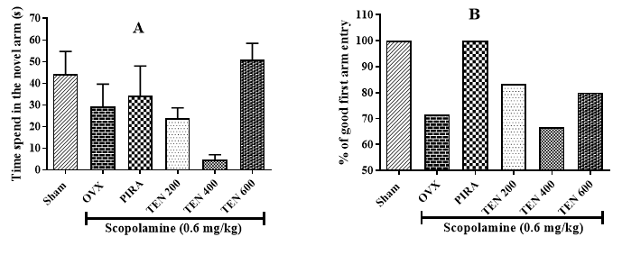
biomedres-openaccess-journal-bjstr

Figure 1:

A. Effects of Tenghõ’s drink on the time spent in the novel arm and

B. The percentage of good first arm entry in the Y maze.

Effects of Tenghõ’s Drink on Spatial and Long-Term Memory Evaluate by the Morris Water Maze Test: The analysis of the latency time of entry into the target quadrant in the training session of the MWM test shows that compared to the Sham group, ovariectomized animal that received only Scopolamine showed an increase of this parameter (Figure 2A). This increase was significant (p < 0.05; p < 0.01) in day 2 and 3. Animals treated with Tenghõ, as well as Piracetam, showed a decreased latency time of entry into target quadrant compared to negative control group (Figure 2A). This effect was significant in day 3 (p < 0.01) and day 4 (p < 0.05) with the drink at the doses of 200 and 400 mg/kg (Figure 2A). In the day of test session (day 5), compared to the negative control group, the treatment with Tenghõ’ drink induced a non-significant decrease of the latency time of entry into the target quadrant. (Figure 2B) presented the effect of Tenghõ’ drink in the number of entry into the target quadrant at the day of test session (day 5). No significant difference was observed between Sham and OVX groups. Compared to the negative control group, Tenghõ at doses of 200 and 400 mg/kg as well as Piracetam, induced a non-significant increase in the number of entry into the target quadrant. At the day of test session (day 5), compared to the negative control group, ovariectomized animal treated with Scopolamine only induced a non-significant reduction of the percentage of time spent in the target quadrant (Figure 2C).

The administration of Tenghõ at doses of 200 and 400 mg/kg as well as Piracetam, induced an increase of the percentage of time spent in the target quadrant in comparison to the negative control group. The effect was significant (p < 0.01) with the drink at the dose of 200 mg/kg (Figure 2C). Data are expressed as mean ± S.E.M., n = 5 per group. Sham: Animals having undergone white surgery, which received distilled water; OVX: Ovariectomized animals, which received scopolamine and distilled water; PIRA: Ovariectomized animals, which received scopolamine and Piracetam 300 mg / kg; TEN 200, TEN 400 and TEN 600: Ovariectomized animals, which received scopolamine and Tenghõ’ drink at the doses of 200, 400 and 600 mg/kg respectively. #p < 0.05, ##p < 0.01 vs. Sham; *p < 0.05, **p < 0.01 vs. OVX (one-way Anova followed by Dunnett’s post test).

biomedres-openaccess-journal-bjstr

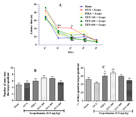
Figure 2: Effects of Tenghõ’s drink on latency time of entry into (A), the number of entry into (B) and the percentage of time spent (C) in the target quadrant of the Morris water maze.

Effect of the Tenghõ’s Drink on Some Oxidative Stress Parameters: The analysis of (Figure 3) shows that compared to Sham group, animals of negative control group presented a nonsignificant change in the MDA level (Figure 3A) and a significant (p < 0.01; p < 0.05) decrease of the of SOD and Nitrite levels and the Catalase activity Figure 3A, B and C respectively). The treatment of ovariectomized rat with Tenghõ’s drink induced a significant (p < 0.05) decrease of the MDA level (dose 400 mg/kg, (Figure 3A), and a significant (p < 0.01) increase of the of SOD (dose 600 mg/ kg, (Figure 3B) and Nitrite (all tested dose, (Figure 3C) levels and the Catalase activity (dose 200 mg/kg, (Figure 3D). Data are expressed as mean ± S.E.M., n = 5 per group. Sham: Animals having undergone white surgery, which received distilled water; OVX: Ovariectomized animals, which received scopolamine and distilled water; PIRA: Ovariectomized animals, which received scopolamine and Piracetam 300 mg / kg; TEN 200, TEN 400 and TEN 600: Ovariectomized animals, which received scopolamine and Tenghõ’ drink at the doses of 200, 400 and 600 mg/kg respectively. #p < 0.05, ##p< 0.01 vs. Sham; *p < 0.05, **p< 0.01, ***p< 0.001 vs. OVX (one-way Anova followed by Dunnett’s test).

biomedres-openaccess-journal-bjstr

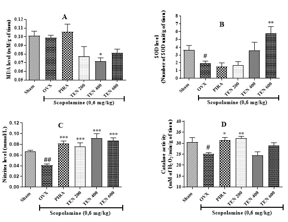
Figure 3: Effects of Tenghõ’s drink on the MDA (A), SOD (B) and Nitrite (C) levels and the Catalase activity (D).

biomedres-openaccess-journal-bjstr

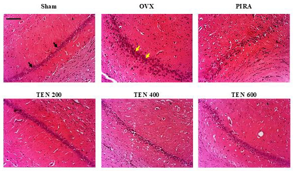
Figure 4: Effects of Tenghõ’s drink on microarchitecture (H&E staining) of CA1 region of the hippocampus.

Effects of Tenghõ’s Drink on the Microarchitecture of the Hippocampus: (Figures 4 & 5) shows the H&E and cresyl violet stain respectively of the hippocampal cells (CA1 region). Compared to Sham group, Scopolamine induced neurodegeneration in animals of negative control group. We noted an increase in the necrotic cells (yellow arrow) in the CA1 regions of the hippocampus of OVX group (Figures 4 & 5). The treatment with Tenghõ’s drink, as well as Piracetam reversed the neurodegenerative effect of Scopolamine in the CA1 region of the hippocampus. OVX group presented more necrotic cells (yellow arrow) than TEN 200, TEN 400 and TEN 600 groups (Figures 4 & 5). Photomicrographs of H&E stain of CA1 region of the hippocampus, X200, scale bar, 20 μm. Sham: Animals having undergone white surgery, which received distilled water; OVX: Ovariectomized animals, which received scopolamine and distilled water; PIRA: Ovariectomized animals which received scopolamine and Piracetam 300 mg / kg; TEN 200, TEN 400 and TEN 600: Ovariectomized animals, which received scopolamine and Tenghõ’ drink at the doses of 200, 400 and 600 mg/kg respectively. Black arrow = normal cells and yellow arrow = necrotic cells.

biomedres-openaccess-journal-bjstr

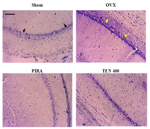
Figure 5: Effects of Tenghõ’s drink on microarchitecture (cresyl violet staining) of CA1 region of the hippocampus.

Photomicrographs of cresyl violet stain of CA1 region of the hippocampus, X100, scale bar, 20 μm. Sham: Animals having undergone white surgery which received distilled water; OVX: Ovariectomized animals which received scopolamine and distilled water; PIRA: Ovariectomized animals which received scopolamine and Piracetam 300 mg / kg; TEN 200, TEN 400 and TEN 600: Ovariectomized animals, which received scopolamine and Tenghõ’ drink at the doses of 200, 400 and 600 mg/kg respectively. Black arrow = normal cells and yellow arrow = necrotic cells.

Effects of Tenghõ’s Drink on the Microarchitecture of the Cingulate Cortex: As shown on (Figure 6), Scopolamine induced neurodegeneration in animals of OVX group in comparison to Sham group. We noted an increase in the necrotic cells (yellow arraw) in the cingulate cortex of OVX group (Figure 6). The treatment with Tenghõ’s drink, as well as PIRA reversed the neurodegenerative effect of Scopolamine in the cingulate cortex. Animals of OVX group presented more necrotic cells (yellow arrow) than those treated with Tenghõ’s drink (Figure 6). Photomicrographs of cresyl violet stain of cingulate cortex, X100, scale bar, 20 μm. Sham: Animals having undergone white surgery which received distilled water; OVX: Ovariectomized animals which received scopolamine and distilled water; PIRA: Ovariectomized animals which received scopolamine and Piracetam 300 mg / kg; TEN 200, TEN 400 and TEN 600: Ovariectomized animals, which received scopolamine and Tenghõ’ drink at the doses of 200, 400 and 600 mg/kg respectively. Black arrow = normal cells and yellow arrow = necrotic cells.

biomedres-openaccess-journal-bjstr

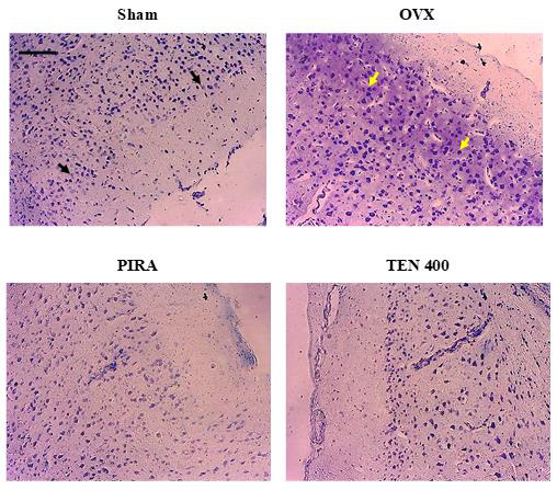
Figure 6: Effects of Tenghõ’s drink on microarchitecture (cresyl violet staining) of cingulate cortex.

biomedres-openaccess-journal-bjstr

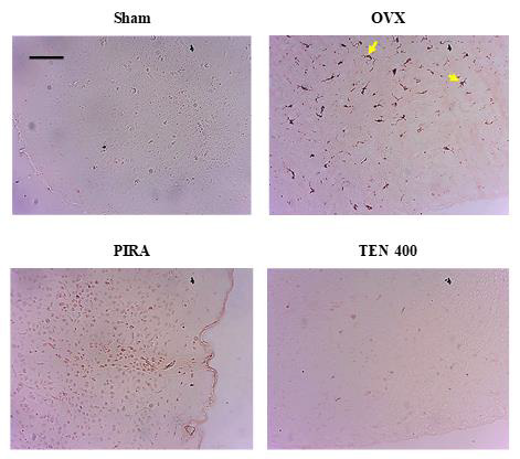
Figure 7: Effects of Tenghõ’s drink on Iba-1 expression in the cingulate cortex.

Effects of Tenghõ’s Drink on Ionized Calcium-Binding Adapter Molecule 1 And Glial Fibrillary Acidic Protein Expression in the Cingulate Cortex: Compared to Sham group, the administration of Scopolamine in ovariectomized rat resulted in an increase in Iba-1 and GFAP-positive microglia and astrocytes respectively (yellow arrow) in the cingulate cortex (Figures 7 & 8). The animals treated with Tenghõ’s drink showed less Iba-1 and GFAP expression of activated microglia and astrocytes in the cingulate cortex (Figures 7 & 8). Microglia activation (yellow arrow) as shown by amoebic isoforms was more in OVX group than groups of animals treated with the Tenghõ’s drink (Figure 7). Astrogliosis (yellow arrow) was greater in OVX group than groups of animals treated with the Tenghõ’s drink (Figure 8). Photomicrographs of Iba-1-immunolabelled microglia in cingulate cortex, X100, scale bar, 20 μm. Sham: Animals having undergone white surgery, which received distilled water; OVX: Ovariectomized animals which received scopolamine and distilled water; PIRA: Ovariectomized animals which received scopolamine and Piracetam 300 mg / kg; TEN 200, TEN 400 and TEN 600: Ovariectomized animals, which received scopolamine and Tenghõ’ drink at the doses of 200, 400 and 600 mg/kg respectively. Yellow arrow = activated microglia.

biomedres-openaccess-journal-bjstr

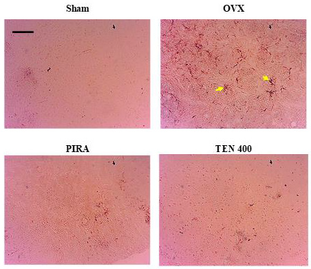
Figure 8: Effects of Tenghõ’s drink on GFAP expression in the cingulate cortex.

Photomicrographs of GFAP-immunolabelled astrocyte in cingulate cortex, X100, scale bar, 20 μm. Sham: Animals having undergone white surgery, which received distilled water; OVX: Ovariectomized animals which received scopolamine and distilled water; PIRA: Ovariectomized animals which received scopolamine and Piracetam 300 mg / kg; TEN 200, TEN 400 and TEN 600: Ovariectomized animals, which received scopolamine and Tenghõ’ drink at the doses of 200, 400 and 600 mg/kg respectively. Yellow arrow = activated astrocytes.

Discussion

The hypothesis of specific neuroanatomical and neurophysiological effects of estrogen on the brain may explain the correlation between estrogen deficiency and cognitive disorders such as dementia of the Alzheimer’s Disease (AD) Genazzani, et al. [20]. To accentuate the effects of this model of neurocognitive disorders, the administration of Scopolamine was associated to ovariectomy. Thus, the present study aimed to assess neuroprotective effects of Tenghõ’s drink on memory impairment and neuroinflammation induce by Scopolamine on ovariectomized Wistar rat. Neuroprotective effects of the Tenghõ’s drink on spatial, short-term and long-term memory were evaluated using Y-Maze and the Morris Water Maze test respectively. The obtained results on the assessment of short-term memory showed that the injection of Scopolamine at the dose of 0.6 mg/kg for 14 days in ovariectomized rats induced a decrease of the time spent in the novel arm and the percentage of good first arm entry compared to the normal control. These effects marked a slight impairment of spatial and short-term memory compared to the normal control (Sham). Indeed, estrogen deprivation resulting from menopause or ovariectomy causes neurocognitive impairment as early as 2 weeks post-ovariectomy Djiogue, et al. [21]. In addition, Scopolamine creates an animal model for AD by disrupting the cholinergic system and altering learning and memory processes Kim, et al. [22]. Scopolamine would increase acetylcholine activity and decrease performance in all memory tasks, confirming the occurrence of cognitive impairment in the animals Xiao, et al. [23].

However, the treatment of ovariectomized rat, receiving Scopolamine, with the Tenghõ’s drink as well as Piracetam induced an increase of the time spent in the novel arm at the dose of 600 mg/ kg. The drink also induced an increase of the percentage of good first arm entry at the dose of 200 and 600 mg/kg. This suggests that Tenghõ’s drink would improve the spatial and short-term memory of animals and could be endowed with secondary metabolite able to reversed the adverse effects induced by estrogen depletion and Scopolamine on memory. These secondary metabolites could act as Piracetam on the central nervous system to improving learning, memory and brain metabolism Katarzyna, et al. [24] by facilitating cholinergic and excitatory amine neurotransmission Vernon [25]. The Morris Water Maze is a commonly used experimental method to assess spatial learning and memory in animal models Hosseini, et al. [26]. Using the Morris water maze test, we found that injection of Scopolamine (0.6 mg/kg) in ovariectomized rats impaired spatial and long-term memory. Scopolamine induce a significant increase of latency time of entry into the target quadrant in the training session (day 1 to 4) as well as a reduction of the percentage of time spent in the target quadrant (day 5) compared to the normal control. Indeed, an earlier study by Markowska [27] reported that after ovariectomy, cognitive impairment was progressive.

Estrogen deprivation is likely to initiate or worsen degenerative changes caused by oxidative stress and reduce the brain’s ability to maintain synaptic connectivity and cholinergic integrity, leading to cognitive decline in the elderly and in people with diseases Gandy, et al. [28]. In addition, Scopolamine causes inhibition of cholinergic signaling Vogel [29]. In contrast, compared to OVX group, the treatment with Tenghõ’s drink improved spatial memory in the Morris water maze. This was marked by a significant reduction of latency time of entry into the target quadrant at the doses of 200 and 400 mg/kg, and a significant increase of the percentage of time spent in the target quadrant at the dose of 200 mg/kg. Several plant-derived compounds show anticholinesterase activity in preclinical studies Konrath, et al. [30] and may thereby improve cognitive decline. Indeed, these neuroprotective effects could be due to the compounds contained in the drink. For example, Garlic found Allium sativum, has been studied in the prevention of AD. Its administration in laboratory animals appeared to be able to improve memory and cognitive functions and increase serotonin activity Mathew [31,32]. Similarly, luteolin (flavonoid) and Apigenin (flavone) found in parsley (Petroselinum crispum) has shown a clear neuroprotective effect by inducing preventive activity on neuroinflammation and improved memory and learning abilities Mecocci, et al. [33-35].

Oxidative stress is one of the main factors involved in the pathogenesis of neurodegenerative disorders, including AD Liu, et al. [36]. Compared to Sham group, injection of Scopolamine 0.6 mg/kg for 14 days in ovariectomized rats induce a significant decrease of the SOD and Nitrite levels and the Catalase activity. No significant change in MDA level was observed. Scopolamine treatment has been associated with elevated oxidative stress Jang, et al. [37]; it induces a decrease of the brain activity of antioxidant enzymes Abhinav, et al. [38]. In addition, excessive oxidative stress can lead to cell death (apoptosis) Floyd, et al. [39]. The treatment of ovariectomized rat with Tenghõ’s drink as well as Piracetam induced a significant decrease of the MDA level at the dose of 400 mg/kg, and a significant increase of the of SOD level at the dose of 600 mg/kg, a significant increase of Nitrite level at all tested dose and a significant increase of the Catalase activity in comparison to OVX group. These effects suggested that the drink is endowed with antioxidant properties. Indeed, many plants, including vegetables and fruits, are natural antioxidant bases that can protected again oxidative stress and play a key role in chemoprevention of diseases that have their etiology and pathophysiology in reactive oxygen species (ROS) Kavya, et al. [40]. Some compounds contained in the Tenghõ drink, such as garlic (allium sativum) are believed to have antioxidant effects.

Garlic exerts its antioxidant effect by scavenging ROS, enhancing activity of antioxidant enzymes and increasing glutathione levels in cells Santhosha, et al. [41]. These positive effects can also be attributed to antioxidants such as flavonoids Rohini, et al. [42] contained in the drink. Brain histology, using H&E and cresyl violet stain, was performed to observe changes in the microarchitecture of hippocampus and cerebral cortex. Compared to Sham group, Scopolamine induced neurodegeneration in animals of negative control group (OVX). This was marked by increase of the necrotic cells in the CA1 regions of the hippocampus and cingulate cortex of the animals of OVX group. It is known that scopolamine induces apoptosis of nerve cells Jahanshahi, et al. [43]. Thus, changes in the rat hippocampus as well as cortex would induce changes in synaptic transmission, resulting in relevant changes in cognitive functions Knafo, et al. [44]. Compared to OVX group, the treatment with Tenghõ’s drink, as well as Piracetam reversed the neurodegenerative effect induced by Scopolamine in the CA1 region of the hippocampus and cingulate cortex. These results suggested that the drink could has neuroprotective effect in hippocampus and cerebral cortex.

The neurodegeneration observed with histological analysis was confirm by immunohistochemistry analysis of Iba-1 and GFAP for microglia and astrocytes expression respectively. It is well known that microglia activation and astrogliosis are characteristic features of CNS lesions and recruitment of microglia is reportedly regulated by astrocytes Gudi, et al. [45]. Compared to Sham group, the administration of Scopolamine in ovariectomized rat resulted in an increase in Iba-1 and GFAP-positive microglia and astrocytes respectively in the cingulate cortex. These results suggested that the administration of Scopolamine for 14 days in ovariectomized rats induced neuroinflammation. Microglial activation is one of the earliest events in AD pathology resulting in an increased of pro-inflammatory effects and the main cause of neurotoxicity Heneka, et al. [46]. Alzheimer’s Disease is associated with specific damage due to astrocytes, which may occur in the early stages of the disease and contribute to cognitive abnormalities Verkhratsky, et al. [47]. The animals treated with Tenghõ’s drink showed less Iba-1 and GFAP expression of activated microglia and astrocytes in the cingulate cortex. Microglia activation as shown by amoebic isoforms was more expressed in OVX group than groups of animals treated with the Tenghõ’s drink. Astrogliosis was greater in OVX group than groups of animals treated with the Tenghõ’s drink.

These results suggested that the drink could induce an inhibition of neuroinflammation causing by estrogen depletion and Scopolamine administration. The effects of the drink could be due to flavonoids compound found in some plant in the drink. Indeed, flavonoids could modulate neuroplasticity in terms of neurogenesis and synaptogenesis Spencer, et al. [48-49]. Flavonoids could also interact with neuronal and glial cell signaling pathways Moosavi, et al. [50] to perform several functions: promoting peripheral and loco regional vasodilation modulating cerebral blood flow Spencer, et al. [48-49], exert antioxidant and anti-inflammatory activity in biological systems that mitigate neuronal and endothelial damage González, et al. [51,52], act as hormonal mimetic that induce possible beneficial neurodegenerative changes (Kridawati et al., 2016).

Conclusion

The aim of the present study was to assess neuroprotective effects of Tenghõ on memory impairment and neuroinflammation induce by Scopolamine on ovariectomized Wistar rat. The results obtained showed that the Tenghõ’ drink improve short-term and long-term memory evaluate by Y maze and Morris water Maze tests respectively. The drink protected the brain against oxidative stress, microglia activation and astrogliosis in the hippocampus and cerebral cortex. Taken altogether, these results could justify the empirical use of Tenghõ’ drink for the treatment of inflammation. This drink could be considered as a functional food.

For More Articles: Biomedical Journal Impact Factor: https://biomedres.us

Open Access Journals on Preventive Medicine

Pharmacy Students and COVID-19; A Perspective Study on their Awareness, Data Resources and Information Accuracy

Introduction

By the end of December 2019 and up to now, everyone in this world had been impassable by COVID-19 pandemic, but education process of medical and pharmacy students had been more rediscovered by COVID-19 in unique ways. Nowadays, COVID-19 has emphasized the importance of continued learning for the profession of the pharmacist’s expanded role and value to the health care team. Also, it was a chance for pharmacy teachers to re-evaluate problems and shortage in recent curricula and study pharmacy students themselves. Yet, doubt about their sources of knowledge and their interests on outbreaks topics as COVID-19 has persistently affect methods of pharmacy curricula preparation and practices. Many theories disclose the importance of the knowledge quantity and quality within the pharmacy schools’ curricula and suggest continuous developing a curriculum to enable graduates to cope with the modern world [1-3].

Unique challenge as COVID-19 pandemic was a new shock wave of considering the curriculum and earned learning skills are educational vehicles for the medical and pharmacy student’s own journey of becoming a professional in searching for information and its evaluation. It is well known that the main purpose of a pharmacy degree program is to provide students with the skills and experiences they will need to practice as a drug information provider not as information bulk. Because, the face of an explosion of knowledge and rapid changes in healthcare management and treatment, much energy is being expended to ensure that the content is up-to-date and evident based to practice. This process must be managed to avoid overcrowding of the curriculum and teach student how to safely reach information and think about it [4]. Further, the vast amount of content to be covered, pharmacy curricula may rely on delivering the content through lectures where the students tend to remain unresponsive so, it also should be avoided and replaced by practical sessions [3].

The lectures are often supported by tutorials or laboratory practical to give students the opportunity to develop the necessary skills and to apply the knowledge learned. So, pharmacy and medical students had been studying a huge amount of information in different curricula, which nowadays seems boring and uninterested. The learning of skills is the only way to make pharmacy student devoted with information especially in the recent days we face a crisis as COVID-19. COVID-19 is novel coronavirus disease that appeared at the end of 2019 in Whun, China. It has proved as one of the most serious pandemics and fetal in mankind’s history. It has influenced a huge number of individuals over worldwide, making alarm and congestion in all circles of life and in its way, it revealed the problems of education methods and learning defects between medical and pharmacy students. Like other viruses of the Coronavirus family, origin of virus is RNA virus [5]. The main clinical manifestations of the disease are very close to the Severe Acute Respiratory Syndrome (SARS) infection which are; malaise which occurs in 99% of the infected persons, dry cough, dyspnea and bilateral patchy infiltration on imaging [6].

The overall mortality rate of COVID-19 is 2% which is much lower than that of (SARS) and Middle East Respiratory Syndrome (MERS) [7]. The virus firstly, named as 2019-nCoV and afterward changed it to extreme intense respiratory condition coronavirus 2 (SARS-CoV-2) due to its similarity to SARS in structure and manifestations. The most significant broadcast method of transmission that is right now established upon, is human-tohuman by means of respiratory beads or direct contacts [8].

During the first half of 2020, Egypt confirmed cases of COVID-19 is rising day by day mostly the last report of laboratory-confirmed COVID-19 cases and deaths in Egypt are as follows: until July 30, 2020, a total of 90.000 cases were confirmed, of whom 5.000 died due to COVID-19 and 200.000 recovered and were discharged from hospitals [9,10]. This study designed for examination the quality of knowledge about COVID-19 between pharmacy students and their resources of information. The analysis of pharmacy students’ knowledge included simple exam about basic information about the COVID-19, preventive behaviors and its broadcast in their country. Since COVID-19 is currently spreading in Egyptian hospitals and all over the world we must learn how student deal with it. In addition.

Methods

This cross-sectional study was conducted on Egyptian pharmacy students, faculty of pharmacy, Deraya University to study their COVID-19 related knowledge, preventive behaviors, and risk perception according to their interests and sources of information they adapted to follow. 1070 participants were encounter to the study. The survey covered the domains of student characteristics, awareness, information sources, knowledge and perceptions related to COVID-19. The developed draft survey instrument was distributed to ten randomly selected faculty members to assess its readability and validity before pretesting. Then questioner was sent to some experts and finally it was revised following the experts’ opinions and suggestions. Questionnaire was prepared in English language as a MCQ model following the instructions and guidelines of WHO that had five parts:

a) General information of Corona virus.

b) Knowledge on manifestations; it included knowledge on signs and symptoms of COVID-19, knowledge on the protective ways to prevent transmission COVID-19, knowledge on COVID transmission.

Protection and treatment COVID-19; it was subdivided into two portions:

i. Perception towards COVID-19.

ii. Perception about the impact of COVID-19.

c) Prediction towards COVID-19 second wave, and

d) COVID-19 broadcast in their country.

Questioner was conducted from 20th to 28th of July 2020, within a week in faculty of pharmacy. Correct answers given to the knowledge measurement questions are scored as 1 point, and participants can get 0 to 20 points in total. And it was subdivided into three groups fair (0-5), good (6-10), very good (11-15) or excellent (16-20).

Ethical Considerations

Confidentiality of the study participants’ information was maintained throughout the study by making the participants’ information anonymous and asking the participants to provide honest answers. Eligible participants in this survey were voluntary and was not compensated. The study was performed following the Helsinki Declaration as revised in 2013. We informed all participants about the aim of the study.

Data Analysis

The obtained data were coded, validated, and analyzed using SPSS version 24 (IBM, Armonk, NY, USA). Descriptive analysis was applied to calculate the frequencies and proportions. The chisquare test was used to investigate the level of association among variables. A p-value of less than 0.05 was considered statistically significant. Graph Pad for Windows version 9 was used for the statistical analysis of the research data. The normality of the distribution of the data was evaluated using Shapiro Wilk values. We performed frequency, percentage, mean, and standard deviation for reporting demographic data of the participants. Additionally, twoway ANOVA-test and analysis of variance were used for comparing the continuous data averages.

Results

biomedres-openaccess-journal-bjstr

Table 1: Demographic Characteristics of participants.

A total of 1070 students participated, 39.7% were male and 60.3% were female students. Their age ranged from 17 to 25 years. The majority of the students (40.9%) were from rural area, (34.1%) came from urban area. Table 1 show demographics of participants including the mean. Main objective of study revealed in Figure 1,that show the percent of correct answers about COVID-19 among participants in questioner. Figure 1 assume more than 60% of participants were incorrect answers while less than 25% of participants gave good answers of all questions correctly that was significantly different (p=0.00002). For understanding these results, first we asked about their resources then, if they interesting on COVID-19 in addition to the importance of information they studied from faculty curricula. Regarding resources; it was some while surprising.

biomedres-openaccess-journal-bjstr

Figure 1: Percent of participants with fair and good answers about medical data of COVID-19.

biomedres-openaccess-journal-bjstr

Figure 2: Percent of participants adapted to use social media, faculty curricula or medical websites as resource of information about COVID-19.

According to Figure 2; approximately 60% of the participants reported that they used social media (Facebook, Twitter, WhatsApp, YouTube, Instagram,) to obtain information about COVID-19 Moreover, nearly 25% of the participants sometimes obtained information about COVID-19 from medical website. Regarding interesting; Figure 3, show More than 42% of participants were strongly response about their interesting on COVID crisis, while (26%) were fair and very strong response about their interesting on COVID crisis To more studying about participant relation to social media they were asked about their activity rating. Figure 4 show that 40% of students were active on social media (more than 6 hours/day) while, 25% were fair on social media and 5% of them were not respond on social media. In addition; it was found 70% of participants denied any role of their curriculum as a resource for COVID-19 information as illustrated in Figure 5. Finally, resulted disclose that 45% of students are not convinced of online education during COVID-19 quarantine.

biomedres-openaccess-journal-bjstr

Figure 3: Percent of participants interested on searching about COVID-19 data.

biomedres-openaccess-journal-bjstr

Figure 4: Percent of activity strength on social media between participants.

biomedres-openaccess-journal-bjstr

Figure 5: Percent of participants encourage online learning during the pandemic.

Discussion

In this paper we describe pharmacy education problems in Upper Egypt that revealed in COVID-19 pandemic. Results highlighted major issues in pharmacy practice including deficiencies in curriculum. The shortage of the correct data about COVID was discussed, including the problems of online learning. The information presented in this paper may stimulate discussion and critical analysis and planning, and will be of value in further adaptation of the pharmacy education to desired educational outcomes. The major cause for revealing these problems was a questioner about COVID-19 that was evaluated by experts. Results of the questioner were surprising as it confirmed thar high percent of participant give wrong answers about COVID-19 and categorized as fair. After further analysis of data, we can return this fairness to the following; 60% of participants considered social media as a valid resource of information, 25% of participants were not interested about COVID-19 pandemic and finally 70% of participant owing their bad results to shortage in their curricula.

These findings were make-sure by studying the activity of participants on social media that show high activity that was come with previous study and ascertain Millennial students have grown up with technology and it is viewed as a natural part of the environment [11]. Additionally, research is needed on pharmacy students’ attitudes toward authority figures judging character, professional attitudes, and employability based on one’s online persona. Our results demonstrate variability of information resources used by participants. As a result, we found different levels of correctness. We included a special question about curricula and it was not surprising that students ascertain useless of traditional methods of illustration and useless content of current curricula thar earlier reported Millennial students have grown up with technology and it is viewed as a natural part of the environment [11].

Additionally, research is ascertain the useless of content and illustration methods in the present curricula as earlier reported by many [3,4,12]. At the end the debate about on-line learning in our countries still a challenge for governments and education leaders as our participant still complain from online leaning in contrast with others [13]. As pharmacists and pharmacy students are the first entities who may have close connection with the affected people. Lack of proper related knowledge or false knowledge in this populace can make them miscalculate the situation, increase stress and exaggerate the level of cases and may interrupt the appropriateness of their medical decisions in addition, Medical staff have always been at risk of contagious diseases and be a source of infection. So their knowledge is a big encounter.

Limitation

We could not conduct pilot study for post-testing of this questionnaire for shortage of time.

Conclusion and Recommendation

Results exposed major problems in both pharmacy curricula and methods of education among medical students in Egypt and Arab countries. It is recommended to give a special consideration for searching and information resources starting from the first grade, afford more prospects to argue drawbacks of social media information and instruct students how to evaluate information before dissemination.

For More Articles: Biomedical Journal Impact Factor: https://biomedres.us

Open Access Journals on Regenerative Medicines

Pandemic Prevention

Introduction

Measures

Infrastructure and International Development: Robust, collaborating public health systems that have the capacity for active surveillance for early detection of cases and to mobilize their health care coordination capacity may be required to be able stop contagion promptly. After an outbreak there is a certain window of time during which a pandemic can still be stopped by the competent authorities isolating the first infected and/or fighting the pathogen. A good global infrastructure, consequent information exchange, minimal delays due to bureaucracy and effective, targeted treatment measures can be prepared. 2012 it has been proposed to consider pandemic prevention as an aspect of international development in terms of health-care infrastructure and changes to the pathogenrelated dynamics between humans and their environment including animals. Often local authority carers or doctors in Africa, Asia or Latin America register uncommon accumulations (or clusterings) of symptoms but lack options for more detailed investigations. Scientists state that “research relevant to countries with weaker surveillance, lab facilities and health systems should be prioritized” and that “in those regions, vaccine supply routes should not rely on refrigeration, and diagnostics should be available at the point of care”.

Technologies

Pathogen Detection and Prediction: In a 2012 study it is claimed that “new mathematical modelling, diagnostic, communications, and informatics technologies can identify and report hitherto unknown microbes in other species, and thus new risk assessment approaches are needed to identify microbes most likely to cause human disease”. The study investigates challenges in moving the global pandemic strategy from response to preemption. Some scientists are screening blood samples from wildlife for new viruses. The international Global Virome Project (GVP) aims to identify the causes of fatal new diseases before emergence in human hosts by genetically characterizing viruses found in wild animals. The project aims to enlist an international network of scientists to collect hundreds of thousands of viruses, map their genomes, characterize and risk-stratify them to identify which ones to pay attention to.

However, some infectious disease experts have criticized the project as too broad and expensive due to limited global scientific and financial resources and because only a small percentage of the world’s zoonotic viruses may cross into humans and pose a threat. They argue for prioritizing rapidly detecting diseases when they cross into humans and an improving the understanding of their mechanisms. A successful prevention of a pandemic from specific viruses may also require ensuring that it does not re-emerge – for instance by sustaining itself in domestic animals. Pathogen detection mechanisms may allow the construction of an early warning system which could make use of artificial intelligence surveillance and outbreak investigation. Edward Rubin notes that after sufficient data has been gathered artificial intelligence could be used to identify common features and develop countermeasures and vaccines against whole categories of viruses.

It might be possible to predict viral evolution using machine learning. In April 2020 it was reported that researchers developed a predictive algorithm which can show in visualizations how combinations of genetic mutations can make proteins highly effective or ineffective in organisms – including for viral evolution for viruses like SARS-CoV-2.` In 2021, pathogen researchers reported the development of machine learning models for genome-based early detection and prioritization of high-risk potential zoonotic viruses in animals prior to spillover to humans which could be used for virus surveillance for (i.a.) measures of “early investigation and outbreak preparedness” and, according to the study, would have been capable of predicting SARS-CoV-2 as a high-risk strain without prior knowledge of zoonotic SARS-related corona viruses. An artificial “global immune system”-like technological system that includes pathogen detection may be able to substantially reduce the time required to take on a biothreat agent.

A system of that sort would also include a network of welltrained epidemiologists who could be rapidly deployed to investigate and contain an outbreak. Funding for the United States’ PREDICT government research program that sought to identify animal pathogens that might infect humans and to prevent new pandemics was cut in 2019. Funding for United States’ CDC programs that trained workers in outbreak detection and strengthened laboratory and emergency response systems in countries where disease risks are greatest to stop outbreaks at the source was cut by 80% in 2018. Despite recent advances in pandemic modeling, experts using mostly experience and intuition are still more accurate in predicting the spread of disease than strictly mathematical models.

Crispr-Based Immune Subsystems: In March 2020 scientists of Stanford University presented a CRISPR-based system, called PAC-MAN (Prophylactic Antiviral Crispr in human cells), that can find and destroy viruses in vitro. However, they weren’t able to test PAC-MAN on the actual SARS-CoV-2, use a targeting-mechanism that uses only a very limited RNA-region, haven’t developed a system to deliver it into human cells and would need a lot of time until another version of it or a potential successor system might pass clinical trials. In the study published as a preprint they write that it could be used prophylactically as well as therapeutically. The CRISPR-Cas13d-based system could be agnostic to which virus it’s fighting so novel viruses would only require a small change. In an editorial published in February 2020 another group of scientists claimed that they have implemented a flexible and efficient approach for targeting RNA with CRISPR-Cas13d which they have put under review and propose that the system can be used to also target SARS-CoV-2 in specific.

There have also been earlier successful efforts in fighting viruses with CRISPR-based technology in human cells. In March 2020 researchers reported that they have developed a new kind of CRISPR-Cas13d screening platform for effective guide RNA design to target RNA. They used their model to predict optimized Cas13 guide RNAs for all protein-coding RNA-transcripts of the human genome’s DNA. Their technology could be used in molecular biology and in medical applications such as for better targeting of virus RNA or human RNA. Targeting human RNA after it’s been transcribed from DNA, rather than DNA, would allow for more temporary effects than permanent changes to human genomes. The technology is made available to researchers through an interactive website and free and open source software and is accompanied by a guide on how to create guide RNAs to target the SARS-CoV-2 RNA genome. Scientists report to be able to identify the genomic pathogen signature of all 29 different SARS-CoV-2 RNA sequences available to them using machine learning and a dataset of 5000 unique viral genomic sequences. They suggest that their approach can be used as a reliable real-time option for taxonomic classification of novel pathogens.

Testing and Containment: Timely use and development of quick testing systems for novel virus in combination with other measures might make it possible to end transmission lines of outbreaks before they become pandemics. A high discoveryrate is important for tests. For instance this is the reason why no thermal scanners with a low discovery-rate were used in airports for containment during the 2009 swine flu pandemic.[39] The German program Infect Control 2020 seeks to develop strategies for prevention, early recognition and control of infectious diseases. In one of its projects “HyFly” partners of industry and research work on strategies to contain chains of transmission in air traffic, to establish preventive countermeasures and to create concrete recommendations for actions of airport operators and airline companies. One approach of the project is to detect infections without molecular-biological methods during passenger screening. For this researcher of the Fraunhofer-Institute for cell therapy and immunology are developing a non-invasive procedure based on ion-mobility spectrometry (IMS).

Surveillance and Mapping

Viral Hotspots and Zoonotic Genomics: Monitoring people who are exposed to animals in viral hotspots – including via virus monitoring stations – can register viruses at the moment they enter human populations – this might enable prevention of pandemics. The most important transmission pathways often vary per underlying driver of emerging infectious diseases such as the vectorborne pathway and direct animal contact for land-use change – the leading driver for emerging zoonoses by number of emergence events as defined by Jones et al. (2008). 75% of the reviewed 1415 species of infectious organisms known to be pathogenic to humans account for zoonoses by 2001. Genomics could be used to precisely monitor virus evolution and transmission in real time across large, diverse populations by combining pathogen genomics with data about host genetics and about the unique transcriptional signature of infection. The “Surveillance, Outbreak Response Management and Analysis System” (SORMAS) of the German Helmholtz- Zentrum für Infektionsforschung (HZI) and Deutsches Zentrum für Infektionsforschung (DZIF), who collaborate with Nigerian researchers, gathers and analyzes data during an outbreak, detects potential threats and allows to initiate protective measures early. It’s meant specifically for poorer regions and has been used for the fight against a monkeypox outbreak in Nigeria.

Syndromic Surveillance and Border Control: Expert on infectious diseases at the Johns Hopkins Center for Health Security, Amesh Adalja states that the most immediate way to predict a pandemic is with deeper surveillance of symptoms that fit the virus’ profile. The scientific and technological ways of quickly detecting a spillover could be improved so that an outbreak can be isolated before it becomes an epidemic or pandemic. David Quammen states that he heard about the idea to develop technology to screen people at airport security points for whether or not they carry an infectious disease ten years ago and thought it was going to be done by now. Thermometers whose measurement data is directly shared via the Internet and medical guidance apps have been used to plot and map unusual fever levels to detect anomalous outbreaks. Various forms of data-sharing could be added to health care institutions such as hospitals so that e.g. anonymized data about symptoms and incidences found to be unusual or characteristic of a pandemic threat could enable high-resolution “syndromic surveillance” as an early warning system. In 1947, the World Health Organization established such a global network of some hospitals. Such sharing and off-site evaluation of symptoms and possibly related medical data may have complementary benefits such as improving livelihoods of workers who work with livestock and improving the accuracy, timeliness, and costs of disease prognoses.

Mutation Surveillance: In December 2020 during the COVID-19 pandemic national and international officials reported mutated variants of SARS-CoV-2, including some with higher transmissibility and worldwide spread. While mutations are common for viruses and the spread of some of the virus’ mutations have been tracked earlier, mutations that make it more transmittable or severe can be problematic. Resources for disease surveillance have improved during the pandemic so that medical systems around the world are starting to be equipped to detect such mutations with genomic surveillance in a manner relevant to pandemic mitigation and the prevention of sub-pandemics of specific variants or types of variants. As of December 2020, contemporary measures such as COVID-19 vaccines and medications seem to be effective in the treatment of infections with the tracked mutated variants compared to earlier forms that are closer to the original virus/es.

Policy and Economics: A 2014 analysis asserts that “the window of opportunity to deal with pandemics as a global community is within the next 27 years. Pandemic prevention therefore should be a critical health policy issue for the current generation of scientists and policymakers to address. A 2007 study warns that “the presence of a large reservoir of SARS-CoVlike viruses in horseshoe bats, together with the culture of eating exotic mammals in southern China, is a time bomb. The possibility of the reemergence of SARS and other novel viruses from animals or laboratories and therefore the need for preparedness should not be ignored”. The US’ National Security Council Directorate for Global Health Security and Biodefense, which worked on preparing for the next disease outbreak and preventing it from becoming an epidemic or pandemic, was closed in 2018.

Environmental Policy and Economics: Some experts link pandemic prevention with environmental policy and caution that environmental destruction as well as climate change drives wildlife to live close to people. For instance the WHO projects that climate change will also affect infectious disease occurrence. A 2016 study reviews literature on the evidences for the impact of climate change on human infectious disease, suggests a number of proactive measures for controlling health impacts of climate change and finds that climate change impacts human infectious disease via alterations to pathogen, host and transmission. Studies have shown that the risk of disease outbreaks can increase substantially after forests are cleared. According to Kate Jones, chair of ecology and biodiversity at University College London, the disruption of pristine forests driven by logging, mining, road building through remote places, rapid urbanisation and population growth is bringing people into closer contact with animal species they may never have been near before, resulting in transmission of diseases from wildlife to humans.

An August 2020 study published in Nature concludes that the anthropogenic destruction of ecosystems for the purpose of expanding agriculture and human settlements reduces biodiversity and allows for smaller animals such as bats and rats, who are more adaptable to human pressures and also carry the most zoonotic diseases, to proliferate. This in turn can result in more pandemics. In October 2020, the Intergovernmental Science-Policy Platform on Biodiversity and Ecosystem Services published its report on the ‘era of pandemics’ by 22 experts in a variety of fields and concluded that anthropogenic destruction of biodiversity is paving the way to the pandemic era and could result in as many as 850,000 viruses being transmitted from animals – in particular birds and mammals – to humans. The increased pressure on ecosystems is being driven by the “exponential rise” in consumption and trade of commodities such as meat, palm oil, and metals, largely facilitated by developed nations, and by a growing human population.

According to Peter Daszak, the chair of the group who produced the report, “there is no great mystery about the cause of the Covid-19 pandemic, or of any modern pandemic. The same human activities that drive climate change and biodiversity loss also drive pandemic risk through their impacts on our environment.” Stanford biological anthropologist James Holland Jones notes that humanity has “engineered a world where emerging infectious diseases are both more likely and more likely to be consequential”, referring to the modern world’s prevalent highly mobile lifestyles, increasingly dense cities, various kinds of human interactions with wildlife and alterations of the natural world. Furthermore, when multiple species that are not usually next to each other are driven to live closely together new diseases may emerge. Research shows that abundant animals, plants, insects, and microbes living in complex, mature ecosystems can limit the spread of disease from animals to people. The United Nations is formulating nature-focused action plans that could help to stop the next pandemic before it starts.

These strategies include conserving ecosystems and wilderness that are still untouched by human activity, and restoring and protecting significant areas of land and ocean (i.e. through protected areas). Protected areas (which may hold wildlife) also limits human presence and/or limits the exploitation of resources (including non-timber forest products such as game animals, furbearers). An article by the World Economic Forum states that studies have shown that deforestation and loss of wildlife cause increases in infectious diseases and concludes that the recovery from the COVID-19 pandemic should be linked to nature recovery, which it considers economically beneficial. A report by FAIRR global investor network found that more than 70% of the biggest meat, fish and dairy producers were in danger of fostering future zoonotic pandemics due to lax safety standards, closely confined animals and the overuse of antibiotics. Some have recommended food system-change, behaviour change, different lifestyle choices and altered consumer spending including moving away from factory farming and towards more plant-based diets. Some traditional medicines (i.e. traditional African medicine, TCM) still use animalbased substances.

Since these can trigger zoonosis, a possible prevention could be changes to handbooks for practitioners of such traditional medicines (i.e. exclusion of animal-based substances). Senior adviser and veterinary epidemiologist at the National Food Institute at the Technical University of Denmark Ellis-Iversen states that in agricultural animal health “outbreaks of exotic disease in wellregulated countries rarely get big because we identify and control them right away”. New York City’s Bronx Zoo’s head veterinarian Paul Calle states that usually emerging infectious diseases from animals are the result from wildlife consumption and distribution on a commercial scale rather than a lone person hunting to feed their family. Dennis Caroll of the Global Virome Project states that the “extractive industry — oil and gas and minerals, and the expansion of agriculture, especially cattle” are the biggest predictors of where spillovers can be seen.

Biotechnology Research and Development Regulation: Toby Ord, author of the book The Precipice: Existential Risk and the Future of Humanity which addresses this issue, puts into question whether current public health and international conventions, and self-regulation by biotechnology companies and scientists are adequate. In the context of the 2019–2020 coronavirus pandemic Neal Baer writes that the “public, scientists, lawmakers, and others” “need to have thoughtful conversations about gene editing now”. Ensuring the biosafety level of laboratories may also be an important component of pandemic prevention. This issue may have gotten additional attention in 2020 after news outlets reported that U.S. State Department cables indicate that, although there may be no conclusive proof at the moment, the COVID-19 virus responsible for the COVID-19 pandemic may, possibly, have accidentally come from a Wuhan (China) laboratory, studying bat coronaviruses that included modifying virus genomes to enter human cells, and determined to be unsafe by U.S. scientists in 2018, rather than from a natural source.

As of 18 May 2020, an official UN investigation into the origins of the COVID-19 virus, supported by over 120 countries, was being considered. United States’ president Donald Trump claimed to have seen evidence that gave him a “high degree of confidence” that the novel coronavirus originated in the Chinese laboratory but did not offer any evidence, data or details, contradicted statements by the United States’ intelligence community and garnered a lot of harsh criticism and doubts. As of 5 May, assessments and internal sources from the Five Eyes nations indicated that the coronavirus outbreak being the result of a laboratory accident was “highly unlikely”, since the human infection was “highly likely” a result of natural human and animal interaction. Many others have also criticized statements by US government officials and theories of laboratory release. Virologist and immunologist Vincent R. Racaniello said that “accident theories – and the lab-made theories before them – reflect a lack of understanding of the genetic make-up of Sars-CoV-2.”

Virologist Peter Daszak stated that an estimated 1–7 million people in Southeast Asia who live or work in proximity to bats are infected each year with bat coronaviruses. In January 2021, the WHO’s investigations into the origin of COVID-19 was launched. In early 2021, the hypothesis of a laboratory cause of the pandemic received renewed interest and expert consideration due to renewed media discussion. Martin Rees, author of the book Our Final Hour which also addresses this issue, states that while better understanding of viruses may allow for an improved capability to develop vaccines it may also lead to an increase in “the spread of ‘dangerous knowledge’ that would enable mavericks to make viruses more virulent and transmissible than they naturally are”. Different accelerations and priorizations of research may however be critical to pandemic prevention. A multitude of factors shape which knowledge about viruses with different use-cases, including vaccine-development, can be used by whom. Rees also states that “the global village will have its village idiots, and they will have global range”.

Food Markets and Wild Animal Trade: In January 2020 during the SARS-CoV 2 outbreak experts in and outside China warned that wild animal markets, where the virus originated from, should be banned worldwide. On January 26 China banned the trade of wild animals until the end of the coronavirus epidemic at the time. On February 24 China announced a permanent ban on wildlife trade and consumption with some exceptions.[103] Some scientists point out that banning informal wet markets worldwide isn’t the appropriate solution as fridges aren’t available in many places and because much of the food for Africa and Asia is provided through such traditional markets. Some also caution that simple bans may force traders underground, where they may pay less attention to hygiene and some state that it’s wild animals rather than farmed animals that are the natural hosts of many viruses. UN biodiversity chief, bipartisan lawmakers and experts have called for a global ban of wet markets and wildlife trade. Jonathan Kolby cautions about the risks and vulnerabilities present in the massive legal wildlife trade.

International coordination The Global Health Security Agenda (GHSA) a network of countries, international organizations, NGOs and companies that aim to improve the world’s ability to prevent, detect, and respond to infectious diseases. Sixty-seven countries have signed onto the GHSA framework. Funding for the GHSA has been reduced since the launch in 2014, both in the US and globally. In a 2018 lecture in Boston Bill Gates called for a global effort to build a comprehensive pandemic preparedness and response system. During the COVID-19 pandemic he called upon world leaders to “take what has been learned from this tragedy and invest in systems to prevent future outbreaks”. In a 2015 TED Talk he warned that “if anything kills over 10 million people in the next few decades, it’s most likely to be a highly infectious virus rather than a war”. Numerous prominent, authoritative, expert or otherwise influential figures have similarly warned about elevated, underprepared or contemporary risks of pandemics and the need for efforts on an “international scale” long before 2015 and since at least 1988 [1-3].

Some have provided suggestions for organizational or coordinative preparedness for pandemic prevention including a mechanism by which many major economic powers pay into a global insurance fund which “could compensate a nation for economic losses if it acts quickly to close areas to trade and travel in order to stop a dangerous outbreak at its source” or, similarly, sovereign or regional-level epidemic-insurance policies. International collaboration including cooperative research and informationsharing has also been considered vital. According to Senator Dianne Feinstein called for the creation of a new interagency government entity, the Center for Combating Infectious Disease which would combine analytical and operational functions “to oversee all aspects of preventing, detecting, monitoring, and responding to major outbreaks such as coronavirus” and get provided with data and expertise by the Centers for Disease Control and Prevention. John Davenport advises to abandon widespread libertarian ideology which, according to him, “denies the importance of public goods or refuses to recognize their scope.”

According to the CDC, investing in global health security and improving the organization’s ability to prevent, detect, and respond to diseases could protect the health of American citizens as well as avert catastrophic costs. Dennis Carroll argues for a “marriage” between scientific discovery and political decision-making and policy formulation. Artificial induction of immunity and/or biocides. Outbreaks could be contained or delayed – to enable other containment-measures – or prevented by artificial induction of immunity and/or biocides in combination with other measures that include prediction or early detection of infectious human diseases. In a preprint published on March 24, 2020 researchers suggested that the unique transcriptional signature of SARS-CoV-2 in the human immune system may be responsible for the development of COVID-19: SARS-CoV-2 did not induce the antiviral genes that code for type I and type III interferons. This could be relevant for the development or repurposing of treatments [4-6].

Vaccination: Development and provision of new vaccines usually takes years. The Coalition for Epidemic Preparedness Innovations, which was launched in 2017, works on reducing the time of vaccine-development. The Global Health Innovative Technology Fund (GHIT) is a public-private partnership fund which involves a national government, a UN agency, a consortium of pharmaceutical and diagnostics companies, and international philanthropic foundations to accelerate the creation of new vaccines, drugs and diagnostic tools for global health. It is unclear whether vaccines can play a role in pandemic prevention alongside pandemic mitigation. Nathan Wolfe proposes that pathogen detection and prediction may allow establishing viral libraries before novel epidemics emerge – substantially decreasing the time to develop a new vaccine. Public health surveillance expert and professor at Harvard University, John Brownstein says that “vaccines are still our main weapon”. Besides more rapid vaccine development it may also be possible to develop more broader vaccines. Misinformation and misconceptions about vaccines including about their side-effects may be a problem.

Culling: Experts warned that depleting the numbers of species by culling to forestall human infections reduces genetic diversity and thereby puts future generations of the animals as well as people at risk while others contend that it’s still the best, practical way to contain a virus of livestock.

Prevention Versus Mitigation: Pandemic prevention seeks to prevent pandemics while mitigation of pandemics seeks to reduce their severity and negative impacts. Some have called for a shift from a treatment-oriented society to a prevention-oriented one. Authors of a 2010 study write that contemporary “global disease control focuses almost exclusively on responding to pandemics after they have already spread globally” and argue that the “waitand- respond approach is not sufficient and that the development of systems to prevent novel pandemics before they are established should be considered imperative to human health”. Peter Daszak comments on the COVID-19 pandemic, saying “the problem isn’t that prevention was impossible, It was very possible. But we didn’t do it. Governments thought it was too expensive. Pharmaceutical companies operate for profit”. The WHO reportedly had mostly neither the funding nor the power to enforce the large-scale global collaboration necessary to combat it. Nathan Wolfe criticizes that “our current global public health strategies are reminiscent of cardiology in the 1950s when doctors focused solely on responding to heart attacks and ignored the whole idea of prevention”.

For More Articles: Biomedical Journal Impact Factor: https://biomedres.us

Open Access Journals on Biomedical Research

Study of COVID 19, COVID 19 Vaccination and its Impact

Introduction

COVID-19 has rapidly become a major public health crisis, affecting 86.4 million individuals, and causing 1.9 million deaths globally by January of 2021. The US has reported more than 21 million cases and 357,000 deaths as of 5 January 2021 [1]. To curb this pandemic, apart from effective public health measures such as social distancing, wearing face masks, hand washing, and avoidance of crowded indoor spaces, educating the general population, efficacious vaccination is emerging as essential to mitigating disease and death [2-6]. Despite unprecedented movement restrictions, social distancing measures, and stay-at-home orders enacted in many countries, the COVID-19 pandemic has caused devastating morbidity and mortality. However, the vast majority of the global population remains susceptible to COVID-19, highlighting the need for an effective vaccine. To mitigate the mounting burden of COVID-19, vaccine development has occurred at an unprecedented pace. As of December 31, 2020, safety and efficacy results for a number of vaccines have been reported, and Phase III clinical trials for several other candidates are underway [5]. Results from two large efficacy trials (Pfizer – BioNTech, Moderna) indicate a vaccine efficacy of over 90% against symptomatic and severe disease, exceeding the preferred population-based efficacy specified by the World Health Organization and the United States (US) Food and Drug Administration (FDA).

These vaccines have received emergency use authorization by the FDA, and vaccination has already started in the US with prioritization of healthcare workers, long-term care residents, and high-risk individuals. This compels an urgent need to understand the potential population-level impact of vaccination on COVID-19 transmission and disease outcomes [6]. COVID 19 has emerged as greatest challenge that has weakened the very basis of human existence. It has devastated economies and created unparralled human needs. It has overstretched health systems that has been seen never before. Even plague of middle age Europe and Spanish flu were less devastating than COVID 19 pandemic. The study aimed at showing the impact of vaccination on people.

Objective

Study of impact of COVID 19 vaccination.

Methodology

• Study design: Prospective study design.

• Study duration: Two weeks.

• Study setting: Multi centric study, hospital and community based.

• Study tool: A Predesigned and pretested proforma validated by a pilot study.

• Sampling: Simple random sampling.

• Exclusion criteria: Preprocedural cases.

Data analysis

The data was received from the answered questionnaires and was plotted on excel 2013. The data was analyzed statistically with the help of statistical software SPSS v19. All the continuous variables of the study were represented by the descriptive statistics and all the categorical variables in the term of frequency and percentage.

Result

(Tables 1-4).

biomedres-openaccess-journal-bjstr

Table 1: Total Subjects.

biomedres-openaccess-journal-bjstr

Table 2: Vaccination Status.

biomedres-openaccess-journal-bjstr

Table 3: Vaccine Acceptance.

biomedres-openaccess-journal-bjstr

Table 4: Side Effects of Vaccination.

Discussion

COVID-19 outbreaks have caused significant global morbidity and mortality, in addition to undermining the economic and social well-being of individuals and communities. Despite this devastating toll, the majority of the population remains susceptible to SARSCoV- 2 infection. Thus, vaccine development has been a high priority. The scale and speed of vaccine development efforts have been unprecedented, and highly protective vaccines are beginning to be distributed. This study shows that COVID-19 vaccines with 95% efficacy in preventing disease, even if they conferred limited protection against infection, could substantially mitigate future attack rates, hospitalizations, and deaths. Given the limited population-level immunity to COVID-19, vaccination remains a key preventive measure to reduce disease burden and mitigate future outbreaks. Our study suggests that a vaccine could have a substantial impact on reducing incidence, hospitalizations, and deaths, especially among vulnerable individuals with comorbidities and risk factors associated with severe COVID-19. Thus, mobilizing public health resources is imperative to achieve the proposed goal of distributing 100 million vaccine doses over 100 days in the US population by the incoming administration.

Our findings support the Advisory Committee on Immunization Practices recommendations, highlighting that a targeted vaccination strategy can effectively mitigate disease burden and the societal impact of COVID-19. We also find that, even with the relatively rapid roll-out simulated here, it may take several months to control COVID-19 at the population level. Moreover, this impact is achieved in the context of continued public health efforts and is not possible without diligent attention to the other aspects of infection prevention and control such as masking, hand hygiene, testing, contact-tracing, and isolation of infected cases. If current vaccination programs are accompanied by widespread relaxation of other measures, a much higher coverage will be necessary with a significantly higher distribution capacity. Nevertheless, our results are an encouraging signal of the power and promise of vaccines against COVID-19.

Summary

Vaccination with a 95% efficacy against disease could substantially mitigate future attack rates, hospitalizations, and deaths, even if only adults are vaccinated. Non-pharmaceutical interventions remain an important part of outbreak response as vaccines are distributed over time. A multicentric study was carried out two sites. A total of 160 patients were studied, 80 in a hospital set up and 80 in community. It was observed that severity of symptoms in cases who had received vaccination was less as compared to unvaccinated lot. Also, the vaccination was viewed positively by the majority of the respondents.

For More Articles: Biomedical Journal Impact Factor: https://biomedres.us

Open Access Journals on Medical Research

Study of COVID 19, COVID 19 Vaccination and its Impact

Introduction

COVID-19 has rapidly become a major public health crisis, affecting 86.4 million individuals, and causing 1.9 million deaths globally by January of 2021. The US has reported more than 21 million cases and 357,000 deaths as of 5 January 2021 [1]. To curb this pandemic, apart from effective public health measures such as social distancing, wearing face masks, hand washing, and avoidance of crowded indoor spaces, educating the general population, efficacious vaccination is emerging as essential to mitigating disease and death [2-6]. Despite unprecedented movement restrictions, social distancing measures, and stay-at-home orders enacted in many countries, the COVID-19 pandemic has caused devastating morbidity and mortality. However, the vast majority of the global population remains susceptible to COVID-19, highlighting the need for an effective vaccine. To mitigate the mounting burden of COVID-19, vaccine development has occurred at an unprecedented pace. As of December 31, 2020, safety and efficacy results for a number of vaccines have been reported, and Phase III clinical trials for several other candidates are underway [5]. Results from two large efficacy trials (Pfizer – BioNTech, Moderna) indicate a vaccine efficacy of over 90% against symptomatic and severe disease, exceeding the preferred population-based efficacy specified by the World Health Organization and the United States (US) Food and Drug Administration (FDA).

These vaccines have received emergency use authorization by the FDA, and vaccination has already started in the US with prioritization of healthcare workers, long-term care residents, and high-risk individuals. This compels an urgent need to understand the potential population-level impact of vaccination on COVID-19 transmission and disease outcomes [6]. COVID 19 has emerged as greatest challenge that has weakened the very basis of human existence. It has devastated economies and created unparralled human needs. It has overstretched health systems that has been seen never before. Even plague of middle age Europe and Spanish flu were less devastating than COVID 19 pandemic. The study aimed at showing the impact of vaccination on people.

Objective

Study of impact of COVID 19 vaccination.

Methodology

• Study design: Prospective study design.

• Study duration: Two weeks.

• Study setting: Multi centric study, hospital and community based.

• Study tool: A Predesigned and pretested proforma validated by a pilot study.

• Sampling: Simple random sampling.

• Exclusion criteria: Preprocedural cases.

Data analysis

The data was received from the answered questionnaires and was plotted on excel 2013. The data was analyzed statistically with the help of statistical software SPSS v19. All the continuous variables of the study were represented by the descriptive statistics and all the categorical variables in the term of frequency and percentage.

Result

(Tables 1-4).

biomedres-openaccess-journal-bjstr

Table 1: Total Subjects.

biomedres-openaccess-journal-bjstr

Table 2: Vaccination Status.

biomedres-openaccess-journal-bjstr

Table 3: Vaccine Acceptance.

biomedres-openaccess-journal-bjstr

Table 4: Side Effects of Vaccination.

Discussion

COVID-19 outbreaks have caused significant global morbidity and mortality, in addition to undermining the economic and social well-being of individuals and communities. Despite this devastating toll, the majority of the population remains susceptible to SARSCoV- 2 infection. Thus, vaccine development has been a high priority. The scale and speed of vaccine development efforts have been unprecedented, and highly protective vaccines are beginning to be distributed. This study shows that COVID-19 vaccines with 95% efficacy in preventing disease, even if they conferred limited protection against infection, could substantially mitigate future attack rates, hospitalizations, and deaths. Given the limited population-level immunity to COVID-19, vaccination remains a key preventive measure to reduce disease burden and mitigate future outbreaks. Our study suggests that a vaccine could have a substantial impact on reducing incidence, hospitalizations, and deaths, especially among vulnerable individuals with comorbidities and risk factors associated with severe COVID-19. Thus, mobilizing public health resources is imperative to achieve the proposed goal of distributing 100 million vaccine doses over 100 days in the US population by the incoming administration.

Our findings support the Advisory Committee on Immunization Practices recommendations, highlighting that a targeted vaccination strategy can effectively mitigate disease burden and the societal impact of COVID-19. We also find that, even with the relatively rapid roll-out simulated here, it may take several months to control COVID-19 at the population level. Moreover, this impact is achieved in the context of continued public health efforts and is not possible without diligent attention to the other aspects of infection prevention and control such as masking, hand hygiene, testing, contact-tracing, and isolation of infected cases. If current vaccination programs are accompanied by widespread relaxation of other measures, a much higher coverage will be necessary with a significantly higher distribution capacity. Nevertheless, our results are an encouraging signal of the power and promise of vaccines against COVID-19.

Summary

Vaccination with a 95% efficacy against disease could substantially mitigate future attack rates, hospitalizations, and deaths, even if only adults are vaccinated. Non-pharmaceutical interventions remain an important part of outbreak response as vaccines are distributed over time. A multicentric study was carried out two sites. A total of 160 patients were studied, 80 in a hospital set up and 80 in community. It was observed that severity of symptoms in cases who had received vaccination was less as compared to unvaccinated lot. Also, the vaccination was viewed positively by the majority of the respondents.

For More Articles: Biomedical Journal Impact Factor: https://biomedres.us

Open Access Journals on Biomedical Research

The Effect of Abrasive Peeling of Wheat-Triticale Grinding Grain Mixture on the Yield of Intermediate Grinding Products and Flour

Introduction

The actual directions of development of one of the most important branches of the processing industry – flour milling – are both the improvement of technologies for processing traditional crops (wheat and rye) and the development of new technologies for processing non-traditional crops, such as triticale [1-7]. One of the main areas of development of the industry is the development of new and improvement of traditional technologies and the creation of processed products of various types of grain with a given composition and properties, incl. and products of deep processing [8-12]. In addition, the direction of joint processing of grain of various crops, including those based on wheat and triticale, is very promising. Triticale is the first grain crop created by man and obtained by crossing wheat (lat. Triticum) and rye (lat. Secale). The use of triticale as a food crop is an interesting, promising direction not only for flour milling, but also for other food and processing industries. This is confirmed by the increased interest in this culture, both on the part of researchers and food producers, not only in our country, but also abroad. Bakery products with the use of processed products from the central part of the triticale grain endosperm are characterized by increased nutritional value due to a higher content of protein and essential amino acids the main limiting acid, lysine [13-16]. The combination of the positive properties of rye – a high content of biologically active aromatic substances and wheat – the rheological properties of the dough, make it possible to produce food products of mass consumption from triticale grain processing products and mixtures based on it. At the same time, the technological properties of baking flour obtained from various grain mixtures, including wheat-triticale grain grinding mixture, remain little studied. Peeling of the wheattriticale grain mixture during varietal bakery grinding is carried out to maximize the cleaning of the grain surface from dust, dirt, mold, bacteria, as well as to reduce and simplify the length of the technological scheme [1-3]. Removal of surface shells with the use of shelling machines allows, in addition, to reduce the number of torn and grinding systems and to shorten the technological process of processing the grinding wheat-triticale grain mixture into flour.

When using abrasive peeling in the finished product, the number of shell particles decreases, and its appearance improves [1-2]. The ash content of the grinding grain mixture of wheat and triticale after peeling is reduced.

Removing shells allows you to:

1. Get a more solid and hygienic clean product.

2. To receive baking flour with a higher whiteness index from tattered systems.

3. Significantly reduce the number of grinding and sieve systems, simplify the technological scheme of grinding.

In addition, it should be noted that in the process of peeling, not only impurities are removed from the surface of the grain, but also part of the fruit and seed coats. This, on the one hand, has a positive effect on reducing the grain moisture process, but on the other hand, due to the exposure of the endosperm and injury to the grain germ, it can lead to the loss of its viability, which is not given enough attention. In this regard, additional studies of the peeling process and its effect on the properties of wheat grain are required [3]. The purpose of our research is to determine the effect of abrasive peeling on the yield of intermediate grinding products and flour during the processing of a hulled wheat-triticale grain mixture with varietal bakery grinding.

Materials and Methods of Research

In studies conducted at the Department of “Grains, Bakery and Confectionery Technologies” of the Federal State Budgetary Educational Institution of Higher Education “MGUPP” and at the Department of Food Technologies and Restaurant Business Organization at the Oryol State University. I.S. Turgenev conducted experiments to determine the effect of the degree of peeling of the wheat-triticale grain mixture on the yield of intermediate grinding products. The objects of research were the wheat variety “Radmira” and the triticale variety “Nemchinovsky 56”, bred by the breeders of the Federal State Budgetary Scientific Institution “Federal Research Center “Nemchinovka” and differing from other wheat varieties in the increased protein content of the 2020 harvest. The main physicochemical and chemical parameters of the initial wheattriticale grain mixture are as follows: moisture content – 11.2%, ash content – 1.83%, protein content – 13.2%, gluten content – 23.8%, gluten quality – 79 units device, glassiness – 46% and the falling number – 354 seconds. When preparing a wheat-triticale grain mixture for laboratory grinding as a hydrothermal treatment (HTT), a mandatory operation for varietal grinding, cold conditioning was used as the most common method and the cheapest way. After hydrothermal treatment, before grinding wheat-triticale grain mixtures, abrasive peeling was carried out. For grinding, an MLP-4 laboratory grinding mill with cut rollers with back-to-back corrugations was used.

The main mechanical and kinematic indicators of the MLP-4 mill with cut rollers are as follows: productivity – up to 100 kg / h, the speed of the rapidly rotating roller is 4.5 m/s, the differential is 1.75, the location of the flutes is back-to-back, the number of flutes per 1 linear centimeter is 8 pieces, the slope of the flutes is 8%. The gap between the rollers on the I torn system was 700 μm, on the II torn system – 300 μm, on the III torn system – 150 μm and on the IV torn system – 100 μm. When conducting research to determine the effect of the number of shells removed during abrasive peeling of wheat-triticale grain mixtures on the yield of intermediate grinding products, laboratory grinding of shelled wheat-triticale grain mixtures was carried out with preliminary removal of shells in the amount of 2.5%, 5.0%, 7, 5%, 10% and control sample without peeling. Further, laboratory grinding was carried out and 4 out of 5 main, groat-forming tattered systems were modeled when grinding the initial wheat-triticale mixture and hulled wheat-triticale grain mixtures. The data obtained to determine the effect of abrasive hulling on the grain-forming ability of hulled wheat-triticale grain mixtures are presented in (Tables 1-5). As can be seen from (Table 1), the yield of intermediate products of grinding during the processing of the original wheat-triticale grain mixture without peeling, sent for grinding-to-grinding systems, was 63.6%, the yield of wheat-triticale flour was 12.0%, the yield of the end product sent on the V tattered system, amounted to 19.3%.

biomedres-openaccess-journal-bjstr

Table 1: Yield of intermediate products of grinding and flour of the initial wheat-triticale grain mixture without peeling.

biomedres-openaccess-journal-bjstr

Table 2: The yield of intermediate products of grinding and flour during the processing of hulled wheat-triticale grain mixtures with the removal of 2.5% of the shells.

biomedres-openaccess-journal-bjstr

Table 3: The yield of intermediate products of grinding and flour during the processing of hulled wheat-triticale grain mixtures with the removal of 5.0% of the shells.

biomedres-openaccess-journal-bjstr

Table 4: The yield of intermediate products of grinding and flour during the processing of hulled wheat-triticale grain-mixtures with the removal of 7.5% of the shells.

biomedres-openaccess-journal-bjstr

Table 5: The yield of intermediate products of grinding and flour during the processing of hulled wheat-triticale grain mixtures with 10% shell removal.

As can be seen from (Table 2), the yield of intermediate products of grinding during the processing of hulled wheattriticale grain mixture with the removal of 2.5% sent for grindingto- grinding systems was 67.4%, the yield of wheat-triticale flour was 12.1%, the yield of of the product sent to the V torn system amounted to 17.8%. As can be seen from (Table 3), the yield of intermediate products of grinding during the processing of hulled wheat-triticale grain mixture with the removal of 5.0%, sent for grinding-to-grinding systems, was 65.3%, the yield of wheattriticale flour was 12.5%, the yield of of the product sent to the V torn system amounted to 17.1%. As can be seen from (Table 4), the yield of intermediate products of grinding during the processing of hulled wheat-triticale grain mixture with the removal of 7.5%, sent for grinding-to-grinding systems, was 67.6%, the yield of wheat-triticale flour was 13.3%, the yield of of the product directed to the V torn system amounted to 16.9%. As can be seen from (Table 5), the yield of intermediate products of grinding during the processing of hulled wheat-triticale grain mixture with the removal of 10.0%, sent for grinding-to-grinding systems, was 68.7%, the yield of wheat-triticale flour was 14.1%, the yield of of the product directed to the V torn system amounted to 15.4%. Thus, according to the results of the studies, it was found that the highest yield of intermediate products of grinding and flour during the processing of wheat-triticale grain mixture is obtained when 10% of the shells are removed and is 82.8%, which is 6.9% more compared to the original non-husked grain.

Conclusion

Thus, according to the results of the studies, it was found that abrasive peeling with the removal of up to 10% of the shells of wheat-triticale grain mixtures before grinding into varietal baking flour has a positive effect on the grain-forming ability and leads to an increase in the yield of intermediate coarse dunst products of grinding and an increase in the yield of flour on torn systems. The highest yield of intermediate products of grinding and flour during processing of the initial wheat-triticale grain mixture is obtained by removing 10% of the shells and is 82.8%, which is 6.9% more compared to the original non-hulled wheat-triticale grain mixture.

For More Articles: Biomedical Journal Impact Factor: https://biomedres.us

Open Access Journals on Microbiology Research

Synthesis of CVD Diamond Nanoparticles and Cytotoxicity Evaluation in Murine Metastatic Melanoma Cells

Introduction

Diamond has a diversity of extraordinary properties that continue to attract scientific interest in the search for new technological applications [1-3]. This widespread interest has led to new approaches to grow and process diamonds and diamond films [4]. A series of applications have been developed based on the combination of diamond properties and the multiplicity of film properties obtained by combining the microstructure, morphological surface, impurities, and surfaces [4,5]. Diamond Nanoparticles (DNPs) are a class of carbon-based nanomaterials of increasing interest in science and technology [3], including their use as a drug delivery [4]. DNPs are inert [5], optically transparent, photoluminescent [6], and biocompatible [7]. The application of DNPs as a drug delivery platform for cancer therapy is based on the known passive tumor targeting due to the enhanced permeability and retention (EPR) effect of NPs with 30–100 nm. The efficiency of DNPs as drug delivery has been proven to be a result of increased vascular permeability provided by DNPs [4,8]. Additionally, DNPs have been shown to remain inside the cell for a longer period of time, increasing the efficiency of chemotherapeutic treatment. These effects are probably a result of the cell internalization of DNPs by endocytosis [8], which may eventually carry also external compounds into the cell due to their complexation with DNPs [9]. Since DNPs can bind tightly to a variety of molecules and deliver them directly to a tumor [10], the use of DNPs as delivery agents considerably reduces the toxic side effects of chemotherapy [11]. Therefore, DNPs are a useful tool in the search for better drug administration against cancer via an induced permeability of vascular barrier [3,12].

As with all commercial applications, cost-efficient production methods are as important as the application itself. Current research has focused on how DNPs can be synthesized through explosions. More recent studies indicate that the synthesis of detonation DNPs has already been optimized for a commercial scale, with rigid structure clusters that can reach hundreds of nanometers or even several micrometers [12]. Therefore, for most of its applications, particularly in biology and medicine, DNPs must be purified after synthesis [13]. Other ways to synthesize may be classified in two approaches: “bottom-up”, when atoms are aggregated to originate the nanomaterial, and “top-down”, when the material is removed from a bulk structure [14,15]. Pulsed laser ablation used in. this study is a top-down approach and is gaining special attention within the scientific community [16-18]. This study focused on producing low-cost CVD-Diamond Nanoparticles (CVD-DNPs) and their cytotoxicity, to evaluate a possible application as drug delivery platform. To synthesize CVD-DNPs, the Synthetic CVD-diamond was obtained by an innovative technology manufacturing from the CVDVale company. The CVDVale uses hot-filament chemical vapor deposition (HFCVD), which makes the CVD diamonds suitable for implantable medical, dental, and drug delivery interests. This work addresses the use of LASER ablation and evaluates the impact of this technique on the particle size, morphology, and cytotoxicity of CVD-DNPs.

Materials and Methods

CVD Diamond Productions

For diamond film growth, the HFCVD reactor employed composed of a set of 6 tungsten filaments, with 125-μm diameter and 4-mm equidistant, maintained at a temperature of approximately 2200°C. The total gas pressure was 50 Torr during the 3 h of growth. The reactive gas mixture consisted of 2% CH4 and 98% H2. The ramp downturn off period was 1h. The sample was placed on the substrate holder at a distance of 5 mm for growth at 700°C, which was approximately 0.43 μm/h and 10 mm for growth at 550°C. An approximately 30 to 50 micrometer microcrystalline diamond was grown in columnar structure without re-nucleation. This is process was used at CVDVale, which is a company specialized in the CVDdiamond coating.

Synthesis and Preparation of the CVD Nano-Diamond Suspensions

To perform the laser ablation process, we used CVD-diamond film obtained via the HFCVD technique provided from the CVDVale Company. CVD diamond film, 30 μm thick, was macerated using an agate mortar and pestle, and sifted in a granulometry sieve of 200 mesh (0.074 mm of mesh opening). CVD-diamond 5 mg/mL aqueous suspensions from Deionizer Millipore Milli-Q system (40 mL volume) were prepared and irradiated by pulsed laser ablation of ytterbium-doped fiber (Yb) (λ=1062nm) using the PRO Marking (Pulsed Fiber-Yb laser), as shown in (Figure 1). The CVD-DNPs were ablated for 30 (i) and 60 (ii) min. Next, the colloidal suspension was centrifuged in a Hettich Rotina 4500 RPM for 0 and 60 min, respectively, to remove aggregates, shown in (Table 1). Hydrofluoric acid(HF) was used to remove SiO2 contamination from the sample due to abrasion in the Agate mortar. To reduce the size of CVD-NDs, at 30 (i) and 60 (ii) min, the colloidal suspensions were centrifuged for 0 and 60 min, respectively, as shown in (Table 1). The CVDDNPs were obtained by the laser ablation method. Using a gaussian laser beam, pulsed average power 20 W, pulse time of 200 ns, and frequency of 45 (KHz), the irradiance is obtained by:

Where,

biomedres-openaccess-journal-bjstr

Figure 1: Draft pulsed laser ablation of ytterbium doped fiber (Yb) (λ=1064nm) PRO Marking (Pulsed Fiber-Yb laser).

biomedres-openaccess-journal-bjstr

Table 1: CVD-DNPs synthesis by pulsed laser ablation of ytterbium doped fiber (Yb) (λ=1064 nm).

and where the minimum beam Ø is 0.04 mm, obtained by:

Where,

𝜆=1064 𝑛𝑚 ((𝑏𝑒𝑎𝑚 𝑙𝑒𝑛𝑔𝑡ℎ)), 𝑓=190 𝑛𝑚 (𝑓𝑜𝑐𝑎𝑙 𝑑𝑖𝑠𝑡𝑎𝑛𝑐𝑒),and where 𝑤0=10 𝑚 𝑚 (𝑏𝑒𝑎𝑚 𝑑𝑖𝑎𝑚𝑒𝑡𝑒𝑟 “in”);

Which results in:

However, considering that this beam radiated particles of 10 micrometers of edge during the experiment, these microparticles when irradiated in the highest energy region of the beam (central region) were probably ablated.

Characterization Techniques

Dynamic Light Scattering (DLS) and Zeta Potential: Hydrodynamic diameter, size distribution, and ζ-potential values were obtained through the dynamic light scattering (DLS) technique. The equipment used for this analysis was the DelsaTM Nano C by Beckman Coulter, belonging to ICT-UNIFESP multi-user NAPCEM laboratory. For each sample, the dilutions were made in deionized water and the measurements were performed in triplicate to obtain mean and standard deviation values, both generated by the equipment software.

Raman Scattering Spectroscopy

The Raman scattering spectra were obtained using the Horiba Scientific equipment with a helium cadmium laser excitation (325 nm), from LAS/INPE. This vibrational spectroscopy technique is employed to determine molecular structures, quantification, materials identification, and the degree of crystalline network disorder information. Raman spectroscopy was also used to identify different forms of carbon.

X-Ray Diffractometry: The diffractograms were obtained using the PAN alytical brand system from the X’PertPro series, from LAS-INPE and operated at 45kV and 40 mA. This technique was used to identify and quantify crystalline phases, orient crystallites, determine single cell parameters, and residual stress [19]. FT-IR (Fourier Transformed Infrared Spectroscopy): The infrared spectra were acquired by Fourier transform infrared spectroscopy using a universal attenuated total reflectance sensor (FT-IR-UATR) (Perkim Elmer Spectrum, model Frontier). The FT-IR spectrum was an average of 32 scans at a speed of 2 s per scan in a range of 400–4000 cm-1. The resolution of the spectrometer was of 4 cm-1.

Field Emission Scanning Electron Microscopy (SEM-FEG): CVD-DNPs were physically and morphologically characterized using a scanning electron microscope. The micrographs were performed in collaboration with the LAS/INPE group and obtained with Field Emission Scanning Electron Microscopy by Tescan Mira 3, MIRA 3 model, which was coupled to the X-Ray dispersive energy spectroscopy (EDS) system.

Transmission Electron Microscopy (TEM): The micrographs were obtained using the Transmission Electron Microscope MET Tecnai G2 Spirit Bio TWIN 120 kV (FEI) with a digitally controlled system, CompuStage Single-Tilt tilt support, Olympus- SIS Veleta CCD 120/200 kV digital camera, tungsten emitter (W), TIA (TEM Imaging and Analysis) program for image visualization, in collaboration with the Institute of Advanced Studies of the Sea (IEAMar), UNESP.

In vitro Assays

Cell Culture and Cell Line: The B16F10-Nex2 cell line studied here as a tumor cell model was kindly provided by the Laboratory of experimental biology of cancer (LABEC-UMC, BR). Tumor line B16 was isolated from spontaneous melanoma in C57Bl/6 animals. Fidler [20] obtained gradually more aggressive and invasive strains after successive in vivo passages, numbering them from F1 to F10. The most aggressive strain was B16F10, obtained from the Ludwig Institute for Cancer Research. B16F10-Nex2 cells were maintained at 5% CO2 at 37°C and grown in medium RPMI-1640 (GIBCO) composed of vitamins, amino acids, salts, D-glucose, 24 mM sodium bicarbonate, 40 mg/mL gentamicin (GIBCO), and supplemented with 10% fetal bovine serum (SFB) (Cultilab), pH 7.4. For cell washing, the buffer was PBS (Phosphate Buffer Saline) with composition of 140 mM of NaCl, 2.7 mM, KCl, 10 mM of Na2HPO4, and 1.8 mM of KH2PO4, pH 7.4). Since B16F10-Nex2 cells are adherent, trypsin (GIBCO) was used to release cells from culture flasks and plates.

Analysis of Cell Viability: The cell viability assay was based on the indirect measurement of mitochondrial activity of cells after incubation with the materials under study. In viable cells, the salt 3-[(4,5-dimethylthiazol-2-yl)-2,5-diphenyltetrazolium bromide] (MTT) (Sigma-Aldrich) was reduced and forms formazan, which is a purple insoluble salt. The formazan was quantified by absorbance measurement after solubilization in organic solvent. In this study, B16 cells were cultivated in RPMI1640 culture medium and plated in 96-well plates at the concentration of 103 cells per well (300 μL). After 12 h, the supernatant was removed, and the cells were incubated with 50 μL of 5 different concentrations of CVD-DNPs suspension. After the incubation period of 24 and 48 h, the culture medium was removed and 100 μL of MTT solution (2 mg/mL, PBS solvent) was added in each well. The MTT was removed 3 h later, and the formazan salt was solubilized with 200 μL of DMSO. The solution was left to rest for 30 min,followed by measurement of absorbance at 540 nm in a plate reader (Biotek). Absorbance of the wells where the cells were incubated in the absence of NDs were considered as 100% of viability.

Statistical Analysis: The assays were performed in quintuplet, expressed as mean ± standard deviation and mean ± mean standard error of MTT assays. All the assays were statistically analyzed by Graphpad Prism® software, using the one-way analysis of variance (ANOVA), followed by the Bonferroni test to compare with the control group. A probability value equal or less than 0.05 was considered statistically significant.

Results and Discussion

The relationship between the morphology of diamond crystals and the conditions of their crystallization has been the subject of great scientific debate in the last two centuries. Based on the analysis of external morphology and diamond surface, Evdokimov [21] concluded that more than one growth and dissolution process can occur in the same crystal, revealing precisely the same surface shapes, and may represent overlapping stages of growth and/or dissolution. In our diamonds, the symmetry of their structure was demonstrated by the morphology of the material viewed with SEMFEG analysis, which found faceted morphology of the CVD Diamond crystals (Figure 2). Particles irradiated by the beam in area 1 are heated and in area 2 are ablated with consequent size reduction, as illustrated in (Figure 3).Water suspensions of laser ablation CVDDNPs were used to determine the size distribution of the obtained particle through the centrifugation process at 4500 rpm, performed at 0 and 60 min (Table 2).The decreased particle size (Figure 4) was due to the laser ablation and centrifuging time, in which the disagglomeration and stability of CVD-DNPs occurred. This size is very close to that of a single diamond nanocrystallite [22], indicated by the left dislocating curves at 30–60 min ablation time and 0–60 min centrifugation time, where the medium hydrodynamic radius was 54 and 57 nm (Figures 4a-4b). The non-centrifuged CVDDNPs exhibited the medium size of 72 and 82 nm (Figures 4c-4d), respectively. Furthermore, a wider distribution curve was observed with laser ablation time, and centrifugation for 60 minute provided the precipitation of larger nanoparticles and average diameter of NDs in suspension. The high stability of aqueous suspension of CVD-DNPs was indicated by the low polydispersity index (PI) (0.2) and a small increase in the mean value of hydrodynamic diameter during the observation period. The high stability was provided by the high charge density on the surface of the NDs, as suggested by the high Zeta-potential (-36.39 and -30.94 mV), respectively (Table 3).

biomedres-openaccess-journal-bjstr

Figure 2: Sample surface in CVD Diamond before laser ablation

(a) Magnification of (100 kx);

(b) Magnification of (500 kx).

biomedres-openaccess-journal-bjstr

Figure 3: Schematic representation of irradiance (w/a) as a function of beam diameter.

biomedres-openaccess-journal-bjstr

Figure 4: Particle size distribution of the

(a) 60 min laser ablation and 60 min centrifugation;

(b) 30 min laser ablation and 60 min centrifugation;

(c) 60 min laser ablation;

(d) 30 min laser ablation without the centrifugation process.

biomedres-openaccess-journal-bjstr

Table 2: Particle size distribution data for samples centrifuged for 60 min and different laser ablation times.

biomedres-openaccess-journal-bjstr

Table 3: Stability of the aqueous suspension Zeta potential CVD-DNPs laser ablation.

According to studies by Koniakhin [23], these z-potential values evidence the produced colloidal stability. Measures were taken in colloidal solutions before and after centrifugation. From the measurements carried out on these solutions, a comparison between the results of the mean hydrodynamic size measurements using SEM and TEM analysis was possible. Purity identification and contaminant ND content were employed to elucidate the characteristics used SEM/TEM [24], XRD [25], Raman Spectroscopy [26], and FTIR [27]. (Figure 5) shows images after laser ablation for 60 min and after centrifugation for 60 min, respectively. The morphological change in the diamonds was due to the process (Figure 5a). The obtained images illustrate the aggregates of CVD formation and the uniform particle size. In addition, the ablated and centrifuged CVD-DNPs are significantly smaller in size. The software image J was employed to analyze the SEM-FEG images, which allowed us to measure the size of CVD-DNPs through a statistical count to find the mean size value of the size distribution for each sample. For each case, using a sample of 100 particles, we obtained the particle size distribution curve, as shown in (Figure 5b). The average particle size of the CVD-NDs before and after centrifugation was 50.62 ± 14.28 nm.Note the morphological difference in (Figure 6) of the average particle size, morphology, and dispersion of CVD-DNPs after ablation as obtained by transmission electron microscopy (TEM), after centrifugation (Figure 6a). The synthesized CVD-DNPs are agglomerated with rounded shape due to the performed process. (Figure 6b) is the image analyzed with the image J software to estimate size distribution. Thus, analyzing four regions of each sample were, a statistical calculation was done by adding the areas of the CVD-DNPs found in each of the two images taken for each sample. The medium particle size for the samples after centrifugation and laser ablation was 20.50 ± 6.19 nm. In addition, the discrete difference in the ND size evidenced by the Dynamic Light Scattering (DLS) and the Scanning Electron Microscopy (SEM), does not indicate that the results found were.

biomedres-openaccess-journal-bjstr

Figure 5: SEM images of the particles

(a) Sample after 60 min of laser ablation, 60 min centrifugation and purification (increase of 500kx);

(b) Image distribution of particle sizes, with average size of 50.62 ± 14.28 nm.

biomedres-openaccess-journal-bjstr

Figure 6: TEM images of the particles

(a) Sample after 60 min of laser ablation, with 60 min centrifugation and purification (20 Kx);

(b) Image granulometric distribution, with average size of 20.50 ± 6.19 nm;

Incompatible, because the second method provides only a small portion of the nanoparticles isolated from the medium. With transmission electron microscopy (TEM), the diameters found were between 10 and 20 nm. According to the results, laser ablation time and the centrifugation process produces diamond nanoparticles with nanometric size [28]. The dispersive energy spectroscopy (EDX) was used to analyze the chemical composition of the CVDDNPs before and after the laser ablation and centrifugation processes. For this analysis, a drop of the colloidal solution was placed under a carbon tape. Due to equipment limitations, we chose a sample region of EDS with larger particles (Figure 7). EDX spectrum with 5 kV energy from laser ablation sample (Figure 8a) shows the mass concentration, where we obtained the same 97.1% carbon and 2.9% oxygen values. Figure 7b presents the mass concentration, where we obtained the same 77.8% carbon and 22.2% oxygen values; and (Figure 7c) exhibits the mass concentration, where we obtained the same 78.3% carbon and 21.7% oxygen values, indicating that carbon is present in these samples. Oxygen concentration is possibly due to high reactivity; in both CVD-DNPs groups, the concentration remained the same. According to Pearce [28], EDX spectra indicated the presence of only C and O; the purified NDs also confirmed the absence of impurities. This result supports those of Yang et al. [29] and confirms that diamond may be produced by ablation under water. Results indicate that, as expected, the final material does not present contamination in the CVD-DNPs from the ablation techniques.

biomedres-openaccess-journal-bjstr

Figure 7: EDS graph

(a) CVD diamond;

(b) CVD-NDs;

(c) 30 min laser ablation and 60 min centrifugation, after acid treatment to remove SiO2 from the sample.

(i) CVD-NDs;

(ii) 60 min laser ablation and 60 min centrifugation after acid treatment to remove SiO2 from the sample.

biomedres-openaccess-journal-bjstr

Figure 8: XRD graphics:

(a) CVD diamond before laser ablation,

(b) CVD-DNPs

(i) 30 min laser ablation and 60 min centrifugation,

(c) CVD-DNPs

(ii) 60 min laser ablation and 60 min centrifugation,

(d) CVD-DNPs (i) 30 min laser ablation and 60 min centrifugation, after acid treatment to remove SiO2 from the sample, and

(e) CVD-DNPs

60 min laser ablation and 60 min centrifugation after acid treatment to remove SiO2 from the sample;

The Raman spectra of all ND colloids suspensions after the laser ablated CVD-DNPs processing, were measured using a laser Raman spectrometer employing 25% of laser power with wavelength of λ = 532 nm (Figure 9). The material was characterized by Raman spectroscopy, which provides the information of photoluminescence frequency of different carbon structures [30]. The size of the nanodiamonds was also studied using Raman spectroscopy, which also made it possible to determine molecular structures as well as quantify and identify materials and degree of crystalline network disorder. The spectral analysis of the CVD-diamond sample showed the characteristic peak of sp3 hybridization at 1332 cm [31] of a residual stressless diamond [32] (Figure 9a). The band of amorphous carbon, centered at 1550 cm-1, much wider than diamond, was also observed. The peaks, found for all CVD-diamond samples and CVD-DNPs analyzed, confirm the literature data that indicated the Raman shift at 1332.5 (cm-1) for carbon in the formation of crystalline diamond, and amorphous carbon at 1550 (cm-1) [33]. The evident luminescent background is due to its visibleluminescence spectrum, as well as to the symmetry of the carbon atoms in the form of graphite (sp2 hybridization) on the CVD-diamond crystal and to the continuous emission because of expected crystal defects in CVD-diamond. The laser ablation process reduced the size of the diamond crystals and their luminescence effect in the Raman spectrum, (Figures 9b-9c) respectively, characteristic of structural diamonds.

biomedres-openaccess-journal-bjstr

Figure 9: Raman spectra intensity (u.a) Vs of

(a) CVD diamond before laser ablation,

(b) CVD-NDs,

(c) 30 min laser ablation and 60 min centrifugation,

(i) CVD-NDs

(ii) 60 min laser ablation and 60 min centrifugation.

Another feature is the principle of incident and dispersed light, where the intensity of the Raman lines is dramatically increased when the scattered light intensity between the incident photons is equal to the permissible electronic interband transition energy [33-36]. Both spectra indicated that the diamond had a high degree of purity, due to the low G-band intensity, which is related to a low graphite inclusion in the diamond, proving the technological efficiency of the process. Therefore, the laser ablation process showed no difference, indicating that the resulting material was not modified. To identify the contamination present in CVD diamond films and CVD-DNPs, a detailed FTIR analysis was performed on each sample as well as those formed the with laser ablation synthesis. This analysis verified whether there were changes in the chemical structure resulting from ablation, since it provides the possibility of functionalization of ND surface to promote biological or materials application. The FT-IR spectra of the CVD-Diamond sample and the CVD-DNPs obtained herein are depicted in (Figure 10). The spectra are presented in a range between 4000 and 400 cm-1. Above 4000 cm-1, there was no absorption in any of the samples. On the other hand, with the ATR sampling technique used, the absorbances obtained below 400 cm-1 were no longer reliable. Spectrum analysis was performed based on the peak assignment available in the literature [37]. The absence of absorption bands in the FT-IR spectrum of CVD diamond confirmed that this sample did not have contamination (Figure 10a). The FT-IR spectrum of CVDDNP laser ablation (Figures 10b-10c) showed very intense bands at 1089 cm-1 and 777 cm-1, which were attributed to Si-O-Si stretching and bending, suggesting the contamination of the sample with silica due to the agate mortar and pestle, wherein the raw diamond was macerated at the beginning of the process of ND preparation. Despite the contamination with Si-O-Si silica, FT-IR spectrum indicated that NDs obtained herein starting from laser ablation could be eliminated by acid treatment with HF followed by washing with deionizedwater and centrifuged for 300 min. The success of cleaning step could be certified by the absence of Si-O-Si silica absorption band in the FT-IR spectra of cleaned CVD-DNPs obtained herein as depicted in (Figures 10d-10e). Based on the identification and quantification of crystalline phases, the determination of single cell parameters, the orientation of crystallite, and the determination of residual stress [38] and its possible contaminants, we used an X-Ray diffractometer to identify and quantify crystalline phases. The material was characterized by x-ray diffraction before and after laser ablation (Figure 8), using the Highscore software. The observed characteristic diffraction peaks of the diamond, for angles of 2ϴ = 43.9°, 75.3°, and 91.5°, were related to the diffraction of the planes (111), (220), and (311), respectively, as shown in (Figure 8a), with Plane (111) being the most intense. This analysis verified that the CVD-DNPs did not present any contamination after laser ablation and purification process of the CVD-DNPs.Using the Scherrer equation [38-40] and the values of the width at half height of the most intense diamond, considering λ=1.54056, it was possible to calculate the crystallite size (Eq.1):

biomedres-openaccess-journal-bjstr

Figure 10: FTIR spectra of the:

(a) CVD diamond before laser ablation,

(b) CVD-DNPs

(i) 30 min laser ablation and 60 min centrifugation,

(c) CVD-DNPs

60 min laser ablation and 60 min centrifugation,

(d) CVD-DNPs

(ii) 30 min laser ablation and 60 min centrifugation, after acid treatment to remove SiO2 from the sample, and (e) CVD-DNPs

(iii) 60 min laser ablation and 60 min centrifugation after acid treatment to remove SiO2 from the sample. The FT-IR spectrum was an average of 32 scans at a speed of 2 s per scan in a range of 400–4000 cm-1.

(iv) The resolution of the spectrometer was of 4 cm-1.

Where, D = average size of the crystallite,

𝑘 = dimensionless form factor,

β = line widening in radians,

θ = Bragg angle, and

λ = X-ray wavelength.

The mean size was determined from the total half-width maximum (FWHM) of the X-ray diffraction peak [41].

The FWHM value includes errors originating from noise and equipment conditions, such as the width of the X-ray diffraction slit. In general, it is difficult to calculate the deconvolutions when the observed signals have high level noise. According to table 4, to obtain the FWHM value of each peak, we used the Voigt line shape approximation by the sum of a Gaussian-type distribution and a Lorentzian width [42]. Sample ablated for 60 minutes had a wider peak, as shown in (Table 4) for all diffraction planes exhibiting a smaller particle size. Likewise, (Figures 8b-8c) shows the orientation of crystallite (111) in the characterization of CVD Diamond after the laser ablation process, and (Figures 8d-8e) indicates that the purification process was efficient for cleaning the CVD-DNPs. In accordance with theoretical studies reported by Telling [43], the Diamond cleavage energy was lower for Plane (111), meaning that there was a priority for the cleavage of the CVD diamond in these crystalline phases. The experimental results presented are coherent with the literature. All observed peaks are in accordance with JCPDS (Joint Committee on Powder Diffraction Standards) no. 00-006-0675 (ND). Considering that one of the main applications of DNPs under investigation in the biomedical field has been drug delivery for cancer therapy [44,45], we evaluated the cytotoxicity of the DNPs prepared herein. The MTT assay was employed for this and offers a quantitative, convenient method to evaluate whether a material affects cell viability.

biomedres-openaccess-journal-bjstr

Table 4: Full width at half maximum (FWHM) of the diffraction peaks after laser ablation of CVD-DNPs.

The extension of MTT reduction by mitochondria of viable cells is proportional to the absorbance of formazan [46]. The cytotoxicity of CVD-DNPs was evaluated against murine melanoma cells B16F10. The MTT assay was performed by incubating cells for 24 h and 48 h with CVD-NDs at 5 different concentrations: CVD-DNPs (0.05 μg/ mL), (25 μg/mL), (50 μg/mL), (125 μg/mL), and (250 μg/mL). As depicted in (Figure 11), CVD-DNPs were not cytotoxic to B16 cells after 24 h of incubation at any of the studied concentrations. In this assay, all the values of cell viability were equal or above 70%, considered the standard deviation, which is the limited value accepted to consider a material as non-cytotoxic. This observation is in agreement with the results reported by Schrand et al. [47]. In their work, NDs with 2–10 nm of diameter were not cytotoxic to neuroblastoma cells or macrophages at concentrations in the range of 5–100 μg/mL.Nevertheless, CVD-DNPs showed increased cytotoxicity after 48 h of incubation. At the range of 0.05-125 μg/ mL cell viability decreased in function of CVD-DNPs concentration. Interestingly, at the highest concentration of 250 μg/mL, the cell viability was high (76%), indicating non-cytotoxicity at this condition. Increased cytotoxicity after a longer incubation time was not observed by Gismondi, et al. [45], after incubation of B16 and HeLa cells with NDs; however, the authors used smaller DNPs (4-5 nm) and a higher concentration range (5–200 μg/mL). Our results were in line with previous observations of the group and the information found in the literature [48-50]. The cytotoxicity of CVD-DNPs at low concentration is a consequence of the interaction between small nanoparticles with cell membrane, inducing loss of membrane integrity and cell death. On the other hand, when CVDDNPs are at higher concentration, they are prone to form large aggregates, mainly due to the proteins of the culture medium, which adsorb on ND surfaces. These large structures are not able to insert into cell membrane; thus, the cell viability is not compromised. Therefore, as low cytotoxicity is one of the key features required for a drug delivery platform [47], the NDs prepared in this work have the potential to be used in the cancer therapy, such as melanoma. Notably, the concentration of DNPs suspension has to be carefully controlled. Another alternative is the application of these small DNPs to prepare larger structures wherein several kinds of drugs could be loaded, as already reported [45,47,51].

biomedres-openaccess-journal-bjstr

Figure 11: B16 F10 murine metastatic melanoma cells viability assessed by MTT assay in cells cultivated for 24 h and 48 h with CVD-NDs produced by laser ablation technique (a–b) at 5 different concentrations. The results are presented as average ± standard error for n = 5 (One Way ANOVA test and Tukey multiple comparisons). Incubation of CVD-NDs without cells followed by the incubation with MTT and measurement of absorbance was performed to evaluate possible interfering signals from CVD-NDs (CVD-NDs control).

Conclusion

The synthetic diamond-CVD, which has the same physical and chemical properties as natural diamonds, was useful to synthesize DNPs by Laser ablation (ytterbium doped fiber laser). It was possible to obtain DNPs with average hydrodynamic diameter of 54 nm and a particle size distribution between -2–10 nm. These CVDDNPs properties favor the adsorption or complexation with other compounds, and the cell internalization due to their small size. The cost of diamonds is become less, encouraging the use of this important class of materials for a variety of applications, especially biological ones. The cell viability assay evidenced low cytotoxic of CVD-DNPs. The incubation of murine metastatic melanoma B16-F10 cells with CVD-DNPs for 24 h and 48 h resulted in cell viability of 70–80% at 250 μg/mL. The low cytotoxicity against tumor cells indicated the potential use of CVD-DNPs as drug delivery platforms for antitumoral therapy. The conjugation of photosensitizers (PS) CVD-DNPs and cytolocation will be investigated in future stages of this research.

For More Articles: Biomedical Journal Impact Factor: https://biomedres.us

Open Access Journals on Medical Research

Cutaneous Melanomas: Current Concepts and Advances in Immunohistochemistry Applied for the Diag

Introduction

Cutaneous melanoma (cM) is a malignant and potentially lethal tumor developing from the transformation of melanocytes normally resident in the basal layer of the skin epidermis and forming with the keratinocytes the epidermal-melanin unit [1,2]. The annual incidence and morbidity of cM are constantly increasing worldwide (the number of newly diagnosed cases has more than doubled since 1973), probably due to population aging, the increase of risk factors as chronic sun damage and the improvement of diagnostic tools; besides, unlike other malignancies, cM affects a higher proportion of younger patients (median age: 57 years), with the sex preponderance that varies in different age groups [female preponderance in younger age groups (4:10 in 20-30-year-olds); male preponderance (16:10 in >85-year-olds)] [3,4]. cM is also the most lethal cutaneous tumor, with mortality rates ranging between 3.5/100,000 in Australia and 1.7/100,000 in Europe [3,4]. This review aims to present and summarize all the data related to the immunohistochemistry of cM, discussing its application for diagnosis, prognostic characterization and treatment of this deadly disease.

Diagnosis

Histological Exam

Despite an everyday increasing understanding of molecular biology and the etiology of cM, the diagnosis of cM is mainly performed by the pathologists with the histological exam rendered on hematoxylin and eosin (H&E) slides [5,6]. The differential diagnosis between cM and cutaneous nevus (cN) is based on the identification and the assessment of numerous morphological criteria. Nevertheless, none criteria are completely specific of cM and all of them could be potentially found also in cN, some criteria are found only in specific cN and cM subtypes, and others are qualitatively assessed and so suffer from low interobserver agreement [5,6]. Besides, new histological entities of cN and cM are identified every day based on different clinical-pathological and molecular backgrounds [5-8]. As result, the diagnosis of cM remains one of the most difficult of the surgical pathology and it should be rendered by only dedicated dermatopathologists that integrate the histological exam rendered on H&E slides, with available clinical, immunohistochemical and molecular data [5,6,9,10].

Immunohistochemistry

Despite the continuous development of molecular-genetic diagnostic techniques, immunohistochemistry remains the most frequently performed and cost-effective tool to implement the histological exam for the diagnosis of cM. In this review, we analyzed the immunohistochemical markers preferentially adopted by us and the other surgical pathology laboratories for the diagnosis of melanocytic lesions, along with their expression profile, the routinary use and clones, the potential diagnostic pitfalls and the ongoing research topics. For a more practical purpose, we divided them into four major classes (in italic, we reported the markers subsequently described):

• Melanocytic differentiation markers (S100, SOX10, HMB- 45, Melan A/MART-1, MITF, Tyrosinase, KBA 6.2, NKI/beteb, PNL2, MC1R, CD146/Mel-CAM, NKI/C3, p75NGFR)

• Markers useful for the differential diagnosis between CN and CM (HMB-45, Ki67, p16, p21, p53, PRAME, NKI/beteb, 5-hmC, PTEN, PHH3, H3KT and H3KS)

• Markers useful for the identification of specific histological subtypes of CN and CM (BRAF V600E, c-Kit/CD117, ROS1, ALK, pan-TRK, BAP-1, β-catenin, PRKAR1A, NF1, IDH1)

• Double stains (DS) (HMB-45/Ki67, MART-1/Ki67, D2-40/ MITF, D2-40/S-100, D2-40/SOX10, D2-40/MART-1, CD34/ SOX10, HMB-45/PRAME, MART-1/PRAME, MART-1/PHH3)

Some of these markers could belong to more than one class (HMB-45) and have been discussed only in one of them. A summary of the main application fields of the immunohistochemical markers most frequently adopted for the diagnosis of cM is presented in (Table 1). Illustrative examples of the immunohistochemical markers adopted in complex routine diagnostic cases are shown in Figure 1.

biomedres-openaccess-journal-bjstr

Table 1: Summary of the main application fields for the immunohistochemical markers most frequently adopted for the diagnosis of cM.

biomedres-openaccess-journal-bjstr

Figure 1: cM: cutaneous melanoma; DS: double staining; NN: nodal nevus.

• Desmoplastic cM (1A-1D):

A case of ulcerated desmoplastic cM with marked desmoplasia, atypical spindle cells and rare mitoses (1A: H&E, original magnification x100). This case turns out positive for SOX10 (1B: CD34/SOX10, original magnification x100; CD34: brown, SOX10: red), S100 (1C: S100, original magnification x100) and p53 (1D: p53, original magnification x100). Note as DS CD34/ SOX100 shows the absence of lympho-vascular invasion (1B), without SOX10(+) cells inside the vessels (labeled with CD34). • NN (1E-1H):

A small intra-capsular NN that histologically resembles cN, with bland nuclei and absence of mitoses (1E: H&E, original magnification x200). This NN is positive for SOX10 (1F: SOX10, original magnification x200), MART-1 (1G: MART-1/Ki67, original magnification x200; MART-1: red, Ki67: brown) and p16 (1H: p16, original magnification x200). Note as DS MART- 1/Ki67 shows the absence of proliferating melanocytic cells (1G), without MART-1(+)/Ki67(+) cells; by contrast, it shows proliferating lymphocytes MART-1(-)/Ki67(+) within the lymphoid follicles.

Melanocytic Differentiation Markers

S100: The S100 protein family comprises about 25 members encoded by different genes located on chromosome 1q21 and involved in a wide variety of cellular processes (cell growth, cell cycle regulation, protein secretion, etc.) [11-14]. The most commonly used antibodies against S100 in routine practice are mouse and rabbit monoclonal antibodies [clones SHB1, 9A11B9 and SP127 (used in our laboratory)] direct against the S100B protein subtype [15,16]. S100 is probably the most historically known and commonly used melanocytic differentiation marker in surgical pathology laboratories, being expressed in almost all cN and cM (also desmoplastic cM) [17-20]. Its sensitivity ranges between 93% and 100% in the published series, with a characteristic staining pattern in both the nucleus and the cell cytoplasm; however, S100 is not highly specific being also expressed by several soft tissue tumors (nerve sheath tumors, adipocytic tumors, chondroid tumors, notochordal tumors and many others), hematopoietic disorders (Langerhans cell histiocytosis) and others tumors (glial tumors, sex cord-stromal tumors, myoepithelial carcinoma and other salivary gland tumors) [17-25]. For this reason, we always recommend using S100 in conjunction with other melanocytic (HMB-45, MART-1) and case-by-case selected immunohistochemical markers, in specific diagnostic settings (metastasis of unknown primary, primary cutaneous tumors with undifferentiated morphology). On the other hand, taking into account the high sensibility of S100, this marker has been largely used for the detection of melanoma metastases (MMs) in sentinel lymph node biopsy (SLNB) [26,27]. However, as S100 could label histiocytic and dendritic cells in lymph nodes, in the last years we always added HMB-45 and recently started to substitute it with SOX10.

SOX10: The sex determinant region Y box 10 (SOX10) is a member of a family of approximately 20 transcription factors encoded by a gene located on chromosome 22q13.1 and involved in the development of neural crest, peripheral nervous system and melanocytes [28,29]. At present, several antibodies anti-SOX10 are commercially available, among which clones 1E6 (used in our laboratory) and A-2 [30-35]. SOX10 is universally accepted as the most sensitive marker for cN and cM (98%-10 in metastatic CM, 78%-100% in desmoplastic CM) with the advantage of not staining dendritic cells and/or histiocytic cells in lymph nodes; as result, it is largely preferred to S100 in for the evaluation of SLNB with the updated EORTC protocol and the characterization of unknown primary metastatic and/or primary cutaneous undifferentiated tumor [27,30-33]. However, similarly to S100, SOX10 exhibits a low specificity being potentially expressed by a large number of tumors (carcinomas and soft tissue tumors) and it should be always used in conjunction with other immunohistochemical markers depending on the diagnostic scenarios [32,34-35].

The staining pattern of SOX10 is nuclear and provides a cleaner signal compared to cytoplasmatic (HMB-45, MART-1) and cytoplasmatic/nuclear (S100) melanocytic markers; for this reason, in our personal experience, it results more appropriate for the highly pigmented lesions, the evaluation of the nuclear profile (useful for the assignment of melanocytic dysplasia according to WHO 2018 criteria) and the correct estimation of intra-epithelial pagetoid spreading. An additional advantage of SOX10 is the potential application for the differential diagnosis between proliferating fibroblasts of scar [SOX10 (-)] and the residual component of desmoplastic cM [SOX10(+)] in excisional enlargements [36].

HMB-45: The name HMB-45 (human melanoma black) originated to indicate the immunogen associated with the monoclonal antibody and targeting PMEL17/gp100, which is a membrane-bound melanosomal protein encoded by a gene located on chromosome 22q13.1 and involved in the intracellular organization of melanosomes [37,38]. The most frequently adopted antibody (also in our laboratory) to detect HMB-45 in routine practice is the monoclonal mouse antibody, clone HMB- 45 [38]. HMB-45 has a lower sensibility as melanocytic marker if compared to S-100 and SOX-10 (73%-100% in primary cutaneous cM, 58%-95% for metastatic cM and only 9-15% in desmoplastic cM), so the latter should be preferred for the immunohistochemical characterization of unknown primary metastatic and/or primary cutaneous undifferentiated tumor [38-40]. Nevertheless, HMB-45 is negative in most of the tumors that could histologically mimic cM and be positive for S-100 and SOX-10, so we often add it to the immunohistochemical panels adopted in these diagnostic settings [32,41]. HMB-45 could turn out positive in PEComa and related tumors, melanotic schwannoma, clear cell sarcoma, sex cordstromal tumors, MiT family translocation renal cell carcinomas, pheochromocytoma and rare cases of salivary gland tumors (it reacts with the fibrillar matrix in stage II melanocytes and should be more appropriately considered an organelle-specific marker rather than a lineage-specific marker) [42-45].

In the melanocytic lesions, HMB-45 strongly reacts with the junctional and intraepidermal melanocytes and, in our experience, it is the best marker for the evaluation of the junctional component, with the intensity that correlates with the grade of the dysplasia in dysplastic cN [46,47]. By contrast, the dermal component of cN is completely negative for HMB-45 and/or tends to retain it only in the superficial portion and loses it with maturation, differently from the dermal component of cM (mainly nevoid cM) that retains the stain (diffusely or patchy/focal with isolated and/ or clustered cells in both superficial and deep parts of the lesion) [46,47]. However, dermatopathologists are aware that this axiom has several exceptions in routinary diagnostic practice: 1) blue cN, deep-penetrating cN and other benign dermal melanocytosis are usually HMB-45(+); 2) nevoid cM could be completely HMB- 45(-) in the dermal component exhibiting the so-called “pseudomaturation” [46-50]. An additional diagnostic field for HMB-45 is the differential diagnosis between nodal nevi (NN) [HMB45(-)] and MM [HMB45(+)] in the pathological evaluation of SLNB [51]. Nevertheless, according to the literature data and also in our experience, p16 and PRAME [NN: p16(+) and PRAME (-); MM: p16(-) and PRAME (+)] have much more sensibility and specificity than HMB-45 in this specific diagnostic set [51,52].

Melan A/MART-1: Melan A/MART-1 is a melanoma-associated antigen recognized by autologous cytotoxic T lymphocytes, encoded by the MLANA gene located on chromosome 9p24.1 and involved in the formation and trafficking of melanosomes [53]. At present, several antibodies anti-MART-1 are commercially available, but the most commonly used in routine practice and for research purposes are the mouse monoclonal antibodies clone M2-7C10 and A103 (used in our laboratory) [54]. Like HMB-45, also MART- 1 shows a lower sensibility compared to S-100 and SOX-10 (85%- 97% in primary cM, 57%-86% in metastatic cM and only 0-7% in desmoplastic cM) and it is negative in the majority of tumors that could be immune-histologically be confounded with cM; as result, in our daily practice routine, we often use MART-1 alone and/or in combination with HMB-45 (and obviously with S-100 and SOX-10) in the above-mentioned diagnostic settings [49,50,55,56]. MART- 1 strongly reacts with the junctional, intraepidermal and also dermal melanocytes in both cN and cM and, we always performed it in conjunction with HMB-45 to evaluate the silhouette of the melanocytic lesion (symmetry/asymmetry), estimate the depth of invasion in cM, and assess the lympho-vascular invasion, the adnexal involvement and the peri-adnexal extension [46,49,50,54].

However, dermatopathologists should be aware that:

1) cN with neurotization and/or stromal metaplasia, congenital cN and hyper-maturating cN could completely lose or show a gradual diminishing of expression of MART-1;

2) MART-1 could be expressed by adrenal cortical tumors, PEComa and related tumors, mesotheliomas, salivary gland tumors and sex cord-stromal tumors (interestingly, some authors showed as these tumors do not produce MART-1 RNA and so concluded that this “apparently paradoxical” positivity is related to an immunologically cross-reaction with unrelated antigens) [45,50,54,57,58].

Because of its high sensitivity for melanocytic lesions, MART- 1 is a useful marker for the pathological evaluation of SLNB, to identify but not to differentiate, NN and MM [both MART-1(+)] [59,60]. Besides, MART-1 has the advantage (over S-100 and HMB-45) to not be expressed in histiocytes and dendritic cells and, as result, it is frequently used in association with the other immunohistochemical markers for the evaluation of SLNB [59,60].

Markers Useful for the Differential Diagnosis between cN and cM

Ki67: Ki67 is a protein associated with cell proliferation and encoded by the MKI67 gene located on chromosome 10q26.2 [61]. It is expressed during all active phases of the cell cycle (late G1, S, G2, and mitosis, but not in G0 and early G1) and it is a reliable tool to evaluate the growth fraction of a cell population [61]. At present, the antibody adopted in the vast majority of laboratories (and also in our) do detect Ki67 is the mouse monoclonal antibody, clone MIB1 (it is often used as a synonym of Ki67, sometimes creating linguistic confusion) [62,63]. Several authors showed as Ki67 shows significant differences between cN and cM [49,50,62-65]. Specifically, conventional, Spitz, congenital, blue and dysplastic cN exhibit positivity in about 1-3% of cells, usually disposed at the dermal-epidermal junction with no/scattered positive cells in the deep part of the lesion (“dermal hot-spot” with Ki67<5%) [62-65]. By contrast, cM shows a higher percentage of positive cells (>15%) and a different staining pattern, with clustered positive cells in the deeper part of the lesion (“dermal hot-spot” with Ki67>5%) and/or a random pattern of staining [62-65].

Although in 2018 WHO Classification of Skin Tumors, Ki67 is strongly recommended for the differential diagnosis between dysplastic cN (<5%) and superficial spreading cM (>30%), in our personal experience it is quite impossible to find “early” superficial spreading cM (those that raise more diagnostic problems with dysplastic cN) with a so high Ki67. Besides, the pathologists should be aware of several diagnostic pitfalls in the application of Ki67 to the diagnosis of melanocytic neoplasms; namely, cN with a high Ki67 index (recurrent/persistent cN, traumatized cN, proliferative nodules in congenital cN, etc.), cM that could display a Ki67 similar to that of cN (especially nevoid cM), and cN for which it is difficult to evaluate Ki67 only in the melanocytic component (cN with a high inflammatory component as halo cN, Meyerson cN, regressed cN) [62-67]. To reduce these pitfalls, several authors elaborated “combined scoring systems” (integrating Ki67 with other markers to obtain a predictive score) and/or DS (Chapters 2.2.4) that allow evaluating Ki67 only in the melanocytic component [68,69]. In our laboratory, we adopt DS (MART-1/Ki67 and HMB-45/Ki67) and we found that more than the absolute value of Ki67, should be taken into account:

1) unusual, deep and/or asymmetrical staining pattern of the dermal component;

2) Ki67(+) deep dermal cells with pleomorphism atypical nuclei;

3) Ki67(+) intraepithelial cells exhibiting pagetoid spreading (personal observation, data unpublished).

p16, p21 and p53: p16/INK4a (p16), p21/WAF-1 (p21) and p53 are all proteins involved in the regulation of the cell cycle and encoded by CDKN2A, CDKN1A and TP53 genes, located on chromosomes 9p21.3, 6p21.2, and 17p13.1, respectively [70,71]. p16 and p21 belong to the CIP/KIP family of kinase inhibitors and play a critical role in cell cycle progression and senescence, mainly cooperating with Rb (“p16/Rb pathway”) and p53 (“p53/ p21 pathway”); p53 is a master regulator of the cell cycle, apoptosis and genomic stability through several mechanisms (activation of DNA repair proteins, arrest of the cell cycle at the G1/S, initiation of the apoptosis and senescence response to short telomeres) [70,71]. Several antibody clones (E6H4, JC8 and G175-405) have been developed for the detection of p16 but the most commonly used in surgical pathology laboratories (and in our laboratory), is the mouse monoclonal E6H4 [49,50,72,73]. p16 attracted great interest in the field of melanocytic pathology since it has been shown that the biallelic/homozygotic inactivation of CDKN2A gene and the corresponding loss of immunohistochemical expression is a molecular step able to distinguish cM [p16 (-)] from cN [p16(+)] [72-75]. Numerous studies showed that almost cN stain (61%- 100%) for p16 with a typical “mosaic/puzzle” staining; by contrast, only 12-80% of cM are p16(+) [72-75].

Nevertheless, the major limits of these studies are the criteria used to define p16 positivity (nuclear, cytoplasmatic, or both; percentage of positivity; the pattern of staining) and the differences between the cohorts (different histotypes of cN and cM, different stages of cM, primary VS metastatic cM; etc.) [72-78]. It is well known that specific histotypes of cN and cM preferentially show loss of p16, due to the relevance of the biallelic inactivation of CDKN2A for their oncogenesis process [78]. Besides, CDKN2A biallelic inactivation is recognized to be a late molecular step in in the oncogenesis of cM, mainly involved in the advanced/metastatic stages (the percentage of metastatic cM p16(+) ranges between 0% and 41%); as result, p16 is not useful for the differential diagnosis of superficial lesions (superficial spreading cM and dysplastic cN), which represent the majority of routine diagnostic dilemmas [77,78]. In our experience and in line with most of the literature data, the diagnostic scenarios in which p16 is mainly useful are the followings: 1) the evaluation of dermal and/or nodular atypical melanocytic lesions/ melanocytomas (atypical Spitz tumor, atypical cellular blue tumor, atypical proliferative nodule arising in congenital cN), where p16 loss reflects the biallelic inactivation of CDKN2A (also proved by molecular techniques) and represents a strong criterion of malignancy; 2) the identification of a more aggressive phenotype acquired by the primary cM, as p16 loss is characteristic of the advanced/metastatic cM; 3) differential diagnosis between NN and MM in the evaluation of SLNB [26,27,51,52,72-79].

Although some studies showed that PRAME is superior to p16 to discriminate NN from MM, in our experience p16 remains a reliable diagnostic tool in this diagnostic setting [51,52,78,79]. Interestingly, we found very exceptional cases of cM that show a “paradoxical” diffuse and/or clonal overexpression of p16, representing a potential diagnostic pitfall and reflecting complex cell cycle deregulation that results in the intracellular accumulation of p16 protein [80]. p21 protein exhibits an opposite pattern compared to p16, with over-expression observed in cM and noor hypo-expression in cN [81,82]. However, this molecule and the underlying molecular mechanisms are less known compared to p16 and the immunohistochemistry for p21 is not frequently adopted in routine practice but mainly for research purposes [81,82]. At present, we use p21 (clone 4D10, mouse monoclonal) in our daily routine as an additional diagnostic tool only in selected scenarios for which the literature data are more substantial, such as Spitz lesions (especially in acral sites) and mucosal melanocytic lesions [81-85]. Although the alterations of the TP53 pathway are very frequent in cM, from a molecular point of view these could underlie numerous genetic, epigenetic and post-translational alterations, whose effects on the protein production (and therefore on our capability to immunohistochemically detect it) are very complex to predict [6,49,50,86,87].

Furthermore, the alterations of the TP53 pathway are a late event in the carcinogenesis of cM (therefore not so useful in the routine practice for the diagnosis of the most problematic cases) and rarely could also affect cN and melanocytic lesions with unpredictable biological potential [6,49,50,73,80,86-88]. At present, we use p53 (clone DO-7, mouse monoclonal) in our daily routine as an additional diagnostic tool only in the context of desmoplastic melanoma, especially for the differential diagnosis between neurofibroma-like desmoplastic cM and neurofibroma [89].

PRAME: PRAME (PReferentially expressed Antigen in MElanoma) is a tumor-associated antigen identified through T-cell clones obtained from a patient with metastatic CM and encoded by the PRAME gene located on chromosome 21q11.22 [90]. PRAME is expressed in several normal tissues and tumors, with a large variety of functions in oncogenesis, immune response, apoptosis and metastases [91-94]. It became of great interest in the field of melanocytic tumors as it proved to be expressed (and so immunohistochemically detectable) in cM but not in cN, so potentially being the marker able to solve one of the most problematic issues of the surgical pathology [95]. Over the last years, several antibodies against PRAME have been developed, but the most commonly used in routine practice and for the evaluation of melanocytic tumors is the rabbit monoclonal, clone EPR20330 [95]. Lezcano et al. developed a score based on tumor cells with nuclear stain (0: 0%, 1+: 1-25%, 2+: 26-50%, 3+: 51-75%, 4+: ≥ 76%) and showed that it has a high sensibility and specificity in distinguishing cM and cN (4+: 87% of metastatic cM, 83.2% of primary cM, 93.8% of in situ cM, 94.4% of acral cM, 92.5% of superficial spreading cM, 90% of nodular cM, 88.6% of lentigo maligna melanomas, 35% of desmoplastic cM and only 1 case of Spitz cN; 0-1%: 86.4% of all cN, 100% of NN, 100% of solar lentigo) [95]. The same authors found a 90% of concordance between PRAME score and cytogenetic tests results, supporting this marker as an important ancillary test (cheaper and faster but not completely interchangeable with cytogenetic tests) for the diagnosis of complex melanocytic lesions [96].

Subsequently, other authors tested this antibody in the most problematic areas of the melanocytic pathology (atypical Spitz tumors, pauci-cellular lentigo maligna, nevus-associated cM, resections margins of lentigo maligna, NN and MM, etc.) and found very promising results; however, they adopted different cut-offs and raised the problem to correctly identify the exact percentage of positive cells able to differentiate cN from cM and whether different percentages need to be adopted for different melanocytic lesions [97-102]. Besides, these results need to be validated in large case series with long-term follow-up able to prove the real nature of ambiguous melanocytic tumors and many other aspects have to be clarified before the adoption of this marker as the “answer to all our problems” (how to interpret “intermediate” results? How to interpret PRAME results in cases of a discordant molecular test?). Besides, PRAME is expressed in many other tumors (germ cell tumors of the testis, lymphomas, peripheral nerve sheath tumors, ovarian carcinomas, etc.) but not in the majority of desmoplastic cM (one of the most challenging melanocytic lesions), and we already suggested great caution before the adoption of PRAME as “panmelanoma” marker [91-95,103]. We recommend using PRAME in conjunction and/or with DS adopting a melanocytic marker (HMB- 45 or MART-1), only in appropriately selected diagnostic settings, and integrating this result with the histologic exam, the other immunohistochemical analyses and the molecular techniques in “really-difficult-to-diagnose” melanocytic lesions. In our practice, we adopt this marker as an adjunctive diagnostic tool especially for

1) Ambiguous melanocytic lesions (atypical Spitz tumors VS Spitz cM, high-grade dysplastic cN VS early cM in situ, etc.);

2) Differential diagnosis between NN and MM in selected difficult cases;

3) More accurate evaluation of surgical resection margins in lentigo maligna;

4) Distinction between the dermal “nevoid” component of nevoid cM and dermal cN in nevus-associated cM.

Markers Useful for The Identification of Specific Histological Subtypes of cN and cM (BRAF V600E, c-Kit/CD117, ROS1, ALK, pan-TRK, BAP-1, β-catenin, PRKAR1A, NF1, IDH1): Over the last years, the growing research in the field of molecular biology made it possible to identify as specific clinical-pathological entities are characterized by specific molecular alterations and 2018 WHO classification of melanocytic lesions is mainly based on their molecular background and its correlation with the entity of UVdamage. As result, the search of these genetic alterations has become fundamental to identify and characterize these new histological entities, thus allowing a more detailed diagnosis and prognostictherapeutic stratification (many of these molecular alterations identify potentially targetable therapeutic targets). Since these genetic alterations lead to an over- and/or aberrant expression of specific molecules and these latter are associated with welldefined histological features of the melanocytic lesion, an expert dermatopathologist could suspect a specific genetic alteration just from the H&E exam and prove it with the immunohistochemistry [104-117]. In our routine practice, we do not use standard panels but the choice of the immunohistochemical panels is performed case-by-case based on the H&E exam. Specifically, the antibodies we use in our laboratory and the specific histological entities related to their over and/or aberrant expression are the following:

– BRAF V600E: melanocytic lesions in intermittently sunexposed skin (superficial spreading cM, simple lentigo, conventional and/or lentiginous cN, dysplastic cN), deep-penetrating cN, BAP1-inactivated melanocytic lesions, pigmented epithelioid melanocytoma (PEM), acral melanocytic lesions (especially cM), nodular cM (less frequent), nevoid cM (less frequent) • c-Kit/CD117: acral melanocytic lesions (especially cM), lentigo maligna

• ALK, ROS1, pan-TRK (NTRK1, NTRK2, NTRK3), RET, MET: Spitz lesions (also Reed cN), acral melanocytic lesions (especially cM)

• β-catenin: deep-penetrating cN

• PRKAR1A: PEM

• BAP-1: BAP1-inactivated melanocytic lesions, cM arising in blue cN and atypical cellular blue tumor (rare cases)

• NF1: lentigo maligna, desmoplastic cM, acral melanocytic lesions (especially cM)

• IDH1: recently introduced category of melanocytoma

Double Stains (DS)

(HMB-45/Ki67, MART-1/Ki67, CD34/SOX10, HMB-45/ PRAME, MART-1/PRAME): Over the last years, the development and application of DS have greatly increased in surgical pathology, due to the more detailed assessment of specific histopathological features (compared to the respective single stains) and the saving of time, money and histological material [118]. Specifically, in the field of melanocytic pathology, the most commonly used DS are those combining Ki67 with cytoplasmic melanocytic markers (HMB-45 and MART-1), thus allowing to more correctly assess the proliferation index only in the melanocytes (ignoring lymphocytes, keratinocytes and endothelial cells) [119]. In our experience, these DS (HMB-45/Ki67 and MART-1/Ki67) are particularly useful in lesions almost exclusively junctional/intraepithelial and in lesions with a high inflammatory infiltrate (halo cN, highly regressed cM, etc.). Other promising DS are those that allow to correctly evaluate the presence of lympho-vascular invasion (D2-40/MITF, D2-40/ SOX10, D2-40/S-100, D2-40/MART-1), even if the obtained results and the superiority compared to single stains and H&E are partially discordant [120-122]. We are currently leading a study aimed to evaluate the accuracy of the DS CD34/SOX10 (“pan-vascular marker” and “pan-melanocytic” marker) to identify the lymphovascular invasion and predict survival compared to H&E [123]. As just clarified (Chapter 2.2.2.3), our working group has recently developed two DS combining PRAME (nuclear) with HMB-45 and MART-1 (cytoplasmatic) that showed very encouraging results and become part of the immunohistochemical panels used routinely in our laboratory [123]. In our experience, these DS (HMB-45/PRAME and MART-1/PRAME) are particularly useful in the following diagnostic scenarios:

a. Lesions almost exclusively junctional/intraepithelial (allowing not to evaluate keratinocytes)

b. Lesions with a high inflammatory infiltrate (allowing not to evaluate lymphocytes)

c. Differential diagnosis between NN and MM, especially in SLNB

d. Metastasis of unknown primary tumor and/or primary

cutaneous tumor with undifferentiated morphology, especially with limited available histological material.

Conclusion

Here we summarize the current concepts and advances on the application of immunohistochemistry in the diagnosis of cN and cM. Despite continuous progress in the genetic classification of melanocytic lesions, there is still a need for improvements in the correct immunohistochemical characterization and diagnosis of this deadly disease. Hopefully, this diagnostic progress could result in the improvement of the therapeutic choices and the reduction of mortality and morbidity by cM.

For More Articles: Biomedical Journal Impact Factor: https://biomedres.us

Open Access Journals on Medical Research

Curved Alveolar Bone Distractor

Introduction

The novel device presented is related to oral and maxillofacial instruments [1], particularly curved distractors for oral and maxillofacial reconstruction, which offer improved anatomical conformity for reconstruction, easy access for regular adjustments, and light weight for convenience of use and ease of manufacture (Figures 1 & 2). Commonly used surgical procedures for anterior alveolar defects, such as cleft alveolar defect repairs, require bone augmentation for esthetic and functional requirements to furnish a foundation for the completion of dental reconstructions, such as dental implants. However, some patients do not possess the requisite physiological foundation of soft and hard tissues for reconstruction the shape of the dental arch to facilitate dental implants placement. Typically, the oral anatomy of one patient differs from another to varying degrees. Distraction Osteogenesis (DO) is a procedure that remedies such deficiencies by inducing additional or new bone and soft tissue growth at the deficient target area [2-5].

A typical DO procedure augments bone and soft tissues by transecting a bone segment adjacent to the target area using a distractor device that maintains a preselected separation between the transected sections and transfers gradually the aimed sectioned bone segment towards the opposing side of the bone defect with the activation of the distractor by preferably 1 mm each day [6]. The distractor is incrementally activated over time until the desired separation and induced growth is attained in the distracted area.

Materials and Methods

Description of the Distractor

The presented curved distractor permits necessary adjustments to accommodate the specific anatomy of a patient’ dental arch. (Figure 1) illustrates components of the presented distractor and example of its use for augmenting a defect in an anterior upper alveolar defect. (Figure 2) shows a prototype of the curved alveolar distractor fixed on a model of a phantom upper dental arch illustrating the application of the distractor on an anterior defect site. The curved alveolar bone distractor includes an elongated curved and threaded traction rod supported on opposite ends by anchor brackets. The anchor brackets fix the traction rod onto the bony foundation of a patient’s jaw. The endcaps cap the opposite ends of the traction rod to prevent dislodging and define the extent of the working length of the traction rod. A traction bracket freely slides along the traction rod, while the traction bracket is fixed to a movable bony segment. A translator nut is threaded onto the traction rod to abut against the side of the traction bracket. The curved shape of the traction rod ensures that the distraction occurs along a curvilinear path such that the new bone and tissue growth conforms more to the natural contours of the patient’s jaw.

The selective rotation of the translator nut pushes the traction bracket to move the movable bony segment at a predetermined distraction distance. When assembled and installed, the working components of the curved distractor are exposed in the oral cavity to the facial side for easy access. Tools were provided to operate the translator nut. The proposed anterior alveolar bone defect is aimed to gain a new bone matching the curvature of the dental arch. A targeted and sectioned bone segment in one side of the defect was distracted through the curvature of the curved activating rod towards the opposing side conforming the curvature of the dental arch (Figures 1 & 2).

biomedres-openaccess-journal-bjstr

Figure 1:

A. Is a perspective view of the curved distractor device in the patient mouth. The anchor plates is fastened with mini- screws to the alveolar bone at both sides of the defect in the maxilla.

B. The moving bone segment is fastened to the mesh plate with mini-screws.

C. Its bracket is attached to the curved activation distraction bar.

D. The translator nut.

E. Is placed left to the sectioned bone segment.

F. Pushing the bone segment in increments during its rotation allowing gradual movement of the bone segment towards the other side of the maxilla following the curved path of the distraction bar.

G. Note a pair of first and second endcaps threaded onto the ends of the traction rod to prevent dislodging of the distractor components.

Surgical Procedure

The following is an example of a surgical procedure. Under local anesthesia, the labial mucoperiosteum of the alveolar bone on both sides of the defect was surgically exposed together with the labial mucoperiosteum of the bony segment that was preselected as the freely moving part. The freely moving bony segment was separated from the adjacent fixed bony part by a surgical saw starting labial until it reached palatal. The traction bracket was adapted to the labial surface of the freely moving bony segment, for example, by trimming and/or shaping the mesh plate or shaping the mounting ring as necessary. The traction bar passed through the mounting ring with the mesh plate in place. The translator nut was threaded onto the traction rod from the side of the surgically created bony cut line mesial to the mounting ring and distal to the anchor bracket. The translator nut was assembled between the anchor brackets and adjacent to the mounting ring. The ends of the traction rod were inserted through the mounting rings in a passive manner the anchor plates were then shaped by bending to conform or adapt to the surfaces of the non-moving bone at both sides of the defect before fixing the anchor plates and mesh plate.

biomedres-openaccess-journal-bjstr

Figure 2: Shows photographs of prototype of the curved alveolar distractor fixed on a phantom model of upper dental arch illustrating the application of the distractor on a defect site at the premaxilla.

A. The proposed alveolar bone defect gained new bone matching the curvature of the dental arch by distracting an aimed sectioned bone segment in one side of the defect

B. distract it through the curvilinear of the curved activating rod towards the opposing side.

The traction rod was supported by the mounting rings during adaptation of the anchor plates and mesh plate. The anchor plates were then fixed with fasteners, such as self-drilling screws, on the labial surface of the non-moving parts of the jaw. Subsequently, several fasteners were used to fix the mesh plate to the labial surface of the moving bony segment. After fixing the anchor plates and mesh plate, the freely moving bony segment was completely separated from the adjacent fixed bony surfaces at the surgical bony cut line using a small chisel. After verifying the stability of all components of the curved distractor, the endcaps were tightened to the ends of the traction rod. The translator nut was rotated using a wrench or pinlever provided with the surgical kit for approximately two counterclockwise revolutions, equating to a distance of approximately 1 mm. This process verifies the action of the curved distractor and the smooth traction of the freely moving bony segment. The soft tissue layers were sutured back, and the distraction process was then commenced at a typically recommended rate of 1 mm per day until the targeted distraction distance was reached. When the distraction process and healing duration is complete, the curved distractor may be easily disassembled and removed with minor surgery.

Discussion

Distracting the hard and soft tissues to augment alveolar defect in in the anterior dental cannot be by any means result in an ideal aesthetic contour and functional bone foundation to reconstruct dental missing teeth by using the straight distractor devices. Currently used distractors to augment anterior alveolar maxillary and mandibular defects are typically constrained to distract the bone in a straight line or are dependent on tooth bearing [2, 3] tend to be heavy owing to their relatively large components, not stable as they depend on their weak anchors attached to the adjacent teeth and may be designed with components submerged under the submucosal layers. These factors may lead to a final reconstruction that does not comply with the correct curvature of the jaw arch, patient discomfort, losing of their components in the dental anchored dependent distractors or unforeseen complications as their components covered by the soft tissue layers. Difficulties in solving problems with the distractor bar activation and difficulties in performing adjustments may also arise from the submerged portions of the typical distractor owing to limited accessibility. Moreover, the area for reconstruction may not follow a curvilinear line.

To this end, a curved alveolar bone distractor that solves the aforementioned problems is presented in this paper. The exposed Curved distractor components permits necessary adjustments to accommodate the specific anatomy of a patient. The bone anchored distractor ensures that distraction process and the distracted bone segment moves with stability. Thus, the curved distractor serves as a relatively simple, convenient, and easy-to-use device for distraction. The curved shape of the traction rod ensures that the induced growth of new bone and tissue follows a curvilinear path that closely matches the contours of the patient’s jaw. The working components of the curved distractor, such as the traction rod and translator nut, are exposed to the facial side of the patient’s mouth so that they can easily be accessed by the oral surgeon for periodic adjustments of the distraction distance. Moreover, the components of the curved distractor are constructed from relatively lightweight materials, which substantially reduces potential discomfort and complications for the patient.

For More Articles: Biomedical Journal Impact Factor: https://biomedres.us