Open Access Journal on Medical Research

Influence of Gabapentin and Atorvastatin, or its Association, in Mechanical Hyperalgesia and Motor Function Assessed on Crushing Nerve Injury Model

Introduction

Peripheral nerve lesions are frequently observed in the medical clinic and are a global health problem, which results in functional deficits in approximately 2.8% of all trauma patients1 [1] and are the main reason for absenteeism in Brazil [2]. These patients suffer from muscle weakness, loss of touch sensation, and increased likelihood of developing neurotrophic ulcers. In most cases surgical repair of the injured peripheral nerve is necessary for functional recovery and, in the United States, this represents an annual expenditure of $7 billion [3]. The main consequences of nerve damage are sensory changes, including neuropathic pain, as well as functional loss related to motor changes. Neuropathic pain leads to common symptoms such as pain in shock or burning, tingling and altered sensations [4,5], and its neurobiological mechanism is quite different from that observed in acute pain, been related to central and peripheral sensitization mechanisms, from which the sensory neurons are sensitized by different compounds and present a decrease in their activation threshold for the nontoxic stimuli [6].
These sensitizing compounds are produced by different cells, both centrally and locally, leading to a paradigm shift in the understanding of neuropathic pain, which has been based on the perception that neurons are not the only type of cell involved in the etiology of this condition. Thus, immune cells such as macrophages, neutrophils and lymphocytes, as well as glial cells such as microglia, astrocytes and endothelial cells, contribute to sensitization in pain pathways, both in the periphery and Central Nervous System (CNS) [7]. Inflammation also contributes to the painful phenomena during the neuropathy, being able to generate the phenomena of allodynia and hyperalgesia; allodynia being the pain that arises after mild stimuli that do not usually cause pain, whereas hyperalgesia refers to the intensification of pain before a painful stimulus [8,9]. In this context, pro-inflammatory or anti-inflammatory cytokines, as well as neurotrophic factors exert influence on painful phenomena and nerve regeneration, which in turn influence the motor capacity of the affected limb. Among Interleukins (IL), IL-10 is an antiinflammatory cytokine that can inhibit the production of other proinflammatory cytokines, and its expression is increased in the distal portion of the sciatic nerve seven days after crushing [10,11].
Sciatic nerve crushing model is a model of axonotmosis, produced by the application of a compressive force to a given nerve, in which the myelin sheath and axons are interrupted, but the basal lamina of Schwann Cells (SC) remains intact [12]. The fibers distal to the lesion undergo degeneration 48 to 96 h after nerve transection, through a process known as Wallerian Degeneration, the macrophages enter the endoneurium to remove cellular debris, and recovery occurs when the axons send growth cones down from the basal lamina tubes originally preserved toward the final organ [13,14]. In this type of model, evaluations of histomorphometric and functional parameters (muscle contraction force) as well as sensory parameters (allodynia and mechanical hyperalgesia) are common [15]. These aspects may be influenced by factors such as Brain-Derived Neurotrophic Factor (BDNF) [16], and Insulin-like Growth Factor-1 (IGF-1) [17], which contribute to nerve regeneration by having functions such as cell-cell adhesion, cytoskeletal reorganization, neurite growth, synaptic plasticity, neuronal survival and neurogenesis, among others [18].
The use of gabapentin (GABAP) to treat painful conditions associated with nerve damage is a consensus in clinical practice, and it has been shown that the drug has improved aspects of nerve regeneration in an animal model of crushing followed by stretching in rats [19]. On the other hand, atorvastatin (ATORV) also promoted neuroprotection in different animal models [20]. Despite this, the possible benefit of the association of these two drugs in different outcomes associated with nerve injury by crushing in mice has not been investigated in preclinical studies. In view of this, the objective of the present study was to evaluate the possible influence of the treatment with GABAP and ATORV, alone or in association, on different aspects observed in a sciatic nerve crush model in mice.

Material and Methods

Animals

All experiments were conducted using male Swiss mice (25-35 g), housed at 22 ± 2°C, under a 12 hrs light/12 hrs dark cycle (lights on at 6:00 a.m.) and with free access to food and water. Mice were acclimated to the laboratory for at least 1 h before the tests that were carried out between 8:00 and 12:00. Animals were used only once throughout the experiments. All animal care and experimental procedures were carried out in accordance with the National Institutes of Health Animal Care Guidelines (NIH publications No. 80-23) and were approved by the Ethics Committee for Animal Use (CEUA)-UNISUL, under protocol number 16.026.5.01.IV. The number of animals and intensity of noxious stimuli used were the minimum necessary to demonstrate the consistent effects of treatments. Experimenters were blinded to treatment conditions.

Chemical Reagents

GABAP (1-[Aminomethyl]-cyclohexaneacetic acid) and caffeine (1,3,7-trimethylxanthine) were purchased by Sigma Chemicals Co. (St Louis, MO, U.S.A.) and were dissolved in sterile saline solution (0.9%), immediately before the experiments. Atrovastatin was purchased by Medley (São Paulo, SP, Brazil) and dissolves as GABAP.

Crushing-Induced Nerve Injury

Mice were kept anesthetized by the inhalation of 2% isoflurane plus 100% oxygen. To obtain nerve injury, the sciatic nerve was located through a 2 cm long rectilinear cutaneous incision on the lateral side of the thigh, from the major trochanter to the knee. After exposure, the nerve was crushed in an area of 5 mm in length proximal to its trifurcation, using a clamp specially made for this purpose, calibrated for an approximate static load of 5.0 g. After this, the nerve was reattached to its bed of origin and the surgical wound was closed by planes with isolated 5-0 gauge nylon stitches (Ethicon, Edimburg, UK). In the control group, the nerve was exposed but not crushed (sham group).

Drug Infusions

From days 2 to 21 after crushing of the nerve animals were treated with GABAP (10, 30 and 100 mg/kg, intragastric [i.g]), ATORV (3 or 10 mg/kg, i.g) or sterile saline (0.9% NaCl solution) and mechanical hyperalgesia or grip force were evaluated at different time points after its administration. Sham animals were subjected to the same procedures but were only treated with saline (0.9% NaCl solution).

Study Outlines

The main outcomes of this study were the mechanical hyperalgesia evaluated by the von Frey test and the grip force. For the evaluation of mechanical hyperalgesia, mice were individually placed in clear Plexiglas boxes (9 cm × 7 cm × 11 cm) on elevated wire mesh platforms to allow access to the ventral surface of the right hand paw. The withdrawal response frequency was measured following 10 applications (manually, duration of 3 s each) of a 0.6 g von Frey hair (VFH, Stoelting, Chicago, IL, USA), responsible to produce a mean withdrawal frequency of about 20% [21]. Data are presented as the percentage of the animal’s response in 10 applications, with 100% being 10/10 and 0% being 0/10 responses. A significant increase in the number of responses, at the different periods of times as mentioned above, was interpreted as mechanical hyperalgesia. Indirect evaluation of motor function was performed by the grip force test of the limb subjected to crushing. The apparatus consists of a force transducer connected to a small metallic support, from which the difference (Δ) of the force (in g) that the animal exerted during the holding of this support with the member that suffered the injury was recorded to the member that has not undergone the crushing procedure. The apparatus was calibrated prior to each experiment and the animals were habituated to the experiment room for at least 1 h prior to the test.

Evaluation of the Effectiveness of the Association of GABAP + ATORV on Hyperalgesia and Motor Function in the Sciatic Nerve Crush Model of Paw Edema

In order to confirm the antihyperalgesic effect of GABAP in this model, different animals were submitted to the nerve injury procedure as described above and were treated daily between day 2 and day 21 after the sciatic nerve crush procedure with i.e. vehicle (saline solution, 10 ml/kg) or GABAP administration at doses of 10, 30 and 100 mg/kg for selection of the dose to be used in this study. These doses of GABAP and observation periods were chosen from a study conducted in our laboratory [22], also considering the minimum time required for the installation of the neuropathic pain process in the model. After chosen better dose of GABAP to be used in this study, the possible benefit of ATORV alone (3 mg / kg) or in combination with GABAP (30 mg/kg) was investigated at doses of 3 or 10 mg/kg, these doses were selected from previous work [23- 28]. At days zero (basal), 7, 14 and 21 days after injury, the animals were evaluated in relation to mechanical hyperalgesia and grip force, as previously described.

Comparison of the Levels of Anti-Inflammatory Cytokine (Il-10) and Neurotrophins (BDNF and IGF-1) from the Sciatic Nerve in Animals Under Different Treatments

The next protocol was carried out to investigate, if possible, changes in the levels of cytokines and neurotrophins in the nerve could be related to an increase of the late hyperalgesia (neuropathic pain) observed in the model, as well as possible influences of the different treatments on these markers. On the 7th day after the induction of the nerve injury by crushing, animals were euthanized and the sciatic nerve was removed by incision. The distal portion of the nerve was homogenized with PBS containing Tween 20 (0.05%), 0.1 mM phenylmethylsulfonyl fluoride, 0.1 mM benzomethonium chloride, 10 mM EDTA, and 2 ng/ml aprotinin A. The solution formed was centrifuged at 3,000 x g for 10 min at 4°C, and the supernatant was stored at -80°C for a maximum of 2 weeks. Cytokine or neurotrophin levels were determined using specific ELISA kits (R&D systems) according to the manufacturer’s recommendations.

Statistical Analysis

Results are presented as the mean ± standard errors of the mean (S.E.M.) for each group. A statistical comparison of the data was performed by two-way ANOVA followed by Bonferroni’s test or one-way ANOVA followed by Newman-Keuls’s test when appropriated. P-values less than 0.05 were considered to be statistically significant.

Results

In the present study crushing of the sciatic nerve induced a behavior of mechanical hyperalgesia in relation to the sham group, already observed from the 7th day after the procedure (Figure 1, Panel A). On the other hand, the treatment of animals with GABAP at doses of 30 and 100 mg/kg (intragastric) promoted an antihyperalgesic effect when compared to the vehicle group. The antihyperalgesic effect of GABAP was observed on the 7th day after the nerve injury (Figure 1, Panel B), remaining until the highest evaluation time used in this study, 21 days after the injury (Figure 1, Panel C). In this last observation period, the percentage of inhibition of the hyperalgesic effect induced by the lesion after treatment with 30 mg/kg GABAP (12.50 ± 5.26% response) in relation to the vehicle group (65.71 ± 8.41% response) was 81.0%. On the other hand, in the present study GABAP, at doses of 30 and 100 mg/kg, did not influence the effect of reduction of grip strength of the affected limb induced by nerve injury in the periods (Figure 2, Panel A). On the 21st day after sciatic nerve injury (Figure 2, Panel B), the mean values of the right hind paw grip strength difference compared to the left hind paw for the different groups were sham: 0.76 ± 1.23 g, vehicle: -5.19 ± 0.92 g, GABAP 10 mg/kg: -4.48 ± 0,69 g, GABAP 30 mg/kg: -5.01 ± 2.08 g and GABAP 100 mg/kg: -4.78 ± 1.00 g.

biomedres-openaccess-journal-bjstr

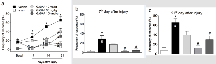
Figure 1: Influence of GABAP on the hyperalgesia observed in the sciatic nerve crush model in mice. Data are expressed as the mean ± standard error of the mean (n= 8 animals). (*)represents the comparison with the sham group; (#)represents the comparison with the vehicle group. Two-way (Panel a) or One-way ANOVA (Panels b and c), followed by the Dunnets’s test; P  0.05.

biomedres-openaccess-journal-bjstr

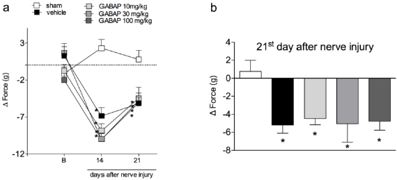
Figure 2: Influence of GABAP on the grip force in mice. Data are expressed as the mean ± standard error of the mean (n= 8 animals). (*)represents the comparison with the sham group. Two-way (Panel a) or One-way ANOVA (Panel b), followed by the Dunnets’s test; P  0.05.

When animals were treated with a combination of GABAP (30 mg/kg) with different doses of ATORV (3 and 10 mg/kg), as shown in (Figure 3, Panel A) there was no difference statistically significant on this antihyperalgesic effect in the tests performed on day 7 (Figure 3, Panel B). On the 21st day after the injury, the percentages of inhibition of the hyperalgesic effect induced by the lesion after the different treatment groups were sham: 8.57 ±4.04 % of response, vehicle 76.00 ± 11.66% of response, GABAP 30 mg/kg: 26.67 ± 4.21% of response, ATORV 3 mg/kg: 36.67 ± 10.85% of response, GABAP+ATORV 3 mg/kg: 8.57 ± 4.04% and GABAP+ATORV 10 mg/kg: 10.00 ± 4.47% of response (Figure 3, Panel C). Regarding motor function, ATORV alone (3 mg/kg), as well as GABAP 30 mg/kg) did not influence the effect of reducing the grip force of the affected limb induced by the nerve injury in the evaluated periods (Figure 4, Panel A). On the other hand, as can be seen in Figure 4, on the 21st day after sciatic nerve injury (Panel B) the mean values of the right hind paw grip force compared to the observed at paw of the animals treated with ATORV 3 mg/kg+ GABAP 30 mg/kg (-1.29 ± 1.22 g) or ATORV 10 mg/kg+GABAP 30 mg/kg (-1.72 ± 1.74 g) were no longer statistically significant in relation to the sham (2.00 ± 1.38 g) and vehicle (-4.19 ± 0.57 g) groups.

biomedres-openaccess-journal-bjstr

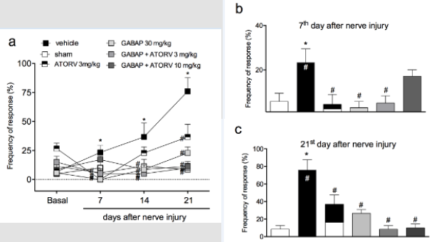
Figure 3: Influence of ATORV, alone or in combination with GABAP on the hyperalgesia observed in the sciatic nerve crush model in mice. Data are expressed as the mean ± standard error of the mean (n= 8 animals). (*) represents the comparison with the sham group; (#)represents the comparison with the vehicle group. Two-way (Panel a) or One-way ANOVA (Panels b and c), followed by the Dunnets’s test; P  0.05.

biomedres-openaccess-journal-bjstr

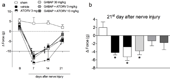
Figure 4: Influence of ATORV, alone or in combination with GABAP on the grip force of the affected limb at different time periods in mice. Data are expressed as the mean ± standard error of the mean (n = 8 animals). (*)represents the comparison with the sham group. Two-way (Panel a) or One-way ANOVA (Panel b), followed by the Dunnets’s test; P  0.05.

The results shown in Figure 5 demonstrate that the levels of BDNF (105.30 ± 12.53 pg/mg protein) and IGF-1 (399.60 ± 61.30 pg/mg protein) were increased in animal groups (11.51 ± 3.69 pg/mg of protein and 25.84 ± 4.91 pg/mg of protein, respectively) submitted to the nerve injury by crushing (vehicle groups). In addition, for BDNF (Figure 5, Panel A) treatment with either GABAP (34.92 ± 5.92 pg/mg protein) or ATORV alone (33.77 ± 4.20 pg/ mg protein), or in combination (30.33 ± 5.88 pg/mg protein), promoted a reduction in the levels of this neurotrophin in relation to the group with nerve damage (data above). Despite this, no statistically significant differences were observed in the mean values of the three treatment groups. On the other hand, in the case of neurotrophin IGF-1 ±, while the treatment of animals with GABAP (388.30 ± 38.57 pg/mg protein) or ATORV (306.50 ± 16.72 pg/mg of protein) alone did not change in relation to the group that suffered the nerve damage (data above), the association of the two drugs caused a statistically significant reduction in relation to this group (210.40 ± 23.92 pg/mg protein). The percentage inhibition of IGF-1 levels in the group treated with drug combinations was 47.35% relative to the vehicle group. Finally, no changes in IL-10 levels were observed between any of the study groups (Figure 5, Panel C).

biomedres-openaccess-journal-bjstr

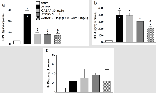
Figure 5: Influence of ATORV, alone or in combination with GABAP on the levels of IL-10, BDNF and IGF-1 in the sciatic nerve. Data for BDNF and IGF-1 are expressed as mean ± standard error of the mean (n= 8 animals), while for IL-10 are presented as median ± interquartile range. (*) represents the comparison with the sham group; (#) represents the comparison with the vehicle group. One-way ANOVA followed by Dunnets’s test; P  0.05.

Discussion

The mechanisms responsible for nerve regeneration and chemical factors are not completely elucidated. Some patients already use some medications of continuous use, including statins in older patients which can be associated to other drugs to relieve pain and prevent neuropathies, such as gabapentinoids. The effects of these drugs and their combinations are under invetigated, justifying the interest in assessing the effect of the association of ATORV with GABAP on parameters of sensory and motor activity in a model of crushing-induced nerve injury. Our data that GABAP was antihyperalgesic on crushing model agree with the literature, since the GABAP also showed reduction of sensorial information in other studies involving thermal hyperalgesia or motor function. Besides of this, it broadens those in the literature, since no records were found for the effects of GABAP on mechanical hyperalgesia and grip strength in mice after sciatic nerve crush injury. In respect to that observed to ATORV in the present study, it was observed that this drug alone promoted antihyperalgesic activity in the model between days 7 and 21 after the injury. The association of ATORV to GABAP did not alter the antihyperalgesic effect of GABAP alone on day 7, but the increase in antihyperalgesic action on the 21st day was evident.
These findings reinforce the benefit observed with the treatment of animals with ATORV in models where their neuroprotective action has been investigated; in rats with ischemia and multiple sclerosis, the drug protected the neurons from the injury caused by free radicals or limited the secondary damage after spinal cord injury. Regarding the possible mechanisms associated with this effect of gapapentinoids on hyperalgesia and motor recovery, it could involve aspects of nerve regeneration. For ATORV, it is known that statins, particularly ATORV and sinvastatin, have been extensively studied in vivo for their positive neuroprotective effect on different models [25-27]. Thus, a synergistic effect between these two drugs may be suggested, especially if we evaluate our other findings for the association of ATORV to GABAP on the grip strength and BDNF levels, as discussed below. Statins are also related to pleiotropic effects such as anti-inflammatory, antiproliferative, antithrombotic properties and antioxidants.
In the present study, ATORV alone promoted an antihyperalgesic effect, this being another fact that had not yet been reported in the literature. However, since the mean values of hyperalgesia for both ATORV and GABAP, alone, reached values close to the control, it was not possible to observe a possible synergistic effect between both regarding this parameter. However, although in the grip force test the association of ATORV to GABAP did not promote a statistically significant improvement over this parameter, a rather important trend in this sense was noted by the fact that in this group the mean values of loss of grip strength decreased significantly in relation to the GABAP group alone and were very close to the values of the control group. The almost significant effect of the association of ATORV with GABAP on motor recovery is striking, since in addition to the actions of these two drugs on nerve protection mentioned above, another action that could add to this would be a possible control of the inflammatory process; this idea is reinforced especially if we consider that the antihyperalgesic effect of both GABAP and ATORV are already observed on the 7th day after the injury, that is, a period in which the inflammatory component is more present.
Studies have shown that the suppression of the release of inflammatory cytokines after experimental injury of the peripheral nerve reflects a positive effect on neurological recovery. Despite this, it should be remembered that some researchers have also reported that statins may be toxic to neurons; in these cases, the toxicity seems to depend on factors such as the structural characteristics and the concentration of the agent employed, as well as the cholesterol content of the affected neural cell. In the present work, a finding that reinforces a possible anti-inflammatory action of the drugs ATORV and GABAP is that the levels of BDNF and IGF-1 in the nerve, increased after the nerve injury by crushing, after treatment with both drugs, alone or in association, have been reduced. The determination of the possible influence of the treatments with these two agents on these two mediators is justified, since studies focusing on the speed of axonal regeneration in the distal part of the nerve have so far focused on three groups of factors, including trophic factors such as IGF-1, IGF-2 and BDNF, as well as the neurotrophic factor derived from glial cell line and cytokines (IL- 6 and IL-10). It is known that BDNF and IGF-1 are induced in the distal part of the injured peripheral nerves.
Thus, the fact that in the present study, the levels of BDNF in the nerve have reduced after treatment with ATORV and GABAP may suggest that by treatment with them there is a control of the inflammatory process that decreases the need for expression of this factor, even occurring for IGF-1 when treated with the combination of ATORV and GABAP. Confirmation for a possible anti-inflammatory effect of treatments in the present study was not possible since no changes were observed in the levels of IL-10 for any treatment in relation to the control group (sham). This is an anti-inflammatory cytokine that can inhibit the production of other proinflammatory cytokines, and its expression is increased in the distal portion of the sciatic nerve seven days after its crush injury.

Conclusion

the findings of present study demonstrate the beneficial effect of GABAP or ATORV, alone, on mechanical hyperalgesia in the sciatic nerve crush model in mice. In addition, an important effect for the association of these drugs was observed on the grip strength in this model, and the influence of the two agents on the inflammatory process following the nerve injury may serve as a possible mechanism of action for the observed effects. As a perspective, it is suggested that the association of substances of different pharmacological classes, such as GABAP and ATORV, in products aimed at the treatment of comorbidities associated with nerve lesions can be evaluated in humans, mainly because they are already used in the population that is more neuropathic conditions, and epidemiological studies such as clinical trials may confirm the potential benefit raised here.

For More Articles: Biomedical Journal Impact Factor: https://biomedres.us

Biomedical Open Access Journals

Chitosan Derivatives – Promising Sustainable Biopolymers for Quality Healthcare Textiles

Introduction

The consumers are now increasingly aware of the hygienic lifestyle and there is necessity and expectation for high quality healthcare textiles. That’s why it is one of the most important, continuously expanding and growing fields in technical textiles. Healthcare textiles represent structurally designed and accomplished for a new health and hygiene-related textiles products for the well-being of mankind which applications is diverse, ranging from hospitals, hotels to personal care products. Because of their importance, a number of chemicals have been employed to impart antimicrobial activity to textile materials. These chemicals include inorganic salts, organometallics, phenols and thiophenols, onium salts, antibiotics, heterocyclic compounds with anionic groups, nitro compounds, urea and related compounds, formaldehyde derivatives, amines and synthetic dyes [1]. However, with the public’s enhanced awareness of eco-safety, there has been considerable debate about their use, because majority of such agents are toxic to humans and are not environmentally friendly. In addition, another big concern is that some of these agents are being increasingly resisted by microbial pathogens. Therefore, the role of textile finishers has now become increasingly demanding and has strengthened the interest in alternative ecofriendly and biodegradable finishing agents [2-5].
Chitosan is the deacetylated derivative of chitin that is the second most abundant polysaccharide found on earth next to cellulose. When chitin is deacetylated over about 60% it becomes soluble in dilute aqueous acids and is referred to as chitosan. Chitin is the main component in the shells of crustaceans, such as shrimp, crab [6-9]. Huge amounts of crab and shrimp (prawn) shells have been abandoned as wastes by worldwide seafood companies. This has led to considerable scientific and technological interest in chitin and chitosan as an attempt to utilize these renewable wastes. The applications of chitosan include uses in a variety of areas, such as pharmaceutical and medical applications, paper production, textile dyeing and finishing, fiber formation, wastewater treatment, biotechnology, cosmetics, food processing, and agriculture [10,11]. There is a greater demand for antimicrobial finishes on textile goods. Chitosan, a natural biopolymer, has many chemical attributes, especially its cationic nature, to make it an interesting candidate for these applications. However, the major problems of chitosan are its loss of the antimicrobial activity under alkaline conditions due to its loss of the cationic nature. But chitosan derivatives will overcome that problem due to retain antimicrobial activity in wide range of pH that could have a strong economic, social, and environmental impact, especially in our country.
In view of these ecological, environmental concerns and to overcome the drawback of chitosan, we are going to explore chitosan derivatives which will in turn help to develop next generation healthcare textiles. This will be followed by a focus on some recent developmental works pertaining to antimicrobial finishing of textiles using various “green chemistry” approaches in order to provide safe and novel antimicrobial textiles for aesthetic, hygienic, and healthcare applications in the near future. The specific objectives of the present project are: preparation of sustainable chitosan derivatives from prawn shell waste, modification of cotton fabric with chitosan derivative and antimicrobial assessment and different characterization of modified cotton fabric as for healthcare textiles. The prepared chitosan and its derivatives will be applied on cotton fabric and the modification will be confirmed using FTIR, TGA and DTA techniques. As quality healthcare textile, antimicrobial test, water vapor transmission rate, swelling test, and tensile strength of finished and unfinished cotton fabric will be investigated.

Experimental

Materials

Shrimp shell was collected from Mongla (near Sundarban forest), Khulna, Bangladesh that are waste of shrimp processing area. Cotton fabrics were collected from Keya spinning mills Ltd., Bangladesh. Escherichia coli and Staphylococcus aureus bacteria are collected from “Biochemistry and Molecular Biology” department of Rajshahi University. All chemicals that were used in the present investigation were of reagent grade.

Methods

Processing of Prawn Shell Waste: Prawn shell was collected, washed, dried, and ground to 40-60 mesh using a hammer mill. The ground prawn shell was then ready for a series of chemical treatment for extraction of chitin. Through a set of chemical treatments demineralization, de-protenization and decolouration of chitin was obtained. Later deacetylation of chitin gave chitosan [12,13].
Preparation of TMC: TMC was synthesized through nucleophilic substitution with CH3I as a reagent, sodium iodide as catalyst and sodium hydroxide (NaOH) as base with N-methyl pyrrolidone (NMP) as solvent [14].
Preparation of CMTMCC: CMTMCC was synthesized by reductive carboxymethylation of TMCC that was accomplished by a chemical reaction between TMCC and monochloroacetic acid in the presence of sodium hydroxide. An amount of 0.1 g of dried TMCC was suspended in 10 mL of NMP or isopropanol as solvent and left to stir overnight at 45°C. After 20 min, under continuous stirring, 0.5 mL of 50% NaOH was slowly added to the reaction mixture that was stirred for another 45 min. Subsequently, 0.12 g of monochloroacetate (molar ratio 1:3 to TMC) was added for about 25 min and reaction was left for 3 h at 60 °C under a nitrogen atmosphere in order to avoid TMC degradation. Afterwards, pH was adjusted to neutral with 1M HCl followed by filtration, washing with 70% ethanol and subsequently with anhydrous ethanol. CMTMC precipitate was dried under vacuum, dissolved in distilled water and dialyzed for 3 days against water, changing dialysis medium twice per day. The dialyzed CMTMCC was lyophilized and later analyzed [15].

Results and Discussion

Yield Percentage of Chitin, Chitosan and its Derivatives

The percentage yield was calculated from the ratio of the dried raw sample and the weight of obtained product. This obtained percentage yield is shown in Table 1. The yield percentage of chitosan, TMCC and CMTMCC was 66.5, 102 and 93 % respectively.

biomedres-openaccess-journal-bjstr

Table 1: Yield % of chitosan and its derivatives.

FTIR Analysis

In the FTIR spectrum of the prepared chitosan shown in Figure 1a, there were two absorption peaks at 1631 cm-1 and 2919 cm-1, which correspond to the N−H bending of the primary amine, and the presence of hydroxyl groups respectively. Obvious changes of the FTIR spectra are observed after quaternization of chitosan with methyliodide. In the FTIR spectrum of TMCC, a characteristic peak at 1460 cm-1 indicates the C-H bending of trimethyl ammonium group thus shows evidence of the quaternary ammonium salt group. It should be also noted that the N−H bending (1630 cm-1) of the primary amine disappeared due to the change of the primary amine to the secondary aliphatic amine [16]. A new peak at 1740 cm-1 was also attributed to the quaternary ammonium in TMCC which is shown in Figure 1b. In addition, the spectrum shows a broad band at around 3400 cm 1, probably due to the increased number of hydroxyl groups. The FTIR spectrum of the CMTMCC is shown in Figure 1c. The characteristic peaks of amine (N-H) vibration appeared at 1594 cm-1 for chitosan. Two new absorption peaks at 1401 cm-1 and 1610 cm-1 were attributed to the methyl group of ammonium and carbonyl group in CMTMCC.

biomedres-openaccess-journal-bjstr

Figure 1: The FTIR Spectra of, (a) Chitosan; (b) TMCC; (c) CMTMCC.

The profiles of FTIR spectroscopy of the chitosan, TMCC and CMTMCC synthesized from chitosan are almost similar but have different characteristic peak which shows evidence of the conversion of chitosan to TMCC and CMTMCC.The FTIR spectra of unmodified, chitosan, TMCC modified and CMTMCC modified cotton fabrics were shown in Figure 2. The obtained spectra of both types of fabrics were mostly similar except the new additional peak in the modified cotton fabrics. In case of chitosan modified cotton fabric a new peak at 1631 cm-1 was appeared due to the N-H bending of primary amine. Similarly, characteristic peaks at 1460 cm-1 (C-H bending of trimethyl ammonium group) and 1610 cm-1 (carbonyl group) were attributed to TMCC and CMTMCC modified cotton fabric.

biomedres-openaccess-journal-bjstr

Figure 2: The FTIR spectra of, (a) Unmodified; (b) Chitosan modified; (c) TMCC modified; (d) CMTMCC modified cotton fabric.

Surface Morphology

Surface morphology of the unmodified and modified cotton fabric were evaluated using three dimensional images getting from scanning electron microscope Figure 3. Unmodified cotton fabric exhibits regular surface as well as no adherence. Besides modified fabrics were overlaid with mentioned modifier which reveals little bit irregular, or microstructure roughness helps to increase surface area as well as antimicrobial functionality [17]. Conglomerated granule was evidently visible on the fiber surface which proofs the attachment of applied modifier on the fabric.

biomedres-openaccess-journal-bjstr

Figure 3: The SEM images of, (a) Unmodified; (b) Chitosan modified; (c) TMCC modified; (d) CMTMCC modified cotton fabric.

Antimicrobial Activity

Minimum Inhibition Concentration (MIC) describes the minimum concentration of the modifier in which concentration the multiplication of the bacteria is inhibited. For being different in structure, different modifiers have variation in antimicrobial activity. The Table 2 & Figure 4 show that antimicrobial activity of chitosan, TMCC and CMTMCC treated modified fabric. Chitosan have antimicrobial activity against both E. coli and S. aureus, because of the positive charge on the C-2 of the glucosamine monomer below pH 6. The exact mechanism of the antimicrobial action of chitosan, and their derivatives is still unknown, but different mechanisms have been proposed. Interaction between positively charged chitosan molecules and negatively charged microbial cell membranes leads to the leakage of proteinaceous and other intracellular constituents. Chitosan acted mainly on the outer surface of the bacteria. At a lower concentration the polycationic chitosan does probably bind to the negatively charged bacterial surface to cause agglutination, while at higher concentrations, the larger number of positive charges may have imparted a net positive charge to the bacterial surfaces to keep them in suspension [18,19]. Chitosan interacts with the membrane of the cell to alter cell permeability. But the problem is that it has lower solubility above pH 6 and also its lower attachment capacity on cotton fabric [20]. For TMCC, the mechanism of antimicrobial activity using on cotton fabric is almost similar to chitosan. The slight difference in antimicrobial activity of chitosan and TMCC is due to having positive charge in nitrogen atom and its higher solubility in water than chitosan.

biomedres-openaccess-journal-bjstr

Table 2: Antimicrobial activity of chitosan, TMCC and CMTMCC treated modified fabrics.

biomedres-openaccess-journal-bjstr

Figure 4: Antimicrobial activity of unmodified and modified cotton fabric against S. aureus and E. coli stain.

The antimicrobial action is believed to occur when the compounds are absorbed onto the bacterial cell surface, increasing the permeability of the lipid cell membrane and causing death through the loss of essential cell materials. In addition, these derivatives of chitosan are generally more active against grampositive and gram-negative bacteria than chitosan. This effect is believed to be due to adsorption of the polymers onto the bacterial cell surface and membrane with subsequent disruption of membrane integrity. Antimicrobial activity generally increases as the content of the quaternary ammonium moiety increases comparatively than chitosan. CMTMCC has better antimicrobial activity than chitosan and TMCC. Here the mechanism is approximately same to them but the difference is that the attachment capacity of CMTMCC (MIC=5.8mg/L for E. coli and 3.3 mg/L for S. aureus) is more than chitosan and TMCC on the cotton fabric.

Wicking, Moisture Absorption, Moisture Liberation and Swelling Test Result

Wicking Test: Wicking is the spontaneous flow of a liquid in a porous substrate, driven by capillary forces. Because capillary forces are caused by wetting, wicking is a result of spontaneous wetting in a capillary system. The wicking test results for untreated cotton fabric, chitosan, TMCC and CMTMCC modified cotton are given in Table 3. Untreated cotton fabric shows lower wicking height than the treated cotton fabrics due to the absence of the coating of chitosan, TMCC, and CMTMCC having channel like structure on cotton fabric. Chitosan and its derivatives show higher wicking height (cm). TMCC and CMTMCC have also higher wicking height (cm) comparatively than chitosan treated fabric. The reason for this is TMCC and CMTMCC have channel like structure same as chitosan and also have the presence of positive charge which helps to attract water molecules.

biomedres-openaccess-journal-bjstr

Table 3: Wicking, moisture absorption, moisture liberation and swelling test result of unmodified and modified cotton fabric.

Moisture Absorption: The properties of cotton fiber are strongly affected by moisture content because it is a hygroscopic fiber and absorbs or desorbs moisture from the surrounding atmosphere. In general, the fibers that absorb the greatest amount of moisture are the ones whose properties change the most. The main types of properties affected are: dimensional, mechanical and electrical. The molecular structure of the cotton fiber includes hydroxyl groups that attract water molecules and hydrogen bond with the cellulose [21]. Ability to retain a liquid in the fabric is known as absorption. Absorption consists of several parts, first the fiber surface is wetted and the liquid is transported into the voids between the fibers and are absorbed into the fibers and diffuse [22]. The moisture absorption test results are given in Table 3. The rate of moisture absorption in untreated fabric sample decreased as compared to the treated fabric due to the absence of channel like structure. On the other hand, chitosan, TMCC, and CMTMCC treated cotton fabrics show more moisture absorption (%) due to the presence of hydroxyl groups (-OH) and channel like structure. Also for having the good attachment capacity on cotton fabric CMTMCC treated cotton fabric showed higher moisture absorption (%).
Moisture Liberation: The moisture liberation ability is one of the crucial factors for wounds healing, because wound dressing manages the moisture on the surface and bed of the wound by absorbing and releasing exudates. The Moisture liberation test results of untreated and treated cotton fabrics are given in Table 3. Untreated fabric can easily liberate moisture because of having low capillaries. On the other hand, chitosan, TMCC, and CMTMCC treated cotton fabrics have channel like structure and for this it is unable to moisture liberation.
Swelling Test: Moisture/liquid transport in textile fabrics is one of the critical factors affecting physiological comfort. In conditions where wearers sweat a lot (e.g. high-level bodily activity), it is not only desirable for the fabric next to the skin to absorb liquid rapidly but also to transport it through the fabric promptly to avoid the discomfort of the fabric sticking to the skin. The comfort afforded by textile fabrics can be improved by understanding the liquid transport mechanism [23]. The swelling capacity of an antibacterial fabric plays an important role in the antibacterial activity, wound healing capacity, and for biomedical application due to their high water/solvent holding capacity. They can further absorb a slight to moderate amount of the wound exudates by swelling which helps in fast healing of the wound. The swelling test results are list in Table 3. Swelling % of untreated fabric is lower than others because of lacking the cross-linked channel structure to hold water for being swollen. Then the swelling capacity of CMTMCC is more than others for having not only the presence of hydrophilic groups in the film networks – as like as chitosan and TMCC which assist in improving the swelling characteristic of the fabric but also N atom containing positive charge and higher attachment capacity to the fabric.

Water Vapor Permeability (WVP) Test

Bolton studied a variety of dressings and determined that a WVTR of about 840 g/m2/day is required to maintain a moist wound surface [24]. Water Vapor permeability occurs mainly for having different vapor pressure on both sides of the cotton fabrics. The WVP test results are given in Table 4. WVP of untreated cotton fabric is high because covering of fabrics with modifiers isn’t occurred on it. For Chitosan, TMCC, and CMTMCC treated fabrics WVP comparatively increases due to the presence of hydroxyl groups. Here after applying Chitosan, TMCC, and CMTMCC on cotton fabric hydrophilic properties are raised and channel like structures are formed which attract water vapor and help to pass away to the outer side of the fabric to saturate the outer side. Among of them CMTMCC have higher WVP because it further has higher attachment capacity on cotton fabric causes more water vapor permeability through cotton fabrics. Fabrics used for wound dressing should have neither higher nor lower WVP value. So for this CMTMCC treated cotton fabrics are more suitable to provide moist wound condition.

biomedres-openaccess-journal-bjstr

Table 4: WVP (g/m2/day) test of unmodified and modified cotton fabric.

Tensile Strength Test

The tensile strength is one of the most important physical properties of the fabrics. The tensile strength of the fabric contributes greatly to its quality as well as its life. The strength of the fabrics depends on of many different factors that affect it. Molecular structure, temperature, composition, are some of the factors that are responsible for the increase or decrease in the tensile strength of fabric. It is well known that chitosan significantly imparts stiffness in fabric and affect tensile strength of fabric. In this study cotton fabrics are bio-scoured in presence of cellulase enzyme and for this hydrolysis is occurred which causes the loss of tensile strength of cotton fabric. Then to regain the tensile strength the cotton fabrics are treated with chitosan and its derivatives. Tensile strength test results are listed in Table 5. Tensile strength of untreated fabric is low due to hydrolysis during bio-scouring with cellulase. For the chitosan, TMCC, and CMTMCC treated cotton fabrics, tensile strength increases because of binding between fibres and yarns. Chitosan, TMCC, and CMTMCC to bind fibres and yarns and increases tensile strength. Among the samples, CMTMCC treated fabric have higher tensile strength than others.

biomedres-openaccess-journal-bjstr

Table 5: Elongation % and Tensile strength test (N) of unmodified and modified cotton fabric.

Thermal Analysis

Figure 5 shows TGA and DTA curves of unmodified-, chitosan modified-, TMCC modified- and CMTMCC modified cotton fabrics. It can be seen that the initial decomposition temperature of unmodified cotton fabric is 225OC as shown in Table 6. Also, for DSC curve it is observed that there is a exothermic peak at 450°C which corresponds the burning of the char present in cotton fabric during oxidation. Figure 5b represents TGA and DTA curve for chitosan treated cotton fabric, from this Figure it is obtained two exothermic peaks are seen which correspond to the burning of the incorporated hydrogens and the burning of the char of cotton fabric. The initial decomposition temperature of chitosan cotton fabric is 310OC as shown in Table 6. Now from Figure 5c it is obtained that at 50°C phase transition is occurred, at 80°Can endothermic peak is appeared due to melting behavior and at 340°C another endothermic peak is occurred for the same at 50°C. In this Figure the Initial decomposition temperature was 315°C. And Initial decomposition temperature of CMTMCC modified cotton was 290°C as shown in Table 6.

biomedres-openaccess-journal-bjstr

Figure 5: TGA and DTA curves of, (a) Unmodified cotton; (b) Chitosan modified cotton; (c) TMCC modified cotton; (d) CMTMCC modified cotton.

biomedres-openaccess-journal-bjstr

Table 6: TGA and DTA data for unmodified and modified cotton fabric.

Conclusion

Different experimental methods were established that chitosan, TMCC and CMTMCC were successfully attached on cotton fabric by pad-dry-cure method. Among the samples, CMTMCC treated fabric have higher tensile strength than others. The rate of moisture absorption in untreated fabric sample decreased as compared to the treated fabric. Antimicrobial activity is the main factor for medical and health care textiles. In this study, chitosan derivatives TMCC and CMTMCC treated cotton fabric exhibited excellent antimicrobial performance. It is expected that this sustainable chitosan derivatives-based healthcare textile retaining durable antimicrobial activities that will be used in hospitals, hotels and personal cares textiles as not only fashion and comfort but also safeguard for human health without disturbing environment. At the same time, the project will help to utilize the by-product or wastage of prawn processing industries.

For More Articles: Biomedical Journal Impact Factor: https://biomedres.us

Journal of Scientific and Technical Research

Antioxidant Capacity and Antitumoral Activity of Citrus Paradisi Essential Oil

Introduction

Phytomedicine has been of increasing interest in recent years. Over 65 % of the world’s population relies on traditional and medical approaches to treating diseases [1]. The return to phytotherapy is more than using an herb to treat an illness or a crude herbal preparation rather than a specific isolated component. Plants are considered essential resources for researchers to prove and develop new drugs. Generally, using plants in the treatment of disease has a long history [2,3]. Thus plants have been primary resources for producing traditional drugs effective in treating cancer and inflammatory diseases [4]. In addition, plants containing essential oils have bioactivity against a host of bacteria, fungi, viruses [5,6]. Currently, essential oils of medicinal plants are increasingly used thanks to their antibacterial, anti-inflammatory, antiseptic, antiviral, antifungal, antioxidant, and antitumor potential [7]. Essential oils are also used in other areas of economic interest, such as cosmetics, perfumes, and aromatherapy [7,8]. Fragrant and aromatic plants such as members of the Asteraceae, Lamiaceae, Rutaceae, and Verbenaceae produce essential oils, which historically have been important in traditional medicines [5]. Citrus species have beneficial pharmacological activities, including antibacterial, vascular protectant, antispasmodic, analgesic, antipyretic, antiinflammatory, and antitumoral effects [9-11].
Citrus peel essential oils have also been searched for their natural antioxidant and antimicrobial properties [12,13]. Furthermore, studies have shown the synergistic effect of two or more ingredients of essential oils against various human pathogens. It is necessary to know more about Citrus paradisi essential oil (CPEo) antioxidant, cytotoxic, and antitumor activity. Therefore, the DPPH, ABTS, FRAP, and NO• techniques studied the antioxidant potential in a chemical system. Furthermore, to check the antitumor activity, MTT was realized using HeLa and MCF-7 tumor cells.

Material and Methods

Samples

In this study, the grapefruit (citrus paradisi) was collected in March 2018 from Sfax, Tunisia. Sfax is one of the biggest coastal cities in Tunisia. It is located in the eastern part of the country. The climate in this area is arid to semiarid with irregular and torrential precipitations [14]. After the collection, it was washed, peeled off and cut into small pieces.

Hydrodistillation

The zest of citrus paradisi was hydro distilled using a Clevengertype apparatus to recuperate the essential oils for 2h to produce the volatile constituents. The distilled essential oils were dried over anhydrous sodium sulfate; then separated from the distillate by liquid-liquid extraction using cyclohexane solvent. The recuperated oils were stored at + 4 °C.

Antioxidant Activity: Chemical System

DPPH Radical Scavenging Assay: Different aliquots from the stock solution (200μl in 1 ml Me OH) of essential oils were mixed with 500μl of 0.2mM diphenyl picryl hydrazine (DPPH) final volume brought to 1mL. The mixtures were vigorously shaken and allowed to stand in the dark for 30min at room temperature. The absorbance was measured by spectrophotometry (LKB BIOCHROM® ULTROSPECPUS 4054 UV/VIS) at 517 nm against a control sample without DPPH. The percentage of radical scavenging activity was calculated using the following equation:

DPPH scavenging effect (%) = (A0- A1)/A0 × 100

A0: The absorbance of the control at 30min A1: The absorbance of the sample at 30min.
ABTS+ Radical Scavenging Effect: The antiradical activity was performed by the ABTS+ free radical decolorization assay as developed by Re, et al. [15]. The 2,2-azino-bis-3- ethylbenzothiazoline-6-sulfonic acid (ABTS) was prepared as an aqueous stock solution (7mM). The ABTS radical cations (ABTS+) were produced by the reaction of the ABTS stock solution with 2.5mM of ammonium persulfate methanolic solution. First, the reaction mixture is incubated in the dark for 16h at room temperature. Then, the solution is diluted to an absorbance of 0.7 ± 0.02 at 734nm to form the working reagent. Next, the reaction mixtures containing 100μl of the sample at different concentrations and 900μL of reagent were incubated at 30 °C for 6min. Finally, the antioxidant power of each sample was expressed as the inhibition percentage calculated according to the following formula:
ABTS+ scavenging effect (%) = (A0- A1)/A0 × 100
A0: the absorbance of the control at 6min
A1: the absorbance of the sample at 6min.
Ferric-Reducing Antioxidant Power (FRAP): This method is based on the plant’s ability to reduce ferric iron (Fe3+) to ferrous iron (Fe2+). The mechanism is known to be a marker of electron donor activity [16]. For 1mL of the sample at different concentrations, 2.5mL of a solution phosphate buffer (0.2M, pH 6.6) and 2.5mL of 1 % K3Fe (CN) 6 potassium ferricyanide solution were added. The mixture is incubated at 50 °C for 20min and then cooled to room temperature. Then, 2.5mL of 10 % trichloroacetic acid (TCA) is added to stop the reaction, and then the tubes are centrifuged at 3000rpm for 10min. 2.5ml of the supernatant are then added to 2.5mL of distilled water and 500μL of a 0.1 % solution of iron trichloride (FeCl3, 6H2O) [16]. The absorbance reading is performed against a blank at 700 nm using a spectrophotometer. Ascorbic acid is used as a positive control. The increase in the absorption capacity of the components indicates the increase in the reduction of iron.
Scavenging Activity of Nitric Oxide (NO·): NO scavenging activity of the essential oils was determined as previously described [17]. Briefly, 0.1mL of the essential oils (0–0.3mg/ml in DMSO) was incubated with 0.5mL of sodium nitrite (0.01mg/mL in 100mM sodium citrate pH 5) at 37 °C for 2h. After incubation, 0.5mL of Griess reagent was added, and the absorbance was read at 540nm using a spectrophotometer (Pharmacia, Uppsala, Sweden). The equation obtained the percentage of RNS scavenging:
NO· Scavenging effect (%) = (A0- A1)/A0 × 100
A0: The absorbance of the control
A1: The absorbance of the sample.

Cytotoxicity Activity

Cell Lines and Culture Conditions: In this study, cancerous cells were used: HeLa and MCF-7. HeLa is a transformed cell line expressing the HPV18 virus (human papillomavirus) [18]. MCF-7 (Michigan Cancer Foundation-7) was isolated from a 69 years old woman with metastatic disease [19]. Hela and MCF-7 cell lines were supplied by ATCC (Manassas, VA, USA). All cells were grown in RPMI 1640 medium (Gibco) supplemented with 10 % (v/v) foetal calf serum (FCS) and 2mM L-glutamine in tissue culture flasks (Nunc). They are incubated at 37 °C, 95 % air and 5 % CO2.
MTT: For the test of cytotoxicity activity, the MTT (3-(4,5-dimethylthiazol-2-yl)-2,5-diphenyltetrazolium bromide) (Sigma-Aldrich, St. Louis, MO, USA) test was done [20]. First, HeLa or MCF-7 cells in 96-well plates were exposed to different concentrations of CPEo (3 wells for each concentration) and incubated for 48h at 37 °C. Then, 20μl of MTT was added to each well. After 4h of incubation at 37 °C, the supernatant was removed, 180μL of DMSO/Methanol (1V/1V) were added to each well to solubilize the formazan crystals. Finally, the plates were shaken 15min at room temperature, and the absorbance was detected at 570nm with a spectrophotometric plate reader.

Statistical Analysis

The statistical studies are carried out using the program SPSS (19.0). The t-Student test carried out the comparison between the averages. The results are represented as mean ± standard deviation (SD).

Results

The Yield of Grapefruit Essential Oils

The yield of the extraction is defined as the ratio between the mass of essential oil obtained and the mass of the plant material.
Y (%) = A/A1 ×100
A: Quantity of extracts recovered in g.
A1: Quantity of dry vegetable matter used for extraction expressed in g.
CPEo yield was 1.32g Eo / 100g dry matter. This result proves that the zest of the fruit was rich in Eo.

Antioxidant Effects

DPPH Radical Scavenging Assay: The curve relating to DPPH radical scavenging shows that the inhibition percentage of the free radical increases with the increase of concentration, either for BHT or CPEo (Figure 1). In effect, Citrus paradisi essential oil has a significant antiradical effect attending 54 % and can be qualified such having an important antioxidant activity.

ABTS+ Radical Scavenging Effect: The results illustrated in the curve show that the percentage of inhibition gradually increases with the concentration of grapefruit essential oil. At a concentration of 1mg/mL, the inhibition rate reaches 83 % (Figure 2).

Ferric-Reducing Antioxidant Power (FRAP): The FRAP assay is usually used to measure the capacity of the sample to reduce the ferric complex to the ferrous form. The FRAP assay confirmed the antioxidant activity of CPEo, with values equal to 66 % (Figure 3).

biomedres-openaccess-journal-bjstr

Figure 1: Antiradical activity against the radical DPPH in the percentage of inhibition CPEo. Aliquots of various concentrations of CPEo (0, 0.2, 0.4, 0.6, 0.8, 1mg/mL) and standard BHT were mixed with DPPH and incubated in the dark. The antioxidant activity of CPEo was measured using a spectrophotometer at 517nm. Results were expressed as mean inhibition percentage (%) ± standard deviations (n = 3). BHT was used as the reference standard.

biomedres-openaccess-journal-bjstr

Figure 2: Anti-radical activity against the radical ABTS in percentage of inhibition of CPEo. Various concentrations of CPEo (0 to 1mg/mL) and acid ascorbic were mixed with FRAP reagents. The reduction of ferric ion (Fe3+) to ferrous form (Fe2+) by CPEo produces an intense blue light revealed as a change in absorption at 700nm. Results were expressed as mean inhibition percentage (%) ± standard deviations (n = 3). Ascorbic acid at various concentrations (0 to 1mg/mL) was used as standard.

biomedres-openaccess-journal-bjstr

Figure 3: Inhibition (%) of standard Vit C and CPEo by Ferric Reducing Antioxidant Power (FRAP) assay. Various concentrations of CPEo (0 to 1mg/mL) and acid ascorbic were mixed with FRAP reagents. The reduction of ferric ion (Fe3+) to ferrous form (Fe2+) by CPEo produces an intense blue light revealed as a change in absorption at 700nm. Results were expressed as mean inhibition percentage (%) ± standard deviations (n = 3). Ascorbic acid at various concentrations (0 to 1mg/mL) was used as standard.

NO· Scavenging Assay

The capacity of citrus paradisi essential oils to scavenge NO was also measured. CPEo was checked for its inhibitory effect on nitric oxide production. The inhibition on free radical percentage increases with elevated concentrations of essential oil and Vitamin C (Figure 4).

biomedres-openaccess-journal-bjstr

Figure 4: Inhibition (%) of standard Vit C and CPEo by Ferric Reducing Antioxidant Power (FRAP) assay. Various concentrations of CPEo (0 to 1mg/mL) and acid ascorbic were mixed with FRAP reagents. The reduction of ferric ion (Fe3+) to ferrous form (Fe2+) by CPEo produces an intense blue light revealed as a change in absorption at 700nm. Results were expressed as mean inhibition percentage (%) ± standard deviations (n = 3). Ascorbic acid at various concentrations (0 to 1mg/mL) was used as standard.

Cytotoxicity Activity

The MTT test studied the cytotoxic effect on HeLa and MCF-7 cells. Cells were cultured in 96-well plates for 48 h, in the presence and absence of the different concentrations of CPEo. The percentage of cytotoxicity was calculated. Our results showed an antitumor potential of the grapefruit essential oils on both cancer cells: Hela and MCF-7 according to the concentrations used: from 0.78 to 25μg / mL (Figure 5).

biomedres-openaccess-journal-bjstr

Figure 5:

(a) Study the cytotoxicity of the essential oil of the grapefruit outer wall in the HeLa
(b) and MCF-7 cell lines.
Cytotoxic activity of citrus paradisi essential oil on cancer cell lines. MTT assessed cell viability. The percent growth reduction was calculated from the extinction difference between treated cell culture and the control. Results are the means of three repetitions.

Discussion

The current study was designed to extract the essential oil from grapefruit. It was obtained by hydro distillation, which is the oldest method and the most used because it is very easy to achieve. Our results agree with studies, which confirmed that Citrus is rich in essential oil, and their yields varied from one plant species to another, ranging from 0.2 to 2.0 % [21]. Other work has shown that yield of Citrus paradisi essential oil collected in September in southern Taiwan was attending 0.37 %, and others were collected in October from Iran were 0.85 % [22,23]. So the difference in EOs yields may be related to the plant’s origin, the environmental conditions, time of collection, and extraction method [24]. The CPEo analyzed in this work showed potent radical scavenging activity. We have demonstrated that CPEo has an interesting antioxidant activity highlighted by the DPPH, ABTS+, FRAP, and NO. Our results agree with the literature, which showed that citrus EO possesses an anti-free radical activity DPPH of 17.7 to 64 % [7]. Other studies on grapefruit seed oil showed a high antioxidant potential (61 %) [25]. Furthermore, the effect of citrus paradisi essential oils on ABTS free radical was determined and exhibited a higher radical scavenging activity. Our results corroborate with other studies showing a significant antioxidant effect of C. paradisi EO against ABTS radicals [22]. In addition, the CPEo had a significant FRAP value, which was in accord with the results obtained by Jang et al. that confirmed the higher FRAP values in the peel of grapefruit [26]. The superoxide anion assay is commonly used to evaluate the superoxide anion radical-scavenging ability of plant extract. Therefore, CPEo was checked for their inhibitory effect on nitric oxide production in the present study, attending 47 %. Our results agree with studies that confirmed that the superoxide values of grapefruits varied from 36.83 % to 50.31 % in peels [27]. The antitumor potential of CPEo was tested on HeLa and MCF-7 tumor cells. Our results showed a considerable antitumor activity dependent on essential oil concentration. This result agrees with the work of Zu et al., which shows that essential oil from grapefruit exhibited an antiproliferative effect on MCF-7 cells [28]. Furthermore, Monajemi et al. studied the impact of different concentrations of essential oils of other species of the citrus family as Citrus limon, Citrus medica, and Citrus sinensis on MCF-7 and Hela [29]. They proved a significant decrease in viability in a dose-dependent manner in both tumor cells [29].

Conclusion

Citrus paradisi essential oils are well known for their flavor and fragrance properties and numerous aromatherapeutic and medicinal applications. Accordingly, the essential oils of Citrus species exhibited intense antioxidant activity. In addition, the essential oil of Citrus paradisi showed an antitumor effect against both cancers cells, HeLa and MCF-7.

For More Articles: Biomedical Journal Impact Factor: https://biomedres.us

Open Access Journals on Surgery

A Review Study on Legs Lameness and Weaknesses Assessment Methods in Commercial Broiler Farming in Pakistan

Introduction

The poultry business has expanded dramatically over the last fifty years, with more than 35 billion broiler birds produced each year [1]. Due to the economic benefits of increasing body weight, the need for enhanced growth rate and output has resulted in differential growth of body components, such as rapid muscular growth [2]. This expansion, however, is not accompanied by skeletal development. Lame broilers have trouble walking and, as a result, they can’t go to the feeder or drinker when they are hungry or thirsty [2]. Their quality of life is being harmed by their laziness. The presence of lameness in birds is significantly linked to their weight and fast development [3]. Furthermore, the broilers may experience discomfort as a result of mobility issues. It can reduce bird’s activity and cause a variety of issues, such as hock burns and chest dirtiness [4]. Broiler chicks are protected against frailty by a rapid development rate coupled by appropriate feeding, good management, ideal lighting and temperature, and a disease-free environment [5]. Leg diseases are caused by the lack of any of these components in combination with inherent weight-bearing features [3]. Infectious, developmental, metabolic, and degenerative diseases can all cause leg weakness. Leg ailment is associated with a higher rate of morbidity than mortality [3]. The handicapped bird, on the other hand, suffers from discomfort, is unable to obtain food and water, and eventually dies from inanition [6]. As a result, the initial goal of this study is to evaluate broiler leg deficiencies and offer readers with a brief explanation of the elements that influence this condition [7]. Nutritional deficiencies, mechanically produced injuries, toxins, genetic abnormalities, infections infectious illnesses, sex, weight and development rate, age, feed conversion efficiency, handling, and mobility are all variables that contribute to leg weakness [8]. The second goal of this research is to examine broiler lameness and lameness evaluation techniques.

Leg Problems

Broiler nutritional and physical density rose as a result of the food modifications. Higher nutrient intakes, as well as a combination of these two variables, have resulted in a significant increase in broiler growth rates [9]. To use this genetic potential for growth, the time until the chick reaches a live weight of 1500 g was lowered from 16 weeks to 4 weeks. Good highly concentrated feed in pellet form and intensive lighting cycles are necessary [10]. A high body weight has also been chosen for the creation of more breast muscles, in addition to a selection for quick growth to rap to accomplish. This causes the chicks’ conformation to alter, which can lead to bone deformities [11]. In addition to the welfare issues caused by leg difficulties, the producer will suffer a financial loss. Animals with serious issues have a poorer feed efficiency and develop at a slower rate. These animals’ carcass quality will deteriorate as well [12].

Occurrence

The majority of broiler lameness is caused by skeletal anomalies, although this is difficult to explain. Increases in the mortality rate, “the cull,” the percentage of rejected chicks, and “downgrades from trimming” [13] may result from the costs and losses incurred as a result of bone abnormalities. Broiler losses due to skeletal anomalies are considerable (Garner et al., 2002). At some stables, up to 90% of the birds in slaughter age show some degree of lameness [14], and roughly 30% of the birds are just too badly handicapped [15].

Behavioral Changes

A roaster’s day is spent for the most part. The hens appear to sleep around a quarter of the time and do nothing more than half of the time. A healthy chicken spends 76% of its time laying down. This proportion rises with age, and it appears to be substantially greater with a higher level of lameness (up to 86%).

Defects and their Types

Broiler lameness is caused by a variety of factors, including abnormalities and illness. Heavy, disease-free birds wander about her as though she’s in pain and want to stay. This might imply that the discomfort is caused by body weight and strain on the bones and tissues [16].

Abnormalities of the Bones

Nutritional deficiencies, mechanically produced injuries, toxins, and genetic flaws are all possible causes of bone abnormalities [17]. All of these features are more important in fast-growing chicken breeds. They require it for their quick expansion. Because their bones are not fully formed, they are more prone to damage as they consume more and more specialized nutrients. As a result, bone anomalies are prevalent in commercial broiler production.

Valgus-Varus

A “knock-kneed” look is caused by valgus deviation. When the tars metatarsus is lined up with the tibiotarsus, the tars metatarsus deviates laterally. It’s the most frequent type of long bone deformity, and it’s also the most dangerous [18]. This aberration gives the impression of being ‘bowlegged.’ The tars metatarsus differs in that it now wonders whether she will be positioned on a line with the tibiotarsus. This aberration is less frequent, but it can cause serious walking problems [19].

Dyschondroplasia of the Tibia (Td)

TD appears to be a dissociation of growth plate chondrocyte proliferation and leg extension during endochondral calcification [20].

Rickets

This is most likely the most well-known bone disease in poultry. When bone mineralization is decreased, this occurs. A lack of calcium and phosphorus, or an imbalance between the two, is the most prevalent cause [21]. ‘Rickets’ can affect both rapid and slowgrowing chicks, although they are exacerbated by the increased requirement for nutrition during growth.

Necrosis of the Femoral Head

The reasons of this condition are unknown, but it has the potential to impact the entire house. The incapacity of the broilers to stand up is the most obvious sign [22]. It is the end of the femur when an autopsy is performed on the birds. Although broilers can respond to vitamin D3 in drinking water, this approach does not always effective [23].

Chondrodystrophie

Shorter bones with various anomalies can occur in poultry lacking in water-soluble vitamins. If the deficit is severe enough, the supporting gastronomies tendon might slide cartilage [24].

Dermatitis due to Contact

Feet burns, also known as contact dermatitis, have been more common in broilers during the last few decades. Contact dermatitis is thought to be induced by the ammonium chemical’s action on the urea in the litter. There is also evidence that the occurrence and severity of contact dermatitis are related to the litter and air quality, and hence to welfare factors other than pain [25]. Because of the amount of time spent sitting and the poor quality of the litter, contact dermatitis is evident. The amount of time the chicks spend sitting and reclining rises with age, from 75% in the first week to 90% at five weeks [26]. Footpad dermatitis is a form of contact dermatitis that causes sores on the soles of chickens’ feet. Swelling and necrotizing tissue can develop or be seen in more severe instances [27]. This might pose a food safety issue since these lesions could be utilized as a bacterial entry point. As a result, the carcass quality suffers [28].

Viruses and Infectious Diseases

Pathogens

Reovirus, Mycoplasma sinoviae, Staphylococcus aureus, and Retroviruses are the most common infections that cause lameness.

Reovirus

Avian reoviruses, which cause viral arthritis and tenosynovitis (tendon sheath inflammation), are common in the broiler business. The virus is thought to propagate by ‘avian egg transmission,’ especially because the infection was discovered in seemingly normal commercial chicken embryos. Mild to moderate lameness characterizes the clinical picture, with enlarged “hocks” and a significant increase in fluid in the “hock” joints [29].

Mycoplasma Synovial

Mycoplasma Synoviae causes respiratory infections in hens and turkeys, as well as airsacculitis and synovitis. M. synoviae’s effects were originally discovered in the 1950s, and the organism is still known. The pathogenicity of distinct strains of M. synoviae is a distinguishing feature of the organism. The disorders of the respiratory tract and/or synovitis appear to be unaffected by any of these [30].

Retroviruses

The avian leucosis viruses are among the most significant retroviruses. They are further split into subcategories. One of the most economically important diseases in broilers is avian leucosis virus subgroup J (ALV-J). The nodes are made up of myelocytes with eosinophilic cytoplasmic granules. Other forms of lesions, such as lengthy sarcomas with aberrant feathers and myelocyte infiltration in the bones and periosteum of the sternum and ribs, may develop [31].

Staphylococcus Aureus

In the commercial Poultry, Staphylococcus aureus is a major source of disease. S. aureus infection can result in septicemia (the prevention of harmful bacteria and their toxins in the blood), bone and joint infections, abscesses, and dermatitis, among other clinical manifestations [32]. Such diseases affect animal welfare and result in financial losses at the slaughterhouse owing to downtime, lower production, and corpse contamination [33].

Mycotoxins

Mycotoxins are one of the dietary variables that induce skeletal illnesses including Rickets, Articular Gout, and Tibia Dyschonroplasia, as well as a number of other bone diseases in broilers [34]. Because mycotoxins such as aflatoxin, ochratoxin, and fusarium toxin are poisonous to the liver and kidney, they cause rickets by preventing vitamin D3 conversion and absorption [35].

Lameness

Lameness is the leading cause of poor wellbeing in broiler hens, according to European Commission research [36]. Lameness is a broad term for a variety of broiler chicken injuries caused by infectious and non-infectious sources [37]. In the broiler business, lameness is also a major issue. The financial losses caused by lameness in commercially raised broilers are significant [38]. The cost of lameness in the United States was estimated to be between $80 million and $120 million in 2010 [39].

Causes Lameness

Infectious diseases, heredity, sex, weight and growth rate, age, feed conversion efficiency, nutrition, handling, and movement are all variables that contribute to lameness. These aspects will be explored in greater detail further down [40]. The genetic selection and management of non-infectious and non-nutritional bone abnormalities are the most important variables here [40].

Weight and Growth Rate

The quickly rising weight will place additional demands on the juvenile skeleton, and the resulting form change may affect the forces generated throughout the run. The high ‘Gait Score’ in broilers might be due to discomfort, biomechanical issues, or a combination of the two [41]. Observed at the connection between lameness, weight, growth rate, and age. Weight and growth rate were found to be significant variables in the development of lameness [42].

Gender

Lameness affects both males and females. Male chicks, on the other hand, have greater difficulties than female chicks, even when body weight is taken into account. The male had a ‘gait score’ that was nearly half a unit higher [43].

Genetics

There is evidence that the prevalence of ‘leg weakness’ differs between different commercial breeds that perform similarly in other areas [44]. Various genetic studies have revealed that the aetiology of lameness is bone anomalies with a degree of heredity [45]. Lameness appears to be more frequent in some breeds than in others. As a result, positive selection against these traits is feasible [46].

Age

It was discovered that with age, one’s ability to walk correctly diminished [47]. The hens could still run well at four weeks of age. Less than 1% of these birds got a gait score of 4 or 5. At 6 weeks of age, broilers walked substantially poorly and could run significantly worse. The pace at which the chicks deteriorated was faster between 4 and 6 weeks than between 6 and 7 weeks [47].

Feed (Nutrients)

Nutrients have been demonstrated to be crucial for proper skeletal development. To avoid leg problems in broilers, they need a well-balanced feed. A lack of water-soluble vitamins, manganese, or zinc, for example, might result in shorter bones with valgus anomalies. In addition, a shortage of nutrition in fast-growing hens might aggravate ‘rickets’ [48]. Biotin deficiencies in feeding regimens resulted with an increase in footpad dermatitis [49].

Feeding Schedule and Composition

The development of bone abnormalities that are not induced by infection can be reduced by reducing the growth rate by dietary restriction. It’s possible that calorie restriction works by decreasing muscle tissue growth, allowing bone tissue to grow at a faster pace. A study was conducted to see if changing the feed pattern or the early feeding pattern would have an effect [50]. A reduced incidence of TD, less “hock burn, a better stride, lower body weight, and improved feed conversion were all linked to fewer meals per day. It was hypothesized that this was due to a well-organized eating schedule, which resulted in increased activity [49].

Management

Bedding, ventilation, heating, lighting schemes, feeding, occupation, and cage enrichment are only a few examples of management. Consultants, according to management, are deficient in these factors, which is the primary reason of the high prevalence of foot pad dermatitis. Producers are urged to enhance ventilation and begin reasonably early. It’s also a good idea to use thin layers of litter and switch from straw to wood shavings every now and again. Scraping the hens would be easier, and it would be easier to keep it dry and ventilated [51].

Litter

Litter on the ground is crucial for keeping chickens. An excellent litter material is one that can absorb and release a significant amount of moisture. Wine wood shavings and straw are the most often utilized materials in practice [52]. It was also shown that chicks that sat on wood shavings were more active than those which were on straw. This might also be an opportunity to be less lame [53].

Heating and Ventilation

Leg problems are more common when the weather is either hot or too cold. When there is a low occupancy, it is clear that the air has minimal impact on the quality of the chicks. The environment has an effect at a greater occupancy. The proportion of chicks with sick feet is smaller, and they are less dirty [54].

Lighting

To enhance food intake and development rate, the broiler industry uses a continuous lighting or almost continuous illumination (23 h light: 1 h dark) standard [47]. Light may have a variety of effects on behavior, physiology, and well-being. Different light times have been observed to impact the lameness and development of chicks in research [5].

Density (Placement)

High occupancy may be used in a variety of ways to gain the most economic benefit. However, the capacity of the surface is restricted due to the negative impacts on development and quality. The chicks’ exterior quality is clearly influenced by their employment. The quantity of chicks with red heels is substantially smaller and the redness of the soles is notably less at the lower occupancy of 16 chicks per m2. Furthermore, with low stocking densities, there are fewer variations for less dirty chicks [5].

Enrichment

From 33 days, it was proven that birds that were taught had less bone defects [3]. The sober environment may aid in the movement of the young ladies. Various initiatives to improve the surrounding environment have been attempted. Enriching the broiler house can increase activity and enhance the health of hens’ bones, as well as their wellbeing [55].

Assessment of Lameness

Manual grading of bird movement and other farm activities are among the traditional ways for determining the gait score as a lameness indicator. Nonetheless, scoring the actions of broiler chicks is challenging. Fully automated image analysis approaches have numerous potentials for lameness assessment as compared to older methods. Because it is becoming cheaper, an automatic video image approach to assess activity as an indication of lameness in broilers is becoming more common [51]. It is also a noncontact technique, allowing for more frequent data collection during the lives of broiler chicks. When data is automatically analyzed in realtime, there is no need for large data storage [20]. Non-invasive and non-intrusive measures can also be taken constantly and automatically throughout the life cycle of birds. It also avoids the biosecurity risk of having individuals visit various commercial farms to conduct visual gait grading for boiler chicks [16].

Manual Assessment (Gait Scoring System)

The first manual evaluation approach for evaluating the gait issue in birds by visually examining and assigning gait scores to each broiler chicken [56]. This technique assigns a score ranging from zero (no leg issues) to five (totally paralyzed) based on the following factors. 0 (healthy broiler); 1 (broiler moves quickly but has a slight walking deficiency); 2 (broiler moves quickly but has a significant walking deficiency); 3 (broiler moves quickly but has a significant deficiency); 4 (broiler cannot move quickly and has a serious difficulty); and 5 (broiler cannot move quickly and has a serious difficulty) (the broiler cannot move anymore).

Bristol’s System of Scoring

The Gait Scoring Guide from the University of Bristol is frequently used to measure walking ability. The idea behind this rating system is the same as the Gait Scoring System. Each broiler is given a score from 0 to 5 based on a set of criteria. Experts determine the score [57]. Although this approach is popular due to its simplicity, it is still very subjective. It is dependent on the observer’s knowledge and experience. Other research has found that the visual gait score’s repeatability isn’t fully dependable. According to other research, just the movement, not the pressure produced by the chick, is measured with this approach. As a result, it will not provide objective information on whether or not the animal is in pain [58].

Automated Assessment Systems

System of Pedobarographs

The intensity of the light will be proportional to the amount of pressure exerted [4]. A custom-built pedobarograph was used to do the gait analysis. The emulsion side of the photographic paper pressed closer against the glass when pressure was applied to the surface, distributing the light. On the bottom of the glass, you can see this. In order to protect the birds, the glass plate and the career were both covered with polythene-backed protective sheeting” (Benckhote, Whatman International Ltd). A 45-degree angle mirror is used under the glass to provide a uniform surface. The mirror reflects the split light, which is recorded using a closed circuit camera (Panasonic WV-BP3101B0). Images were uploaded to a Powermac 8100/110 computer with a Scion LG-3 frame grabber card and processed using Scion Image (version 1.57) software after the video was captured on an S-VHS recorder (Panasonic AG-7355) (Scion Corporation, Maryland, USA). There were 12 frames per second in this experiment. Depending on the brightness of the point, each pixel is assigned a value between 1 and 254. After that, the system may be calibrated to correspond to applied pressure [59]. The following gait characteristics were assessed and compared between the groups:
1. Acceleration (m/sec)
2. Step rate (steps per minute)
3. Depth of the step
4. The length of each step
5. The angle of the step

Video Recordings

The behavior of broiler chickens examined in relation to lameness in another research [26]. Between the ages of 39 and 49 days, comparisons were made between healthy broilers and lame birds. Healthy birds slept 76% of the time and stood and/or moved 24% of the time. Broilers with a gait score of 3 spend 86% of their time laying down. Broiler age was similarly linked to an increase in lying occurrences [39].

Latency to a Lie Detector Test

Another approach for determining lameness in broilers was reported by [4]. The length of time that birds stood in water was assessed, and the results were compared to previous findings. There was a significant (P<0.001) connection between bird gait ratings and LTL. In a broiler house, over 750 chickens between the ages of 32 and 45 days were examined. Almost all of the healthy broilers could stand for at least 15 minutes, whereas the majority of the lame broilers were able to sit down in less than five minutes.

Avian Gate Force Plate Research

Another method was developed by (Thapa et al. [58] to define the ground reaction force of birds. While the broilers walked on the experiment setting, the ground reaction force was measured. GRF patterns showed significant growth shifts. The force plate was shown to be an acceptable study instrument for capturing broiler ground response force patterns.

Broiler Lameness May Be Detected Using Precision Livestock Husbandry Techniques

Automatically monitoring broiler chicken activity is one of the simplest techniques to detect lameness in broiler homes. Created a completely automated monitoring system to track the activity of broilers at various gait scores [26]. An automated video recording system was used to record the activity. The created system then automatically evaluated the pictures of the birds with six various levels of lameness. The findings revealed that there was a strong link between the lameness determined by an expert and the activities captured by an image monitoring technology [26]. There was also a significant decrease in the activity of broilers with a high gait score (GS4&GS5). As a result, it was determined that this approach may be utilized in broiler houses as an indication of high lameness levels (GS4&GS5). Another study [31] used image analysis to establish a novel approach for estimating the geographical usage of broilers. The lameness and the mobility captured by the image monitoring technology had a significant connection in both tests. The findings also revealed that there was a substantial correlation between broiler spatial usage and lameness degree and activity. As a result, it was also determined that the spatial usage of broilers may be used as a sort of activity indicator and criteria for determining lameness [4,60,61].

Conclusion

Leg weakness encompasses a wide spectrum of anomalies related to a variety of etiological reasons, as detailed in this research. It is clearly impacting the development and end-weight of broiler chickens, resulting in a significant financial loss for the farmer. As previously stated, many distinct factors impact broiler chicken leg weakness, which must be taken into account while handling broilers. Leg weakness can be avoided by altering the surroundings and eating habits. Artificial lighting and restricted nutrition can also slow down development rates. However, effective mental management techniques should be used.

For More Articles: Biomedical Journal Impact Factor: https://biomedres.us

Journals on Biomedical Science

Influence of Nanoparticles on the Growth, Development and Germination of Plants and Their Subsequent Effects on the Environment: A Review

Introduction

Nanotechnology is an innovative captivating and advancing discipline of science. Innovations and developments in the field of nanotechnology might unfold new usages in the discipline of agriculture and plant biotechnology. It endorses enhanced research in many fields. Recent breakthroughs in the field of nanotechnology bestowed high-tech platforms and information for many usages in defense sectors, medical science, electronics, and aerospace [1]. It is acknowledged that nanomaterials impart several exclusive magnetic, mechanical, optical, chemical and physical attributes that bestow numerous pros and sanctions them to be extensively utilized in food, bioengineering, materials, medicine and chemicals, and in many other areas. Owing to accomplishment of use of nanotechnology in these fields, curiosity has been raised in introducing this technique of nanotechnology in food systems and agriculture [2]. The engineered nanomaterials are capable to pierce into the leaves and cells of plants, and they ca also transfer chemicals and DNA into the cells of plant [3-5]. This attribute of nanotechnology confers novel prospects in biotechnology for alteration in particular genes and gene expression in the certain plant cells. Recently, there has been an escalation in usage of nanoparticles in biofuel industry, biotechnology, plant production for non-edible usages and management of crops due to advent of findings of the distinctive applications of nanomaterials on the cells of plants and on entire plants. Though, toxicity instigated by nanoparticles to living beings and their influence on the environment is one of the vital concerns that has to be attended to direct studies on efficacious usages of nanoparticles [6].
The aquatic and terrestrial ecosystems may be adulterated by the substantial production of engineered nanoparticles for various uses according to the latest studies [7]. The modifications in structure and physiological attributes and substantial size reduction of artificial nanoparticles may bring about arbitrary effects on humans, animals, and plants [8-10]. Nanotechnology has huge aptitude to bestow a prospect for the plant science researchers and scientists of other disciplines to exploit novel tools for penetration of nanomaterials into plant cells that might ameliorate present roles and supplement new ones [11]. In this review article, we will elaborate on the contemporary advancements in plant science that emphasizes on the probable harmful and helpful functions of nanoparticles (NPs) contributing to the development and growth in plants and as biomolecule delivery vehicles.

Cytogenetic Studies in Plants Involving Nanoparticles

Numerous areas such as medicine, electronics and cosmetics involve the use of NPs [12]. Queries that how the employments of NPs may influence the environment regardless of its apparent advantages still exist. NPs become part of the environment during production, use or disposal in-deliberately because of its vast useage. Toxic effects are discovered in plants and other organism due to excessive production of NPs [13]. As a consequence of this phytotoxicity in crop plants human health can face excessive threats via the food chain [14]. Regardless of the fact, phytotoxicity caused by NPs in plant have been witnessed by various researchers [15-17]. Surface charge, pH and the environment size have also a huge role on the toxicity of NPs [18-20]. There are four groups into which the engineered NPs are divided: Carbon-based materials, Dendrimers, Composites and Metal-based materials [21]. NPs impinges the growth of higher plants, for example, corn, cucumber, and soybean when grown in soil treated with nano- scale alumina (nano-Al2O3) exhibits the inhibition of root elongation [22] Table 1. Phytotoxic characteristics of various NPs on several plant species are investigated through various reports, such as the reduce leaf growth and transpiration was observed in maize seedlings when cultivated in soil treated with TiO2 [20].

biomedres-openaccess-journal-bjstr

Table 1: Harmful role of nanoparticles on plant growth and physiology.

CuO NP is preferred by the researcher more than NPs for medical usages owing to its biocidal and antibacterial characteristics [23-25]. Eminent toxic effects with durable existence after 72 h of the treatment is shown by Cu NPs when incorporated in algae contradictory to its bulk [26]. Mung bean and wheat when cultivated on agar with Cu NPs, resulted in decreased shoot dry matter and seedling length enhancement [27]. Figure 1. Hydroponically grown cucumber plants show a substantial upsurge in the production of ROS, enzyme peroxidase (POD), catalase (CAT) and super dismutase (SOD) that further enhances the phytotoxic effect of Cu NP [28-30]. Figure 2.

biomedres-openaccess-journal-bjstr

Figure 1: Role of Ag NPs in cytotoxicity and genotoxicity, morphology, and physiology of plants.

biomedres-openaccess-journal-bjstr

Figure 2: Application of nanoparticles in the areas of herbicides, fertilizers, disease control, insecticide, and pesticide for sustainable agriculture.

Beneficial Aspects of Nanoparticles, their Instigated Phytotoxicity and Harmful Effects on the Environment

Being a novel science, nanotechnology is an uppermost trending field in the scientific society owing to the vast utilizations and employments of nanoparticles (NPs) in numerous research areas and industries. NPs have been employed in diverse fields like cosmetics, electronics, and medicine [31-35]. There are vulnerable queries on environmental effects of NPs that are extensively used in daily life, regardless of the evident gains offered by NPs. During disposal, utilization and production, NPs are excessively liberated into the environment due to their broad use [36-39]. Escalated concentrations of NPs create detrimental impacts on living things counting plants [40-42]. Phytotoxicity pose harms to the health of humans via food chain afterwards [43]. There is not much inclusive knowledge in spite of various research displaying phytotoxicity in plants by NPs [44-47]. NPs having both useful and harmful bearings on the plants have been described. For instance, peas demonstrated stimulated growth of roots in contrast to control plants when applied with ZnO NP [48,49]. TiO2 and SiO2 augmented the fertilizer nutrient and water absorbance in soybean (Glycine max) through enhancing the activity of nitrate reductase which influence antioxidant system [50] Figure 3. Spinach growth has been exhibited to enhance by increased nitrogen metabolism and photosynthesis using a specific amount of TiO2 NP [51]. On the contrary, treatment with powders of nanoscale alumina (nano- Al2O3) caused impediment of root enlargement in carrot, cucumber, cabbage, and corn [52-54]. Treatment of corn seedlings with TiO2 NP demonstrated lowered growth of leaves and transpiration rate [55-57]. Mung bean and wheat plants cultivated on agar plates were used to analyze phytotoxicity caused by CuNP. They were found to diminish the growth rate of shoot and seedling [58].

biomedres-openaccess-journal-bjstr

Note: (O2: singlet oxygen, (O2.-): superoxide, H2O2: hydrogen peroxide, (HO.): hydroxyl radical, (SOD): superoxide dismutase, (CAT): catalase, (APX): ascorbate peroxidase, (MDA) monodehydroascorbate, (DHA) dehydroascorbate).

Figure 3: Signaling crosstalk of Ag NPs application in plants.
a) Under normal environments peroxisomes, chloroplasts and mitochondria generate byproducts which produce ROS including Single oxygen, superoxide, hydrogen peroxide and hydroxyl radical.
b) Under stress environment oxidative stress is created in plant cells treated with Ag NPs. Huge damage is inflicted by the generation of ROS principally by transferring electron that results in cell death.
c) Plant utilizes its defense mechanism to elude the generation of ROS. When plants are treated with Ag NPs, enzymatic antioxidants SOD, CAT and APX are stimulated and safeguard plants from oxidative stress. SOD give Fe, Mn and Cu-Zn that modify the deleterious O2.- into a less harmful H2O2. CAT convert H2O2 in H2O and O2. APX convert H2O2 in water (H2O) by oxidizing ascorbate into MDA and DHA. d) When Ag NPs are applied in Arabidopsis, it results in gene upregulation that elevate the expression of genes coding for Peroxidase, SOD, Cytochrome, P-450 dependent oxidase and eventually diminish the metal and oxidative stress. Conversely, applying Ag NPs also result in gene downregulation and diminish gene expression levels of those genes that assist in pathogenic and hormonals stimuli.

Although CuNP have numerous usages, they may also confer undesirable effects to the environment. Largely, the harmfulness of the NPs is determined by pH, surface charge and size of the environment [59]. Moreover, Kim described the phytotoxic influence on hydroponically cultivated plants of cucumber treated with CuONP with a 50 nm size. The plants displayed a prolific enhancement in ROS enzymes; superoxide dismutase (SOD), peroxidase (POD), and catalase (CAT) [60] Figure 2. In different research, CuONP considerably lessened the development and growth of plants of cucumber grown on soil-plant microcosm [61] Table 1. While earlier studies suggested that supplementary investigation is necessitated to study the CuNP phytotoxicity on various plants.

Favorable and Unfavorable Attributes of Silver Nanoparticles, their Transport and Accretion in Arabidopsis Thaliana

Nowadays, there are several products of nanotechnologies that are available in market. Their large availability and usage in commercial market products raises alarms about their significant influence on health of humans, animals, and ecosystems. As these products are engineered, their health results have significantly been observed in wastewater and it has also been noticed that these particles are released from some products [62]. Engineered nanoparticles vary in size form 1-100 nm and they are pollutants which exhibit ambitious physio-chemical properties e.g., large surface area and confinement of surface [63]. Ambitious physiochemical attributes of engineered nanoparticles might result in varying environmental noxiousness and behaviors [64-66]. Toxicity of engineered nanoparticles depend upon surface confinement and thus can be lowered by altering functions of surface. Dissolution plays role in altering determination of toxic engineered nanoparticles in case of metal nanoparticles. Plant specific toxicity of TiO2 nanoparticles in seedling of maize was due to lowered hydraulic conductivity which result from obstruction in pores of root cell walls [67]. In case of metallic nanoparticles, there are no cases of attempts being made to discriminate metallic nanoparticles present on tissues of plants. Of all the nanoparticles, silver nanoparticles are usually used in industries such as fabrics, food, medical dressing etc. as silver nanoparticles consist of some properties that are antimicrobial in nature [68]. Some silver nanoparticles that are used in environmental case studies are monovalent silver (Ag+) and elemental silver (AgO) [69]. It was assumed that monovalent silver was the source of silver toxicity, but the results were indecisive. Toxicity caused by silver nanoparticles have been researched thoroughly in animals [70-76]. However, very less is known about toxicity caused by silver nanoparticles in plants [77]. As plant are sessile in nature, their roots absorb not only water and nutrients but also pollutants and contaminants from an ecosystem.
Therefore, questions arise in mind that how silver nanoparticles will be transported in root system and can they be harmful to plants in turn causing damage to humans and animals as these are the last consumers of plant and derivative products. Silver nanoparticles exhibited phytotoxicity in Lemna paucicostata and Lolium multiflorum [78-80]. Copper nanoparticles are observed to cause phytotoxicity in bean and wheat [81], whereas zinc oxide nanoparticles were found attached to surface od root and their transportation in plant body was not observed [82]. Arabidopsis thaliana is a plant which has a very short life cycle i.e., 6-8 weeks and is considered a long day flowering plant, therefore, its small size makes it more feasible for study. Genome size of Arabidopsis is very small and consist of 157 mega base pairs [83-86]. When genome of Arabidopsis was developed in year 2000 in program Initiative 2000 [87], it has been studied by various scientist for research in plant biology [88].
The Arabidopsis Information Resource was founded in year 2001 [89]. The benefit of studying Arabidopsis is that its findings can be practiced in many other crops that may also have a different or bigger size of genome e.g., soybean which has a genome of 100 megabase [90-92], maize which size of genome is 2500 mega base [93] and can also be practiced in plants whose genomic data is not available e.g., cucumber. Arabidopsis life cycle is finely studied and characterized, and its reproductive and vegetative stages are well known [94]. Due to these benefits, Arabidopsis is usually used to study the influence of nanoparticles [95]. Therefore, to determine the influence of silver nanoparticles on the plants and plant derived products, more research is required on transportation of silver nanoparticles in biomass of plants and on regulation of genes [96].

Impact of Various Nanomaterials on the Seedling Growth and Germinations of Seeds

Currently, huge development in the discipline of Nanotechnology and Nanoscience was observed which allows us to develop engineered nanoparticles having different shapes, forms, and sizes [97]. As nanoparticles were using on huge scale, an interesting connection between agriculture and engineered nanoparticles was created which is quite striking by using them as a source of fertilizer and to cope with environmental hazards as well as for vibrant agriculture [98-100]. Modern study on nanoparticles indicates that plant shows different physiological responses during germination due to ENPs but considerable variations between nanoparticles and plants were reported which effect root growth and seed germination capability. For instance, in order to increase seed germination in fennel TiO2 nanoparticles will be deployed [101]. If 400 and 1600 mg L–1 of nanoparticles of ZnO is used it will boost up the germination level of cucumber [102-105]. T able 1. According to Lahiani et al states that soybean, barley, and maize germination could be enhanced by using MWCNTs [106] Table 2. While on the contrary shoot weight, seeds germination and shoot length of rice is decreased by using CuO NPs [107] Figure 1, in mud Lepidium sativum seed germination would also been inhibited by using CNTs Table 1, Pennisetum glaucum germination efficiency is increased by using Ag NPs [108] Table 2. In contrast to this, modern study stated that NPs effect plant’s growth but does not influence the germination. E.g., number of wheat roots will by decreased by using graphene as it does not influence seeds germination in wheat [109]. Root elongation of maize was inhibited by using CuO nanoparticles although it does not influence seed’s germination of maize [110].

biomedres-openaccess-journal-bjstr

Table 2: Beneficial effects of nanoparticles on plant physiology and growth.

Note: (MWCNTs: Multi-walled carbon nanotubes)

Role Of C60 Fullerenes and Multi-Walled Carbon Nanotubes in Plants

The domain of nanotechnology is flourishing continuously. It is assessed that the market value of nanotechnology would probably exceed $3000000 in 2020 [111]. One of the main reasons of this upturn is gaining knowledge about inimitable physical and chemical properties at nanometer scale which allow us to make enquiries, increase development and improve their application by drawing a line between nanometer scales and their bulk products efficiently. Of the total commercially accessible products like cosmetics, plastics, surgical instruments, and fabrics engineered nanomaterials were using most commonly [112]. As engineered nanomaterials were being used on a significantly large scale as well as due to their improved usage it is certain to release them in the environment. But still scientific community agree upon this fact that we are still short of information about nanomaterial especially its effects on the environment which leads to the development of econanotoxicology and nanotoxicology domains as we are unbale to build a framework which monitor risk or toxicity related concern in the ultimate onsumer’s product [113-116]. For food processing or production and in agriculture, engineered nanoparticles were using which is prominent problem. Engineered NPs were developed in agriculture to
(i) Generate minimum waste
(ii) Water and energy will be saved in sufficient amount as agricultural products will give us high yield and available to us before its actual maturity.
Main objective of this technique is to present cheap, wellorganized, sustainable, and benign agricultural practices [117].
We are still facing short of information about nanomaterials used in agricultural system especially their outcomes as their use causes contamination in food chain and influenced the human health in varieties of ways [118-120]. Inorganic nanostructure does not reach the level of carbon nanomaterial, one of the main reasons is that we can easily produce them as well as bring changes due to their distinctive chemical and physical properties [121]. According to Miralles et al if we want to improve germination and roots elongation of alfalfa and wheat MWCNTs will be very helpful, but the problem arises during uptake and transfer of MWCNTs to different parts [122]. For appropriate plant growth and cell division genes expression play a very crucial role, therefore Khodakovskaya and his colleagues studied and comes to know exactly the same thing and also defined that MWCNTs have the potential to escalate the growth rate in tomato plants and tobacco cells [123]. Nonetheless, many researchers illustrated that for numerous plant species carbon nanomaterials are injurious [124-126]. Common relationship between inorganic modification, organic chemicals and engineered nanomaterials in agricultural system is still hidden. Carbon nanomaterials have an ability to make a strong bond with inorganic and organic substances owing to their hydrophobic nature and big surface area [127-130]. The probable relationship can be complicated it may limit efficiency of pesticides which give rise to economic issue, or it may increase contaminants accumulation which yield in food safety issues. In cottonwood 82% increase in trichloroethylene uptake was observed by Ma and Wang by applying C60 [131]. Kelsey and White discovered that C60 exposure had negligible influence on the weathered DDE accretion in the soil by earthworm and pumpkin [132].

Deploying Nanotechnology for Elevating the Levels of Plant Growth and Crop Protection

Currently, beneficial, and important inventions are introduced in agriculture so that enhancing difficulties related to food security and production via sustainable means can be overcome [133- 138]. With escalating population of the world excessive food is required, these innovations are pivotal elements in achieving this by utilizing various other resources simultaneously such as synthetic and natural resources. As there are several problems emerging in the field of agriculture the only solution for these problems in general is, nanotechnology. Researchers are interested in utilizing nanotechnology for diminishing the rift in molecular or atomic structures and material with huge size. Important research related to nanotechnology are conducted to highlight its use in agriculture in past twenty years [139-141]. To enhance the production of various crops, fruits and vegetables fertilizer played a significant role, but in-addition to the benefits there are some lethal disadvantages of using fertilizers extravagantly. When used excessively fertilizers decreases the availability of good land for producing crops by amending the chemical nature of soil. As less agrochemical use-age is the demand of Sustainable agriculture therefore it is mostly preferred for securing various species which are about too extinct and to save environment [142]. The capability of Agri-inputs to help regulate the site aimed nutrient availability is enhanced remarkably by reducing the agriculture input i.e., utilizing nanotechnology for achieving a striking production of crops.
Enhanced crop production and excessive plant protection is now attainable certainly because of the aid provided by nanotechnology. As the major constrain in crop production is climate change and the fragility of ecosystem so to overcome these problems, we have to adopt nanotechnology enabling the plants to excessively adapt the continuous changes in water content, alkalinity, temperature, salinity, and the accumulation of toxic metals causing environmental pollution [143]. Moreover, the human command on health of plant and soil is maintainable only by introducing precision farming which is made possible sable only by utilizing and devising nano sensors. These nano sensors are used for examining the soil conditions, environmental pollution, seepage of agrochemicals, various diseases Figure 3 and for measuring the crop growth [144]. A wider specific area which is significant for maintainable development of agricultural system is attainable by nano-material engineering which is the modern research track as it is assist the innovation of agriculture fields with advanced technology is. Modern industrial agriculture is facing various problems and the only solution to them is nanotechnological inventions related to agriculture which can diminish these obstacles by introducing diminutive technological fixes [145]. Hence nanotechnology is the only replacement we have against the orthodox technologies for coordinating the strategies of management as well as for diminishing the ambiguities.

Plant Growth Amelioration Using Carbon Nanotubes

It is noteworthy that low dosage of specific nanomaterials stimulates physiological processes in plants. In case of spinach, an optimum concentration of TiO2 nanoparticles augmented the plant growth by effecting rate of photosynthesis [146]. In ryegrass [147] and onion, cucumber [148] the root growth was boosted by carbon nanotubes. Multiwalled carbon nanotubes (MWCNTs) are showed to stimulate the gene expression which are crucial for the cell division and plant development [149-150] and ameliorate the growth in tomato [151]. Conversely, single walled nanotubes (SWCNTs) have the ability to pierce the cell walls and plasma membranes in tobacco plants [152]. This piercing trait of NPs has incited curiosity in the expectation of using NPs delivery systems in plants [153]. DNA can be transmitted into plants by penetrating the cell walls by utilizing gold-capped mesoporous silica nanoparticles (MSNs) and exercising bombardment method [154]. Herbicides can be provided to the plants using nanocapsules. This practice is likely to provide increased penetration across the tissues of plant and allow measured and perpetual herbicidal release [154-156] Figure 3.
The use of nanoporous silica beside urease enzymes allow the better regulation of ammonia release from urea fertilizer [157] Figure 3. In another case, a plant growth regulator (NAA) is liberated precisely using zinc-aluminum-layered double- hydroxide nanocomposites [158]. Robust uptake and diffusion of carbon nanotubes has been exhibited using C70 (fullerene) in rice [159]. Whereas some researchers found out that the carbon nanoparticle fullerene (C60) had a minor influence on the functioning and anatomy of the microbial community in soil [160]. Growth of young tomato seedlings has been elevated by inducing single walled carbon and multi walled carbon nanotubes in a nanotube supplemented medium [161]. The tomato plants acquiring CNTs show more height and give double times the fruit and flower per plant in contrast to plants grown in controlled environment and procuring activated carbon [162]. Subjection of tomato seedlings to escalated negative surface charged, well-dispersed and efficient CNTs exhibited paramount increase in growth [163]. The tubular structured crystalline CNTs with surface thickness of only about 25 nm in contrast to relatively larger non-crystalline activated carbon materials may boost surplus uptake and synergy with biological system [164].
Root growth is boosted by carbon nanotubes (CNTs) as they enhance the global histone acetylation in meristem region in the roots of rice by modulating the related genes [165]. The cell walls in rice root contains multi-walled CNTs while Intercellular spaces in rice roots comprises of Single-walled CNTs (SWCNTs) [166]. SWNTs and MWCTs modulate various processes such as the photosynthetic rate and chlorophyll contents are enhanced by regulating the related gene as well as the development and leaf growth is modulated at a specified concentration of 20 mg/L [167]. The escalation of antioxidant enzyme activities and gibberellin content in addition with the reduction in abscisic acid both are performed by single walled and multiwalled CNTs [168]. In rice, the hydrogen bonds between DNA nucleobases and water are reduced by Singlewalled CNTs [169] Table 1. In contrast to the control, treatment with 500 mg/kg C60, MWCNTs and rGo extensively escalates the concentrations of IAA, BR, and gibberellin acid 4 (GA4) in the roots of plant [170]. GO nanosheets hinders the growth of roots, contents of plant endogenous hormone and cell wall synthesis [171]. Several mechanisms depend on CNTs such as the cell interactions depends on different types of CNT related variables like impurities [172], size [173-175], functional group densities [176], duration of exposure [177] conjugated surfactant [178], and concentration [179]. In addition to the membrane hyper-polarization, apoptosis [177], oxidative stress [180], and aggregation of cells [181] is elicited by CNTs.
The visual image of nanomaterials in tissues and cells of plants the accurate delivery of chemicals and DNA into plants can be performed because the initiative work with NPs has facilitated the proficient DNA and chemicals transfer into the plants [182] and the visualization of nanomaterials in the tissues of plants [183] and cells [184]. However, there are several factors still to be appraised such as the risks of nanoparticles released into the environment incidentally. The protoplasts extracted from plant tissues have physiological activities identical to that of an infant cell as well as they maintain their cell differentiation and identity according to various studies [185]. To study several stress responses systems in plants for cell biology and genetic engineering, protoplast is used as a model system [186-188].

Gene Delivery in Plants Via Mesoporous Silica Nanoparticles as Transporters

The usage of nanomaterials in biological and medical research has acquired great curiosity lately. Several kinds of inorganic nanoparticles with distinctive chemical and physical characteristics, counting semiconductors, metals, metal oxides, silica, and carbonbased materials, are manufactured for transfer or tracking functions. Amongst these, mesoporous silica nanoparticles have acquired consideration recently [189,190]. MSNs have enriched textural properties, counting a large pore volume and surface area (>1000 m2 g1), tunable pore size (2–20 nm) and simply functionalized surfaces. MSNs usages have been exhibited in numerous biomedical fields, like protein and enzyme transfer [191-193], RNA or DNA transfection [194-196], in stimuli-responsive transfer of drug [197- 199], multi-practical theranostic agents [200] and cell markers for bioimaging (MRI and fluorescence) [201]. The reaction of living targets to MSNs counting biocompatibility, biodegradability and cytotoxicity have been researched in mammal cells.
Conversely, in plant sciences, cell walls of plants inhibit the usage of nanoparticles contrasting the mammalian system. Presently, greater proportion of research emphasize over the phytotoxicity caused by nanoparticles [201-203] and the impact of nanoparticles on the plant development. In plant sciences, regardless of inadequate research, it has been narrated that cell of plant may uptake extremely little nanoparticles, amid them being CdSe/ZnS quantum dots, single and multi-walled carbon nanoparticles, carbon-coated magnetic nanoparticles and anatase TiO2alizarin red S nanoconjugates. The tiny nanoparticles reach the cells via different modes in plants, like making new pores, ion channels, attaching to a carrier protein etc [200]. However, it has been displayed that the devoured nanoparticles might function as transport vehicles for the whole plant according to some studies. There are a small number of research demonstrating the nanoparticles-mediated transfer of biomolecules into the plant cells [194-200].
Cells of plants that lack a cell wall are referred to as protoplasts and thereby nanoparticles macromolecules can be engulfed using endocytosis. Unluckily, many of the studies utilized suspension of culture cells as objects exclusive of subsequent investigation of the usage of differentiated tissues. Conversely, the separation of protoplast is a laborious method, and it is complicated to develop transmuted protoplasts and cultured cells into the whole plants. Mechanical forces, like ultrasound or biolistic method are the main basis of present practices counting nanoparticles for piercing the cell wall obstructions for transferring biomolecules into plants. An ultrasonic method is principally deployed for culture cells, nonetheless, it is uncomplicated to operate and less costly. Lately, by utilizing the gene gun method, gold-capped MSNs are displayed to transport chemicals, protein, and DNA to the cultured or extracted cells. Still, nanoparticles coated with the biomolecules just aim at the plant surface tissues and bombardment method is relatively expensive. The lower activity rate of endocytosis and impediment of cell walls constrains the usage of nanomaterials in plant cells. A biomolecule transferring method using nanoparticles could assist plant biotechnology exponentially, as a method like this endows a less costly and uncomplicated procedure to attain many resolutions in high throughput research, particularly after these nanomaterials penetrate the whole plants suddenly.
However, till now, merely calcium phosphate and poly dendrimer nanoparticles have demonstrated that these nanoparticles can be procured into the cells of plants through uncomplicated culture techniques, and they function as transporters without any additional assistance for transferring genes except for cell-piercing peptides, of non- nanomaterial-based particles [203]. Yet, according to these findings, the process of nanoparticles uptake, impact of the nanomaterials on the target cells following the uptake, and the subcellular dissemination of nanomaterials inside the cells were not directed.

Usages of Nanotechnology in Crop Protection and Growth

Currently, beneficial, and important inventions are introduced in agriculture so that enhancing difficulties related to food security and production via sustainable means can be overcome. With escalating population of the world excessive food is required, these innovations are pivotal elements in achieving this by utilizing various other resources simultaneously such as synthetic and natural resources. As there are several problems emerging in the field of agriculture the only solution for these problems in general is nanotechnology. Researchers are interested in utilizing nanotechnology for diminishing the rift in molecular or atomic structures and material with huge size. Important research related to nanotechnology are conducted to highlight its use in agriculture in past twenty years. To enhance the production of various crops, fruits and vegetables fertilizer played a significant role, but inaddition to the benefits there are some lethal disadvantages of using fertilizers extravagantly. When used excessively fertilizers decreases the availability of good land for producing crops by amending the chemical nature of soil. As less agrochemical useage is the demand of Sustainable agriculture therefore it is mostly preferred for securing various species which are about too extinct and to save environment. The capability of Agri-inputs to help regulate the site aimed nutrient availability is enhanced remarkably by reducing the agriculture input i.e., utilizing nanotechnology for achieving a striking production of crops.
Enhanced crop production and excessive plant protection is now attainable certainly because of the aid provided by nanotechnology. As the major constrain in crop production is climate change and the fragility of ecosystem so to overcome these problems, we must adopt nanotechnology enabling the plants to excessively adapt the continuous changes in water content, alkalinity, temperature, salinity, and the accumulation of toxic metals causing environmental pollution. Moreover, the human command on health of plant and soil is maintainable only by introducing precision farming which is made possible sable only by utilizing and devising nano-sensors. These nano-sensors are used for examining the soil conditions, environmental pollution, seepage of agrochemicals, various diseases and for measuring the crop growth. A wider specific area which is significant for maintainable development of agricultural system is attainable by nano- material engineering which is the modern research track as it assists in the innovation of agriculture fields with advanced technology. Modern industrial agriculture is facing various problems and the only solution to them is nanotechnological inventions related to agriculture which can diminish these obstacles by introducing diminutive technological fixes. Hence nanotechnology is the only replacement we have against the orthodox technologies for coordinating the strategies of management as well as for diminishing the ambiguities.

Transport and Translocation of Fluorescently Labeled Mesoporous Silica Nanomaterials in Plants

Nanotechnology is a fast-developing area and usages of nanomaterials in medicinal and biotic examination has involved numerous investigation collections to target vital investigation matters like addressed tissue engineering, diagnostics, drug delivery and ecological remediation. There exist diverse kinds of mineral NPs through exceptional structures, as well as metaled, metallic oxides, silica, semiconductors, and carbon-based resources testified for distribution and chasing purposes. Between these, mesoporous silica nanoparticles (MSNs) have influenced as a prolific biomolecule distribution automobile in the systems of mammals e.g., mesoporous silica nanoparticles (MSNs) attained consideration for the usages in carrier vehicles, particularly in animals, where they are used in drug delivery for treating cancer [191]. MSNs are procured into acidic lysosomes of the cells by utilizing endocytosis. They are harmless to the living cells, which makes them a prevailing drug transport aspirant [193]. Furthermore, MSN pores are capped by compatible molecular porters to ensure that an internalized carriage can efficiently extend to the particular target. Amid the porter’s reaction to external or internal stimulus, a perpetual and regulated release structure is attained like initiation of light, changes in temperature and pH, competitive binding, and activation of redox.
The knowhow and usages of MSNs on quantification, sub-cellular localization and uptake mechanism in the plants are narrated sporadically as compared to numerous findings on the uptake pathways and their appliances as a bio-molecule transport cargo in mammalians. Numerous non-porous NPs have been validated for their uptake and noxiousness together in mammalians and plants, however, they are less versatile in contrast to MSNs. The greater proportion of experiments were conducted on protoplasts without call walls, calli and extracted cells cultured in fluid suspension. Conversely, Chang and his associates demonstrated transport of DNA into the endodermis and cortical cells of the roots of Arabidopsis thaliana utilizing 100 nm MSNs, however, in the midair plant parts, no validation of expression of genes was described [197]. MSNs own many distinctive traits which make them suitable biomolecule vehicles for tiny molecules. These include lower degradability and elevated biocompatibility under physiological environments, luxury of surface functionalization, high surface area, big volume of tunable pores, chemical and physical stability Scarcity of information of these nanoparticles concerning transport and translocation within the plants has confined the potential to exploit the distinctive characteristics of MSNs as a transport vehicle in plants. Protoplasts and plants grown in-vitro were regarded as an excellent standard system for examining and grasping the transport of MSNs operationalized with DNA and proteins before running trials for assessing extensive distance MSNs delivery, as demonstrated persuasively by various studies.
The cells deprived of cell walls are referred to as protoplasts and therefore biomolecules, like nanoparticles, might be procured by the cells through endocytosis. Though, altered culture cells and protoplasts of suspension are challenging to regrow into whole plants and the protoplast extraction is a wearying procedure. Mechanical forces, like ultrasound or biolistic method are the main basis of current practices involving nanoparticles for piercing the cell wall impediments for transporting biomolecules into plants An ultrasonic method is basically employed for culture cells, however, it simpler to operate and less costly. Lately, by using the biolistic method, gold-capped MSNs are exhibited to transfer chemicals, protein, and DNA to the cultured or extracted cells [201-203]. In spite of that, nanomaterials coated with the biomolecules purely aim at the plant surface tissues and bombardment method is expensive.

Conclusion and Future Prospects

For many years, non-artificial nanomaterials have occurred in the environment, and they produce fewer toxic effects among plants, humans, and animals. Lately, the determination of toxicity caused by NPs in plants is an extremely necessitated research discipline all over the world. Nanoparticles facilitate the enhancement of growth and development in plants. Though, some recent findings have unveiled a few harmful properties of nanoparticles in living organisms. From this review paper, it is noticeable that engineered NPs are capable to bring about hazardous effects in the living systems and environment. In several countries, laws and regulations have been issued to evade or reduce the potential jeopardizes of engineered nanoparticles. Wider studies are imperative in the discipline of nanotechnology to recognize and evade hazardous nanoparticles. Furthermore, emergent nanomaterials ought to be put to thorough toxicity testing. Biosynthesized nanoparticles are preferable for usage. We can ascertain their phytotoxicity levels through regulating their concentration and size. Nanoparticles have exponential capability to ameliorate development and growth in plants in the future through escalating photosynthetic activity, augmenting uptake of water and nutrients. Though, there is a necessity to augment the use of nanomaterials in agriculture through evolving target-specific and environment friendly nanoparticles to enhance physical parameters, growth of plants without harming the environment.

For More Articles: Biomedical Journal Impact Factor: https://biomedres.us

Biomedical Open Access Journals

Biomass Yields and Nutritive Composition of Leucaena Varieties in Irrigated Lowland of Dassench Woreda of South Omo, South-Western Ethiopia

Introduction

“The overall productive and reproductive performances of livestock in Ethiopia are generally very low” [1-3]. Likewise, the poor-quality feeds and inadequate supply of feed biomass from the grazing-land (natural-pasture) to livestock in South Omo Zone are one of the important element that are contributing to low outputs (milk, meat and growth rates) from the livestock [4,5]. It seems that the nutritive values generated from the grazing-lands are seriously affected by dynamics of pasture-forages [5,6]. Hence, nutritive values from the extensive-grazing-lands do not realize the crude protein and energy demands of livestock. “This is activating to escalation great slow growth rates, high mortality, longer calving intervals and substantial weight loss” [7,8].
The supplementary feeding system is the addition of some feeds that are rich with vital nutrients to low-quality-forage to improve the intake and digestibility of low-quality forages aimed for higher outputs from animals. The evaluating and familiarizing of locally adaptable improved forages-crops to supplement the extensive-pasture-grazing system for pastoral and agro-pastoral areas is only the way to engulf feed shortages [4,5]. The Leucaena species is one of legume fodder trees, which is evergreen, highly branched and have a fast-growing potential, which grows up to a height of 5m to 20m [9]. It well adapted and better yielded in a wide range of rainfall environments which ranging from 650- 3,000mm with warm temperatures ranging from 25°C to 30°C [10]. The average dry biomass yields of Leucaena species are ranges from 3-30 tons/ha depending on soil, temperature, and moisture conditions [9]. The dried leaf of Leucaena species contained crude protein and metabolizable energy values which ranging from (230- 288.6g/kg DM) and (10.27MJ/kg DM), respectively [11,12]. The sheep groups fed on basal diet of grass-hay and supplemented with diets contained dried Leucaena leaves with inclusion levels, ranging from 25-50%, showed higher daily feed intake and weight gain over non-supplemented sheep group [11]. “The Leucaena leaves meal or fresh leaves can also replace concentrate diets, since it increases total daily dry matter and protein intake, and thus improving growth rate of animals” [13,14]. Also, it was reported that the Leucaena foliage inclusion levels between 500g/kg, DM and 750g/kg, DM can be replaced concentrate diet without adversely affecting the growth and milk production performances in goats [15,16]. However, with this notable potential of species, different Leucaena varieties have not been evaluated under irrigated condition for biomass yields and nutritive values. Thus, this study aimed to evaluate the biomass yields and nutritive values of Leucaena varieties for the drought prone areas in South Omo Zone.

Materials and Methods

Experimental Site

On-farm evaluation was carried out from June-December 2019 main planting season. The Sermemiret has laid at 5014’N latitude and 36044’E longitude, and far about 230km from Jinka town (Capital city of South Omo Zone). The area has annual temperature and average rainfall which ranging from the 25-40oC and 350- 600mm, respectively, with erratic rainfall distribution. The altitude of the study area is in the range of 350m to 900m above sea level and soil of planting site is alluvial soil.

Agro-Pastoral Research Extension Group (APREG) Formation

One APREG that comprised about 25 Agro-pastoralists was established with vigorous envelopments of experts from Woreda Livestock and Fisher Development Office by considering irrigationaccess, interest and irrigation capabilities of trial agro-pastoralists. The selected trial Agro-pastoralists and experts were trained on forage agronomy and irrigation water management techniques.

Experimental Design and Variety

The five Leucaena varieties such as DZ-0032, DZ-321, DZdismounts, DZ-235 and DZ-032 were obtained from DZARC. A plot area sized 4m by 3m = 12m having about four rows and two seed per hole planted after treated seeds in water boiled at1000C for one minute to break dormancy of seeds. The spaces between plots and plant were 1m, respectively and the total area of trial site was 12x26m (312m2). Thus, a total of about fifteen plots which each measured 12m2 sized were used in the present study. The randomized completed block design comprising three repetitions each variety was used and plots in each block were randomly assigned per variety. The 3kg/ha seeding rate were used without fertilizer application [17]. The surface irrigation was used, and all the plots were irrigated uniformly as per the intended irrigation schedule [17].

Experimental Site and Data Collection

The trial site was always kept weed-free by hand weeding and hoeing, and agronomic data like plant height and branches per plant were recorded when plants was at 50% blooming stages (28 weeks after planting) by taking nine plants from middle of three rows per plot. The average plant height was recorded from ground to the pitch of the main stem. To measure the biomass yields at 28 weeks, all leaves and sprouts were contemplated from the three collected middle row per plot, and fresh sample was recorded in field by using spring weight balance, and 500g sub-sample per plot was brought to laboratory and cut into small pieces. Then 300g of sub-sampled sample was allocated into oven dried set at 105oc temperature for 24hours for biomass yields determination [18].

Where TFW = total fresh weight kg/plot, DWss= dry weight of sub-sample in grams, FWss = fresh weight of sub-sample in grams, HA = Harvest plot area in square meters and 10 is a constant for conversion of yields in kg/m to t/ha. The BPP for each variety was calculated by counting all main braches on sampled trees and average of sampled branches was considered.

Chemical Analysis

The nutritive values analysis was done at DBARC. Three leavessamples of each variety were put into oven dried which set at 65°C for 48h and crushed to pass comprehensive 1mm sieve size for chemical analysis. The laboratory analysis was conducted for different nutritive values (DM, Ash, CP, NDF and ADF). DM, CP and ash were investigated by “technique of [19]”. The NDF value was evaluated by [20] while the ADF value was evaluated by process of [21].

Data Analysis

The data such as plant height, branches per plant, biomass yields, and nutritive values were subjected to analysis of variances using the Generalize Linear Model (GLM) [22]. The significant differences among the means of varieties were declared at p<0.05 and means were separated using LSD test with following model:
Yijk = μ + Vi + eijk,where, yijk = all dependent variables; μ = overall mean; Vi = the effect of variety; and eijk = random error.

Results and Discussion

Biomass Yields and Agronomic Parameters

The result on biomass yield, plant height and branches per plant of Leucaena varieties at Dassench Woreda are presented in Table 1. The result revealed that DZ-321 variety yielded higher (p<0.05) biomass yields than DZ-dismounts variety, but the biomass yield was similar (p>0.05) to DZ-032 and DZ-235. However, the biomass yield was not significantly (p>0.05) varied among DZ-032, DZ- 0032 and DZ-235 varieties. Moreover, the DZ-321 had taller plant height than DZ-032, DZ-0032, DZ-235 and DZ-dismounths, and but it was not significant (p>0.05) varied among the varieties. Relating to branches per plant, the more (p<0.05) branches per plant were recorded from DZ-0032 variety than DZ-235 variety, but plant height was similar (p>0.05) among the DZ-dismounths, DZ-321 and DZ-032 varieties.

biomedres-openaccess-journal-bjstr

Table 1: Biomass yields, plant height and branches per plant of Leucaena varieties that grown at Dassench Woreda in 2019 planting season.

Note: (Means with the different letters (a, b) in across column for biomass yields, plant height, branches per plant at 50 percent blooming periods are not similar at p<0.05; SEM=Standard error of mean).

The reason for higher biomass yields for DZ-321 variety was justified that variability genomic potential. In supports to result from the present study, the studies stated by [23] and [24] were showed that the difference in biomass yields among the forage crops is ascribed due to variances in hereditary potential of forage species. Moreover, “biomass yields variability for forage species could be reported due to varietal or biological potential” [25]. “These effects have been widely described for legumes in general [26] and Leucaena variety in particular” [27]. The biomass yields obtained from present study for all varieties were similar to reported average biomass yields which ranges from 10-30 tons/ ha under well fertile soil [28]. Also, similar biomass yields were perceived from this study to reported biomass yields of 2-13 tons/ ha in Australia, 14-16 tons/ha, in Brazil, 15-50 tons/ ha, in New Guinea 15-19 tons/ha, in Taiwan from 3-21 tons/ha, in Virgin Islands [29]. However, the biomass yields from this study were higher than previously reported values range from 1.5-10tons/ha under tropical condition [17]. The lower branches per plant for DZ- 235 variety over other varieties are due to lower varietal potential of variety. Similar result to present study was reported for branch per plant by Minson and Hegarty [27], which is indicating that there are variations in branches per plant among species, varieties and eco-types of the Leucaena species.

Nutritive Values

The nutritive values of Leucaena varieties in Dassench Wored are presented in Table 2. The Leucaena DZ-032 and DZ-321 varieties had higher (p<0.05) crude protein (CP) content than DZ-235 variety, but the ash content was similar (p>0.05) to DZ- 0032 variety. Moreover, the DZ-032 contained higher (p<0.05) CP content than DZ-321, DZ-dismounths, DZ-235 and DZ-0032 varieties. However, the DZ-321 and DZ- dismounts varieties had lower (p<0.05) CP content compared to DZ-235 and DZ-0032 varieties, but the CP content was in significant (p>0.05) between DZ-321 and DZ-dismounts varieties. Similarly, DZ-032 variety had low (p<0.05) NDF and ADF than DZ-321, DZ-dismounths, DZ-235 and DZ-0032 varieties, but the DZ-321, DZ-235 and DZdismounths were insignificant (p>0.05) for NDF and ADF content. The higher CP content and lower NDF and ADF for variety DZ-032 in study area is due to “higher-genetic-potential which might be accounted to accumulate the high nitrogen contents from the given environments”. The result on CP content obtained from our study for DZ-032 Leucaena variety was in agreement with the previously reported value of (23%) by Juma, et al. [30], while the CP content values form this study for all Leucaena varieties was lower than previously reported values of 28.86% and 25.7%, respectively, [12,31] for the dried leaf Leucaena species. The protein content of all Leucaena varieties from this study is quite adequate to realize the smallest requirement of ruminant-animals (8–12%) which is suggested by Dhok, et al. [32]. The deficiency of protein can be a major limitation in animal diet that affects the intake and utilization of most tropical forages by animals. Thus, it was suggested that “the minimum CP content required for lactation and growth in cattle is 150g/kg, DM [33], whereas the NRC [34] recommended the minimum requirement of CP is 75g/kg, DM for adequate rumen function in ruminant animals. Moreover, “the feeds sources that containing less than 60g/kg, DM are considered as CP deficient and such feeds cannot provide the minimum level of ammonia (50- 80mg-l) required for maximum microbial growth in the rumen”. Conversely, in the study district the CP content of the range forages were can not provide adequate CP to meet the requirements of the ruminant animal especially during dry period’s higher fibers and lower crude protein contents from our study for DZ-321 and DZ dismounths varieties might be due to environmental factors. “The soluble carbohydrate contents in neutral detergent do not depend only on species, but also on their responses to the environment” [35]. Hence, the CP content reported from our study is higher than minimum recommended level for ruminant production in pastoral and Agro-pastoral production systems. The result from this study is also presented a good opportunity for pastoral and Agro-pastoral communities to overcome the CP deficiency if the communities are participated voluntary to supplement their animals by planting the Leunaena species at farmyard and around homesteads by using irrigation or rain fed. Besides, the physical characteristics of the samples, the responses to the environmental conditions, the genetic-factors and the differences in the phonological development of these eco-types could have influenced [36]. In this respect, “the great diversity in the Leucaena genus exists in the content of NDF (21.5-56.8%) and the cellular content [37]”. The NDF and ADF values obtained from this study were higher than previously reported values which ranged from 31.70-35.74% and 18.20-22.60%, respectively for NDF and ADF by different scholars [31,38,39].

biomedres-openaccess-journal-bjstr

Table 2: Nutritive values of Leucaena varieties grown-up under irrigation at Dassench Woreda in 2019 planting season.

Note: (Means with the different letter (a, b, c) in across column for nutritive values are significantly differed at p<0.05 at 50 percent blooming periods; DM%= Dry matter percent, Ash%= Ash Percentage; CP= Crude Protein; NDF = Neutral Detergent Fiber; ADF= Acid Detergent Fiber; SEM= Standard error of mean).

Conclusion

The DZ-321 variety gave higher biomass yields, while DZdismounts variety gave the lower biomass yields. However, the DZ- 032 variety had higher CP and lower fibers (NDF and ADF), while DZ-dismounts and DZ-321 varieties were giving the lower CP. Thus, it can be concluded that herders in study areas can be planted DZ- 321 variety for higher biomass yields, while DZ-032 variety for higher CP content.

For More Articles: Biomedical Journal Impact Factor: https://biomedres.us

Journals on Medical Research

Effect of Hydrothermal Treatment on the Yield and Quality of Spelt Flour

Introduction

Spelt refers to non-traditional types of plant raw materials, promising for expanding the range of healthy foods, as well as for the manufacture of food additives for functional purposes, including biomodified spelt flour and biomodified spelt cuts [1]. A promising and relevant direction of scientific research is the use of methods of biotechnological impact on spelt processing products with the production of food products for general, functional and therapeutic and prophylactic purposes [2-9]. Spelt grain is superior to wheat, rye and triticale in terms of protein, essential amino acids, vitamins, macro- and microelements, biologically active substances. Spelt flour is limited to lysine and threonine, but it contains lysine 1.7 times more than in wheat baking flour [3]. The protein content in spelt grain is 5% higher than in wheat grain. A sufficiently high protein content makes spelt a valuable source of vegetable protein for the production of various foods based on its processed products. This is especially true in view of the lowcalorie content and imbalance of the diets of the majority of the population of our country [10]. At the same time, a significant drawback of the unpretentious to the conditions of growing cereal culture of spelt is the difficulty of its threshing and subsequent post-harvest processing. The fact is that the grain of spelt, unlike wheat grain, is not completely ground from the ear, but together with the flower and spike shells attached to it, as a result of which certain difficulties arise in the processing of grain into baking flour. It is in connection with this drawback, as well as in connection with low yields, that drought- and cold-resistant spelt has now been replaced by new varieties of high-yielding holozer wheat, more demanding than ordinary spelt, to soils, climate and other growing conditions. Consumption of such a useful crop as spelt is unreasonably low, which is largely due to the limited scientifically based technologies for its processing and the range of products from its processing. The purpose of the research is to determine the optimal parameters of hydrothermal processing of spelt grain in the production of baking flour.

Research Materials and Methods

In experimental studies conducted at the Department of Grains, Bakery and Confectionery Technologies of the Federal State Educational University of Higher Educational Enterprise “Moscow State Unitary Enterprise”, I.S. Turgenev State University of Higher Learning and Federal State Educational Enterprise “N.V. Parakhin OsAU”, spelt of the Runo variety of the 2019 harvest grown in the Oryol region was used as an object of research to achieve this goal. A distinctive feature of the object of research was that the original grain of spelt was passed through a peeling machine with the removal of 10% of the flower and seed membranes. Baseline indicators of the quality of the original spelt grain are presented in Table 1. Grinding of the original spelt grain was carried out on grinding and sorting units MLP-4 with rifled rollers and MLP-4 with smooth, microrolling rollers. Sieving of intermediate grinding products was carried out in laboratory sieving for 90 seconds. The parameters and modes of grinding corresponded to the recommended “Rules for the organization and conduct of the technological process at flour milling enterprises” operating in the Russian Federation. The strip systems use grooved rollers with a grooved back along the back. All grinding and grinding systems use roller machines with micro-rolling rollers. The modes of grinding spelt grain on the I drednaya system are 25-30%. Due to the fact that the initial grain of spelt was peeled, the total extraction of intermediate grinding products on I-III draught systems was increased and amounted to at least 85%. The extraction mode on 1-3 grinding systems is at least 50%. The whiteness of spelt flour was determined according to GOST 26361-2013, ash content according to GOST 27494-87, humidity according to GOST 13586.5-2015.

biomedres-openaccess-journal-bjstr

Table 1: Initial quality indicators of the initial grain of spelt.

Results and Discussions

Cold conditioning was used as a TGT, as the most common method. To study the effect of hydrothermal treatment (GTO) on the yield and quality of spelt baking flour, the initial peeled grain of spelt was moistened to a process humidity of 16-16.5% and served, respectively, 2, 4, 6, 8 and 10 hours. The technological scheme for processing spelt grain into baking flour was based on a patented method for producing flour from triticale grain [11]. The technological scheme of grinding the peeled grain of spelt consisted of 4 dredged (other vessels), 5 grinding (r.s.) and 1 grinding system (ext.s.). The ripped process of the scheme of processing spelt grain into baking flour consists of a stage of cereal (I-III striped systems) and a stage of grinding (IV stripe system). When grinding the original peeled spelt grain, the mechanical and kinematic parameters of roller machines with rifled and microsched shafts, sieving schemes, the dimensions of both flour sieves and the dimensions of the sieves for sifting intermediate grinding products remained unchanged. After the end of each grinding of spelt into baking flour, all the streams of flour were weighed, and the grinding balance was made. Next, the ash content, humidity and whiteness of each stream (a total of 10 streams of spelt flour) were determined. Based on the results of the obtained data on the quality indicators of flour flows, the weighted average ash content and whiteness of spelt flour were calculated, and cumulative ash and whiteness curves were constructed. The process of grinding and forming the quality of flour from the peeled grain of spelt is shown in the form of cumulative ash curves (Figure 1). The presence of 2 stages of flour formation during grinding of spelt grain according to the developed technological scheme was established, which is quite clearly seen from the graphs of cumulative curves of spelt flour.

The first stage in the formation of spelt flour consisted in the extraction of the central part of the endosperm with an output of 70-72% and an ash content of 0.77-0.85%. At the second stage of the formation of spelt flour occurs due to the extraction of the peripheral part of the endosperm and the subaleuronic layer with an output of 15-20% and an ash content of 0.85-1.01%. Analyzing the cumulative whiteness curves (Figure 2), it was revealed that the weighted average whiteness of spelt flour is higher at 6 hours of agitation of the original grain and is 41.2 units of the device. When the initial grain of spelt is 4 hours, the lowest weighted average whiteness of spelt flour is established, which is 37.2 units of the device [12].

biomedres-openaccess-journal-bjstr

Figure 2: Rice. 2 – Cumulative whiteness curve of spelt flour.

Conclusions and Recommendations

1. The optimal time for peeling off the flaky grain of spelt sent for processing into baking flour is 6 hours.
2. When grinding the peeled spelt grain into baking flour, the total yield of spelt flour was 87.4-90.0%, depending on the spelt time from 2 to 10 hours, while all spelt flour obtained in all 5 modes of hydrothermal treatment corresponded to 1 grade of spelt flour.
3. The obtained results can be recommended for use in the construction of a flour mill when calculating bunkers for the removal of spelt grain entering for processing on the roll machine of the I dredal system.

For More Articles: Biomedical Journal Impact Factor: https://biomedres.us

Open Access Journals on Medical Research

Influenza

Short Communication

In the US, influenza (flu) causes 9 to 45 million illnesses, leading to 12,000 to 61,000 deaths annually (10.1001/jama.2020.14772). The World Health Organization estimates that annually there are about one billion infections, 3-5 million severe illnesses, and 300,000-500,000 deaths worldwide (10.1038/s41572-018- 0002-y). Influenza is caused primarily by influenza A and influenza B viruses [1]. Influenza A is the cause of pandemics. A schematic diagram of the influenza A virus is presented in Figure 1. Influenza will continue to be prevalent because current vaccines are safe but only 30-60% efficacious. In contradistinction, COVID-19 vaccines have an efficacy of about 90%. Additionally, unlike coronavirus, new zoonotic influenza strains intermittently migrate to humans. Influenza is a major part of “The New Normal.”

biomedres-openaccess-journal-bjstr

Figure 1: A schematic diagram of influenza A virus.

Co-infection

Overlapping spread of COVID-19 and influenza could be a major strain on the health care system [2]. Their coinfection is not common but may cause more severe disease. Influenza vaccination and therapy are important in addition to COVID-19 vaccination and therapy, especially in persons at increased risk.

Incubation

Influenza’s median incubation period is 2 days. Patients are infectious for a day before and 5 to 7 days after symptoms. COVID-19 has an incubation period of 4-12 days, a median of about 5 days [3]. Patients are most infectious from 2 days before symptom onset. Influenza symptoms peak in 3-7 days. COVID-19 symptoms peak in second or third week. The spread of both viruses is facilitated by transmission by asymptomatic patients.

Transmission

About one-half of influenza A cases are due to aerosol transmission (10.1038/s41598-019-38825-y). Adequate ventilation can reduce it. Used appropriately, surgical masks reduce the concentration of aerosolized influenza virus by about ten-fold and are adequate for prophylaxis against influenza (10.1016/j.jhin.2013.02.007; 10.7326/M20-3213). Transmission by respiratory droplets can also be reduced by masking [4-9]. Transmission by direct contact can be reduced by hand and general hygiene. COVID-19 is transmitted in a similar manner. Social distancing reduces transmission by all the mechanisms. According to the Centers for Disease Control and Prevention (CDC), the precautions utilized for COVID-19 reduced the positivity rate for influenza tests from 16.8% in 2019-20 to 0.15% in 2020-21. Influenza is less contagious and causes less severe disease than COVID-19. Quarantine recommended for COVID-19 patients is not necessary for those with influenza. As children play an important role in transmitting influenza, opening of schools is likely to increase transmission. Healthcare providers should take precautions to avoid infecting themselves and others.

Influenza-Like Illness (ILI)

CDC defines ILI as fever 100°F (38°C) or greater and cough or sore throat that is not due to another known cause such as streptococcal pharyngitis (strep throat). ILI’s causes can be benign such as common cold, i.e., nasopharyngitis caused by rhinovirus or other viruses. ILI’s severe causes include sepsis, meningitis, COVID-19, and SARS. Often there is an abrupt onset. SARS leads to severe disease in most of the infected persons. Influenza and COVID-19 cause mild disease in most of the infected persons (10.1016/S1473-3099(20)30484-9). SARS is currently not prevalent [10]. Severe disease due to COVID-19 and SARS occurs primarily in the elderly. Influenza is more evenly distributed across the age groups. ILI can cause immunosuppression, leading to bacterial pneumonia, necessitating antibiotic treatment (10.1097/ QCO.0000000000000347). After general anesthesia, children with influenza have a longer hospital length of stay and increased risk of requiring intensive care (10.1186/1471-2253-11-16). Routine surgery should be postponed for about four weeks in a patient with ILI. Unvaccinated patients should be offered influenza vaccination after the acute phase of ILI has passed and before the surgery. Before performing urgent surgery, severe causes of ILI should be excluded [11].

Upper Respiratory Tract Infection (URI) Excluding ILI

In cold weather URI is common, especially in children. Often it is due to a common cold, or noninfectious allergic or vasomotor rhinitis. Less commonly, it can be an early presentation of more serious illness including ILI, COVID-19, strep throat, and herpes simplex. Early in infection it is difficult to distinguish between different etiologies of URI. URI may cause sneezing, coughing, headache, malaise, rhinorrhea, sore throat, sinusitis, and bronchitis. Subsequently, bronchi may be hyperreactive for about six weeks. Pulmonary complications associated with surgery in a patient with URI are bronchospasm, laryngospasm, coughing, breath holding, postintubation croup, episodes of desaturation, atelectasis, and pneumonia. Anticholinergics and bronchodilators may not be beneficial. If ILI and other serious illnesses are unlikely, routine surgery can be performed with caution. Perioperatively, adequate hydration and humidification should be maintained. There are no pediatric or adult anesthesia closed claims that implicate URIs including influenza with serious adverse events.

Cardiac Effects

There is a very small incidence of viral myocarditis in patients with URI including ILI. It may lead to serious arrhythmias and refractory heart failure (10.1016/j.jcrc.2018.06.001; 10.1038/ s41569-020-00435-x). Cardiac abnormality should be excluded before performing non-emergent surgery.

Diagnostics Tests

These include nucleic acid amplification via polymerase chain reaction (PCR) and antigen-based immunological assays [12]. A PCR test can be performed even at the point-of-care with results available within an hour (10.1016/S2213-2600(20)30469-0). This can facilitate infection control and utilization of antiviral thrapeutics. It is especially useful for patients who have severe symptoms or are hospitalized. Test for COVID-19 may also be performed if indicated.

Influenza Vaccine

It is the best preventive measure. Despite moderate efficacy, it substantially reduces morbidity and mortality because of the high prevalence of influenza. It is recommended for anyone over 6 months of age. It is especially beneficial in the presence of age <2 years or >65 years, pregnancy, and pre-existing conditions (10.1001/jama.2020.14772). Lack of vaccination in pregnancy not only increases the risk to the mother but also increases risk of preterm birth, fetal death, infant respiratory infections, and hospital admission [13]. The T cell response vaccines elicit is substantially weaker than the antibody response. Children may need two doses of the vaccine, at least four weeks apart. The vaccine should be administered at least one week before surgery. It takes two weeks to develop full effect. As the protection wanes over time, mid- September to mid-October is preferred for vaccination. Influenza and COVID-19 vaccines may be administered together. The vaccine may be administered to surgical inpatients (10.7326/M15-1667).

Available Vaccines

The influenza virus mutates frequently. Quadrivalent vaccines protect against four of the currently most prevalent strains of influenza. The vaccines are altered every year for the predicted prevalent strains. Nine vaccines from four manufacturers are available in the US. Inactivated influenza vaccine is most commonly used. It is approved for persons above 6 months of age. As older individuals have a reduced response, vaccines that have a higher dose or are adjuvanted are recommended for persons above 65 years of age [14]. The vaccine is usually administered intramuscular, but a lower dose intradermal vaccine may be non-inferior (13-10.1001/ jamanetworkopen.2020.35693). Live-attenuated influenza vaccine is administered via nasal spray. It is approved for ages 2-49 years. It may be preferable in some situations such as vaccinating many persons in a community. It should be avoided if the patient or someone nearby has a suppressed immune system. Hence, it is not suitable for inpatients. Recombinant vaccine and cell culture vaccine do not contain egg products. They are especially suitable for persons who need to avoid eggs because of allergy or dietary preferences.

Future Vaccines

Universal vaccines that provide durable response against all influenza strains are in human trials (10.1038/s41591-020- 1118-7). These vaccines generate antibodies against the viral hemagglutinin protein stem (stalk) domain (HA2). Current vaccines generate antibodies against the immunodominant globular head domain (HA1), which is variable and mutates much more frequently [15]. Vaccines utilizing mRNA are also in human trials. They are likely to have greater efficacy but more side effects than current vaccines. A major advantage of the mRNA vaccines is that they can be readily modified to match mutations in the virus.

Benefits of Vaccination for Adults

Influenza vaccine reduces the risk of respiratory and cardiovascular adverse outcomes and mortality among adults, especially in the presence of pre-existing conditions and advanced age. This was confirmed in a meta-analysis of studies on all adults (10.1016/j.arr.2020.101124). Another meta-analysis found that vaccination reduced the risk of adverse cardiac outcomes, especially in patients with more severe cardiac disease (10.1001/ jama.2013.279206). Preoperative vaccination is beneficial. A large study of elderly patients who had major surgery found preoperative vaccination reduced by about one-half the risk of pneumonia, intensive care admission, and death (10.1093/infdis/jix616). Patients also had shorter hospital stays and reduced resource utilization.

Influenza Therapeutics

These include neuraminidase inhibitors oseltamivir, zanamivir, peramivir, and laninamivir; cap-dependent endonuclease inhibitor baloxavir; and matrix protein M2 ion channel blockers (10.1001/ jamanetworkopen.2021.19151). They attenuate viral replication. They provide postexposure prophylaxis. When started within 2 days of symptom onset, they reduce duration and severity of the disease, and complications. They also reduce transmission of influenza virus. However, their efficacy is limited, especially in patients with serious illness [16]. They are expensive and not widely utilized. Although influenza and COVID-19 have similar initial symptoms, their therapeutics are different. Thus, dexamethasone reduces mortality for hospitalized COVID-19 patients on respiratory support but may increase mortality for hospitalized influenza patients (10.1001/ jama.2020.15260).

Influenza Pandemics

They are usually caused by zoonotic influenza A virus strains migrating to humans. The 1918 “Spanish flu” pandemic caused by influenza A H1N1 virus led to more than 40 million deaths worldwide. H1 denotes haemagglutinin subtype 1 and N1 denotes neuraminidase subtype 1. The pandemics of 1957, 1968 and 2009 were caused by influenza A H2N2, H3N2, and H1N1 viruses, respectively [17,18]. The 2009 “swine flu” influenza A H1N1 virus that originated from pigs was antigenically different from previously dominant influenza A H1N1 viruses. It caused 150,000- 600,000 deaths worldwide. Avian influenza “bird flu” caused by H5N1 and H7N9 is not prevalent. Overlapping occurrence of influenza pandemic and COVID-19 could be devastating. Vigilance and prompt action are essential to prevent zoonotic influenza A virus strains from migrating to humans.

Conclusion

Influenza is a prevalent respiratory disease that will continue to affect anesthesia practice in the foreseeable future. Vaccination reduces risk. With appropriate management, the risk of adverse outcomes is low.

For More Articles: Biomedical Journal Impact Factor: https://biomedres.us

Journals on Medical drug and theraputics

The Alarming Toxicity of Ruta Graveolens

Introduction

Worldwide use of Ruta Graveolens as an herb and as alternative medicine has increased dramatically in the past decades without any increased effort to compensate for the toxicity and the unsafe human consumption. While pharmacological studies on drugs that cause cardiac, hepatic, and nephrotoxicity have resulted in drug termination, the same type of studies on herbal medicine are ignored and actions have not been taken to protect the youth from this herb in their productive stages of life. The endemic production of Ruta Graveolens is in India [1], Ethiopia [2], and South Africa [3] where there is a dramatic growth in population that masks the toxic and infertility effect on the younger population. The purpose of this study is to bring awareness that Ruta Graveolens is a toxic plant and should be handled with caution.

Scholarly articles have reported their findings on the abortifacient properties of herbs resulting in miscarriages followed by a likely chance of organ failure, especially kidney failure in pregnant mothers. In herbal medicine, Ruta is an emmenagogues herb that is taken by women to induce and increase menstrual flows and to cleanse the body. It is reported that Ruta has been used as an abortifacient in Europe, Asia, Africa, and South America and is found to be endemic in the Mediterranean countries. Studies over the past twenty-one years on both female and male mammals have shown abortive effects, specifically implantation, premature infant losses, and paralyzing sperm activities. The chemical composition analysis of Ruta Graveolens listed 50-120 different compounds among which Kuzovkina in his 2004 publication found coumarins and xanthotoxins as the toxic infertility drugs known to man. The most active component of Ruta Graveolens extract is chalepensin, widely known as an anti-fertility drug in mammals. In cultural medicine, Ruta Graveolens is still used as an anti-inflammation, anticancer, antioxidant, antidiabetic, antibacterial, and antifungal medicine. Figure 1 shows a picture of Ruta Graveolens grown in our laboratory.

biomedres-openaccess-journal-bjstr

Figure 1: Ruta Graveolens.

Methodology

biomedres-openaccess-journal-bjstr

Figure 2: Treated Onion bulbs with Rue.

In this study, Ruta was extracted by diethyl ether, hydrochloric acid, and sodium hydroxide solutions and was used on the commercially purchased organic onions and the growth rate of onion roots was observed. Rationally, we used onion roots to avoid animal sacrifice. The three extracts were administered in the solution where the onions were grown in the amount of 1, 2, 5, 10, 15, 25 milliliters and the growth rate were measured. Pure water was used in the control sample. A review of literature lists several methods of extraction of phytochemical compounds like coumarins and xanthotoxins which is effectively done with a hot water extract and or a variety of pure and mixed organic solvents of ethanol, hexane, chloroform, acetone, and a combination of these solvents. Separation of the essential oil and other chemicals from the stem, leaves, roots, and seeds of Ruta was done by several planned extraction steps and the results were successful. Figure 2 is the setup of the onion bulbs hanging on sticks and the roots were submerged in water. The submersion water was replenished every day and onions were exposed to open air sunlight. At the end of 15 days, the roots and stems were shaved, dried, and weighed along with the control group.

The following research approach is reviewed below to show that the extraction of Ruta Graveolens by different solvents and different testing methodology compliments the method we chose in this study. Kong et al [4]. performed solvent extraction of 10.4 Kg of powdered whole plant of Ruta Graveolens with petroleum ether and chloroform that percolated overnight. The extracts from the root, stem, and leaves were separated into its components using benzene and methanol. They succeeded in isolating chalepensin, a toxic compound among furanocoumarin which is a pharmacologically active compound in Ruta Graveolens. They purified chalepensin and the yield was 9 grams of the crystal that was tested and confirmed with proton NMR and mass spectroscopy. The effects of infertility due to Ruta Graveolens were tested on 10 Sprague Dawley rats that were randomly selected from a large selection of genetically identical 8 weeks old females. The pregnant rats were kept in an environmentally controlled room, with minimized infections, and a supply of nutrition. Solvent enhanced chalepensin extract was administered to the experimental group and the control group.

Hale et al [5]. studied Ruta Graveolens leaves, seeds, flowers, and stems obtained from credible sources and separated for extraction. The sample was blended, the extraction was conducted by several volatile liquids, and the isolated crystals were separated into fractions and tested using TLC and proton and carbon 13 NMR, and UV-VIS spectroscopic methods. Fresh leaf extraction was purified and isolated using several steps in the extraction process. All the extracts from their experiment inhibited cell division in the root tip of duckweed. Gonçalves de Freitas [6]. studied Ruta Graveolens grown in Brazil. These were air-dried, grounded, and extracted using 70% ethanol with a 25% yield. The selected CF1 mice were supervised in a controlled environment with nutritional supplies and an environmentally controlled laboratory. They administered 1000 mg/kg of extract per body weight to the experimental group and only the vehicle was administered to the control group. The extract was administered to the 10 mice per group at different stages of pregnancy. The mice were sacrificed and checked for the condition of the fetus in their ovaries.

Indian [1] grown Ruta Graveolens was collected, and aerial parts and the roots were separately, dried, and powdered, and made as a paste with water. The plant gave a 20% slurry extract, and the paste was administered to rats and hamsters. The ingestion dose was 8g/kg, and it was administered to rats and hamsters within two weeks of pregnancy. Then healthy female rats and hamsters were used in a controlled environment and experimental and controlled groups were used. At the end of the experimental comparison of body weight, before and after the trial was used, the effect of the toxicity of the Ruta Graveolens on the inner body organs was investigated. Statistical significance using (P,0.05) chi-square analysis was obtained. The results showed increased toxicity and a high rate of fetus mortality. The chloroform extract chalepnisin was found in all parts of the plant, and its toxicity was reported in the pregnant rodents. The results showed a significant increase in infertility effect in the pregnant rats, and the experiment revealed chalepnisin, as the specific compound that has a significant effect on fertility. Finally, Ruta Graveolens leaves grown in Peru [7] were stove dried, pulverized into powder, and produced the collected extract using water as a solvent. The extracted liquid was evaporated and administered as 10 mL injection per mouse. Most of these methods that are published in the literature used rodents and animals’ specimen for their study investigation, and our experimental study of the toxicity of Ruta on plants is a good complement to the collection.

Results and Discussion

This study showed significant effect in the retardation of onion root cell growth in our laboratory. Onion root growth that was treated with different concentrations of ether extracts were graphed in Figure 3. The ruta extract showed its growth inhibitory effect in all three extracts. All the variables like the pH (6.0 + 0.5), temperature (23 + 1oC), and atmospheric pressure were the same for the control group and the experiment. The experiment was stopped after 15 days, the roots and stem were shaved from the onion bulb, and the dried sample was weighed for the 1-10 mL ether extract solution of Ruta Graveolens. The onion bulbs died when any significant Ruta Graveolens extract was added to the water where the plant was partially immersed. In other studies, the toxicity of the Ruta extract has also prohibited other researchers from coming to a firm conclusion Y. C. Kong et al. [4] did not come to a clear conclusion in the Ruta’s anti-infertility and abortion effect, and their results didn’t produce any clear-cut conclusion. The amount of Ruta Graveolens they used in rats was far too toxic to the specimen and organ failure resulted before the infertility rate was observed. The chlorophyll-water extract they used was less toxic, but the cumrain and specifically chalepensin’s anti-implantation and infertility has not been proven.

biomedres-openaccess-journal-bjstr

Figure 3: Root Growth of onion bulb after addition of different volumes of Rue extract by ether.

Anderson Gonçalves de Freitas et al. [6] also found that Ruta Graveolens did not result in infertility and anti-implantation in pregnant mouse. However, their results showed good evidence of fetotoxicity after the seventh DOP pregnant mouse that received the herbal extract. The use of Ruta Graveolens resulted in organ failure, and they concluded that the herb should not be used in humans as a medicinal herb or as a contraceptive. Manoj Gandhi et al. studied the effect of Ruta Graveolens in rats and hamsters for infertility activities. They used methanol and petroleum ether extracts and found that the herb caused acute toxicity in both the rats and hamsters used. In their study, the toxicity effect interfered with the result. Gutiérrez-Pajares et al. [7] exposed pregnant mice to Ruta Graveolens during the first four days of pregnancy. The super ovulated pregnant mice different amounts of the aqueous extract. The administration of Ruta Graveolens to goats was found to attack the kidneys and higher doses were toxic resulting in death. The oil extract from the leaves has the potential toxicity that will result in uterine hemorrhaging. They were cautious to advise that their finding in animals cannot be transferred to humans. Their studies showed that ingested Ruta Graveolens is toxic enough to create an unfavorable environment for embryo implantation and will also retard fetal growth at any stage of fetus development. They isolated the alkaloid components of the herb like flavonoids, acridones, and furanocoumarins that are known growth inhibitors, and they concluded that the herb reduces implantation and growth development in mice. In our experiment, the toxicity level that will kill the onion was determined to be 5 mL of ether extract of Ruta, and we used 1-4 mL of volumes to gather our conclusions. Figure 3 showed 99.4% retardation of onion root growth while 1- and 2-mL extract resulted in 72.4% and 80.8% respective retardation [8,9].

Conclusion

The use of Ruta Graveolens, an unclassified drug, reported in this paper shows that the dosage level used for experimentation can very easily reach the toxic level before recognizing its anti-cell growth characteristics. We were successful to show the growth retardation of Ruta Graveolens in onion bulbs by plant-based research and applying the toxicity of a plant against another plant. The growth rate inhibition may be the root cause of the herb’s damage to unborn babies. In the studies reviewed, the toxicity of the chalepension which is extracted from the herb, led to organ failure and death in rats before any meaningful infertility result was achieved. It will be interesting to see further studies to implement Ruta Graveolens as an organically grown safe and herbal alternative to currently available hormone contraception. Ruta Graveolens is shown here as a plant-growth inhibitor, and this property of the herb may open the door for the development of future organic herbicide development. Furthermore, Ruta’s cell growth inhibition has been studied in anticancer melanoma, and it will be beneficial to see further research in implementing Ruta for other medicinal purposes. In conclusion, the toxicity of Ruta Graveolens is alarming, and this study showed that onion cell growth inhibition was demonstrated.

For More Articles: Biomedical Journal Impact Factor: https://biomedres.us

Journals on Medical Informatics

A Tensional Network in the Knee

The Medium of Haptic Perception

In contrast to the widespread assumption that the neural system generates impulses to the motor units in the skeletal muscle fibers, Turvey [1] place high importance on the fibrous collagenous connective tissues in the body; these are seen as providing a tensional network throughout the whole body, the biomechanical properties of which provide the framework for muscular force transmission as well as for the haptic sensory system. To identify the haptic system’s medium, Turvey focused on connective tissue and the conjunction of muscular, connective tissue net, and skeletal as the body’s proper characterization Turvey [1]. Myers has also posed the medium as a body-wide responsive physiological network—the myofascial meridian Myers [2]. Taking on ‘geometry’ first, cell biologist Donald Ingber placed one final piece of the puzzle: to view the body’s architecture in the light of ‘tensegrity’ geometry Ingber [3]. ‘Tensegrity’ was coined from the phrase’ tension integrity’ by the designer R. Buckminster Fuller (working from original structures developed by artist Kenneth Snelson Skelton [4]. As Snelson describes it Snelson [5], “The sculpture could be put into orbit in outer space, and it would maintain its form. Its forces are internally locked. These mechanical forces, compression, and tension or push and pull are invisible—just pure energy—in the same way that magnetic or electric fields are invisible.”
The tensegrity principle describes precisely the relationship between the connective tissues, the muscles, and the skeleton. For example, weight applied to shank/thigh bones would cause it to slide off its knee joint if it were not for the tensional balances that hold it in place and control its pivoting Swanson [6]. The invariant feature of tensegrity structures encompasses those that stabilize themselves through a phenomenon known as pre-stressing. Architects call this type of pre-stressed structural network, composed of opposing tension and compression elements that self-stabilize its shape by establishing a mechanical force balance, a tensegrity structure. Bio tensegrity is a term introduced by Dr. Stephen Levin and denotes the application of tensegrity’s principles to biological structures Hutson [7]. Tensional forces naturally transmit over the shortest distance between two points, so the elastic members of tensegrity structures are precisely positioned to withstand applied stress. For this reason, tensegrity structures offer a maximum amount of strength for any given amount of material Myers [2]. Furthermore, the invariant feature of a knee tensegrity system (specified by a given set of external forces such as the ground reaction force (GRF)) is a stable equilibrium if the structure returns to the initially given configuration after the application of arbitrarily small perturbations with respect to the functional knee axis (FKA) anywhere within the configuration Ingber [3].
Kim [8] showed how the knee tensegrity system manages the balance between tension and compression during locomotion by utilizing a unique combination of the FKA and GRF stimuli Kim [8]. When deformed by the shank to the ground via GRF, the strain is distributed over the whole structure, not locked into the joint itself by virtue of the assembly Phillips [9]. Thus, a reaction torque is zero on the knee joint if the GRF line of action intersects the joint axis. The configuration can exert a significant force on the ground without overloading the knee joint. This study aims to introduce the conception of the tensional network in muscles, i.e., the action lines that satisfy the given condition as the tensegrity’s structure is characterized by transmitting forces across the bones Kim [10]. Thus, we hypothesized that tensional forces in muscles are derivable from a scalar function, the work function.

N-lines as the Medium of Haptic Perception

Neurophysiologist Nikolai Bernstein (1967) defined coordination as mastering the many degrees of freedom (DOF) of a particular movement by reducing the number of variables to be controlled Turvey [11]. Recently, a contemporary perspective on Bernstein’s concept of synergies has been proposed Profeta [12]. The muscle synergy is equivalent to the complexity of lines, a manifold approximated by individual fibers Kim, et al. [13]. Muscles are not functional units, even though this is a common misconception. Instead, most muscular movements are generated by many individual motor units distributed over some portions of one muscle, plus portions of other muscles. The tensional forces of these motor units are then transmitted to a complex network of fascia sheets, bags, and strings, which convert them into the final joint/body movement Myers [14]. We define an n-line in a body as a member of the tensional network by virtue of some constraint upon the body Phillips [9]. This will mean that, whereas all n-lines in a body will immediately become a tensional network as soon as the body begins to move, not all members in a tensional network in a moving body will have been n-lines before they began to move. The whole regulus of n-lines is a tensional network for the three constraints at the instant (Figure 1a). This single infinity of n-lines are the generators of the regulars of lines, a linear ruled surface, upon the hyperboloid. Given any three lines in a body (protectively independent) which are bespoken by virtue of some existing constraints to be n-lines in the body, the whole regulus of lines to which the three bespoken lines belong are also n-lines in the body.
In (Figure 1), if any four forces along the n-lines are in equilibrium, their lines of action will reside in space as the lines of some regulus. What makes this remarkable is that, while any three of the lines of action are enough to define the hyperboloidal surface, the fourth line of action will be found, not to miss or to intersect that surface, but to lie automatically and exactly upon it. The object of the illustrated apparatus in (Figure 1) is to set up body 2 with 3 DOF with respect to body 1 in such a way that, within it, three n-lines do exist. The 3 DOF speaks about a body’s ICRM, its instantaneous capacity for relative motion with respect to some other body. It should next be clear that, given this infinity of n-lines in (Figure 1a), there is a corresponding infinity of hinges, which are the generators of the other regulus upon the same hyperboloid. The articulation between bones in (Figure 1b) is ball-and-socket type, which can take up the single infinity of hinges as the ICRM, which seem to be continuously registered by the joints; the changes of the angles seem to be episodically registered by their input to the nervous system Gibson [15]. The question to be answered is this: How does a perceiver feel what he is touching instead of the cutaneous impression and the bone posture as such Gibson [15]? Now to answer the question. In brief, The ICRM of the body (with its 3DOF) can be exactly reproduced by the substitution of (6DOF-3) direct points of contact between the body and its frame.

biomedres-openaccess-journal-bjstr

Figure 1:

(a) A regulus of n-lines represents the tensional network. It physically connects bodies 1 and 2. The whole regulus of hinges is an equivalent mechanical substitution for the three n-lines at the instant
(b) The articulation of bones is the ball-and-socket type.

Tensional Network

Skelton [16] defined a tensegrity configuration of rigid bodies as follows Skelton [16]. In the absence of external forces, let a set of rigid bodies in a specific configuration have torque less connections (e.g. via frictionless ball-joints). Then this configuration forms a tensegrity configuration if the given configuration can be stabilized by some set of internal tensile members, i.e. connected between the rigid bodies. The configuration is not a tensegrity configuration if no tensile members are required and/or no set of tensile members exist to stabilize the configuration. (p. 1) which are conjointly reciprocal to the ICRM as indicated by their intersections (at the ⊗ ’ s). A balance of forces happens when the virtual coefficient vanishes, being it the necessary and sufficient condition for knee equilibrium. The original anatomic schematics and lines of action were published previously Kim, et al. [17-19] and are used with the permission of Professor Michele Conconi. The video is available:
h t t p s : / / d r i v e . g o o g l e . c o m / f i l e / d / 1 8 _ Y t s z z T 3 _ IvNIken5uxObj4jmSd0Zs_/view?usp=sharing
Attached to body member 1, in (Figure 2a), by mean of five taut strings, there are five n-lines, exhibiting a body with 1 DOF. It follows that body 2 suffers constrained motion as the tensegrity system moves that the paths in body 1 of all pints in body 2 are predetermined. (Figure 2a) is a stable embodiment of the same tensegrity configuration, hence a tensegrity system, exhibiting a torque less connection between body 1 and body 2. The line n-E-n in (Figure 2a) is an n-line by this definition, for it is a tensional network by virtue of a constraint. A member in the tensional network in a moving body is any straight line that joins two points in the body whose linear velocities are perpendicular to the line. The instantaneous velocity at point E,, is not yet known without analysis. Still, we know that it will occur in one or another of the directions indicated by the planar pencil of possible directions vectors drawn upon the flat surface perpendicular to the n-line at the point there, E (Figure 2a). The intra-articular structures of the tensegrity system of the knee include the muscles, the anterior cruciate ligament (ACL), posterior cruciate ligament (PCL), medial collateral ligament (MCL), lateral collateral ligament (LCL), and articular contact in the medial (P1) and lateral (P2) compartments (Figure 2b) Kim [18]. We have shown that six n-lines or constraints are members of the “tensional network” and are spatially oriented in such ways that by imposing an internal tension or “pre-stress” to reduce the play in the system; this ensures immediate mechanical responsiveness (all others feel, i.e., that movement of one element) and reduces impact fatigue at the joint.

Please refer to the authors ‘ previous works towards a visualization of the linear complex set Kim [13]. Let us look at the case next of six forces in equilibrium. When six forces are in equilibrium, their lines of action in the n-lines in the tensional network will be members of the same linear complex. Line manifold contraction is a linear line complex Jessop [20] defined by screws ICRM (Figure 2b). Using the superposition principle of infinitesimal quantities of DOF Kim [21], we can apply these two types of displacement as a body twist around a screw called Instantaneous Screw Ball [22]. The lines remain within the complex in any screw motion along a line axis, forming a linear complex. Additional cognitive processes or internal representations are not needed to explain these phenomena, as perception and action are coupled. Perceptual systems are active sets of organs designed to reach equilibrium through synergies Smart [23]. Our previous research Kim, et al. [24] introduced the concept of measurable invariance of the knee perceptual organ. In such invariant, six constraints ($) are collectively reciprocal to the instantaneous knee screw (ICRM) indicated by ⊗ (Figure 2b). These metrics predicted the knee synergy model based on synergies Turvey [25]. Moreover, this perspective defines torque-free pure forces based on the tensegrity structure Kim, et al. [26,27,21,24].
It is important to note that this configuration is a tensegrity configuration, as the system is pre-stress able in the absence of external forces, such as ground reaction forces during actual locomotion Skelton [16]. It was shown the knee tensional network (KTN) has six constraints and that it can balance the forces between tension and compression in the joint such that no work results Huang [28]. The KTN can be pre-stressed to obtain the same configuration as if external loads were applied. The selected pre-stress may yield the same configuration in the swing phase (external forces are absent) as in the stance phase (external forces are present) Skelton [9]. Notably, preparedness is not only a reactive aspect of the movement apparatus, but it also relates to anticipatory adjustments that predispose a system to behave in a particular way Profeta [12].

biomedres-openaccess-journal-bjstr

Figure 2:

(a) A set of rigid bodies in a specific tensegrity configuration have torque less connections.
(b) The knee joint synergy is represented by six constraints ($’ , 1,..,6 i i = ).

Ensembles of a Geometric Pattern

It has been shown that a special kind of line in a body, which is determined by the constraints, namely n-line, is a regularly occurring and probably useful kind of line in bio tensegrity. Moreover, an important theorem about the overall layout or tensional network of n-lines in a body has been presented: the beginnings of a geometric pattern that emerged; it has been found in some special cases only, that associated 1 DOF and 3 DOF of a body in tensegrity, there are ensembles of n-lines existing which are, respectively, linear complex and regulus. This study has shown that the appearance or otherwise of n-lines in a body is a mystery. Still, it can be said that throughout the knee tensional network run two forces in the two types of n-lines: the one is mechanically operated by direct contact between bodies Lanczos [29]; the other is the living force exerted by the neuromuscular mechanism. Sylvester has shown that when six n-lines in (Figure 2) are so situated that forces acting along them equilibrate when applied to a free rigid body, a certain determinant vanishes, and he speaks of the six lines so related as being in involution Ball [22]. We shall see in a later investigation that these ensembles of lines figure largely and continuously in the theory of freedom and constraint of the tensegrity.

For More Articles: Biomedical Journal Impact Factor: https://biomedres.us