Effects of Drug Addiction and Abuse on Academic Performance of Students Within the Age of 14-30 Years
Introduction
Drugs are substances that influence the physical and mental state of persons essentially and destructively any substance that can prompt compulsion, abuse and reliance is a medication [1]. The level of enslavement for drugs increments with every day of utilization. On the off chance that drugs are not accessible, the patient shows discriminating withdrawal manifestations when prompt restorative consideration is expected to forestall physical and mental decay, even demise (Psychology) [2]. World Health Organization (WHO 2002 and 2003) characterizes tranquilize as a compound substance planned for indicative, restorative or palliative utilization or for altering physiological elements of man and creature, [south_Asia_Regional_Profile_Sept_2005/08_ bangladesh.pdf]. Opium has been used for medical purposes since 3500 years ago. Morphine was discovered in 1806 and codeine in 1832. Cocaine was extracted from leaves of coca plant in 1860. Injecting morphine and heroin was expanded at the beginning of 20th century [3-5]. Addiction to drugs is one of the saddest tragedies of modern man which threatens his life. Despite this, unfortunately tendency toward these deadly substances, especially opiate substances, is daily increasing, especially among adolescents [6]. Drug consumption in Iran has started thousands of years ago. Over the last century, expanded consumption of heroin and other narcotics, especially ecstasy and cocaine during past decades has complicated the condition of drug abuse in our country [3,7-10].
Studies conducted by United Nations show that more than 180,000,000 people in the world are addicted to drugs [11-14]. On the other hand, quitting addiction is complicated and difficult and is not successful most of the times [15]. Hashish is the most prevalent drug being used in America and many other countries. In North America, south and center of America most problems occur due to consumption of cocaine [16]. Most used drugs in Europe are cannabis, heroin, amphetamine and hallucinatory, besides to ecstasy. In most Asian countries most problematic drugs are cannabis and opiate substances [17,18]. According to a United Nations Office on Drugs and Crime (UNODC) Report (2005), about 200 million people, or 5 percent of the world’s population age between 15 and 65 have used drugs at least once in the last 12 months. Likewise, according to the World Drug (2005) reported that, the use of illicit drugs in all Nations has increased in recent years. For most of Europe and Asia, opiates accounted for 62 percent of all drug treatment sought in 2003. A survey in the Czech Republic showed that 37% of new drug users were teenagers between 15 and 19 years old. In Egypt, drug use – in particular heroin use – is becoming a serious problem and nearly 6 percent of secondary and tertiary school students admit to having experimented with drugs [15]. The history of human race has also been the history of drug abuse [19]. History tells us that the Chinese used Opium as a cure of dysentery before the 18th century. European countries such as Britain and Holland were known to exchange opium growth in their colonies for tea and silk with China [20].
State of Drug Abuse and Drug Addiction Global Perspective
Drug abuse is a global problem that poses a great danger to the lives of individuals, society and political stability and security in many countries [United Nations 1995]. According to the United Nations (2005), the use of illicit drugs has increased throughout the world and the major world trend is the increasing availability of many kinds of drugs among ever widening spectrum of consumers [21-23]. A study carried out by the London School of Economics in 1980, on students learning behavior revealed a relationship between drug abuse addiction and poor academic results [Otieno et al. 1994]. The continent, over recent years has experienced an upsurge in the production, distribution and consumption of drugs with the youth and young adults being most affected [24-26]. In Ethiopia, it is reported that 82 per cent of the street children in Addis Ababa use some kind of a drug [27-29]. According to the United Nations (UN) statistics (2013), 37,000 people in Africa die annually from diseases associated with drug abuse.
State of Drug Abuse and Drug Addiction: Bangladesh Perspective
Adolescents, especially those who are socially weak, may choose drug abuse as a means to integrate themselves into a peer group, and thereby increase self-esteem and decrease anxiety [30]. They feel failure and frustrated. Parental factors exert significant influence on the overall development of the child. It is not only impairing public health, but also corrupting institutions, retarding socio-economic development, and threatening political stability and, in some cases, impacting state security [31-33]. Due to drug addiction, criminal activities are increasing with alarming rate in all over the country. Especially in urban areas like Dhaka, Chittagong, Rajshahi, Khulna, Barisal and Sylhet. According to Family Health International (FHI) about 50 lac people of Bangladesh are drug addicted [34].
Materials and Methods
The present study designed as descriptive, qualitative and cross-sectional type, conducted in various Rehabilitation centers located in Dhaka city. The study population was consist of students in Rehabilitation center aged 14 to 30 years old. Random sampling technique (Probability sampling) was applied for determining sample size and data collection. Data was collected by a self-administered questionnaire, which was pre-tested. The investigation was conducted upon 143 students in various rehab center located in the Dhaka city located in the Dhaka city. The questionnaire and findings are based on the objectives and variables. Data was processed and analyzed using Microsoft Excel and SPSS version 21.00.
Ethical Considerations
Approval letter for conducting research was taken from American International University- Bangladesh (AIUB) during MPH courses
Results and Findings
In the present study, 34.97 percent of the participants were 14-18 year olds, 38.46 percent of the participants were 19-23 year olds, 18.18 percent were 24-27 year olds and 8.39 percent were 28-30 year olds (Figure 1), 79.72 percent of the participants were males, and 20.28 percent of the participants were females. Showed that 15.38 percent of the participants were below SSC, 29.37 percent passed SSC, 34.97 percent passed HSC, 16.08 percent passed bachelors and 4.20 percent passed masters (Figure 2). In the present study, 62.94 percent participants were lived in urban area whereas, 37.06 percent were lived in rural area, 62.94 percent participants were lived in nuclear family and 37.06 percent were lived in join family. The analysis indicates that 37.76% of the respondents’ abused drugs for the first time at the age of 14-18 years old, 33.57% of the respondents abused drugs for the first time at the age of 19-23 years old, 20.28% were abused drugs for the first time at the age of 24-27 years old and 8.39% were abused drugs for the first time at the age of 28-30 years old.
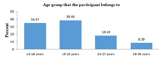
Figure 1: Distribution of the participants by their age.
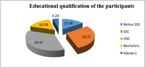
Figure 2: Distribution of the educational qualification of the participants.
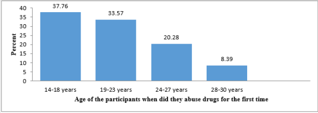
Figure 3: Distribution of age of the participants when did they abuse drugs for the first time.
The present investigation indicates that 37.76% of the respondents abused drugs for the first time at the age of 14-18 years old, 33.57% of the respondents abused drugs for the first time at the age of 19-23 years old, 20.28% were abused drugs for the first time at the age of 24-27 years old and 8.39% were abused drugs for the first time at the age of 28-30 years old (Figure 3). (Figure 4) indicates that 16.78% of the respondents have been addict for 0-2 years, 18.89% of the respondents have been addict for 2-3 years, 38.46% of the respondents have been addict for 3-4 years and 25.87% of the respondents have been addict for more than 4 years. The analysis indicates that 44.76% of the respondents were influenced to take drugs by friends, 28.67% were by classmates, 15.38% were by relatives, 6.29% were by himself and 4.90% were influenced by others (Figure 5). According to the results, the respondents indicated that Yaba was major abused drugs in the age between 14-30 years. 31.47% of the respondent were abused Yaba, 21.68% were abused Phensedyl, 20.98% were abused Cannabis (ganja), 14.69% were abused heroin, 7.69% were abused Sleeping pills (seduxen) and 3.49% were abused other drugs (Figure 6).
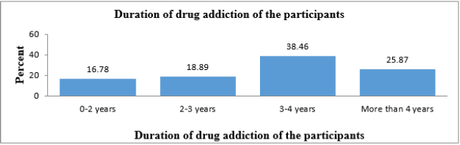
Figure 4: Distribution of the length of time of participant’s taking drugs.
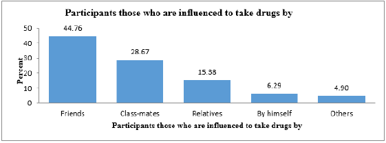
Figure 5: Distribution of participants those who are influenced to take drugs by.
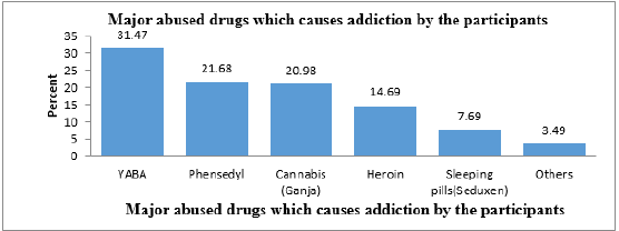
Figure 6: Distribution of major abused drugs which causes addiction by the participants.
The analysis showed that 70.63% of the respondents abused more than one drug at the same time and 29.37% of the respondent abused only one drug at the same time. Figure 7 indicated that Yaba was major abused drugs in the age between 14-30 years. 31.47% of the respondent were abused Yaba, 21.68% were abused Phensedyl, 20.98% were abused Cannabis (ganja), 14.69% were abused heroin, 7.69% were abused Sleeping pills (seduxen) and 3.49% were abused other drugs. The findings summerized in (Figure 8) shows that, 27.27% of the respondent abuse drugs due to peer pressure, 21.68% of the respondents due to frustration, 16.78 % due to mental stress relief,10.49% due to curiosity, 12.59% due to build-up self-esteem& ability to increase performance.6.99% due to sense of euphoria, and 4.20% due to easy access or availability of drugs. The analysis showed that, 56.64% of the respondents had more than one reason of drug addiction and 43.36% of the respondent had only one reason of drug addiction.
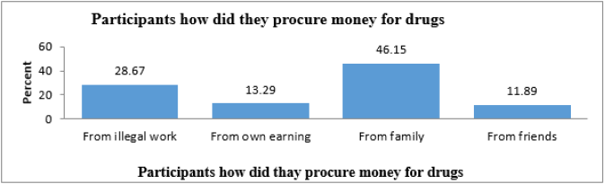
Figure 7: Distribution of participants how did they procure money for drugs.
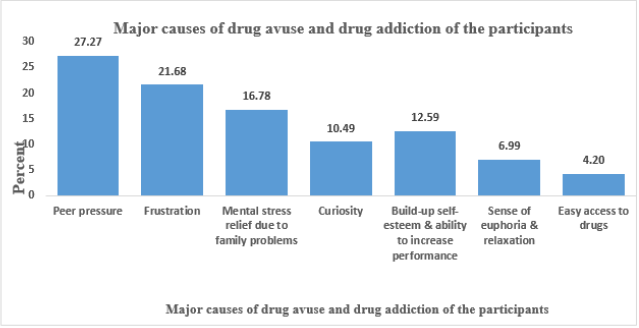
Figure 8: Distribution of the Major causes of drug abuse and drug addiction of the participants.
The analysis showed that 28.67 % of the respondent were procure money for drugs from illegal works, 13.29% were procure money from own earning, 46.15% were procure money from family and 11.89% were procure money from friends. The results on the effects of drug abuse experienced by the respondents indicate that headache, memory loss, gastritis, insomnia, bad tempered and instability were serious effects of drug abuse that the respondents experienced as expressed by the percentage of 5.59, 4.90, 13.29, 32.87, 20.28 and 23.07 respectively. In addition, the study also found out that red eyes, nervousness and fatigue also have been observed due to drug abuse and drug addiction. (Figure 9) shows that 74.83% of the respondent had more than one physiological consequences and 25.17% of the respondent had only one physiological consequences. Figure 9 showed that, 29.37% of the respondent were encouraged to get into the rehab center by friends, 11.19% were encouraged by teachers, 44.75% were encouraged by their families and 14.69% were encouraged by their relatives. It was evident that, 27.28 % of the respondent were engaged with any criminal activities but majority 72.72% of the respondent did not engaged with any kind of criminal activities. The analysis showed that, 70.63% of the respondents abused more than one drug at the same time and 29.37% of the respondent abused only one drug at the same tim.
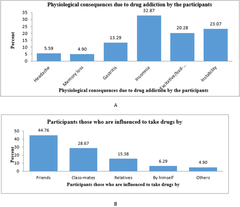
Figure 9:
A. Distribution of physiological consequences due to drug addiction by the participants.
B. Distribution of participants those who are influenced to take drugs by.
It was found that, 28.67 % of the respondent were procure money for drugs from illegal works, 13.29% were procure money from own earning, 46.15% were procure money from family and 11.89% were procure money from friends (Figure 7). The present results reveals on the effects of drug abuse experienced by the respondents indicate that headache, memory loss, gastritis, insomnia, bad tempered and instability were serious effects of drug abuse that the respondents experienced as expressed by the percentage of 5.59,4.90,13.29,32.87,20.28 and 23.07 respectively. In addition, the study also found out that red eyes, nervousness and fatigue also have been observed due to drug abuse and drug addiction. The result of the present study showed that 74.83% of the respondent had more than one physiological consequences and 25.17% of the respondent had only one physiological consequences. (Figure 10) showed that, 29.37% of the respondent were encouraged to get into the rehab center by friends, 11.19% were encouraged by teachers, 44.75% were encouraged by their families and 14.69% were encouraged by their relatives. The third objective sought to find from respondents the likely effects of drug addiction on academic performance.
The results on the effects of drug addiction on academic performance among respondents was severe grade decades, drop out ,low concentration span, discontinuation of academic career, lack of interest in extra curriculum activities and strained relationship with other students.25.87% of the respondent had grade decades, 7.69% had drop out, 41.26% had low concentration span,9.09% had discontinuation of academic career,4.90% had lack of interest in extra curriculum activities and 11.19% of the respondent had strained relationship with other students (Figure 11). The analysis indicates that 28.67 % of the respondent received treatment from rehabilitation centers for 1 year, 19.58% of the respondent received treatment for 2 years, 7.69% of the respondent received treatment for more than 2 years and 44.06% of the respondent received treatment for 6 months (Figure 12).
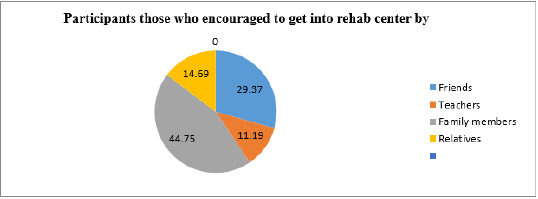
Figure 10: Distribution of participants those who encouraged to get into the rehab center by.
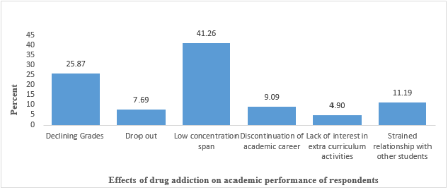
Figure 11: Distribution of effects of drug addiction on academic performance of participants.
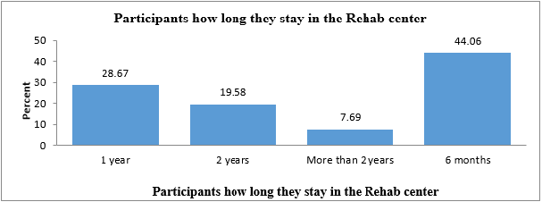
Figure 12: Distribution of the participants’ period of time to receive treatment from Rehab center.
Discussion
According to Family Health International about 50 lac people of Bangladesh are drug addicted. The World Health Organization [WHO, 2003] estimates that, about 50 cror people in the world are affected by the abuse of drugs. Many of the adolescent users begin their experiment with drugs though smoking cannabis (marijuana) cigarettes, use of heroin. Typically, they begin by sniffing it (snorting) and finally injecting it intravenously (shooting the mainline) [35,36]. Findings on how the subjects developed their habits indicated that 50% first took drugs through drug user friends and under pressure, 20% out of frustration and 15% out of curiosity. The study reports that the highest incidence of addiction occurred between 23-26 years old [37]. Due to drug addiction, criminal activities are increasing at an alarming rate all over the country, especially in urban areas like Dhaka, Chittagong, Rajshahi, Khulna, Barisal and Sylhet. The present study tries to explore the effects of drug addiction and drug abuse on academic performance of young students [38,39].
The purpose of this study was to determine the effects of drug abuse and drug addiction on Academic performance of students within age 14-30 years. The study includes the addict’s students who received treatment from drug treatment and rehabilitation centers. The majority of the drug users were male even though the involvement of female was also evident. In the present study 79.72% of the respondents were males and 20.28% of the respondents were females. The present, YABA is the most frequently abused drugs by the respondents. The study also shows that70.63% of the respondents were abused more than one drug at the same time. The study also found that, 56.64% of the respondents had more than one reason of drug addiction and 43.36% of the respondents had only one reason of drug addiction. About 27.27% of the respondents were engaged with any criminal acts [40]. The present study found that insomnia, headache, memory loss, gastritis, bad-tempered and instability were serious physiological effects of drug abuse and drug addiction of the respondents. The current study shows that drug abuse and drug addiction have adversely affected the academic performance of the respondents. Declining grades, drop out, low concentration span, discontinuation of academic career, strained relationship with other students and lack of interest in extra-curriculum activities are the adverse effects of drug addiction which directly related with academic career of the respondents.
This is in agreement with United Nations (2005) view that cognitive and behavioral problems experienced by alcohol and drug-using youth may interfere with their academic performance and also present obstacles to learning. Again drugs abused effect the brain; this result in major decline in the functions carried out by the brain. According to Abot (2005) drugs affect the student’s concentration span, which is further drastically reduced setting in boredom sets in much faster than for non-drug and substance abusing students. The students who abuses drugs is likely to lose interest in schoolwork including extra curriculum activities. The study shows that the respondents were encouraged to get into the Rehab center by Friends, by teachers, by family members and by their relatives [41]. In the present study, 28.67% of the respondents stayed in the rehab center for 1 year to more than 2 years and majority of the respondents were stayed in the rehab center for 6 months. More than half of the respondents (83.22 %) were regret for their actions and (88.21 %) of the respondents want to go back to a healthy life.
Conclusion
Most of the respondents that involved in drug use and abuse fall between the age brackets of 14-23 years. Yaba is the most frequently abused drugs by the respondents. Phensedyl, Cannabis ganja, heroin and sleeping pills (Seduxen) are also abused in significant proportion. They are addicted to drugs because of the influence of peer groups, frustration, mental stress relief, curiosity. The study found out that insomnia, headache, memory loss, gastritis, bad-tempered and instability were serious physiological effects of drug abuse and drug addiction of the respondents. Drug abuse and addiction have adversely affected the academic performance of the respondents. More so, the effects of drug abuse and addiction have resulted into respondent declining grades, drop out, law concentration span, discontinuation of academic career, strained relationship with other students and lack of interest in extracurriculum activities. Government should prevent the cultivation/ sales of deadly herbs that encourage drug abuse. Government should strictly enforce its existing laws against drug abuse through its regulatory agencies [42]. Parents and guardians should endeavor to monitor their children, so that they do not engage in drug abuse. Counseling education should be introduced in campuses to revive those who have already been engaging in the act [43-57].
For More Articles: Biomedical Journal Impact Factor: https://biomedres.us


 )
)