Open Access Journals on Pediatrics

Pathophysiology of Vascular Manifestations in Systemic Lupus Erythematosus

Introduction

Systemic Lupus Erythematosus (SLE) is a potentially fatal, chronic, multisystem autoimmune disorder. As defects can occur in various parts of the immune cascade, clinical presentations may vary significantly [1]. These clinical features range from musculoskeletal to neuropsychiatric, renal, cutaneous, gastrointestinal, pulmonary, cardiac, vascular, haematological, ophthalmological and immunological manifestations [2]. In a prospective, Europewide, decade-long study, the most common symptoms of SLE were arthritis, malar rash, nephropathy, photosensitivity, neurologic symptoms, fever, Raynaud’s phenomenon, serositis, thrombocytopenia and oral ulcers [3]. Notably, coronary heart disease, atherosclerosis and thrombosis are common in SLE [4,5], and small- and medium-vessel vasculitis across affected organ systems prevail in up to 36 % of SLE cases [6]. Adding to the complexity of the disease are varying laboratory abnormalities‚ from hematological and serological changes to negative serology or the occurrence of specific autoantibodies [1]. SLE predominantly affects women between puberty and menopause, which suggests an endocrine influence on disease onset and progression. This is underpinned by the fact that the female-to-male ratio in children is rather low and increases significantly in childbearing age [7].

Apart from hormonal factors, genetic, ethnic, environmental, psychological and socioeconomic influences have also been identified [7,8]. Due to this etiologic diversity, a considerable variation in incidence and prevalence rates among different countries is to be expected. Indeed, North America has the highest incidence and prevalence rates, while African populations show the lowest incidence rates, and Australia has the lowest prevalence. In Europe, incidence rates ranged between 1 and 4.9 per 100.000 person years, and prevalence settled between 16.2 and 110 cases per 100.000 [9]. According to estimates, around 4000 SLE patients can be expected among the Austrian population [10].

While the 5- and 10-year survival rates in SLE improved significantly in the second half of the 20th century (from 75 % to 95 % and from 63 % to 91 %, respectively), this improvement has slowed down from the 1980s onwards, despite further advances in diagnostics and treatment [11]. As direct mortality decreased, long-term complications and comorbidities might be of increased interest. This conclusion is supported by the fact that SLE is generally associated with an increased risk of premature death [1]. Traditionally, SLE has been viewed to have a bimodal pattern of mortality, presenting with peaks of early mortality (<1 year after onset) due to disease activity or infection, and of late mortality (∅ 8.6 years after disease onset) due to cardiovascular events [12]. A substantial number of instruments have been developed in order to facilitate diagnosis and classification of SLE. Diagnosis of SLE has relied on the American College of Rheumatology (ACR) criteria[13] for years but has been challenged by alternative classification concepts since. Earlier this decade, the Systemic Lupus International Collaborating Clinics (SLICC) criteria were introduced and were proven to be equally specific, but more sensitive [14].

Most recently, an initiative for new European League Against Rheumatism EULAR/ACR criteria exhibited promising results in adult-onset SLE (aSLE) [15] but did not yield superior global results in a retrospective study of pediatric-onset (pSLE) cases [16]. Apart from diagnostic criteria, many different disease activity indices have been developed in order to quantify clinical outcomes, compare patient groups cross-sectionally, and measure disease activity longitudinally [17]. Among various scoring systems, the SLE Disease Activity Index 2000 (SLEDAI-2k) is a commonly used index which allows for the prediction of global disease activity and the occurrence of new disease flares [18]. In contrast to the aforementioned tools measuring SLE disease activity, the SLICC/ ACR Damage Index (SDI) assesses irreversible damage to 12 organ systems in SLE patients regardless of their cause, including various organ manifestations related to vascular morbidity [19].

For the onset and progression of SLE, multiple risk factors have been identified. Among the most prominent candidates, tobacco smoking, environmental exposure, gender influences and genetic risk alleles have been proposed [20-22]. With respect to current or recent tobacco smoking, it has been shown to be strongly associated to anti-double-stranded DNA (anti dsDNA) seropositivity and the development of SLE, even though the underlying mechanisms have not yet been completely understood [20]. Occupational exposure to crystalline silica dust has also been demonstrated to strongly correlate with SLE prevalence, as it is believed to increase apoptotic material in a pro-inflammatory environment [23]. In women, estrogen-related exposures (e.g. early age at menarche, oral contraception etc.) were identified to be associated with SLE susceptibility, and explained by immunomodulatory functions of sex steroid hormones [24]. Moreover, a multitude of genetic risk alleles have been identified to contribute to certain SLE subphenotypes, leading to the establishment of a genetic risk score (GRS) for SLE [25].

SLE and Cardiovascular Disease

Patients suffering from SLE are at risk for premature cardiovascular disease (CVD). Apart from manifestations such as venous thromboembolism, Raynaud’s phenomenon, vasculitis, peri-, myo- and endocarditis, valvular disease and aneurysm, most research has been conducted on atherosclerosis and arterial thromboembolic events. Prevalence of atherosclerosis has been confirmed to be significantly elevated in SLE patients in metaanalyses of studies providing data of surrogate markers, namely carotid intima-media thickness and prevalence of carotid plaques [26]. Strikingly, CVD accounts for more than one-third of all deaths among SLE patients [27]. In a prospective cohort study among 119,332 women, the relative risk of a cardiovascular event (myocardial infarction, stroke, coronary artery bypass grafting, angioplasty) in SLE patients was 2.26 after multivariate adjustment for confounding factors, among which “traditional” cardiovascular risk factors [28]. In some studies, up to 50-fold incidence rate increases have been identified for myocardial infarction in SLE patients, e.g. in young women of reproductive age [29].

Both adult- and pediatric-onset SLE have been identified as independent risk factors for premature atherosclerosis [30], coronary heart disease [31], myocardial infarction [32] and/ or death in young premenopausal women [33,34]. Reciprocally, many “traditional” cardiovascular risk factors, such as metabolic syndrome, smoking and hyperhomocysteinaemia are more prevalent among SLE patients [4]. Altered severity of these risk factors could, however, not be demonstrated to independently account for the significantly increased risk for adverse events (coronary heart disease and stroke) in SLE [31]. Additionally, lupus nephritis (LN), one of the most common organ manifestations in SLE, increases odds to develop atherosclerotic plaques by more than two-fold compared to non-LN patients and healthy controls [35]. Apart from atherosclerosis and its associated morbidities in the arterial branch of the vascular system, venous thrombotic events occur in SLE at elevated rates: Different studies identified a 20-year risk of around 10 % for both deep venous thrombosis and pulmonary embolism [36].

Up to two-thirds of SLE patients present with Raynaud’s phenomenon at a given point in time [37], but prevalence differs significantly between sources [38], with one prospective study reporting an incidence rate of around 16 percent during a 10- year period [39]. Among SLE patients, vasculitis presents as a heterogenous class of disease which rarely affects large [40] but more commonly medium or small vessels and can be divided into primary and secondary forms [41]. Vasculitis afflicts different organ systems, which explains a vast array of possible, heterogenic symptoms due to cutaneous, musculoskeletal, mesenteric, hepatic, pancreatic, coronary, pulmonary, retinal, neurologic and renal manifestations [6]. As the most common, strictly cardiac manifestation of SLE, around a quarter of all SLE patients develop pericarditis, with subclinical disease being even more prevalent [42]. In these patients, pericardial effusion, cardiac tamponade, pleuritis and myocarditis occur more frequently [43,44]. Valvular abnormalities, such as valvular thickening, fibrosis or regurgitation, are observed in up to 18 % of SLE patients [45]. A subset thereof are cases of Libman-Sacks, or “atypical verrucous” endocarditis, which most commonly affects the mitral valve, but is typically clinically silent and does infrequently lead to complications [42,46].

Rarely, namely in under 10 % of cases, SLE patients develop clinically overt myocarditis, but subclinical involvement might be more frequent [44,46]. Myocarditis may facilitate the development of nonspecific electrocardiographic (ECG) changes, heart block, arrhythmia, tachycardia, heart failure and cardiomyopathy [44,47]. Whether the latter is primary or secondary in SLE patients is not easily distinguishable, as signs and symptoms are similar regardless of its cause [46]. It is, however, likely that most cardiomyopathies in SLE are secondary and emerge due to coexisting hypertension, coronary artery disease and atherosclerosis, small vessel disease, cardiac microcirculation thrombosis, renal failure, valvular disease or drug toxicity [42,46]. Rates of different conduction tissue abnormalities in SLE diverge widely. While peri- and myocarditis frequently present with tachycardia, atrioventricular and branch blocks occur rarely in adults [42,46]. In infants from anti-Ro and anti-La autoantibody-positive mothers, cross-placental Immunoglobulin (Ig) G transmission can cause transient neonatal SLE syndrome [48]. This condition often involves isolated, first to third degree, permanent heart block with onset in the second or third trimester, which presents with fetal bradycardia, and, in some cases, results in cardiomyopathy [49-51].

In adult-onset SLE patients, numerous cases of aneurysm or dissection of variable severity and of different vessels have been reported with onset at strikingly young ages, among which thoracic [52-54,54] and abdominal aortic [54-56], coronary [57,58], pulmonary [59], carotid [60], basilar [61], cerebral [62,63], hepatic [64] and mesenteric [65] artery aneurysms. Some cases have been reported as early as in childhood [66] and adolescence [67,68]. Generally, reports on aneurysm in SLE are less common than literature on other cardiovascular complications such as pericarditis or atherosclerosis, but some observations have been made. Regarding coronary artery aneurysm in SLE patients, an odds ratio (OR) of 4.09, adjusted for demographics and related risk factors, but excluding traditional CVD risk factors has been described in a retrospective, population-based case-control study [69]. With respect to aortic aneurysm (AA), a similar study has described a non-adjusted OR of 4.5 [70]. In another populationbased retrospective study, incidence rate ratios (IRRs) of 2.63 for AA and 5.7 for aortic dissection were detected along with correlating traditional and non-traditional risk factors, such as SLE disease duration > 3 years and end-stage renal disease [71]. In absolute numbers, AA does not occur commonly in SLE patients, but when it does, it has a substantial negative influence on patient survival [72]. Apart from the higher incidence and mortality of aneurysm in SLE patient’s vs the general population, the age of onset is significantly lower among the former: In two meta-analyses, the median age for onset of AA was around 45 years [72,73], which is around 20 years younger compared to the general population [74]. In addition to that, patients with SLE complicated by abdominal AA are struck with disease around 30 years earlier [56].

Apart from the aforementioned manifestations, antiphospholipid syndrome (APS) is a separate, but often accompanying disease entity to SLE. APS regularly causes thromboembolic events and other major cardiovascular complications, including stroke, deep vein thrombosis, pulmonary embolism and valvular disease [75].

Pediatric-Onset SLE

Incidence and prevalence rates in pediatric-onset SLE are substantially lower than in its adult-onset counterpart: Only 15-20 % of cases of SLE present before the age of 18 years [34] and a mere 8 % develop the disease before the age of 14 years [3], which is why many studies report data from adult populations only [1]. In studies from Europe and North America, the annual incidence rate of SLE in children and adolescents under 16 years of age was less than 1 in 100,000 persons [7,76]. According to a study conducted in the late 1990s, the mean annual incidence rate for SLE in patients under 16 years of age was 0.4 per 100,000 person years across three eastern Austrian regions [76]. Patient survival rates are similar between adult- and pediatric-onset SLE, however, SLErelated deaths are more common in pSLE due to a generally lower baseline mortality in children [33]. This can be explained by the fact that, compared with adult-onset SLE, patients with pSLE tend to undergo a more aggressive clinical course (expressed in higher mean SLEDAI scores), a higher rate of major organ involvement, exposure to tissue damage at a younger age, longer overall disease duration, and an increased risk of developing vasculitis [32,33,77]. As a result, long-term-morbidities play an even more prominent role in pSLE patients [33]. While initial diagnosis at a relatively high age can be associated with a lower risk of long-term morbidity [28], delay in diagnosis of SLE is associated with increased damage to vital organ systems [78]. As cumulative organ damage as per SDI value has been proven to, inter alia, significantly increase with disease duration in childhood-onset SLE [79], this even more so underlines the fact that early diagnosis of SLE is important to prevent major organ damage [80] and to lower the rate of disease flares [81,82].

With longer disease duration, endothelial dysfunction (and therefore early-stage atherosclerosis) progresses over time in both pSLE and aSLE patients [83–85]. As a precursor to morphologic changes of the arterial wall, endothelial dysfunction decreases along with flow‐mediated dilation (FMD), a sonographically determinable surrogate marker which corresponds to the endothelium’s capacity to mediate reactive hyperemia [86]. Moreover, increased carotid artery stiffness, also a suspected precursor to atherosclerosis, has been demonstrated in patients suffering from both active and inactive pSLE, even in those with relatively short disease duration and good disease control [87,88]. In younger pSLE patients, arterial stiffness and consecutive morphologic vascular changes have been shown to correlate with disease activity, but not (yet) with traditional CVD risk factors [89]. Other data suggests that carotid intima media thickness (CIMT), a surrogate marker for atherosclerosis, does, however, correlate with both traditional (age, body mass index, male gender) and nontraditional (Azathioprine, low- and high-, but not medium-dose glucocorticoid treatment) risk factors in pSLE patients (Figure 1) [90]. As disease progresses, reducing the cardiovascular risk for patients suffering from pSLE by optimal control of disease activity and reducing the “add-on” of traditional cardiovascular disease (CVD) risk factors is, by all means, of great importance [26,33,91,92].

biomedres-openaccess-journal-bjstr

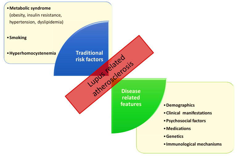
Figure 1: Traditional risk factors and disease-related features in lupus-related atherosclerosis (4)

Pathophysiology

SLE is a chronic autoimmune disease which encompasses many landmark mechanisms of autoimmunity, such as loss of tolerance against autoantigens, lymphoproliferation, polyclonal autoantibody production, immune complex disease (Arthus/type III hypersensitivity reaction) and target organ inflammation [93,94]. On its most basic level, it is caused by an inappropriate immune response against self-antigen, particularly nucleic acid-containing particles. Because several intensively intertwined processes and aberrations interplay in SLE, the origin of its autoimmunity has not been conclusively identified to date. In a highly simplified model of its disease mechanisms, impairments of (apoptotic, necrotic) cellular remnant clearance could be chosen as an arbitrary starting point. From there on, the accruing autoantigens are opsonized by naturally occurring autoantibodies (NAbs) before being targeted by the complement system and phagocytes. Now, many different pathways commence: Local inflammatory reaction by cells of the innate immune system and complement complexes leads to early signs of tissue inflammation, cytokine release, and attraction of additional leukocytes. Antigen-presenting cells (APCs) migrate to lymphatic organs, where they are sampled by lymphocytes in order to establish highly specific cellular and humoral immunization. Developing specificity of immune reactions and rising numbers of autoantigen-specific antibodies perpetuate and extend autoinflammation which peak in vicious disease flares resulting in long-term target organ damage [1,95,96].

As Figure 2 suggests, susceptibility for developing autoimmunity has been associated with a plethora of candidate genes regulating the immune response in various stages. Genetic variants can negatively impact self-tolerance and are typically labelled as lossor gain-of-function mutations. For instance, a loss-of-function in gene motifs coding for parts of the physiological clearance of cell remnants might affect the development of SLE similarly to how a gain-of-function mutation of pro-inflammatory T cell signaling would and vice versa. Some severe cases of SLE present with early onset, which is suspicious for monogenic SLE, a concept of disease entities induced by solitary mutations in single genes [93,97,98]. Apart from these rare and mostly pediatric-onset cases of SLE, multiple hits of different flavors accumulate before a “susceptible” genetic predisposition lapse into active disease Figure 3.

biomedres-openaccess-journal-bjstr

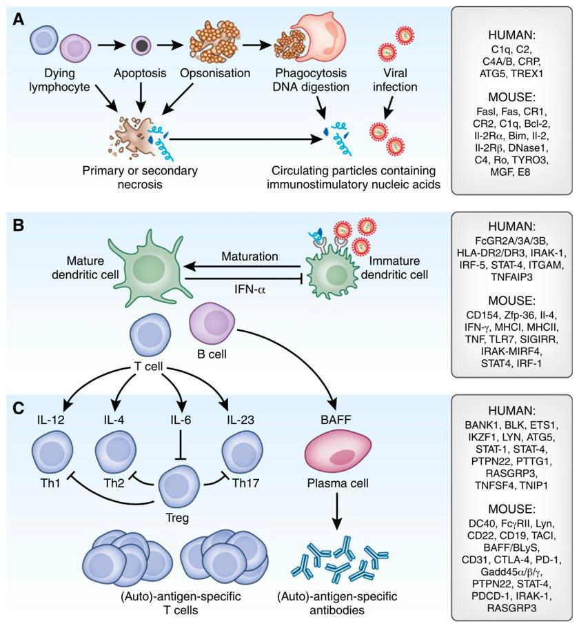
Figure 2: Pathomechanisms of SLE and a selection of candidate genes (93).

These dysregulated autoimmune processes involve both the innate as well as the adaptive immune system, which explains why certain types of antibodies are commonly found in SLE. Foremost, antinuclear antibodies (ANAs) are identified in almost all cases of SLE [99]. As members of the ANA specifity, anti-Smith (Sm) and anti-dsDNA antibodies are two examples of commonly found antibodies in SLE [1].

biomedres-openaccess-journal-bjstr

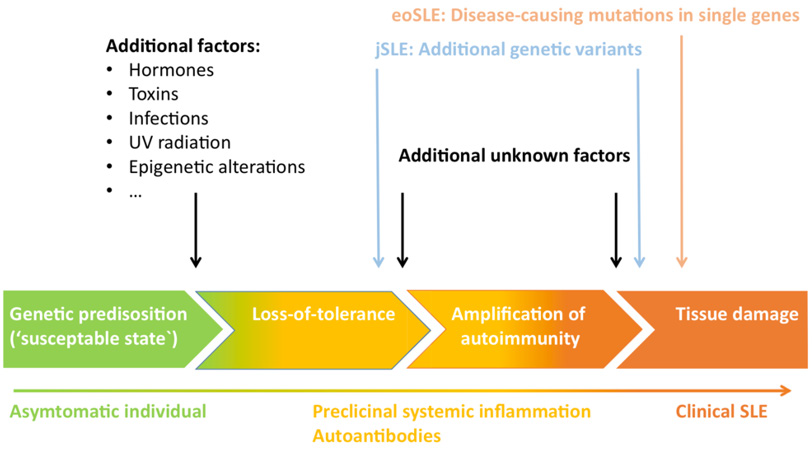
Figure 3: Timeline of SLE disease progression and pathogenic hits (98)

Cell Death, Nuclear Remnants and Clearance

In order to develop immunization against self-antigens, nuclear epitopes have to be accessible to antigen-presenting cells (APCs). In physiologic homeostasis, the overbearing presentation of self-antigens is avoided by rapid cell death clearance, which is a complex process involving components of the complement system, dendritic cells (DCs) and phagocytes, as well as a multitude of other factors such as antibodies and other proteins, enzymes, cytokines and opsonins [100]. Due to unfortunate immunoregulatory defects in SLE, low levels of chromatin in extracellular compartments (e.g., after necrosis, impaired apoptosis or NETosis, see below) cannot be maintained. Consequently, protein antigens and immunostimulatory nucleic acids (acting as immune adjuvants) are targeted and presented by plasmacytoid dendritic cells (pDCs), which serve as a link between the innate and adaptive immune system [93,101]. In other words: If cell debris is not efficiently cleared, a first step towards an excessive immune response is triggered. Apart from extracellular cell debris due to impaired clearance of dysfunctional cells, SLE autoantigens can also present on the surface of apoptotic cells [102]. With regard to reasons for cell death, external noxious influences such as UV radiation of the skin is hypothesized to contribute to apoptosis, secondary necrosis or impaired clearance, but this has recently been subject to discussion [103,104].

Among many different hypothesized dysfunctional factors, complement component (C) 1q deficiency is believed to add to the breakdown of self-tolerance [102]. This shortage might be traced back to impaired local synthesis of C1q in its main producers, namely macrophages (Mφ) and dendritic cells [105]. To further complicate the involvement of phagocytes, functional Mφ/ monocyte (Mo) defects have been identified to contribute to SLE, including decreased phagocytosis, antigen presentation (MHC-II/ HLA-DR, CD80), Interleukin (IL)-1b production, autologous mixed lymphocyte reaction, and T-cell activation [106]. Apart from these defects, increases in pro-inflammatory M1 and M2b, and decreases in anti-inflammatory M2a and M2b subsets skews Mφ populations towards a more autoinflammation-prone profile [107]. Additional mechanisms, such as the inhibition of class A scavenger receptors by autoantibodies, are also believed to impair phagocyte function [108]. Aside from a reduction in cell and immune complex clearance, these functional defects lead to the impaired (co)stimulation of lymphocytes and the accumulation of apoptotic material and immune complexes in target organs (see below).

Innate Immunity

In general, nucleic acid-containing immune complexes and cytoplasmatic RNA and DNA are known to be potential stimuli for the activation of the innate immune system [1]. In SLE, type I interferons (IFNs) are produced in a response mainly mediated via endosomal toll-like receptors 7 and 9 (TLRs) and TLR-independent nucleic acid sensors. Apart from migrating pDCs, which are the major source of IFNα, a variety of cells participate in the production of pathogenic type I-IFNs, among which fibroblasts, epithelial cells and bone-marrow resident neutrophils [109,110]. Belonging to the group of pattern recognition receptors (PRRs), TLRs are membrane receptors located either on the cell surface or intracellularly in the endosomal-lysosomal compartment, notably in dendritic cells (DCs) [111]. Because of their function to bind pathogen-associated molecular patterns (PAMPs), TLRs are generally considered to contribute to antiviral immunity [112] in a host of different cell types. As opposed to myeloid dendritic cells (mDCs), which express functional TLR7 but not TLR9, pDCs express both of these subtypes [113]. Depending on their variant, TLRs recognize different molecular patterns. In particular, TLR7 binds to single-stranded RNA (ssRNA) and imidazoquinoline compounds [114,115], and TLR9 binds unmethylated cytosine-guanine dinucleotide (CpG)-rich DNA [116]. Additionally, TLR7 and TLR9 can be activated by immune complexes internalized into the cytoplasm via the interaction of Fc γ receptor II (FcγRII) and cell fragments [114,116]. Ultimately, TLRs couple nucleic acid detection to essential immune functions, such as the induction of cytokine responses, the presentation of antigen to lymphocytes, and the enhancement of virus-specific antibody responses [117].

Data from murine models suggests potentially pathologic functions of TLRs in SLE (Figure 4). With regard to the production of autoantibodies, TLR7 and TLR9 exert analogous functions – the former propagating anti-RNA (e.g. anti-Sm), the latter facilitating anti-DNA production. Although both TLRs are associated with components of the same downstream signaling pathways, TLR7- deficiency was linked to decreased lymphocyte and pDC activation, as well as ameliorated end-organ (e.g., renal) morbidity. Moreover, TLR9 has been described to downregulate the expression of IFNα [118]. However, in the presence of DNA-containing immune complexes, IFNα seems to be upregulated in a TLR9-dependent mechanism [119]. A similar effect is attributed to TLR7-dependent anti-RNA autoantibodies, which are believed to also enhance the production of type I IFNs. In this respect, opposing roles are attributed to TLR7 and TLR9 in the pathogenesis of SLE [118]. Also, some observations attest a strongly correlated pathogenic function of autologous RNA-containing immune complexes in conjunction with TLR7 in innate immune activation, IFN production and the development of SLE [120].

biomedres-openaccess-journal-bjstr

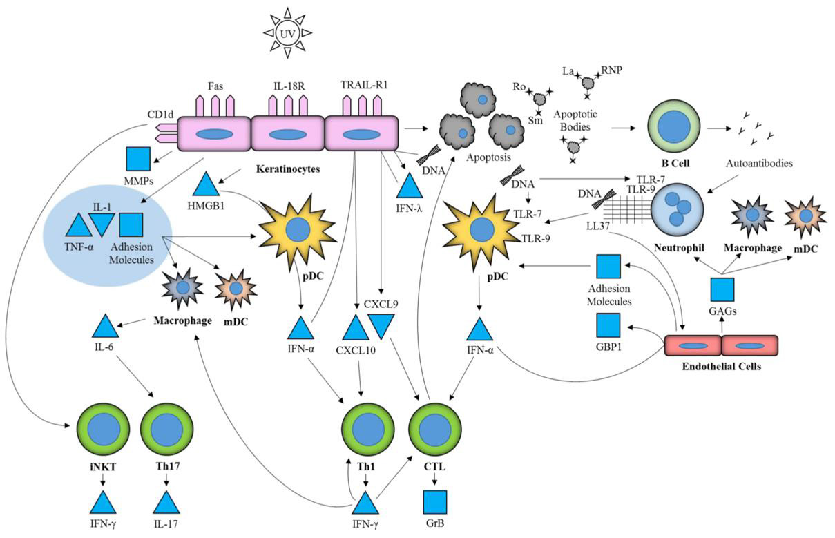
Figure 4: Involvement of TLR7 and 9 in cutaneous lupus erythematosus (103)

Other data suggests that TLR-independent innate immune pathways might also contribute to SLE pathogenesis in cells of the innate immune system: RNA helicases, such as RIG-like Receptors (e.g. RIG-I, MDA5), can bind and destabilize distinct (viral) RNA patterns, and form a signaling complex leading to IFN and IFNstimulated gene expression and the production of type I IFNs and pro-inflammatory cytokines via mitochondrial antiviral-signaling protein (MAVS), nuclear factor-κB (NF-κB), interferon regulatory factor (IRF) 3 and IRF7 (112,121). With regard to DNA recognition, a TLR9-independent mechanism was identified, which can be, for instance, relevant in TLR9-lacking mDCs: In a nutshell, the inhibition of Cyclic GMP-AMP synthase (cGAS) significantly blunted autoimmunity, particularly the expression of IFN-induced genes and inflammatory cytokines, in two autoimmunity-prone murine models (Trex1-/- and DNaseII-/-) [122]. In about two-thirds of adult-onset SLE patients and virtually all pediatric-onset SLE patients, overstimulation of type I IFN-producing cells and vastly elevated levels of IFN-dependent downstream products suggest that type I IFNs are pivotal contributors in the pathogenesis of SLE [123]. These type I IFNs cause the expression of a measurable “IFN signature”, consisting of multiple type I IFN-inducible genes expressed primarily in peripheral blood mononuclear cells (PBMCs) [124,125]. It can be used as a biomarker of disease activity and might be useful in selecting individual treatment regimens [126]. Due to the significance of IFN-I as a primary pathogenic factor in SLE, gain-of-function mutations in the type I IFN pathway are assumed to associate with disease onset and severity [112].

Interferons have many pro-inflammatory key roles in SLE, which overlap among many species of type I-III IFNs, and target cells of both the innate and adaptive immune system. Most notably, IFNα stimulates monocytes and their development into myeloid dendritic cells (mDCs), as well as T-helper (TH) 1 and cytotoxic T CD8+ cells. B cell differentiation, autoantibody production and class switching, natural killer (NK) cell cytotoxicity and NETosis (neutrophil extracellular trap formation) are also facilitated. Moreover, IFNα suppresses the development of TH2, TH17 and anti-inflammatory regulatory T (Treg) cells, is believed to trigger apoptotic death of certain tissue cells, and to impede clearance of nuclear antigens by macrophages [110,127]. Therefore, type I IFNs facilitate pathognomonic processes in lupus, such as an increase in autoantigen loads, the augmentation and presentation of autoantigens via APCs, the production of IgG autoantibodies and inhibition of immunosuppressive activity [127]. In this respect, it is not surprising that type I IFNs are reasonably hypothesized to promote premature cardiovascular disease in SLE [128]. Overall, immune alterations observed in SLE resemble those characteristic of chronic viral infection [93,112,123], such as: Sustained expression of type I IFNs; increased and sustained production of proinflammatory mediators such as IL-6 and 1IL-10 as well as tumor necrosis factor (TNF); altered expression of cell surface receptors including programmed death ligand 1 (PD-L1) and TNF-related apoptosis-inducing ligand (TRAIL, TNFSF10); and a shift in T cell differentiation towards a follicular T helper cell phenotype. These alterations induce, among others, sustained and poorly regulated macrophage activation, impaired T cell function and regulation of cell death, excessive B cell differentiation, the production of autoantibodies and immune complexes and widespread tissue and organ inflammation and damage [1].

In this regard, Epstein–Barr virus (EBV) infection has been highlighted as a potential trigger of the disease. Precisely, defective CD8+ T cell response to EBV infection might lead to an increase in intra- and extracellular EBV DNA, which would induce a shift toward a CD4+ T cell mediated immune reaction, and therefore lead to an increase in the production of autoantibodies (129). Aptly, specific antibodies to EBV nuclear antigens (e.g. EBNA-1) have been demonstrated to effectively cross-react with autologous dsDNA [130]. As opposed to mechanisms linked to viral infection, primarily antimicrobial mechanisms of the innate immune system have recently received attention in the pathogenesis of adultand pediatric-onset SLE, namely cell death and the formation of neutrophil extracellular traps (NETs), most likely after exposure to anti-RNA antibodies [131]. In NETosis, neutrophils actively release chromatin (and therefore autoantigens such as dsDNA), bactericidal proteins and enzymes, and immunostimulatory molecules (e.g. IL-17) into the extracellular space in order to fend off microbial infection [132]. Therefore, NETting neutrophils are believed to, among others, contribute to the activation of innate immunity, the characteristic stimulation of IFNα production of pDCs, and target organ damage [132,133]. A distinct neutrophil subset, namely lowdensity granulocytes (LDGs) are prone to form NETs, which might be due to an increased sensitivity to IFNα stimulation [131]. This subclass is of great interest in SLE, as it has been linked to skin damage, vasculitis, and endothelial cell death [132].

Apart from a potential role of naturally occurring antibodies, the complement system is the main participant of the humoral innate immune system involved in the pathogenesis of SLE [134]. Under physiological conditions, complement has an independent role in cytolysis via its soluble or membrane-bound membrane attack complex. Three (classical, lectin, and alternative) pathways start the complement cascade either after opsonization of immune complexes (via C1q), ligation of recognition molecules (such as mannose-binding lectin, or MBL), or, in case of the alternative pathway, spontaneously (Figure 5). In the further course of the complement cascade, proinflammatory recruitment of leukocytes (through C3a, C5a), and an increase in specific autoantibody production (C3d) facilitate inflammation. Additionally, opsonization (C1q, C3b, C4b) leads to immune complex and cell debris clearance by phagocytes (100,102). In SLE, C1q suppresses the production of SLE immune complex-induced IFNα by directing stimulatory immune complexes to monocytes rather than to IFNα- producing pDCs, when both of these possible targets are present [135]. Accordingly, C1q and a number of additional complement components are decimated in SLE patients by autoantibodies, most frequently by anti-C1q, anti-MBL and anti-C1s autoantibodies [136]. While C1q physiologically attaches to the Fc domains of IgG and IgM antibodies as an entry point into the classical complement pathway, only immune complexes containing both anti-C1q and C1q seem to be correlated with tissue inflammation in SLE, and likely amplify complement activation rather than inhibition [102,136,137].

biomedres-openaccess-journal-bjstr

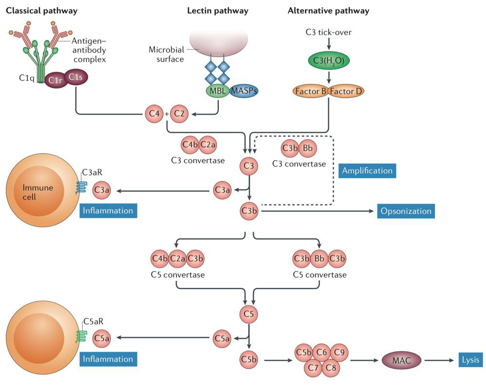
Figure 5: Overview of the complement cascade and its three main pathways (102).

Accordingly, congenital C1q deficiency is highly associated with SLE [103], and defects of C1q production in Mφ and DCs are highly suspect [105]. In conjunction, these findings strongly imply that C1q exerts a protective role in SLE. In contrast to the loss of C1q, other complement components are also diminished in patients’ blood samples. This circulatory complement depletion most commonly manifests as a reduction in C3 and C4, as these components accumulate in inflamed tissues (see below) [102]. While data regarding the impact of anti-MBL activity on disease progression is inconsistent [136,138,139], anti-C1s engages C1s’ proteolytic activity and is therefore believed to be partially responsible for the reduced levels of C4 observed in SLE [136,140], but C1s deficiency was shown not to independently induce SLE [105]. In lupus-prone mice, downstream inhibition of immune cell complement receptors (C3aR, C5aR) is beneficial and reduces symptoms, but a general C3-deficiency is associated with enhanced symptoms [102]. This raises the question whether complement-induced lysis might be beneficial in SLE, while complement-mediated inflammatory response has adverse effects.

Adaptive Immunity

Cells of the adaptive immune system are essential immune mediators in the pathogenesis of SLE as they exert a number of functions, predominantly the production of pathogenic autoantibodies (Figure 6). Beyond the build-up of the pathognomonic humoral adaptive (auto-)immunity, lymphocytes reproduce, adapt, and stimulate immune cells in order to develop, enhance and perpetuate autoimmunity and autoinflammation [95,141]. Considering the previous remarks on the excessive activation of innate immunity in SLE, lymphocyte tolerance to autoantigens is constantly challenged by downstream stimulation. As soon as self-tolerance is lost due to distinct aberrations, autoreactive cells enhance autoimmunity beyond its expected magnitude from innate immunity alone [142]. Thymocytes (T cells) and bursa of Fabricius cells (B cells) are primary drivers of disease chronification, because subsets of these lymphocytes, namely memory T and B cells as well as plasma cells, memorize prior immunization against SLE autoantigens [93]. T cells in SLE deviate from those of healthy individuals in a number of – sometimes contradictory – ways. The T cell phenotype in SLE is distinctly different than in healthy individuals: In SLE, a hyperactive T cell population reacts to low thresholds for activation by secreting pro-inflammatory cytokines, cooperating with B cells, stimulating autoantibody production, and accumulating autoreactive T cells [143,144]. Even though generalized lymphocytopenia is common in SLE, specific T cell populations either proliferate or decline [1]. When considering certain individual thymocyte subgroups, apparent deviations can be discerned in SLE:

biomedres-openaccess-journal-bjstr

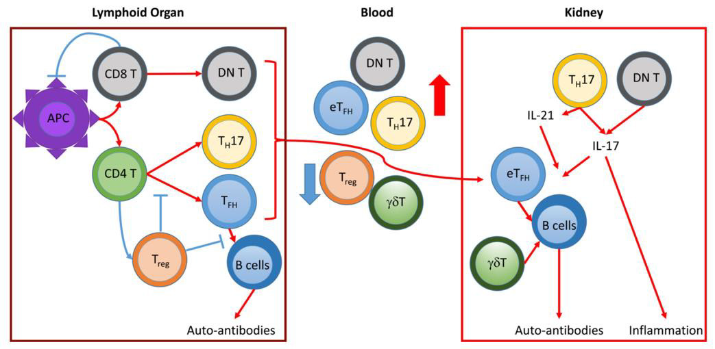
Figure 6: Roles of T cells in SLE and LN pathogenesis (red pathways up-, blue pathways downregulated) (143).

CD4+ T cell populations are generally expanded in SLE. As major contributors to autoimmunity, follicular helper T (TFH) cells proliferate significantly in SLE, providing disease-promoting B-cell targeted help for the production of autoantibodies at elevated levels [145,146]. Specifically, TFH cells promote the differentiation of B cells into memory B cells and plasma B cells, affinity maturation, and class switching. Therefore, they are causal for the increasing specificity of B cells to autoantigens and the concurrent production of autoantibodies [143,147]. Whether numbers of Interferon γ (IFNγ)-producing TH1 cells are in- or decreased in SLE has not been definitively determined and is likely related to IL-2 signaling [148,149]. TH2 cells, on the other hand, seem to rescind as mediators of SLE pathogenesis [150]. An increase in IL-17-producing CD4+ T helper (TH17) cells is accompanied by elevated serum IL-17 levels, which promotes the attraction of neutrophils and the formation of germinal centers, stimulates B cells, reduces B cell tolerance, triggers autoantibody production, and facilitates target organ damage [143,151,152]. Ultimately, CD4+ cell populations are significantly skewed towards TH17 and away from Treg cells [149].

Functional CD4+, CD8+, and γδ regulatory T (Treg) cell deficiency disrupts the physiologic balance between pro- and antiinflammatory stimuli in SLE [143]. Healthy Treg cells are known to regulate a number of processes in the immune system, among which suppression of B and T lymphocyte activation and proliferation, inhibition of autoantibody production of B cells and prevention of immunogenic but facilitating of self-tolerant DC expansion [153]. Different mechanisms have been proposed regarding the reason for deregulation of inflammatory stimuli by Treg cells: For one thing, decreased formation and survival of Treg cells has been identified, and their numbers are generally diminished. For another, defects in their immunosuppressive function are also likely [145] but might be secondary to an acquired resistance of SLE T effector cells to Treg suppression [153]. Another explanation could be the relative depletion of Treg cells, which results from activated, suppression-resistant effector T cells outweighing the number of Treg cells [151,152,154]. Lastly, the influence of TLR-dependent IL-6 expression in APCs has been shown to render effector T cells refractory to Treg suppression, a relevant mechanism given the likeliness of TLR activation by nucleic autoantigens in SLE [155].

Increased numbers of activated CD8+ T cells remarkably present with decreased cytotoxic ability, defective viral antigen responses, and therefore increased risk of infection, as well as a possible role in triggering autoimmunity [143,151]. Apart from increased numbers of functionally impaired CD8+ cytotoxic cells, quantities of NK cells are, generally, diminished (145). TCRαβ+/ CD4 /CD8- double negative (DN) T cells result from inactivation or exhaustion of autoreactive or continuously stimulated CD8+ T cells, which usually exert immunosuppressive functions in chronic infection [143]. In SLE, they have been shown to locally accumulate and contribute to infection via the production of IL-17 (156). Due to their TCRs being comprised of γ- and δ- rather than α- and β-chains, γδ T cells do not detect antigens bound to major histocompatibility complexes (MHC), but rather – similar to antibodies – bind antigens directly and exert a variety of functions [157]. By presenting antigens, producing proinflammatory cytokines, interacting with Treg cells, and promoting autoantibody production via B cell help, γδ T cells have been shown to participate in the development of SLE. Constituting around 10-15 % of T cells in the human blood under physiological conditions, their occurrence seems to be reduced in the peripheral blood but increased in affected target organs of SLE patients [158].

As opposed to thymocytes in healthy individuals, SLE T cells produce a different cytokine profile (Figure 7), which is generally characterized by an increase in IL-6, IL-17, IL-12, and IL-10, but reduced levels of TH1-dependent IFNγ and IL-2, as well as Treg-dependent transforming growth factor- β1 (TGF-β1) [149]. Notably, a decline in IL-2 is associated with impaired formation and survival of Treg cells, as well as elevated levels of proinflammatory cytokines (particularly IL-6) which inhibit Treg cell functions in SLE [151,159]. Conversely, low levels of IL-2 influence T cell subset distribution, inasmuch as skewing T cell populations towards TH17 cells, and impairing cytotoxic functions of NK and CD8+ effector T cells corresponding to SLE disease activity [148]. Also, IL-2 initiates activation-induced cell death (AICD), which is a crucial mechanism in T cells to retain peripheral autoantigen tolerance but is defective in SLE [160]. A complex set of factors influences T cell activation. Its centerpiece is the T cell receptor (TCR) or, respectively, TCR/CD3 complex, which recognizes proteolytically processed short peptide antigens bound to MHCs of both specialized APCs (predominantly MHC-II) and most nucleated cells (MHC-I). In order to be activated rather than to be put in a state of unresponsiveness (anergy), naïve T (TN) cells have to be stimulated by APCs through their T cell receptor/CD3 complexes (TCR/CD3), costimulatory signals provided by membrane molecules (CD86 or CD80 to CD28 in DC:TN ligation), and concomitant cytokines [161]. The latter decide the T cells’ fates, as T cell differentiation depends upon the availability and composition of the local cytokine environment [157].

biomedres-openaccess-journal-bjstr

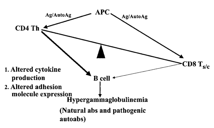
Figure 7: Disbalance of T cell function and autoantibody production (145)

SLE is associated with defects on multiple levels of the T cell signaling cascade, which can be divided into proximal, middle and distal regions ranging from extracellular ligation to protein translation. Therefore, these defects are associated with many different aberrations from premature apoptosis to dysregulated DNA methylation and impaired protein translation [145]. One of the mechanisms affecting T cell activation in a majority of SLE patients is an altered expression of Fc receptor signaling components, in particular a decrease of the T cell receptor ζ chain expressed in healthy individuals and its substitution with the more common TCR γ chain. This pathologic feature of SLE can singularly cause T cell hyperresponsiveness and diminish the TCR/CD3-mediated production of IL-2 in SLE T cells [162]. Compared to controls, hypomethylated CG-rich DNA sequences and promoters of IFNregulated genes were identified in SLE CD4+ precursor T (TH0) cells, priming thymocytes to overexpress interferon-related genes in a rapid type I-IFN response. On the other hand, hypermethylated genes like CD247, which encodes for the TCR ζ chain, have also been identified to participate in SLE T cell dysfunction [163]. As a result, commonly subthreshold interactions between T cells and APCs cause T cell activation in SLE.

Processes responsible for autoantibody production in SLE mainly proceed in secondary lymphatic tissue (Figure 8). As a general principle, activated APCs migrate from peripheral tissues to draining lymph nodes via the lymphatic system (“homing”), where they are sampled by T and B lymphocytes [164]. When a T cell is stimulated by a suitable peptide presented via a MHC, costimulatory molecules and cytokines, it differentiates into one of the many subtypes depending on the local cytokine environment [157]. If it becomes a CD4+ TFH cell, it perpetuates germinal centers by providing survival signals for activated autoreactive B cells (i.e. Centro blasts) together with follicular dendritic cells (fDC), which present autoantigens. In the subsequent process of affinity maturation, B cells are rewarded for increased specificity of their B cell receptor (BCR) to the respective autoantigen by stimulation, facilitating the expression of highly specific autoreactive SLE B cells in a proliferative process [165]. This clonal selection is based on somatic hypermutation, in which BCR/immunoglobulin (Ig) expression is genetically diversified [166]. Long-term exposure to the aforementioned survival signals causes differentiation of autoreactive B cells into memory B or class-switched, long-lived plasma B cells, which produce high levels of pathognomonic IgG autoantibodies [142]. These cells home to protective niches in the bone marrow, where they are maintained by chemokines and products of bone marrow stromal cells. This is of particular relevance in SLE pharmacotherapy, as such cells are refractory to certain immunosuppressive B cell depletion therapies [167].

biomedres-openaccess-journal-bjstr

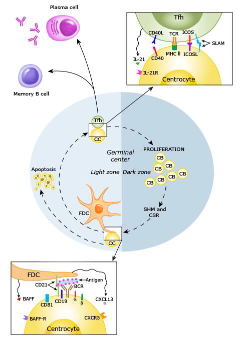
Figure 8: B cell activation in a secondary lymphoid follicle (168)

CD40 ligand (CD40L, or CD154), a surface molecule of the TNF family and TCR costimulant on (mostly CD4+) T cells, is expressed and maintained for longer durations after T cell activation in SLE [169]. As it binds to CD40 expressed on the surface of APCs, CD40L is classically known as an essential part in the co-stimulation of MHC-II antigen-presenting B cells in the process of affinity maturation, differentiation, and class switching. Hence, these mechanisms are pathologically augmented [170] in SLE T:B cell interaction. A therefore alleviated co-stimulation through T cells encounters more susceptible, hyperreactive SLE B cell phenotypes, which further promote disease activity [171]. In addition to their roles in cells of the innate immunity, TLR7 and TLR9 also activate autoreactive B cells in cytoplasmatic compartments (Figure 4.8) which internalize nucleic autoantigens or DNA/RNA-containing immune complexes, either coupled with a BCR Ig receptor [172]. TLR7 acts synergistically with CD40L and other factors in terminal B cell differentiation [173], and might, remarkably, even facilitate a thymocyte-independent mechanism of B cell activation and differentiation [174]. Additionally, TLR7 is an indispensable B-cell intrinsic mediator in the spontaneous formation of new germinal centers which induce systemic autoimmunity [175]. Nonetheless, the contribution of TLR9 in the production of autoantibodies might be ambivalent, as it competes with TLR7 for the intracellular ligand UNC93B1. For when the latter is modified for loss of TLR9 affinity, lethal TLR7-mediated inflammatory disease develops (Figure 9). Autoreactive B cells can be tied to two different sources. The first wave of B cells displaying self-reactive BCRs emanates directly from the early B-cell-repertoire in the bone marrow. Typically, these cells are deleted before exiting the bone marrow or undergo receptor editing [165].

biomedres-openaccess-journal-bjstr

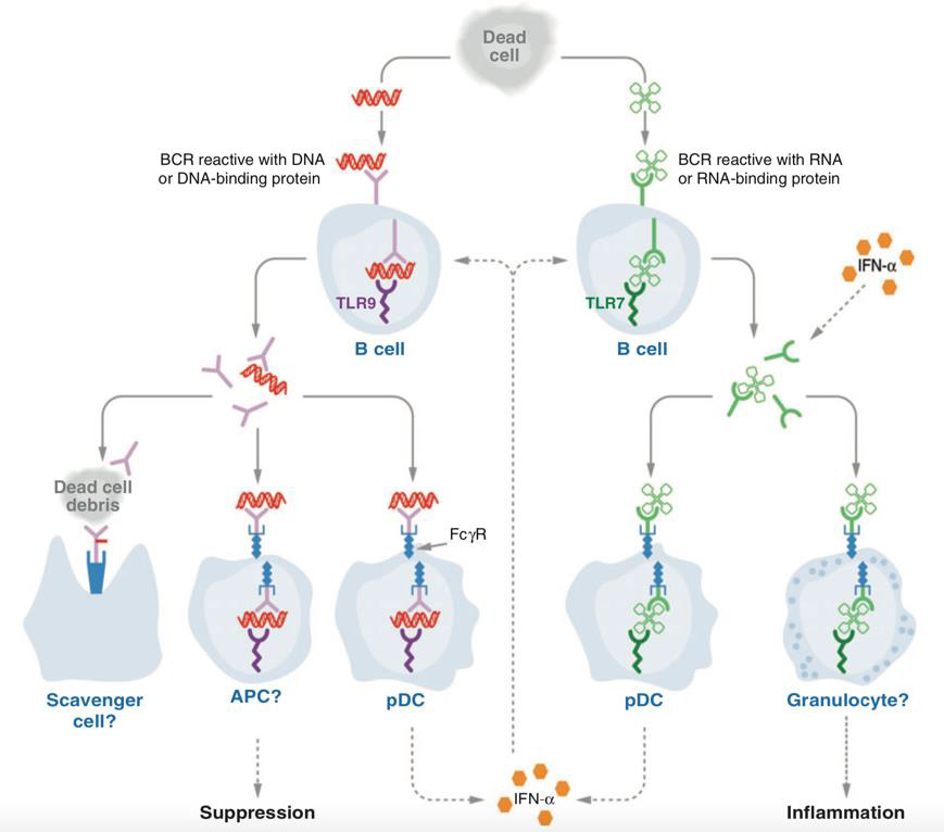
Figure 9: Roles of TLR7 and 9 in B cells (172)

These processes are attributable to central B cell tolerance checkpoints, which can be defect in SLE, resulting in a substantial primary distribution of autoreactive, but otherwise naïve B cell populations [176]. A second wave of autoreactive B cells develops in the process of somatic hypermutation of Ig (BCR) heavy and light chain variable regions. These cells are usually subject to peripheral censoring, which can be impaired by abnormalities in different B cell signaling molecules such as PTPN22, Bruton’s Tyrosine Kinase (Btk), Lyn, or B-cell activating factor (BAFF) [165]. The inhibition of mature, autoreactive B cells also relies on the surface Fc receptor FcγRIIB (CD32b), which is usually upregulated in healthy B and plasma B cells but downregulated in SLE. In normal quantities, it initiates a feedback loop upon binding IgG immune complexes, resulting in antibody homeostasis by regulating naïve B cell progression and therefore limiting the anti-DNA response to the expression of nonpathogenic IgM autoantibodies [177].

Apart from follicular responses, extrafollicular, hyperresponsive B-cell populations shape disease expression in SLE [175]. Initiating the production of somatically mutated and class switched SLE autoantibodies by long-lived plasma cells, extrafollicular B cell responses are a major contributor to circulating antibodies in SLE [143,178]. In contrast to follicular B cells, extrafollicular B-1 and marginal zone B cells generate short-lived circulating plasmablasts and extrafollicular plasma cells, which are suspected to be a main source of dsDNA autoantibodies in disease flares [165,179]. Emanating from ectopic germinal centers and lymphoid aggregates, follicular, long-lived plasma cells might also contribute to persistent autoantibody production outside of secondary lymphatic tissue [165]. Both the occurrence of circulating plasmablasts and antidsDNA antibodies is associated with an expanded population of circulating programmed cell death protein 1 (PD-1)-expressing TFH-like cells in active SLE, which are therefore likely to provide T cell help in both follicular and extrafollicular B cell maturation [143,180].

B cells have been proven to exert functions beyond autoantibody production and the buildup of B cell memory in the pathogenesis of SLE (Figure 10). For one thing, they appear to be vital APCs for the activation and priming of native T cells. For another, they promote autoimmunity of T cells reciprocally via co-stimulation of thymocytes, namely by providing CD80 and CD86 as binding partners of CD28, and CD40 to ligate CD40L. Third, they contribute to autoimmunity by proinflammatory stimulation of a wide range of targets through the excretion a number of cytokines, among which IFNα and IL- 6 [141,181].

biomedres-openaccess-journal-bjstr

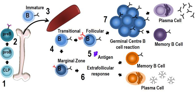
Figure 10: Stages of B cell development (147)

Autoantibodies

Pathogenic processes in SLE rely heavily on autoantibodies targeting self-antigen. Lupus-specific antibodies can be categorized according to their targets, among which DNA and DNA-binding proteins, RNA and RNA-associated proteins, β2-glycoprotein 1, phospholipids and cell membrane proteins. As an example, for pathognomonic Igs, antinuclear antibodies can be detected in serologic samples from virtually all SLE patients [182]. A number of around 180 possibly pathogenic autoantibodies have been identified, which are diverse in their prevalence and effect on disease activity but are mostly not uniquely specific for SLE. This has introduced the question whether SLE might not be a single disease with varied phenotypes but rather a similar phenotype associated with different pathogenic mechanisms. However, some 90 mutual antibodies have been shown in 20 % or more of SLE patients, and around 20 antibodies are common to more than 50 % of SLE patients [183]. Among these, anti-dsDNA and anti-Sm are most specific for SLE [1]. When comparing quantities of circulating autoantibodies, anti-Ro, anti-La, and anti-nucleosome antibodies also stand out [95]. This begs the question of commonalities among “classic” lupus autoantibodies, and indeed, nucleic acid associated antigens seem to be their preferred target. Once again, this is explained by BCR co-stimulation through T-cell derived signals, cytokines, and especially TLR7 [165].

With respect to disease patterns, certain subsets of SLE can be distinguished by related autoantibody clusters [184] or individual autoantibodies [185]. As an example, titers of 13 antibodies have been found to be significantly increased in pSLE cases with proliferative LN, among which dsDNA and C1q autoantibodies. In the same study of autoantigen microarray results, fifty autoantibodies were significantly increased in the sera of pSLE patients, including anti-BAFF, which has curiously been demonstrated to be associated with active disease [186]. Blood samples of SLE patients usually contain elevated levels of IgM, IgG and IgA autoantibodies [187]. This can be explained by the fact that the pathogenic autoantibody profile in SLE undergoes a shift from monoclonal IgM to IgG, temporally corresponding to disease progression and tissue damage (1,142). It depends on plasma cells originating from B cells undergoing affinity maturation and class switch, processes mostly driven by CD4+ TFH cells, TLRs and co-stimulation from receptors and cytokines such as IL-21 and BAFF (see above). To some extent, IgM antibodies are assumed to be a protective factor in SLE, e.g. by binding autoantigens and therefore blocking the formation of pathogenic IgG immune complexes [188]. They are also suspected to decrease IgG autoantibody production by autoreactive B cells, diminish DC activation and act as competitive inhibitors to their IgG counterparts in binding circulating nuclear antigens [187]. In autoimmune arthritis, IgM NAbs have been documented to exert a number of functions to prevent excessive, detrimental autoimmune response: T15-NAbs specific to phosphorylcholine epitopes facilitate the deposition of MBL and C1q onto apoptotic cells, enhancing their phagocytosis, and suppress TLR-induced maturation of conventional DCs. Additionally, T15-NAbs inhibit macrophage and DC secretion of pro-inflammatory cyto- and chemokines. Lastly, in a murine model, infusion of high doses of T15-NAbs or IgM anti-phosphorylcholine (PC)-inducing apoptotic cells was shown to inhibit the development of autoimmune collagen-induced arthritis [189].

In SLE, pathogenic autoantibodies contribute to disease progression in numerous ways, primarily by forming immune complexes which, as in a feedback mechanism, stimulate cells of the innate and adaptive immune system. Apart from their already discussed, essentially “upstream” immunogenic roles, autoantibodies also take part in various mechanisms of tissue inflammation, hence facilitating organ damage. Along with preformed immune complexes, which are deposited in target organs such as skin tissue or renal glomeruli, immune complexes can also be formed in situ by circulating autoantibodies binding to resident epitopes [142]. Autoantibodies generally bind (neo-)autoantigens in cellular debris exposed by NETosis, (aberrant) apoptosis or necrosis [190]. Additionally, immune complexes consisting of mammalian nucleic acids and autoantibodies are potent selfantigens for endosomal TLR7 and TLR9 activation in pDCs and therefore inductors for the excessive expression of IFNα in SLE [114]. When considering the high affinity of TLR7 to bind RNA, the fact that autoantibodies specific for RNA-binding proteins (anti- Ro, anti-La, anti-Sm) are strongly associated with high expression levels of IFN-inducible genes in PBMCs seems reasonable [120]. Consistent with the foregoing, autoantibodies against RNAcontaining antigen are more potent in the induction of type I IFN than such against DNA-containing antigen, which again underlines the proinflammatory predominance of TLR7 over TLR9 in SLE [118].

Different persistence intervals of different autoantibody classes imply different sources: While high-levels of anti-Sm, anti-Ro and anti-La antibodies are consistent with production by long-lived germinal center-derived plasma and memory B cells, transitory presence of anti-DNA antibodies suggests their production by rather short-lived, extrafollicular plasma cells [165].

Organ Damage

In SLE, tissue damage and the accompanying clinical symptoms are consequences of dysregulated autoimmune inflammatory processes and exaggerated or aberrant repair responses by resident tissue cells [191]. Tissue damage cannot be explained exhaustively only by local deposition or formation of immune complexes but depends on additional pathomechanisms of immune effector cells. This can be outlined by reference to the most prominent example of organ damage in SLE, lupus nephritis. LN is explained by deposition of immune complexes in the glomerular and tubulointerstitial compartments, the recruitment of myeloid cells and the release of enzymes from neutrophil granules and reactive oxygen intermediates from macrophages [192,193]. Recently, new concepts like the notion of immune complexes formed in situ or through NETosis (Figure 11) and the direct uptake of antibodies in different glomerular and proximal tubular cells [194] have evolved the understanding of lupus nephritis [93]. In situ, cells of the innate immune system, notably macrophages and dendritic cells, as well as certain localized cells, like glomerular endothelial and mesangial cells, produce large amounts of IFNα and IFNβ and proinflammatory cytokines via a TLR-mediated pathway. This mechanism is believed to contribute to renal damage in LN and the formation of tubuloreticular structures or inclusions [93].

biomedres-openaccess-journal-bjstr

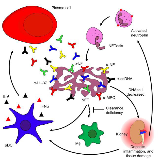
Figure 11: The role of neutrophils in SLE and target organ manifestation (142)

Exposure of extracellular DNA and chromatin fragments associated to glomerular basement membranes precedes severe nephritis and involves downregulation of DNase1 transcription and loss of nuclease activity, which might suggest a pivotal protective role for these enzymes [192]. Regarding opsonization and removal of autoantigens in the extracellular space, a protective role of the C1q has already been discussed earlier, and in the absence of C1q autoantibodies, LN is almost never seen [136]. With regard to SLE target organ damage, however, the complement system shows its Janus-faced nature: Complement factors (i.e. complement component 3 and beyond) have been demonstrated to directly cause immune-complex related renal inflammation and immunopathology [195,196]. Murine models indicate, however, that by immune complex removal, ameliorative effects of the complement system prevail and thus reduce Fcγ-receptor III (FcγRIII)-mediated inflammatory response [94]. As an additional mechanism, even antibody-deficient mice have been demonstrated to develop SLE, and pathogenic effects of B cells beyond antibody production have been identified. Namely, B cells spontaneously activate and co-stimulate pathogenic T cells, and produce a plethora of immune response-inducing cytokines. Among others, TNFα, IFNα and IL-6 mediate local inflammation following paracrine secretion. These cytokines exert a number of functions, either stimulating tissue damage by driving inflammation in an unspecific manner, by polarizing and activating T cells or by affecting lymphoid tissue formation. On the other side of the spectrum, a role for IL- 10 producing regulatory B (Breg) cells might be established, as their product, IL-10, suppresses local inflammatory processes by inhibiting further cytokine production, antigen presentation by macrophages, or TH1 and TH2 polarization [141].

Together, the ligation of TLRs, of complement receptors and fragment crystallizable receptors (FcRs) induce renal cells to express cell adhesion molecules inside the microvasculature and to release proinflammatory cyto- and chemokines [93]. Among these are type I IFNs, IFNγ, IL-6, IL-12, IL-21 and IL-23, which alter the function of local tissue (e.g., endothelial, stromal) cells and activate pathogenic lymphocytes, Mφ, and DCs on-site [1]. Due to the local expression of a limited number of chemokines, various leukocyte (sub) populations are directed into different renal compartments and locally activated [197]. Kidney tissue is foremost infiltrated by cytotoxic CD8+ T cells, TH17 cells and B cells [93]. In proximity to T cell aggregates, B cells form de novo perivascular tertiary lymphoid organs inside the kidney through clonal expansion, selection, and somatic hypermutation, namely T:B cell aggregates or germinal centers, the latter also comprising of antigen-presenting fDC cells [198]. Apart from local inflammation and tissue pathology, these aggregates contribute to intrarenal and systemic autoantibody production [199]. Certain T cell species have been shown to be particularly involved in LN pathology. IL-17 producing, CD3+/ CD4 /CD8- DN and CD3+/CD4+ T cells are generally increased in SLE patients, while TH17 and Treg cells seem to be in homeostatic balance when disease activity remains stable. In the kidney, IL 17 overexpression increases the influx of effector cells, activates B cells, increases autoantibody production and accelerates the formation of germinal centers through induction and promotion of many pathogenic mediators [200]. Additionally, activated Mo and Mφ infiltrate kidney tissue and vigorously participate in renal inflammation and injury [106]. Among Mφ subtypes, it is likely that M1 cells contribute to local inflammatory processes, but the contribution of M2 cells is less clear, as their role in resolving inflammation and tissue repair might “inadvertently” contribute to pathogenesis by proliferation and excess tissue remodeling [201].

It is obvious that LN has been a primary subject of research on target organ damage in SLE. While many different pathways for renal injury have been established, the SLE phenotype develops due to many different factors collaborating in a severely dysregulated immune response. Moreover, varying microenvironments in different tissues are believed to affect the individual contributions of participating cells and their products, e.g. interactions between myeloid cells and T cells [1]. In concert with specialized immune cells (e.g. lymphocytes, Mo/Mφ and APCs), local stromal, parenchymal and endothelial cells have been demonstrated to collaborate in target organ damage [93].

Mechanisms of Atherosclerosis in SLE

Cardiovascular disease is a common phenomenon in many autoimmune rheumatic diseases. Apart from the usual traditional and non-traditional risk factors, the vasculature is affected by harmful autoimmunity-associated, either local or systemic inflammation. Both of these inflammatory processes exhibit proatherogenic effects, promoting the formation of atherosclerotic lesions which progress and destabilize in the course of autoinflammation. Subsequently, atherosclerosis is responsible for conditions such as vessel occlusion, thromboembolism and distension or division of the vessel wall, all of which possible mediators of fatal ischemia. Atherosclerosis is the long-term result of endothelial inability to maintain homeostasis, leading to atherogenesis, a process responsible for the subendothelial accumulation of lipids and fibrous elements in large arteries resulting in a proinflammatory and procoagulant state spanning across all layers of the arterial wall [202,203]. From an inflammatory perspective, atherogenesis can roughly be divided into three stages: leukocyte adhesion, migration and activation. In a first stage, leukocytes adhere to the endothelium after the expression of different adhesion, rolling, and attachment molecules, such as vascular cell adhesion molecule-1 (VCAM- 1), P- and E-selectin, and integrins, which are expressed due to opsonization by anti-endothelial cell (EC) antibodies, inflammation, or either oxidative or mechanical stress [202,204,205].

Inflammation is suspected to be triggered by the accumulation of modified lipoprotein molecules in the arterial intima as a result of altered endothelial permeability. Early local inflammation causes the expression of pro-inflammatory cytokines such as IL-1β and TNFα, mediating endothelial expression of VCAM-1 via a NF-κB dependent pathway. Chemoattractants are of vital significance to trigger the second stage, specifically diapedesis of leukocytes entering the intima at EC junctions. Such chemokines include monocyte chemotactic protein-1 (MCP-1), Eotaxin, and chemokines induced by IFNγ (e.g. IP 10, Mig, I TAC), which attract monocytes, mast cells, and lymphocytes, respectively [202]. The third phase (Figure 12) is characterized by leukocyte activation, distinctively monocyte migration, differentiation into residual macrophages, and subsequent progression towards inflammation-perpetuating foam cells. Phagocytes are then joined by other leukocyte populations of mainly T cells, but also B cells, neutrophils, dendritic and mast cells, which locally release a multitude of, among others, pro-inflammatory, vasoactive, proteindegrading, pro- and antithrombotic substances [203,204,206]. In a continuous process, this most primitive form of atherosclerosis, or “fatty streak” develops into an inflammation-dependent, more sophisticated and enlarged lesion. In this stage, further progression into pathologically variable types of atherosclerotic plaques looms, which are generally vulnerable to progress, disrupt, and eventually cause thrombosis, embolism, or occlusion. [27,204].

biomedres-openaccess-journal-bjstr

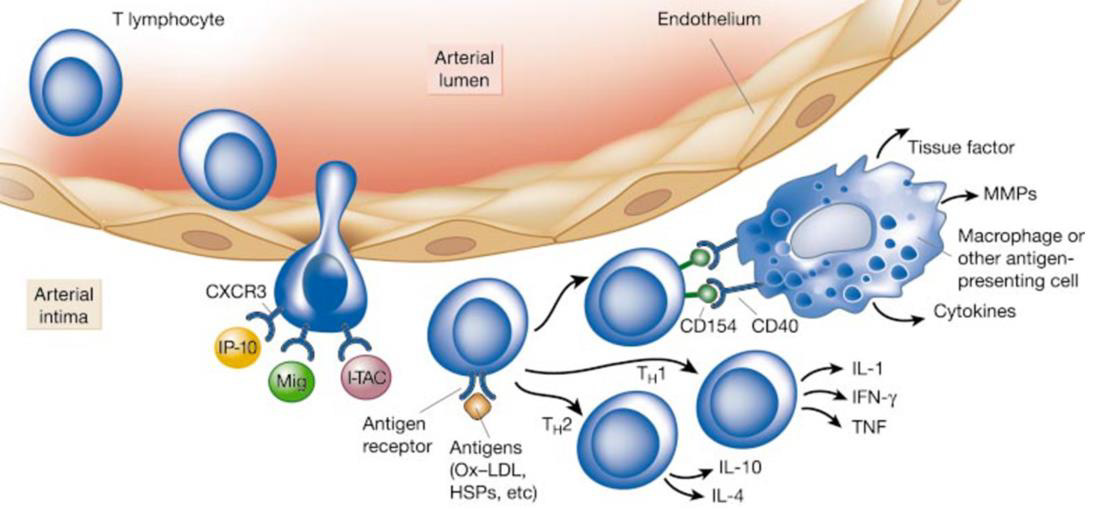
Figure 12: Inflammation in the third phase of atherogenesis (204).

When looking at these most basic processes in plaque formation, progression and rupture, initial anchor points can be discerned tying SLE to atherosclerosis. Endothelial cell necrosis, which might result from elevated levels of inflammatory mediators in the vicinity or cellular cytotoxicity [204], could be facilitated by autoreactive immune cells. This however, results in a typical henand- egg problem: As defective clearance of apoptotic and necrotic cells is present both in SLE and in atherosclerosis [27], antigen for the deposition of autoantibodies or self-reactive immune complexes is readily available. By enhancing autoinflammatory, SLE-associated processes, which result in the upregulation of the local cytokine environments, the process fueled even further. Other peculiarities of SLE, like the increased co-stimulation and recruitment of mononuclear cells via CD40L on TH cells, on which its expression is pathologically elevated, might be a source of autoimmune contribution to atherosclerosis. Indeed, soluble CD40L is increased in both SLE patients and patients suffering from acute coronary syndrome, suggesting its contribution to plaque instability and thrombosis in the setting of atherosclerosis [207]. Above and beyond the mentioned pathways, SLE contributes to atherosclerosis in a number of ways through unspecific upregulation of the immune system, e.g., through cytokine responses, up- and downregulation of the complement pathway or inflammation-associated prothrombotic disposition [202]. Besides the atherogenic involvement of the SLE-governed immune system, its contribution in anti-atherogenic processes is also feasible. In this respect, induced natural killer T (iNKT) cells have been identified as possible mediators of early stage, but not clinical apparent atherosclerosis in SLE.

Acting as disease-ameliorating effectors by responding to lipid antigen presentation in APCs, iNKT cells have been shown to release a wide array of anti-inflammatory cytokines and to polarize macrophages into an anti-inflammatory and anti-atherogenic M2 phenotype [208]. It is, however, not clear if the apparent reduction and dysfunction of iNKT cells in SLE patients impairs a possible, more extensive atheroprotective role [209]. As one of the major tissues affected by SLE pathology, the endothelium is a susceptible target for autoinflammation. It exerts pivotal functions of immunity: ECs express PRRs, produce IL-1 and other inflammatory molecules, internalize ligands such as low-density lipoprotein (LDL) particles, present antigens to specific T cells, recruit leukocytes, increase permeability, and cause edema (210). The endothelium happens to also be extensively involved in atherosclerosis, which is accompanied by loss or disturbance of its various regulatory functions [204,211]. Nitric oxide (NO), a vasodilator whose reduction is associated with endothelial dysfunction, reduces the expression of VCAM-1 by inhibition of NF-κB, the central transcriptional control point in vascular inflammation [204]. Decreased bioavailability of NO links inflammation to endothelial dysfunction, and is caused by elevated levels of TNFα, which downregulates the expression of endothelial NO synthase (eNOS). Reactive oxygen species (ROS), mediated by inflammatory cytokines, further reduce the availability of NO in the vessel wall, therefore causing impaired endothelium-dependent vasorelaxation and increased arterial stiffness [27,84]. Endothelial dysfunction is characterized by an increase in permeability, loss of junctions, secondary apoptosis, and therefore leukocyte diapedesis [205]. Resulting from direct binding of autoantibodies to endothelial cells or from the deposition of pre-formed immune complexes and subsequent autoinflammatory responses, it primarily causes functional impairment and prothrombotic activity of the arterial wall (Figure 13). Therefore, endothelial dysfunction makes for a proinflammatory and proadhesive entry point for subsequent atherosclerotic transformation, linking it to local autoinflammation [211].

biomedres-openaccess-journal-bjstr

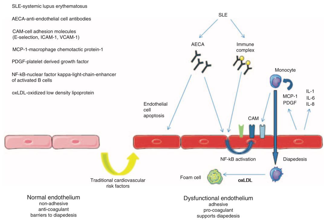
Figure 13: Autoantibodies causing endothelial injury in SLE (205).

A number of processes facilitate the migration of Mo, the uptake of LDL into Mφ and therefore the development of foam cells. On the other hand, the metabolism of triglycerides (TG) and LDL as well as the efflux of cholesterol are impaired in SLE through a decrease of lipoprotein lipase and cholesterol 27-hydroxylase. When exposed to ROS, LDL is converted to oxidized LDL (oxLDL), which stimulates the influx of Mo and the subsequent differentiation into Mφ via MCP- 1 and macrophage colony-stimulating factor (M-CSF). Therefore, oxLDL subsequently leads to its own continued phagocytosis via scavenger receptor CD36, but inhibits phagocytosis of apoptotic cells, possibly further contributing to autoinflammation in SLE [202]. Moreover, in-situ formation and deposition of oxLDLcontaining immune complexes are likely to occur as such complexes have been identified in human atherosclerotic lesions, further driving autoimmunity and -inflammation [212]. Apart from exposure to oxidative stress, increased oxLDL levels can be attributed to several other mechanisms, such as anti-highdensity lipoprotein (HDL), anti-Apolipoprotein A1 (ApoA1) and antiphospholipid autoantibody-mediated, reduced levels of antioxidant HDL and reduced paraoxonase activity in HDL particles [4,208,213]. Among the reasons for elevated levels of oxLDL is also an increase in detrimental pro-inflammatory HDL (piHDL), which occurs during states of systemic inflammation and oxidative stress, and is, conversely, strongly associated with atherosclerosis in SLE, likely contributing to the condition [213-216].

Complement activation might also contribute to the process of endothelial cell activation and recruitment of leukocytes to inflammatory sites [205]. In SLE-associated atherosclerosis, it has been shown to precede the development of spontaneous atherosclerotic lesions [210]. A protective or pathogenic role of C1q in SLE has not yet been fully established [205], but C3, C5 and overall complement activity has been demonstrated to correlate with subclinical markers of atherosclerosis, therefore reflecting activation of the complement system in patients with atherosclerosis and SLE. Interestingly, piHDL particles have been directly correlated to complement levels, suggesting an additional influence of C3 and C4-enriched HDL. Also, the complement membrane attack complex has been demonstrated to correlate to plaque formation, a finding consistent with various traces of complement activation in atherosclerotic lesions in the general population [217]. Considering the above, a contribution of the classical complement pathway in SLE-associated atherosclerosis seems likely, bearing in mind its dependence on antibodies, which have been proven to be associated with both conditions (see below). Additionally, complement induces the release of, among other molecules, MCP-1 from human VSMCs, which are – under physiological conditions – well protected from the exposure to complement by the tunica intima, but become exposed to plasma molecules in inflammation [218].

Among innate immune cells contributing to atherosclerosis, a special role for neutrophils has become apparent, with the main stage being taken by LDGs, a distinct subset of neutrophils in SLE patients with the ability to initiate different atherogenic processes [132]. Among these, LDGs propagate apoptosis, dysfunctional endothelial repair, atherothrombosis, and NET formation [27]. With regard to the latter, neutrophil extracellular traps contain antimicrobial molecules which induce endothelial cell death and vascular dysfunction [132,202]. NETosis-prone LDGs further contribute to accelerated atherosclerosis by disrupting the maturation of endothelial progenitor cells (EPC), by directly inducing endothelial damage or synthesizing increased levels of pro-inflammatory cytokines (IFNα, IL-17) (4,202). Furthermore, NETs drive IFN, IL-1β and IL-17 expression in target myeloid cells, which promote a vicious cycle of tissue inflammation [27]. Moreover, oxidant-generating enzymes in NETs lead to the oxidation of HDL and puts it into a dysfunctional, pro-inflammatory state [213,215,219]. Considering the fact that oxLDL has the ability to mediate NET release [220], another vicious cycle might be driven by dysfunctional removal of oxLDL as a consequence of HDL oxidation, further propagating LDGs to form NETs. Taking into consideration the impaired clearance of cell remnants in SLE, and degradation protective anti-DNAse I and anti-NET autoantibodies, NETs are well protected against cellular clearance [202]. This, in turn, stimulates autoinflammation through the exposure of potent autoantigens such as dsDNA and various SLE-dependent immune complexes [132].

In conjunction with peculiarities of SLE, a generally proinflammatory, self-perpetuating feedback mechanism of NETs can be discerned in atherosclerosis, and unsurprisingly, NETs remain present in mature atherosclerotic plaques, further implying their role in atherosclerotic progression [202]. Decreased numbers and decreased capacity of EPCs and myeloid circulating angiogenic cells (MACs) have been described in SLE patients with atherosclerosis. Hence, their capacity to mediate angiogenesis and therefore revascularization and vascular repair by migration, differentiation and production of growth factors, predominantly vascular endothelial growth factor (VEGF) and hepatic growth factor, is impaired [221-223]. Apart from type I IFNs, lowered vitamin D levels have been associated with these findings [4,222,224]. Vitamin D deficiency has been described as a likely proatherogenic factor in aSLE and pSLE patients, but evidence has so far been inconclusive [87,225]. Even so, vitamin D has been shown to positively modulate endothelial function in SLE patients, whereas in vitro, MAC function has been augmented by calcitriol [226]. As pathognomonic mediators of SLE, type I IFNs have been suggested as major contributors to atherogenesis. They promote foam cell formation, platelet activation and an imbalance between endothelial damage and repair (4,27). By inducing apoptosis of EC, vascular rarefication is promoted.

It is joined by dysfunctional vasculogenesis, correlating to dysfunctional EPCs and depleted numbers thereof, as well as dysfunction of resident endothelial cells [202,222–224]. Type I IFNs recruit Mφ to atherosclerotic lesions, promoting their lipid uptake through scavenger receptors and thus stimulating foam cell formation, enhancing atherosclerotic plaques. Apart from pro-inflammatory stimuli by type I IFNs, they exert prothrombotic effects, prevent maturation of vascular smooth muscle cells (VSMCs), cause VSMC dysfunction, retardation and apoptosis, and increase the risk of cardiovascular events by rupture of destabilized plaques [4,27,128,227,228]. In addition to the stimulation of phagocytes, increased levels of IFNα in atherosclerotic plaques enhance vascular damage directly by cytotoxic T and NK cells or mediated indirectly via TH, most notably apoptosis of ECs and VSMCs via a TRAILdependent mechanism [206]. Consistent with these findings, type I IFNs have been shown to be potent antiangiogenic factors which are independently associated with different markers of subclinical atherosclerosis in SLE patients [128,206,223]. As strong promoters of type I IFN production by pDCs (see above), TLR7 and TLR9 might therefore play an important role in the formation and progression of atherosclerotic lesions [206]. Apart from type I IFN, apoptotic EC microparticles also cause expression of pro-inflammatory cytokines such as IL-6 and TNFα by pDCs and mDCs, which demonstrates another possible self-perpetuating mechanism of atherosclerotic lesions [229].

One of the most overexpressed non-type I IFN cytokines in SLE, IFNγ, contributes to plaque formation by upregulating a number of atherogenic processes, among which antigen presentation or cytokine (TNFα, IL-1) expression [202]. It also mediates the inhibition of collagen production in VSMC and the overexpression of collagenases (Matrix Metalloproteinases, or MMP 1, 8, 13) and gelatinases (MMP 2, 9) in mononuclear phagocytes, endothelial and smooth muscle cells via IL-1β, TNFα and CD40L (204). Therefore, IFNγ is a powerful growth inhibitor for VSMC and EC in the local environment that promotes instability of atherosclerotic plaques [230]. In concert with TNFα, IL-1 induces local inflammation by stimulation of Mφ, induces MMPs, and promotes the production of cell surface adhesion molecules and colony stimulation factors (G-, M-, GM-CSF). TNFα has been found in all stages of atherosclerotic lesions, but may not be specifically etiological in rheumatic disease, as it also presents in atherosclerosis among the general population [230]. Other cytokines might also promote or ameliorate the pathogenesis of SLE. Among these, decreased levels of transforming growth factor β (TGF-β) were associated with arterial wall dysfunction and premature atherosclerosis [202]. More precisely, oxLDL inhibits the activation of latent TGF-β1 and (V)LDL inhibits the binding of TGF-β1 to the type II TGF-β receptor, thereby suppressing signaling. Low levels of this cytokine are strongly associated with increased CIMT, likely resulting from EC and VSMC proliferation accompanied by excessive apoptosis and atherosclerotic disposition [231].

So far, atherogenic processes have been outlined as mainly innate immunity-dependent, but adaptive immunity also plays a major role in atherosclerosis. In addition to circulatory lymphocytes, adventitial artery tertiary lymphoid organs (ATLOs), a form of tertiary lymphatic tissue, develop as a result of chronic local inflammation and contribute to atherosclerosis in SLE locally [203]. ATLOs persist in the connective tissue surrounding atherosclerotic arteries and can range from small T/B cell clusters to lymph nodelike organs harboring lymphocytes and various effector cells of innate immunity. As they can be hugely complex, it has not been fully understood whether ATLOs exert disease-promoting or -ameliorating functions, or both [232].

Atherosclerosis is likely caused by an imbalance between pathogenic T cells and Treg cells but establishing clinical manifestations of this phenomenon has been a challenging task in SLE patients to date [233]. Nevertheless, a disbalance of TH17 and Treg subgroups in patients with atherosclerosis and SLE has been suggested [234], implicating a specific role for a IL-17 mediated response in atherogenesis [212]. Conversely, Treg cells may protect against atherosclerosis by improving endothelial function and inhibiting B cell activation and the production of inflammatory cytokines [27]. Complicating the possible contribution of TH17 cells, it might even reduce vascular T cell infiltration and development of atherosclerosis [144], leaving the question of its precise role unanswered.

Different mechanisms have been described for a potential of SLE B cells to promote atherosclerosis, although various B cell subpopulations seem to exert opposing effects. In SLE, autoantibody production of B cells generally shifts from largely atheroprotective IgM NAbs to largely pathogenic IgG autoantibodies [235]. While both follicular and extrafollicular B cells might contribute to atherosclerosis in SLE by producing class-switched IgG antibodies, follicular B cells predominate in releasing pathogenic IgG antibodies and proatherogenic cytokines [203], likely due to stimulatory effects of BAFF [236]. FcγRIIB has already been discussed as an inhibitory Fc receptor to IgG autoantibodies, that is typically downregulated in SLE (see above). As it inhibits pro-inflammatory signaling in B cells, the lack of FcγRIIB has been proven to exacerbate atherosclerosis [237], which might exactly be its role in SLE. B-cell derived autoantibodies can be directed against a variety of epitopes, e.g. endothelial cells, phospholipids and oxLDL, and traditional SLE antigens such as dsDNA. For some SLE IgG autoantibodies, a distinct pathogenic role in atherosclerosis has been assumed, but no underlying mechanism has yet been identified [238]. In about twothirds of SLE patients, anti-endothelial cell (AEC) autoantibodies are present, exposing a potential to upregulate adhesion molecule expression, induce oxidative stress, or cause apoptosis in EC [4,205,239]. Sub-APS levels of antiphospholipid autoantibodies contribute to a high cardiovascular disease risk profile [35] and may also be directly causative for atherosclerosis in SLE [4].

Anti-β2-glycoprotein I (β2-GPI)-autoantibodies, a subset of antiphospholipid antibodies, have been linked to subclinical atherosclerosis in SLE patients. They facilitate oxLDL foamcell- generating phagocytosis via an anti-β-glycoprotein I (β2- GPI)-dependent mechanism and are also intertwined with increased T cell reactivity, an increase in NETs, and thrombin production [212,240]. While anti-oxLDL antibodies have been loosely associated with atherosclerosis, anti-oxidized palmitoyl arachidonoyl phosphocholine (oxPAPC, a more stable epitope of oxLDL) antibodies do indeed correlate to Intima-Media-Thickness (IMT), and therefore to subclinical atherosclerosis [202,238]. Autoantibodies to lipoprotein lipase (LPL) impair degradation of very low-density lipoprotein (VLDL) conversion to LDL, resulting in a low LDL, low HDL, high VLDL, high TG lupus lipid pattern, which is correlated with disease activity and markers of inflammation [223]. Other populations of autoantibodies such as anti-cardiolipin and anti-dsDNA antibodies have also been demonstrated to correlate with atherosclerotic markers and events in SLE [241], but whether they contribute in an atherosclerosis-specific or general SLE pathomechanisms is yet to be understood. In contrast to pathogenic autoantibodies, the lack of distinct subsets of protective autoantibodies has been shown to be correlated with cardiovascular disease in SLE patients (Figure 14). For one thing, decreased levels of IgM NAbs to phosphorylcholine further impair apoptotic cell clearance in SLE and are associated with carotid plaque formation and increased vulnerability (4,242). Analogously, antibodymediated clearance of LDL particles might also be impaired, which is reflected in decreased serum levels of atheroprotective anti- ApoB-100 IgG and IgM NAbs [243].

biomedres-openaccess-journal-bjstr

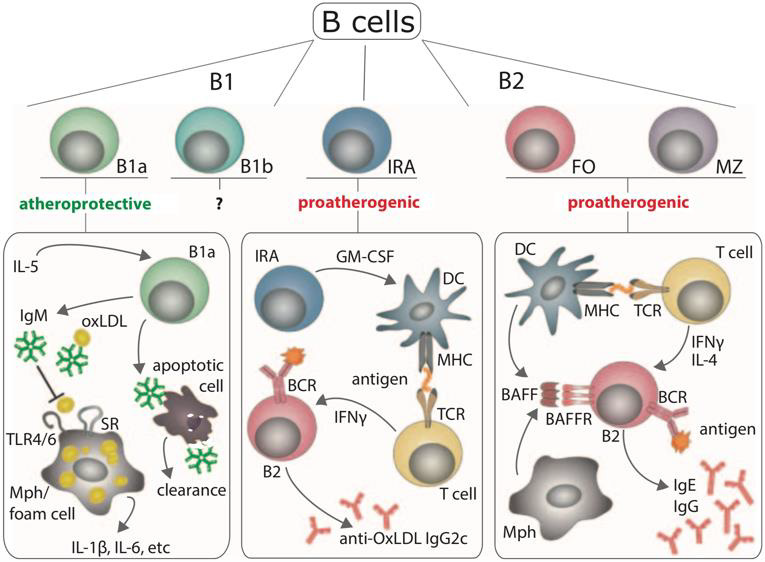
Figure 14: Atheroprotective and proatherogenic roles of B cells, NAbs, and pathogenic autoantibodies (235)

As already outlined previously, data resulting from smaller samples and case studies suggest that, in addition to the wellestablished correlation between SLE and atherosclerosis, patients suffering from SLE are at an increased risk of developing arterial, mostly aortic or coronary aneurysm. Literature on the mechanism of its development in SLE is rather scarce compared to such of atherosclerosis. Formation of AA is described as an immunerelated process in which cells of the innate and adaptive immune system accumulate in the adventitia of the aortic wall [73]. Apart from this, the pathogeneses of AA and dissection have also been linked to atherosclerosis, aortic elastic tissue degeneration and certain vasculitides [72,73,244,245]. This corresponds with the fact that, seemingly, some consent has been reached about at least two different SLE-related disease mechanisms which result in the development of aortic aneurysms. The first of the mentioned pathway is one of atherosclerosis-associated, mostly abdominal AA formation. Sites of aneurysm often display atherosclerosis, which increases the likelihood to rupture plaques as a result of aneurysmatic dilatation [246]. As atherosclerosis and abdominal AA are also positively correlated among both the general population and SLE patients [56,69,72,247], this pathomechanism might be similar to such of non-SLE-related abdominal AA. This is supported by the fact that abdominal AAs are more likely to occur in hypertensive, older SLE patients [71], which might lead to the assumption that general risk factors of abdominal AA also apply to SLE patients. Therefore, abdominal AA might be fueled by secondary hypertension due to damage of renal vasculature in SLE, which has also been demonstrated as an independent risk factor for the development of aneurysm [56].

The second pathway, which is hypothesized to account for most cases of thoracic AA in SLE patients, is believed to result from a SLE-related inflammatory process that leads to a diffuse pattern of aortitis which is associated with a higher prevalence of combined aneurysm and dissection [244,245,248]. This pathway might result from vasculitis-like inflammation [56], which might be similar or even equivalent to either Takayasu’s arteritis or cystic medial degeneration on a case-to-case basis [72,249]. The fact that mostly young and normotensive patients are affected might imply a lower dependence on mechanical stress compared to atherosclerosisassociated aneurysms [71]. This “vasculitis-like” pathway and atherosclerosis might also be closely related by sharing a similar autoinflammatory process [246], which is, however, likely to extend beyond the lamina intima in the case of atherosclerosis. Evidence of aneurysms beyond of the aortic wall in SLE is extremely rare. As an example, a causative relation between SLE and coronary artery aneurysm is suggested, but its mechanism is poorly understood [69]. It might result from severe coronary inflammation, medial degeneration and weakening of the arterial wall by deposition of Igs and complement, suggesting a similar, “vasculitis-like” pathway [42,69,247].

Discussion

This review has demonstrated a substantial array of pathomechanisms contributing to SLE onset and progression. Many of these general mechanisms of disease are reflected in atherosclerotic processes and have been suspected to facilitate atherogenesis. Starting from the most exposed layer of the vessel wall, ECs contribute to autoinflammation both as active mediators and as targets for autoimmunity. Apart from the endothelium’s role as a regulator of mechanical stress and lipoprotein uptake, it also exerts immune functions and influences inflammatory processes. Unless it induces endothelial cell death, autoimmunity in SLE is likely to impair homeostasis and skew ECs towards a more pro-inflammatory, hyporegenerative state. Even though the endothelium has been a primary subject of prior research, VSMCs might also exert pro-inflammatory functions and fail to retain their physiological functionality, primarily leading to a destabilization of the vessel wall. While the reviewed literature has not specifically highlighted other resident cells as possible mediators of SLEassociated atherosclerosis, cytokine-expressing fibroblasts are only one likely, additional option of such contribution.

Possible interfaces between immune cells in SLE and atherosclerosis are versatile. First and foremost, Mo/Mφ play a dominant role in atherogenesis. While their capacity to phagocytose cellular remnants is decreased, foam cell formation is preserved, most likely due to SLE-derived costimulatory influences and the occurrence of pro-inflammatory subpopulations. A distinct subgroup of neutrophils, namely NETosis-prone LDGs, interferes with vessel wall homeostasis in a number of ways, completing their general relevance in SLE as main drivers of NET formation. DCs, T and B cell populations facilitate excessive inflammation by making their SLE-specific, overshooting contributions. Among these, the production of pathogenic autoantibodies, which may even originate from tertiary germinal centers adjacent to atherosclerotic plaques, stands out most prominently. Such autoantibodies are either fairly specific to sites of atherosclerosis, as is the case with AEC, anti-β-GP1 and anti-oxLDL, or rather unspecific. The latter may target autoantigen in areas with a high concentration thereof, such as atherosclerotic plaques. Apart from the production of autoantibodies, upregulated T cell help and excessive cytokine production are just two of many possible cellular contributions to SLE-associated atherosclerosis. Completing the role of the immune system, the complement cascade has been shown to activate in atherosclerotic lesions, suggesting a direct influence on endothelial cells and lipoproteins, as well as more unspecific effects at sites of active inflammation. Beyond its immune phenotype, SLE has been shown to promote the formation of pro-inflammatory and otherwise dysfunctional HDL, oxLDL, and prothrombotic environments, which have been shown to promote atherogenesis.

On par with its underlying disease, SLE-associated atherosclerosis has been discussed as a combination of many virtually independent mechanistic defects, but an indispensable trigger has not been identified for either condition. Because SLE presents as an exceptionally heterogenous disease, the very likely explanation would suggest a general disbalance of immunity, causing homeostasis to tip over as soon as pro-inflammatory outweigh regulatory stimuli. Considering the many possible vicious cycles in SLE, this notion is a compelling explanation for the correlation between disease activity over time and prevalence of atherosclerosis. While some SLE-specific defects have been shown to prevail in distinct cells of the immune system, others span across many systems. Apart from ubiquitous mediators such as type I IFNs, individual contributors to cell signaling reach beyond certain cell types. As such, TLR7 and TLR9 run like a common thread through many parts of autoimmunity in SLE, and therefore constitute likely candidates for further research into the vascular manifestations of SLE.

Undeniably, this review has certain limitations. Notwithstanding the vast amount of literature on the pathogenesis of SLE and SLEassociated atherosclerosis, many questions still remain unanswered. Among these, one certainly concerns the lack of research into the pathogenesis of SLE-associated aneurysms. Notwithstanding the fact that two distinct pathways of aneurysm and dissection formation in SLE have been identified, the corresponding pathomechanisms have not yet been illuminated. Considering the underlying mechanisms, autoimmune processes involving the enzymatic degradation of connective tissue might only be one of many fields of interest. In contrast to this mostly uninvestigated manifestation, independent disease entities such as large- and medium-vessel vasculitides were not reviewed in this thesis due to their self-sufficient nature. These conditions can result from SLE or share certain pathomechanisms with SLE, both of which might translate into their contribution to SLE-associated atherosclerosis or aneurysm. Lastly, as a general principle, iatrogenic interventions were not described as separate mediators of disease because the literature cannot be distinguished between data from treated vs. treatment-naïve individuals. In this regard, it is important to remark that disease-modulating or anti-inflammatory treatment regimens may damage tissue via discrete mechanisms.

Conclusion

This review of the literature discussed the main disease mechanisms in SLE known to date and many peculiarities of these mechanisms. Both longtime knowledge as well as more recent insights into the pathomechanisms of SLE were outlined, resulting in a broad overview as well as a more detailed look into certain aspects of SLE pathogenesis. Strikingly, SLE pathomechanisms could not be delineated as a single, stringent process. Rather, the pathophysiology of SLE presents as a complex and convoluted set of individual immune dysregulations, in which components of the innate and adaptive immune systems partake. Apart from its etiologic diversity, SLE results in profoundly different clinical pictures, which led to the question whether SLE is a single disease entity or a rather a collective term for different conditions. Some of its various manifestations are of vascular nature, the most prominent being atherosclerosis. Along with aneurysm and dissection, atherosclerosis remarkably affects SLE patients at much younger age than the general population and correlates with disease duration and activity. Therefore, these vascular manifestations are likely to be associated to the pathomechanisms of SLE. Indeed, many of such mechanisms have been identified to take part in accelerated atherosclerosis, but no definitive trigger can be singled out. Rather, an interplay of different autoinflammatory reactions likely facilitates many stages of atherogenesis. Although no definitive mechanisms have yet been established, two individual pathogeneses are proposed to cause SLE-associated aneurysms and dissections in different patient cohorts. While the first presumed mechanism is dependent on pre-formed atherosclerotic plaques, the second might share a common autoinflammatory pathway with SLE-associated atherosclerosis.

For More Articles: Biomedical Journal Impact Factor: https://biomedres.us

Open Access Journals on Zoology and Animal Physiology

Use of the Leaf-Aqueous Extract of Pseudopanax Arboreus (Araliaceae) (L.F. Phillipson) is Void of Toxic Effects

Introduction

Over the past two decades, there has been an increase in the use of plant-based medicine worldwide. It is estimated that threequarters of the world’s population use herbal medicine for their healthcare [1]. Interestingly, the World Health Organization (WHO) has encouraged this alternative medicine for the prevention and treatment of diseases and according to estimates put up by this organ (WHO), more than 80% of the population in Africa uses the doctor’s plants to meet their need for care and health [2]. The toxicity of chemical products, the high cost of chemical drugs, the remoteness and /or insufficiency of health centers, especially in rural settings, which limits the genuine handling of public health problems, have favored the use of such drugs [3]. As a result, traditional medicine can be considered an integral part of primary healthcare and is used to improve access to healthcare [4]. Unfortunately, because plants are natural, they are considered to be non-dangerous, and the population use them in many and very different contexts. The products used are often the mixture of plants whose knowledge and requirements of preparation and consumption are not controlled. Thus, although plants may be effective in treating some ailments, they may contain potent chemical compounds that cause adverse effects and toxicity, especially when administered at high doses and for a long period [5,6]. For instance, Peganum harmala is a plant with scientifically proven analgesic, hypoglycemic, anti-nociceptic and anti-parasitic properties, but whose prolonged administration leads to hepatotoxic and nephrotoxic abnormalities. Also, Astragalus hamosus is effective in the treatment of gastrointestinal diseases, respiratory problems and headache, but causes liver and kidney dysfunction at higher doses and following long term administration [7].
It is therefore essential to ensure not only the clinical efficacy and the quality, but especially the safety of any medicinal herbal preparation before making it available to consumers. Toxicity tests are therefore indispensable and accompany the biological activity test in the course of the selection of new molecules [8]. For this, a renewed interest has been brought to phytotherapy to deepen the analysis of its therapeutic efficacy and especially its toxicity aspect. P. arboreus is, belonging to the Family Araliaceae, is commonly used in traditional medicine in the treatment of many ailments such as hypertension, male infertility and male sexual dysfunction. It has already been the subject of several scientific studies in which the sexenhancing properties of the aqueous [9] and methanol [10] extracts of its leaves have been demonstrated. In other investigations, its potentials to reverse clinically induced male sexual dysfunction (MSD) have been proven [11]. Further studies have evaluated the time-response activity of the leaf-methanol extract of the plant [10]. However, till date, no study has been conducted in relation to its toxicological effect, hence the question of whether the aqueous extract of P. arboreus would have toxic effects in the body. This would suggest that in addition to its proven biological efficacy, the aqueous extract of P. arboreus would be devoid of toxic effects. This work therefore had as aim to evaluate the toxicological effect of aqueous extract of P. arboreus in rats.

Materials and Methods

Plant Collection and Preparation of the Aqueous Extract

Fresh leaves of P. arboreus were collected from Ntenako village, Manyu Division, South-West Region of Cameroon, under the guide of a local tradi-practitioner who confirmed the plant’s identity. Its authentication, processing and production of the aqueous extract were done following the same procedures as outlined in our previous study [9].

Chemical Products or Reagents

Products or reagents used in this study included assay kits for triglycerides and total cholesterol (IVD DIALAB, Austria), Creatinine, AST (aspartate aminotransferase), ALP (alkaline phosphatase), ALT (alanine amino transferase) (Elabscience, USA) and Albumin (BioVision Inc, USA). All were purchased and stored under recommended conditions until used.

Breeding of Animals

Animals used were rats of the Wistar Strain of either sex that were bred in the Animal Facility of the Department of Zoology and Animal Physiology of the Faculty of Science, University of Buea under standard conditions of temperature, humidity and light (12H cycle). They were given free access to water and a standard laboratory diet.

Acute Toxicity

In order to assess the toxic nature of a compound, acute oral toxicity is the first step to be carried out [12]. Acute toxicity testing involves the determination of lethal dose, the single dose that kills 50% of the tested group of animals within 24 hours. Acute toxicity studies of the leaf aqueous extract of P.arboreus were carried out in male rats by using Organization for Economic Co-operation and Development (OECD) guidelines [13]. Before oral administration of a single dose of the test substances, the rats were deprived of food for 3 hours. They were randomly divided into 3 groups of 7 rats each. Animals of group 1 were administered 10 ml/kg distilled water to serve as the control; while those of groups 2 and 3 received 2000 and 5000mg/kg of the aqueous extract, respectively. All animals were observed for general behavioral changes (somnolence, convulsion, fatigue, increase heart rate); symptoms of toxicity and mortality after treatment for the first four (critical) hours, then over a period of 24 hours and thereafter, 2 hours daily for 14 days. Meanwhile, body weights were measured daily. Abnormal findings were recorded with the time of onset and disappearance. On the 14th day, all animals were sacrificed and selected organs (lung, liver, heart and kidney) isolated and processed for macroscopic observations [12].

Sub-Acute Toxicity

When treatment related toxicity is not identified in acute toxicity, sub-acute toxicity is assessed to ensure safety after repeated exposure over a relatively long period of time. Sub-acute toxicity study can be used to determine the undesirable effects of continuous or repeated exposure of part of an average life laboratory animal to a plant extract and to provide information of target organ toxicity. Like in the acute toxicity test, sub-acute toxicity study (28- day repeated oral toxicity study) was also carried out according to OECD 407 guidelines [14]. Eight (8) weeks old (110-120g) rats of either sex were divided into 4 groups with 10 animals (5 males plus 5 females) in each group. Animals of group 1 received 10ml/kg of distilled water and served as a control group whereas groups 2, 3 and 4 were given the aqueous extract at 250 mg/kg, 500 mg/kg and 1000 mg/kg body weight, respectively. All animals were observed for 4 hours daily for mortality and morbidity till the completion of the experiment. They were observed for clinical signs and the time of onset, duration of these symptoms, if any were recorded. Body weights of the rats in all groups were recorded once before the start of treatment, once weekly during the treatment period and finally on the day of sacrifice. The amount of food and water intake was recorded daily and expressed as an average for 7 days. Animals were treated orally once a day using the metal oropharyngeal cannula for a duration of 28 days [15].
On the 29th day from commencement of treatment, all animals were terminated for the evaluation of other signs of toxicity. To this effect, they were starved for 24hours, then anesthetized using an overdose of ethyl-ether. The thoracic region was rapidly dissected and blood samples collected through cardiac puncture. Part of it was collected in heparinized test-tubes, whereas the other part was collected in heparin-free test-tubes which allowed coagulation and the subsequent collection of serum. The animals were then sacrificed through cervical dislocation and selected organs including the heart, kidney, liver and spleen isolated. They were freed from all connective tissue moisture, examined for morphological changes such as the presence of any kind of lesions and then weighed using an electronic balance (NVT 1601/1, OHAUS, USA). Both blood samples were preserved at 4oC±1 for hematological and biochemical tests, respectively. As regards hematological tests, red blood cell (RBC), white blood cell (WBC) and platelet numbers as well as the percentage lymphocytes, monocytes, eosinophils and neutrophils were determined using the fully automated hematology analyzer (URIT3300) (Prasanth et al. 2014). The serum was processed for biochemical parameters including creatinine, alanine aminotransferase (ALT) (serum glutamate pyruvate transaminase, SGPT), aspartate aminotransferase (AST) (serum glutamic oxaloacetic transaminase, SGOT), alkaline phosphatase (ALP), albumin, total cholesterol and triglycerides [16,17].

Ethical Consideration

Animals were handled in accordance with the Organization for Economic Cooperation and Development (OECD) guidelines for testing chemicals 423 and 425 (OECD, 2008a&b) and the experimental protocol was approved by the University of Buea Institutional Animal Care and Use Committee (UB-IACUC).

Statistical Analyses

Values were expressed as Mean±standard error of mean (SEM). Mean values were calculated for each animal and quantitative comparisons between groups established from those means. One way ANOVA followed by Duncan test was used to analyse the data with the aid of the SPSS for windows version 20.0 software. Significant levels were tested at p<0.05.

Results

Acute Toxicity

Single oral administration of the leaf-aqueous extract of P. arboreus at 2000 mg/kg and 5000 mg/kg produced no behavioral changes, no signs of toxicity and no mortality in rats after treatment. Since there were no clinical signs of toxicity and death in the tested doses, LD50 value of the extract was found to be greater than 5000mg/kg.

Sub-Acute Toxicity

Following 28 days repeated oral administration of the leafaqueous extract of P. arboreus at 250, 500 and 1000mg/kg doses to rats, there were no treatment related toxicity signs and mortality observed in both sexes of rats treated. No significant (p<0.05) differences in weekly body weight gain were observed between the extract-treated and control rats Table 1. As presented in Table 2, repeated treatment of rats with the leaf-aqueous extract of P. arboreus produced a non-significant (p<0.05) difference in the weekly food intake of the animals, compared to their control counterparts. Like in food intake, similar results were obtained in water intake of animals following 28days repeated treatment with the leaf-aqueous extract of P. arboreus, compared to the control animals Table 3. Sub-acute treatment of rats with the leaf-aqueous extract of P. arboreus did not produce any significant (p<0.05) difference in the weight of organs such as the heart, kidney, liver and spleen, compared to the control animals Table 4. Results of the effects of sub-acute treatment of animals with the leaf-aqueous extract of P. arboreus on hematological parameters are presented in Table 5. According to the table, there were no statistically significant (p<0.05) differences in the hematological parameters measured between the control and extract-treated groups. Subacute administration of the leaf-aqueous extract of P. arboreus did not show any significant changes in biochemical parameters such as alkaline phosphatase (ALP), alanine amino transferase (ALT), aspartate aminotransferase (AST), creatinine, triglycerides, total cholesterol and albumin when compared to control group Table 6.

biomedres-openaccess-journal-bjstr

Table 1: Effects of the Sub-acute (28 days) administration of the leaf-aqueous extract of P. arboreus on the body weight of rats.

Values: Mean±SEM; DW: distilled water; AE: aqueous extract

biomedres-openaccess-journal-bjstr

Table 2: Effects of sub-acute (28days) administration of the leaf-aqueous extract of P. arboreus on weekly food intake (g) in rats.

Values: Mean±SEM; DW: distilled water; AE: aqueous extract; Fem.: females.

biomedres-openaccess-journal-bjstr

Table 3: Effects of sub-acute (28days) administration of the leaf-aqueous extract of P. arboreus on weekly water intake (ml) in rats.

Values: Mean±SEM; DW: distilled water; AE: aqueous extract.

biomedres-openaccess-journal-bjstr

Table 4: Effects of sub-acute (28days) administration of the leaf-aqueous extract of arboreus on the weight of some selected organs.

Values: Mean±SEM; DW: distilled water; AE: aqueous extract.

biomedres-openaccess-journal-bjstr

Table 5: Effects of sub-acute (28days) administration of the leaf-aqueous extract of P. arboreus on hematological parameters of rats.

Values: Mean±SEM; AE: aqueous extract; DW: distilled water %: percentage; μl: microliters.

biomedres-openaccess-journal-bjstr

Table 6: Effects of sub-acute (28days) administration of the leaf-aqueous extract of P. arboreus on biochemical parameters.

Values: Mean±SEM; AE: aqueous extract; ALP: alkaline phosphatase; ALT: alanine aminotransferase; AST: aspartate aminotransferase; DW: distilled water; SGOT: serum glutamic oxaloacetic transaminase: SGPT: serum glutamic pyruvic transaminase.

Discussion

In developing countries, herbal medicines have become famous in healthcare, and some have been falsely considered as safe without understanding the possible health effects and thus commonly used as self-medication [18]. As the use of plant-based products increases, it is important to screen the toxicological profile of these plants to confirm the safety and efficacy of those natural sources. Though P. arboreus is popular in the Bayang folk medicine as a sex enhancer, there is lack of data on its toxicological profile and adverse effects. Therefore, toxicity studies were necessary not only to identify the further range of doses in animal studies, but also to explain the probable clinical signs evoked by its extracts. Hence the present experiment was undertaken to evaluate the possible effects of the short term and long-term administration of its leafaqueous extract. Throughout the 14 days of observation period, no morbidity or mortality was observed in the extract-treated rats. In the present study, the results showed no adverse events in the dose groups 2000 mg/kg and 5000 mg/kg which indicate that the LD50 was greater than 5000 mg/kg. According to the OECD [12], a substance that does not cause mortality at the limit dose of 5000 mg/kg would have a DL50 greater than this limit dose and can be considered non-toxic. When treatment related toxicity is not identified in acute toxicity, sub-acute toxicity is assessed to ensure safety after repeated exposure over a relatively long period of time.
In the repeated dose (28-day) oral toxicity study, there were neither deaths nor treatment-related signs observed in all the groups of animals. After exposure to a few possible toxic substances, there will be changes in body weight gain and internal organ weights which would reflect toxicity [19]. The body weight changes are markers of adverse effects of drugs and chemicals and if the body weight loss occurred is more than 10% of the initial body weight it will be considered as statistically significant [20,19]. There were no significant differences in body weight gain of both control and treated groups. We can therefore deduce that leaf-aqueous extract of P. arboreus is almost non-toxic. Organ weight also is an important indicator of physiological and pathological status of animals. The relative organ weight is fundamental to confirm whether the organ was exposed to the injury or not. The heart, kidney, liver and spleen are the primary organs affected by metabolic reactions caused by toxicants [21]. In the present study, organ weights in all the treated groups of both sexes were not significantly different from those of control groups. Hence, it can equally be concluded that leaf-aqueous extract of P. arboreus is almost non-toxic.
Since proper food and water intake is necessary to the physiological status of the animal and to the achievement of a better response to test substance under investigation, these parameters were measured in our study [17,22]. Our findings revealed that both food and water consumption were not affected by the administration of the extract. Thus, this indicates that there was no interruption in the metabolism of carbohydrate, protein and fat. Analysis of blood parameters is important in the evaluation of risks associated with test compounds under investigation as the changes in the hematological system have a greater indicative value for human toxicity, when the data are converted from animal studies [18]. Repeated treatment of animals with the aqueous extract of P. arboreus for 28 days did not produce any changes in hematological parameters including RBC, WBC and platelet counts as well as the percentage lymphocytes, monocytes, eosinophils and neutrophils, an indication that the extract did not affect the blood cellular components or their production. Transaminases such as serum glutamic oxaloacetic transaminase (SGOT) and serum glutamic pyruvic transaminase (SGPT) (also called aspartate aminotransferase [AST] and alanine aminotransferase [ALT], respectively) are normally present in the heart and liver and their release into blood indicates heart or liver damage.
They are therefore well-known good indicators of liver and heart function and are used as biomarkers to predict the probable toxicity of drugs and xenobiotics [23,24]. Normally, destruction to the liver parenchymal cells will result in an increase of both these enzymes in the blood [25]. Interestingly, there were no changes in the ALT and AST levels in our investigations, which reveal that the extract did not affect the liver function/ or metabolism. Furthermore, determination of plasma proteins like albumin is required in order to assess the synthetic capacity of the liver and decrease in plasma proteins therefore tend to reflect chronic damage [26]; hence, the no alteration in the level of albumin in extract-treated animals is another indication that their liver was not affected. The extract did not equally provoke any change in the serum levels of total cholesterol and triglycerides. The action of the extract in maintaining a stable serum lipid profile could be through the induction of the inhibition of reductase hydroxyl methyl glutanyl CoA (HMG-CoA) leading to reduction of hepatic synthesis and intestinal absorption of cholesterol. Indeed, the inhibition of HMGCoA by flavonoids which have been identified in our plant extract has been reported. This effect of the leaf-aqueous extract of P. arboreus on lipid profile could suggest its beneficial effects against lipid peroxidation and subsequently on oxidative stress [27]. The reduction of blood lipids is an efficient method to prevent and treat cardiovascular affections [28]. This could explain its empirical use in treating hypertension, since arterial hypertension is generally associated to dyslipidemia [29].

Conclusion

Results of our findings indicate that treatment with single oral doses of 2000 mg/kg and 5000 mg/kg of the plant extract did not result in any toxic signs or mortality in the acute toxicity studies; likewise, daily oral administration of the extract of P. arboreus for a period of 28 days did not cause mortality, changes in body weight and body weight gain. Hematological and biochemical examinations proved that the extract is safe. Hence, the no-observed adverseeffect level (NOAEL) of the extract was found to exceed 1000 mg/ kg/day. Overall, it can be concluded that the leaf-aqueous extract of P. arboreus is well tolerated.

For More Articles: Biomedical Journal Impact Factor: https://biomedres.us

Open Access Journals on Medicine

Cropland Water-Soluble Selenium, Groundwater Silicon, Atlantic Rain – Agro-Geological Assessments

Introduction

This study is treating hot water extractable selenium of Finnish croplands in 1978-80, before the era of Se fertilizers. There are many methods for cropland Se determination [1], even “total selenium” can be determined by different selections and orders of strong acids. Se values are usually expressed by mg/kg, but watersoluble Se by mg/l or μg/l. Total Se in plough layer has reported to have been 0.201 mg/kg (N=250) [2], 0.209 mg/kg (N=93) [3], 0.229 mg/kg in 1998 [4]. Generally, Se content has been highest in clays and organic soils (org), lowest in coarse mineral soils (coms). In 1979 Se content in coarse mineral soils (coms) (without silt) was 0.157 mg/kg , (calculated) [2] and in fine sandy soils 0.172 mg/kg (even 1979) [3]; in fine mineral soils [(clay + silt), fims] 0.276 mg/ kg [2] (calculated), in clays 329 mg/kg [2] and 0.290 mg/kg (early spring) [3]; in mull soils 0.228 mg/kg [2] – in peat soils 0.093 mg/ kg [2] – in organogenic soils 0.464 mg/kg [3]. In studies of Yläranta in1983 and 13 yrs after start of Se fertilization [4], Se of organic soils was about double to that in fine mineral soils and that about 50 % higher than respective Se in coarse mineral soils. (Coms in Sippola 1979 originally included silt). Se has been associated with clays and organic matter. Clay fraction consists markedly of micas, e.g., biotite, relatively high in selenium [5]. Biotite K(Mg,Fe)3(AlSi3O10)(F,OH)2, contains Si potassium (K) and magnesium (Mg), but no calcium (Ca) [6]. It is weatherable by normal organic acids [7]. Surprisingly amount of the liberated (molybdate reactive) Si has been only about 1 % relative to the large proportions of cations in the extracts [7]. Colloidal elemental selenium is electrically charged and adsorbed by clay minerals [5], which explains Se clay association.
Water-soluble Se can be determined by shaking or boiling the water [8], the exact method is not always clearly expressed, e.g. “soluble Se” 0.011 mg/l in [2] was extracted by acid ammonium acetate-EDTA, was not an indicator of water-soluble Se as written in [4]. Proportion of total Se: water-soluble selenium composed via hot water extraction, 3-10 % according to (Table 3) (by 7-13μg/kg) in [1] and 1 % according to text in [1], 2 % (by 6 μg/kg), via water shaking method without heating according to the study in 13 EU countries [9], (N = 128) and ca 4 % (by 6-18 μg/kg, in plough layer of different soil-types) by hot water extraction according to [3] , N = 230. Proportion of water-soluble Se can vary from 0.3 to 36 % of total Se, according to several referates in [8]. “Soluble Se” can indicate [acid (pH 4.65) ammonium acetate-Na2EDTA (AAAc-EDTA) extractable Se] [9], which was 5 % when Se.H2O was 2 % of total selenium. “Soluble Se” can even be a synonym for acid ammonium oxalate extractate, by [(0.18 M(NH4)2(COO)2+0.1 M(COOH)2, pH 3.3)], which was 5-10 x higher to Se.H2O, Table 3 in [1]. In study of [9] the soil samples were collected from 13 European countries, samples of [1] from Finland. In the study of [9] plant Se correlated with soil selenium as follows: Water extr. r = 0.33***, AAAc-EDTA extr. r = 0.33***, HNO3-HClO4 digest. r = 0.27**, aqua regia digest. r = 0.23**.
Atmospheric Se: Volatilization of selenium from selenates and selenites in Finland according to [10] for 3 months has been very scanty: generally, < 1 %, anyhow from Carex peat Se losses could reach ca 3 % by treating soils with both lime and organic matter [10]. Metylated selenium compound can volatilize more easier, e.g. even 30 % of the selenium added to fine sand in the form of trimethylselenonium chloride (Se 2,5 mg/kg) volatilized from the soil during 42 days (the trimethylselenonium ion is an important urinary metabolite of dietary Se) [11]. Atmosphere is a great reservoir of Se [12], composed from anthropogenic (62.5 %) and natural (37.5 %) sources. Vicinity of ocean can increase Se content in soil [13]. It is expected that the amount of Atlantic rainfall could effect on atmospheric emission of Se.

Materials and Methods

Groundwater (gw) silicon (Si), calcium (Ca), magnesium (Mg) and potassium (K) values are provided by Geologic Survey of Finland [14]. Rural Centes (RC) ‘(04a). Finska Hushållningss.’and ‘(04b).Åland’ are excluded, because of missing Se.H2O values and ‘(16).Ostrobothnia’ because of small number of Si.gw samples and sulfurous soil [15]. Cropland hot water extractable Se values (Se. H20), (μg/l) (N.B. weight per volume), in 1978-80 are from [16] (Soil fertility Service, Eurofins Viljavuuspalvelu Oy). (Method: dry and milled soil sample was extracted with boiled water at ratio 1:3. leachate was analyzed using CV-AAS equipment) [17]. Original data, (N=1340), were missing values concerning RC’s (04a) and (04b). Exclusion of “(16). Ostrobothnia’ caused ca 5 % (N 27) reduction, approximated by its cropland area of Finnish total value. Whole country mean of [Se.H2O] was 6 μg/l. [Se.H2O] values by humus content were as follows: 0-3 %: 5 μg/l, 3-12 %, 6 μg/l, 12-20%: 7 μg/l, by peat 4 μg/l (remarkable is that values are per volume). Geographic coordinates of the Rural Centers are attained by web search: ‘name of the visually selected central commune’ and ‘geographic coordinates’ or “geographic coordinates dateandtime. info”. In RC. (07) Renko has been after 1980 combined with Hämeenlinna, but data for both are accessible, why the old value was benefited. The order of Rural Centers is the same as by Eurofins Viljavuuspalvelu Oy. By order we must be careful with RC’s (06), (07), (12) and (13) – it is not always the same!
Relative regional proportion of Altantic rain, determined by change in 18O/16O isotope ratio to respective Oceanic standard (VSMOW), δ18O (‰), has been estimated by combining the RC map (in [14]) and map in [18] Figure 1. Data for this study is in Table1.

biomedres-openaccess-journal-bjstr

Table 1: Names of Rural Centers after exclusion, names of central communes, groundwater Si, Ca, Mg, K, (Ca+Mg).

biomedres-openaccess-journal-bjstr

Figure 1: Represents regional δ18O values by Rural Centers. Numbers of RC’s are as given in [14], but RC.(04) is divided into two parts with different mineral element database (to clarify other articles).

Results

Comments on Table 2

biomedres-openaccess-journal-bjstr

Table 2: Regressions of groundwater Si,[ Se.H2O] and δ18O by Latitude, Longitude, Altitude and groundwater.

All associations of [Si] and [δ18O] were significant. Long and Alt explained [Se.H2O] weakly: by 14.5 % (ns) and respectively by 17.3 % (p < 0.05)]. Combined regression by [Lat;Long;Alt] explained better [Si] (by 84 %), Figure 2, than [Se.H2O] (by 65.2 %) Figure 3. [δ18O] behaved rather similarly. [Ca+Mg] and [Mg+K] explained stronger [Se.H2O], (by 68.9 %, Figure 4, and 72.2 %, Figure 6, respectively, p < 0.001) than [Si], (by 52.1 %, Figure 5, and 53.9 %, Figure 7, respectively, p < 0.01). [Se.H2O] was explained 62.7 % by [δ18O] and 61.9 % by [Si] Figure 8, respectively (p < 0.001).

biomedres-openaccess-journal-bjstr

Figure 2: Represents groundwater Si and its regression by geographic factors [Lat;Long;Alt].

biomedres-openaccess-journal-bjstr

Figure 3: Represents cropland [Se.H2O] and its regression by geographic factors [Lat;Long;Alt].

biomedres-openaccess-journal-bjstr

Figure 4: Represents cropland [Se.H2O] and its regression by groundwater (Ca+Mg).

biomedres-openaccess-journal-bjstr

Figure 5: Represents groundwater Si and its regression by groundwater (Mg).

biomedres-openaccess-journal-bjstr

Figure 6: Represents cropland [Se.H2O] and its regression by groundwater (Mg+K).

biomedres-openaccess-journal-bjstr

Figure 7: Represents groundwater Si and its regression by groundwater (Mg+K).

biomedres-openaccess-journal-bjstr

Figure 8: Distribution of children according to presence of disabilities.

Discussion

Remarkable are the high/remarkable associations with geographic factors, especially by [Alt], which has been determined by one point of central commune, which is only a part of each Rural Center. Weathering of rocks in mountains produces sediments that are transported downstream by erosion to create fertile soil in lower parts of the landscape [19]. [Lat], [Long] and [Alt] can be seen as erosion factors: Latitude is associated with temperature (Finnish range of latitude is from 60 to 70 °N). Longitude in general indicates distance to Baltic Sea and soil age [20]. [Alt] means more loss than receiving of minerals via erosion. Combined regression by [Lat;Long;Alt] explained [Si] 84.0 % (p < 0.001), [Se.H2O] 65.4 (p < 0.001) and [δ18O] 95.2 % (p < 0.001). Single associations by these “erosion/dilution factors” were negative, but in combined regressions coefficients of [Alt] were positive by [Se.H2] and by [δ18O], which could suggest, that the losses by erosion could have been partially compensated.

[Se.H2O] was strongly explained by [Mg+K] (72.8 %, p < 0.001), more strongly than [Si] (53.9 %, p < 0.01). [Se.H2O] association can be understood by its association with biotite and clay [5,6]. It even suggests that [Si] has other important sources besides biotite, too. [Si] explained [Se.H2O] by 61.9 % (p < 0.001). This can be understood by [Se.H2O] -humus [16] and humus-[Si] [21] associations. Associations of [Se.H2O] are surprisingly high, range of values is from 4 to 9 (μg/l), and values are integers. [Se. H2O] sample collection, with regional analyses, [16] from period before Se fertilization is worthy of attention. When the results (by Rural Centers) were composed to provincial [Se.H2O] values (not presented here), they explained stronger timothy Se in [2]. than “soluble” (AAAc-EDTA extractable) Se in [2]. Remarkable is the increasing trend in the hot water extractable [Se.H2O] values: 6 μg/l [16] in 1978-80, 7.3 μg/l in 1982-84 [22] before Se fertilization, 7.8 μg/l in 1985-89 (during Se supplementation) [22], (based on data of Soil fertility Service as [16], with sampling time usually autumn [23]), 10 μg/l in 1998 [24] (in early summer, when “timothy had formed a full spike”, ca one month after Se supplementation) and 9 μg/l in 2006 (estimated mean by author from [1], where Se content in sand was 7 μg/kg and in clay 13 μg/kg (by rough volume weight estimate 1 by [4], ca 10-12 months after Se fertilization). Data in [16], as well as in [22] are results of commercial analyses. There are several studies on Se content in soil and plants. But the number of samples has been generally small. “Soluble Se” can be understood at least in three ways:
a) water-extractable,
b) AAAc-EDTA extractable and
c) acid ammonium oxalate extractable
Additionally, water extraction can be performed by shaking or boiling Sampling time-point could affect on results during Se fertilization: e.g. 1 or 12 months after fertilization [24,1]. Label “solube Se” is non-precise and misleading, e.g., in [4]. N of samples in [16] (1340) is higher than in [2] (250), which obviously made the values less sensitive on environmental factors, why it associated better with timothy Se [2] (can be calculated). (Environmental factors are not assessed in this article, because specific data on environmental factors concerning [Se.H2O] is difficult to find). Availability of Se from fertilizers to plants disappears rapidly after fertilization [25], why [Se.H2O] increase between 1980 -2006 suggests on change in active Se reservoir [16,1]. High [Se. H2O] variation in the 1980’s [22] could be explained by sampling and moderately low Se in early summer in 1998 additionally by environmental factors. Possibly rapid turnover of [Se.H2O] could partially explain why increase in [Se.H2O] is lower than in plant Se. Anyhow 50 % increase in “plant-available” [Se.H2O] [16,24] could not predict the possible 30-500-fold increase in plant Se [25] caused by fertilizers.
In Finland there are no satisfactory studies on regional airborne Se depositions. In 1990 Finnish total “anthropogenic” Se fallout from precipitation was approximatedly 18.4 t/a (0.54 g/ha) [26]. Se content in rain was 118 ng/l, and in snow 63.1 ng/l, suggesting on moderate inaccuracy, because consumption of coal and oil was obviously higher during snowing than raining [26]. Support on (some part of) Atlantic Se fallout from precipitation gives Danish rain water (250 ng Se/l) in 1971 [26]. Se association with sulfur is known [26]. So higher sulfur emissions in 1978-80 to 1991 [27] together with inaccuracy in Se determination [26] could suggest on availability of airborne upto Se 1 g/ha/a in 1978-80, cf. 12 g/ ha/a via fertilizers in 1992-2004 [4]. The separate role of airborne Atlantic Se, possibly upto 1/3 of Se fallout [12], could not be determined. Anyhow all atmospheric Se via (common) southwest wind could have had compensated the Se losses – better than by Si – of the hills, which are impoverishing by erosion Table 2. Low content of molybdate-reactive silicon (1 %) in biotite extracts by oxalate [7] can be dependent on aluminium-silicon interactions [29] in acid solution (pH 0.65).

Conclusions

During the time before Se supplementation “plant available Se”, [Se.H2O], obviously worked well. Humus is the home of soil biota, amount of “plant available Se” can be increased 10-15-fold by mycorrhizae [28], which explains the high association between humus and [Se.H2O]. Conclusions: [Si.gw] was more strongly than [Se.H2O] associated with geographic factors, but [Se.H2O] with clay-indicator [Mg+K], as well as with [Ca+Mg]. Positive coefficient of [Se.H2O] with [Alt] in combined regression by [Lat;Long;Alt] suggests “hint-like” that the altitude-erosion of Se could have been compensated by atmospheric Se. High association between [Si] and [Se.H2O] can be explained by their associations with humus. Se fertilization seemed to have influence on [Se.H2O] and its ability to predict plant Se.

For More Articles: Biomedical Journal Impact Factor: https://biomedres.us

Open Access Journals on Biochemistry

Developmental Difficulties Prevalence and Management Capacities Among Children Including Genetic Disease in a Coastal District of India

Introduction

Birth defects (BD’s) are one of the most frequent conditions that pediatricians experience in clinical practice. The major clinical conditions can be grouped as genetic disorders and congenital anomalies [1]. When the child fails to meet developmental milestones related to daily living, it is considered as developmental delay (DD) [2]. Over the past 12 years, the occurrence of DD’s has increased by three percent in children under the age group of five and has reached up to 15 % [3]. Worldwide, 1.5% -19.8% of children have DD. In India, the children who are discharged from the sick newborn unit have a high prevalence of DD’s. The government of India initiated the 4D’s approach through District early intervention center (DEIC) for the treatment and support of these challenged children [4]. For ensuring proper health care for children, it is necessary for early detection and in time management of disorders [5]. If the DD’s are not intervened in time, it can lead to manifested functional disabilities in children. In such cases, the children are being subjected to treatment after the identification of these disabilities [6].
DEIC is engaged with a team consisting of medical officers, Pediatrician, Paramedics, and staff nurses and also provides referral support to children (1month-18 years) diagnosed with health conditions during the screening period. The present study investigates the prevalence of the conditions screened among the children attending the DEIC center at Visakhapatnam for the period of 5 years (January-2016 to December-2020) and also to find out the adequacy of institutional capacities in child health screening.

Materials and Methods

The present study was aimed to assess the burden of DD’s and their trend in the DEIC, Visakhapatnam, and also to analyze the availability of manpower as well as infrastructure, so as to suggest possible improvements in the management of such diseases. Necessary permission was obtained from the institutional ethics committee, Andhra University, and also from the Andhra Medical College in this regard. In this center, BD’s were diagnosed by the pediatrician through a medical examination. As part of the study, the registers of the patients admitted at the health center with DD’s during 2016-2020 were reviewed on the lines of patient sex, age and health condition including genetic disorder. Assessment of the institutional capacities in child health screening was done taking into cognizance of the Rashtriya bal swasthya karyakram (RBSK) norms which were initiated by the government of India. During and after the survey privacy and confidentiality were ensured. SPSS Software Version 19 was used for data entry and analysis (SPSS South Asia Pvt. Ltd, Banglore, Karnataka).
Quantitative and Qualitative research methodologies were utilized to evaluate the administration rehearses on (developmental delays) DD’s at the chosen health facilities. The study instruments were office agenda; record survey for specific administrations over the most recent one year; and semi-organized schedule of interview for administration providers. Security and privacy were kept up during and after the review. Information passage and investigation were done in SPSS Software Version 19 (SPSS South Asia Pvt. Ltd, Banglore, Karnataka).

Results

Total 26423 children with DD’s consulted the DEIC during the investigation time frame. These children were classified under the 4D’s (i.e., defects, deficiencies, diseases, and disabilities) approach. From the results tabulated in Graph 1, it can be observed that children below 6 weeks age group are 4786 (18.11%) numbers, of which 2508 are male and 2206 are female. The age group of one and a half month old to 3 years old, total 6397 (24.20%) kids was there of which 3619 are male and 2778 are female; and in 3 years to 6 years category, 3325 are male and 2006 are female totaling to 5331 (20.17%) numbers. Children between 6 years to 18 years old are 9909 (37.50%) numbers involving 5408 males and 4501 females. The children having conditions under each of the 4D’s are tabulated under the graph 2, 3, 4, and 5 respectively.

biomedres-openaccess-journal-bjstr

Graph 1: Profile of the children attending at DEIC.

biomedres-openaccess-journal-bjstr

Graph 2: Distribution of children according to presence of birth defects.

biomedres-openaccess-journal-bjstr

Graph 3: Distribution of children according to presence of deficiencies.

biomedres-openaccess-journal-bjstr

Graph 4: Distribution of children according to presence of diseases.

biomedres-openaccess-journal-bjstr

Graph 5: Distribution of children according to presence of disabilities.

Graph 2 shows the distribution of children according to the presence of BD’s for the last five years. The different birth defects screened by the center are neural tube defect, down’s syndrome, cleft lip & palate, club foot, developmental dysplasia of the hip, congenital cataract, congenital deafness, congenital heart diseases, and retinopathy of prematurity. Out of 135 BD’s cases identified in 2016, the majority of cases were cleft lip & palate cases with 86 (63.70%) numbers followed by congenital heart disease cases with 33 (24.44 %) numbers. In 2017, the total BD’s are 157 out of which club foot cases were maximum with 57 (36.30 %) numbers and congenital heart diseases cases come next with 47 (29.93%) numbers. In 2018 total number of these cases are 246, of which club foot cases were maximum with 73 (29.67%) numbers followed by congenital deafness cases with 66 (26.82%) numbers. During the year 2019, a total of 298 cases was identified with congenital deafness in a maximum of 95 (31.87 %) numbers followed by cleft lip & palate cases in 66 (22.14 %) numbers. For the year 2020, a total of 126 cases was found, comprising the highest number of club foot cases in 45 (35.71%) children and congenital deafness cases in 36 (28.57%) children. From all the above it is observed that the highest number of BD’s occurred in 2019 and dropped to their lowest among children in 2020. However, while studying the trends in BD’s data for a selected five-year period, it was observed that the prevalence rate of cleft lip & palate, club foot, and congenital deafness cases is much higher.

Graph 3 shows the distribution of children according to the presence of deficiencies. These are severe anemia, vitamin A deficiency (bigot’s spot), vitamin-D deficiency, severe acute malnutrition, and goiter. The number of deficiencies is 1496, 296, 116, 261, and 60 for the years 2016, 2017, 2018, 2019, and 2020, respectively from the center. The deficiencies have diminished from a maximum of 1496 cases during the year 2016 to a minimum of 60 cases in the year 2020. During the total period of the study, it is observed that most of the children are suffering from severe acute malnutrition followed by severe anemia. To overcome this problem, a nutritional rehabilitation center was attached to this institute and is located in King George hospital, Visakhapatnam. With the help of this rehabilitation center, the nutritional status of the needy children is being improved.

Graph 4 indicates the distribution of children according to the presence of diseases. Among the list of diseases, skin conditions, otitis media, rheumatic heart disease, reactive airway disease, dental caries, and convulsive disorders were present. The number of diseases is 3297, 1197, 1190, 1473, and 359 from the center for the years 2016, 2017, 2018, 2019, and 2020, respectively. During the total period of the study dental caries were found in a maximum number of children and the second most common disease found was otitis media. Year by year there is a gradual decline in the number of cases with diseases and this is due to public awareness and stringent steps taken by the government towards diseases.

Graph 5 demonstrates the distribution of children according to the presence of disabilities. Among these vision impairment, hearing impairment, neuromotor impairment, motor delay, cognitive delay, language delay, behavior disorder (autism), learning disorder, attention deficit hyperactivity disorder, and other inabilities like growing up concerns, substance abuse, feel depression, delay in period cycles, torment during the period, agony or copying sensation while peeing and release/Foul-smelling release from the genitourinary zone. 3785 disability cases were found in 2015- 2016, of which there were a maximum of 1548 (40.89%) vision impairment cases followed by common learning disorder cases with 542 (14.31%) numbers. In 2017 the total cases coming under disability are 2787 of which neuromotor impairment cases were the highest with 667 (23.93%) numbers and motor Delay cases were the next with 595 (21.34%) numbers. In 2018 the total cases under this section are 3929, in which language delay cases are topping the list with 781 (19.87 %) numbers followed by neuromotor impairment cases with 667 (16.97%) numbers. During the years 2019 and 2020 the total number of cases under this category are 4590 and 625 respectively of which vision impairment, hearing impairment, and learning disorders have mostly occurred. While studying the data, it is observed from the trends in disabilities for the selected period that the occurrence rate of vision impairment and language delay disorders was much higher.

On investigation of the institutional amenities like manpower and diagnostic services accessible in the DEIC, it is observed that the posts of a pediatrician, dental specialist, physiotherapist, optometrist, audiologist cum speech therapist, early interventionist cum exceptional instructor, lab technician, staff nurse-1, staff nurse-2, and social worker were filled as per the sanctioned strength and they were working throughout the study period. The post of a medical officer, psychologist, and manager stayed empty all through the examination time frame. Pediatricians and 40 % of supporting staff are having knowledge of genetic testing and awareness towards genetic disease management. Regular record maintenance is taking place in this center; however, much information related to genetic disorders is missing. Also observed that the center does not have genetic counselors. It was found that there are no investigation facilities for genetic diseases. Related diagnostic tests were virtually non-existent in the center with the exception of basic blood, serum, and urine diagnostic tests. The needy children are being referred to KGH or private diagnostic laboratories for specific genetic tests and for confirmation.

Discussion

BD’s are persisting throughout the world. Due to the high mortality rate of affected infants in low-income countries, it can be admitted that the impact of birth defects is higher in these countries. Even in the children who have BD’s and still survive, due to no timely intervention, these disorders are causing irreversible lifetime complications with mental or physical disabilities and these children are about 3.2 million in number around the world per each year [7]. The present study found a significantly increased prevalence of BD’s in children in the selected region. Our study revealed that a total of 26,423 cases were admitted through the 5 years period. This prevalence is higher than that reported by prajna Bhide and Anita Kar wherein it was stated that the affected births with surveillance of congenital anomalies are as many as 472,177 in India each year [8].
The findings of the present study reveal that the gender distribution of admitted patients is 14932 (56.51%) male and 11491 (43.48%) female. Such a study is also made in the past by Valla et al and it was identified that the male sex is associated with a high risk of having DD’s [9]. Another previous study by Dabar et al. illustrates that there is no relationship between gender and DD’s [10]. With regard to BD’s, our findings provide evidence of the rise of these defects over time. Majority children had congenital deafness (218; 22.66%) followed by cleft lip & palate (212, 22.03%). Our study confirms the findings of several other studies which reported that the prevalence of congenital deafness is more in India and 63 million people suffer from significant auditory loss, due to a lack of skilled manpower and human resources for the management of these defects [11]. The second most common defect was cleft lip & palate. Our estimates however have to be considered as bestavailable data, as previous analysis on the cleft lip in south India also reported similar findings [12,13].
In respect of deficiencies, during the selected study period the most common deficiency was severe acute malnutrition with 1519 (68.14%) cases followed by severe anemia with 430 (19.29%) cases. Our findings are also supported by one of the previous surveys conducted by measuring weight for height during a ten-year period which states that children under the age of five years are mostly suffering from severe acute malnutrition [14]. Regarding anemia, similar findings reported by Avina Sarna et al., who identified that the prevalence of anemia is higher from newborns to adolescents in India [15].
Regarding diseases, it is observed that most of the children had dental caries. The admission trend of these cases is high in 2016 and then declined in the next year and again rises from 2017 to 2019. Previous findings by Abhishek Mehta et al., are supporting these trends, wherein it was reported that a large number of Indian children have been affected by dental caries [16]. In the present study, we also observed a gradual declining trend of otitis media, skin conditions, and reactive airway disease. Similar findings were observed in the previous studies that the prevalence of otitis media [17], skin conditions [18], and reactive airway disease [19] are low during the past years. This is probably due to the reason that the people are now much aware of these diseases, and they approach the health care personnel in time.
From the side of disabilities, vision impairment has occurred in most of the children in about 3231 (20.55%) numbers and the occurrence was alternatively rising and declining through the years. Similar findings were observed by Murthy et al., that the incidence and prevalence of loss of sight in children is varying during their study period in India [20]. This was followed by language delay with (2438, 15.51%) cases, and their occurrence has risen from 2016 to 2018 and then declined. Overall, these results are slightly higher when compared to the prevalence in developed countries as reported by Wren [21]. The present study reveals that the trend of admission of neuromotor impairment cases was being consistent with a large number up to 2019 and then diminished. In the past, population-based studies reported that the neurological disorders in rural India are higher and were found in about 6-8 million people [22]. The present study divulges that the peak incidence of deafness was during 2018; and this finding corroborates with the study findings of Nagapoornima et al. wherein it was reported that due to failure of timely screening of newborns, most of them are facing hearing impairment in India [23].
Institutional facilities are labor and infrastructural facilities and are important in offering quality assistance to children regarding genetic disease management. In the present study, on examining the manpower we observed that the medical officer, psychologist, and manager were not available throughout the study period. Due to the non-availability of manpower in such key posts, there is a devastating effect on the treatment of the kids with DD’s. The rest of the working staff from DEIC, Visakhapatnam are well aware of their respective duties in the management of these disorders. Further, only basic biochemical tests are being conducted in the center and there are no specific genetic testing facilities available here. Private diagnostic laboratories in the city are being referred to for genetic tests. With regard to public health concerns, Institutional facilities form the foundation for improving public health. The entire public health services are dependent on the availability of basic infrastructural facilities and manpower capacities [24].

Conclusion

Through this study, the institutional facilities at DEIC, Visakhapatnam as well as the high incidence of birth defects in Visakhapatnam district were observed. The study discloses the need for strengthening management services for these disorders in this region so that the prevalence of birth defects can be minimized. National health agencies use such data for the design and evaluation of birth defects. The proposals for better management of birth defects in the selected area are; increasing the number of genetic testing units, improving the skills and expertise of the health care personnel with respect to birth defects, and developing national policies for reinforcing related services. In addition, more extensive studies are needed across the nation to determine the distribution of birth defects and their causes for overall understanding and management of the same.

For More Articles: Biomedical Journal Impact Factor: https://biomedres.us

Open Access Journals on Former Research

Sexual Behavior of Tribulus Terrestris

Introduction

Many of today’s popular dietary supplements come from plants that have been used medicinally since ancient times. One of these botanicals is Tribulus terrestris, which is purported to have a variety of health benefits, including reduced blood sugar, cholesterol, altered hormone levels and increased sexual function and libido. Tribulus terrestris is normally distributed in tropical and subtropical countries in Asia, Africa, south Europe, North Australia and introduced in new world tropics. In UAE common and widespread in Urban areas, roadsides and depressions receiving runoff water (Figure 1) [1]. In UAE the plant is used by some healers as an aphrodisiac, diuretic and hypotensive also used to treat dysuria; it is soothing, analgesic, diuretic, tonic, against colic . The leaves of the plant used to treat enlarged spleen, puerperal fever, sores, diarrhea, nervous exhaustion and cramps, normally the whole plant is used. A transverse section of the stem from the periphery to the pith shows the following: an epidermal layer consisting of small ovoid cells with thick cell walls and the epidermis is covered with thick cuticle.

biomedres-openaccess-journal-bjstr

Figure 1: Tribulus Terrestris.

Attached to the epidermis are numerous one-celled covering trichomes that have comparatively wide lumens and thick cell walls. The epidermis is encircling several layer of cortical colenchyma (about 6-7 layers); the inner layers enclose groups of lignified fibres that have thick cell walls and narrow lumens. The cortex encircles phloem tissues, heavily lignified xylem tissues (vessels, trachieds and fibres) and at the centre are the nearly rounded parenchyma cells of the pith which occupy a wide zone (Figure 2).

biomedres-openaccess-journal-bjstr

Figure 2:

a) Leaf: Detailed transverse section at the midrib region covering and glandular trichomes on the upper epidermis, palisade (one layer) spongy tissues, vascular tissues, lower epidermis (less covering trichomes) coloured particles (in palisade and spongy parenchyma cells). Vascular elements are surrounded by characteristic cells with thick cell walls and wide lumen.
b) From outside to inside (TS of stem): Covering trichomes, epidermal layer (small globular cells covered with a thick cuticle), several layers of cortical collenchyma, groups of lignified fibres of the cortex (purple in colour), phloem tissues, heavily lignified xylem tissues (vessels, tracheids, fibres (dark violet) and at the centre the parenchyma cells of the pith.
c) From outside to inside (TS of stem): Epidermal layer (small ovoid cells with thick cell walls) bearing covering trichomes, cortical collenchyma with thick cells, groups of lignified fibres with thick cell walls and narrow lumens, phloem tissues, vessels, tracheids and xylem fibres (heavily lignified).

Chemical Constituents

Alkaloids, fixed and volatile oils, saponins diasogenin, ruscogenin, resin and sapogenin [2]. Compounds: terrestribisamide, 25R-spirost-4-en-3,12-dione and tribulusterine. N-p-coumaroyltyramine, terrestriamide, hecogenin, aurantiamide acetate, xanthosine, fatty acid ester, ferulic acid, vanillin, p-hydroxybenzoic acid and β-sitosterol in dried fruits [3]. The steroidal saponins: protodioscin, prototribestin, pseudoprotodioscin, dioscin, tribestin and tribulosin. The flavonoid rutin. Furostanol saponin (tribol), and sitosterol glucoside [4]. furostanol saponins from the fruits : terrestroside A [5]. Furostanol glycosides: tribufurosides I and J from the fruits [6]. sitosterol-D-glucoside and a spirostanol type steroidal saponins: Tribulosin [7]. also contains the steroidal saponin: protodioscin [8]. Steroidal saponins in the aerial parts : Protodioscin, neoprotodioscin and prototribestin [9]. Aerial parts: flavonoids, tannins,sterols and/or triterpens and volatile oils. leaves and fruits: flavonoids kampferol, kamferl 3-glucoside, kaempferol 3-rutino- side and tribloside.
Steroidal saponins diosgenin. Chlorogenin and gitogenin, ruscogenin, hecogenin, neotigogenin, and 3-deoxy Δ3-diosgenin, β- sitosterol and stigmasterol. Steroid glycoside dioscin, trillin, diosgenin-Dglycoside and gracillin in addition to Harman and harmine alkaloids from the aerial parts. The flowers contain sterols (stigmasterol, campesterol and β-sitosterol), sapogenins (diosgenin, gitogenin, neogitogenin), flavonoidal aglycons (Kaempferol, quercetin) and reducing sugars (D-glucose, D-arabinose and L-rhamnose).The aerial parts contain steroidal saponins main furostanol bisglycosides [10].

Physicochemical Constituents (%)

Physico chemical constituents carried out on the plant Tribuls terrestris are as follows:

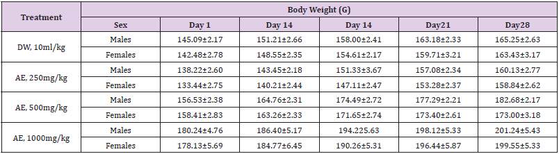
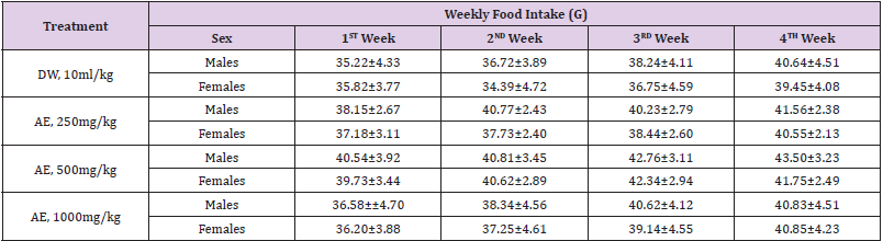
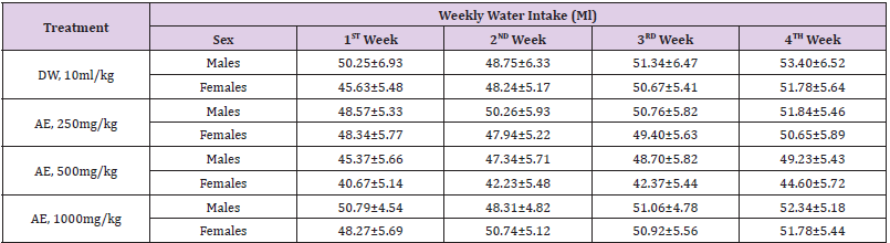
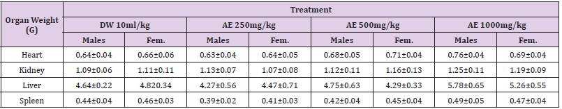
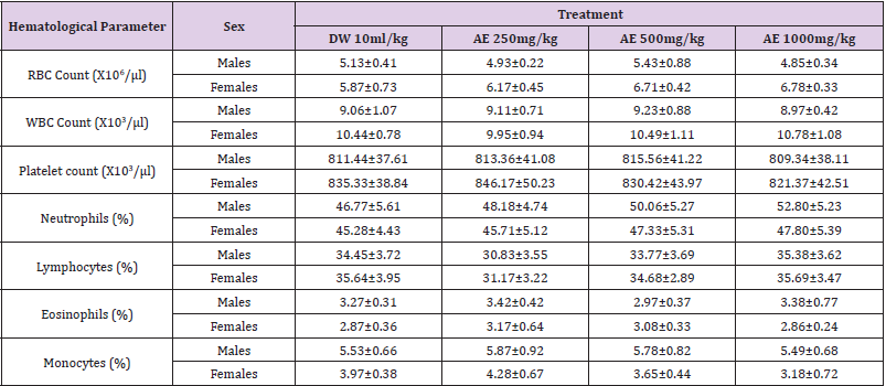
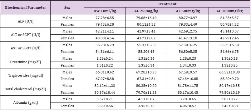
 Loss of weight in drying at 105°C : 7.09Absolute alcohol solubility : 3.20Water solubility : 21.20

 Successive extractives (%)Petroleum ether (60-80℃) : 1.75Chloroform : 1.55

 Absolute alcohol : 5.70Distilled water : 18.00

 Ash values (%)

Total ash : 10.60

Water soluble ash : 3.60

Acid insoluble ash (10% HCl) : 1.30

 pH values (aqueous solution)

pH of 1% solution : 5.98-6.05

pH of 10% solution : 5.47-5.48

Elemental Analyses

(Table 1).

biomedres-openaccess-journal-bjstr

Table 1.

UV Spectral Studies

(Table 2, Figures 3 & 4).

biomedres-openaccess-journal-bjstr

Figure 3: Intestinal Fluid simulated without pancreatic pH=7.5±0.1.

biomedres-openaccess-journal-bjstr

Figure 4: Gastric Fluid simulated without pepsin pH=1.2±0.1.

biomedres-openaccess-journal-bjstr

Table 2.

Thin layer Chromatography (TLC)

Figure 5.

biomedres-openaccess-journal-bjstr

Figure 5: TLC fingerprint of Petroleum ether -60-800 (track 1) and MeOH extract (track 2) Mobile phase.
• A&C: Ethyl acetate, methanol, water (100:13.5:10).
• B&D: Toluene, ethyl acetate (93:7)
• Detection A: UV 254nm
• B&C: UV366nm
• Derivatization D: Vanillin- Sulphuric acid-vis.

Pharmacological and Toxicological Studies

Information reported in the Literature about the plant: The important pharmacological and toxicological activities of the plant Tribulus terrestris reported in various scientific journals have been presented in the present brief review: A mixture of 9 oriental herbs were given including Tribulus terrestris showed an increase in the copulatory behavior parameters in rats, improved the sexual activity and erectile function. The effects of methanolic and aqueous extracts of Tribulus terrestris on rat blood pressure (BP) and the perfusion of mesenteric vascular bed were investigated and found possessing significant antihypertensive activity in spontaneously hypertensive rats [11]. Effect of Saponins from Tribulus terrestris (STT) on the renal carcinoma cell (786-0) in vitro and inhibitory mechanisms studied significantly inhibited the growth of 786-0 in vitro, partially, by apoptosis [12]. Tribulus is a genus of plants found in many warm regions.
The best-known member is T. terrestris (Puncture Vine), a widespread weed and also the source of a dietary supplement claimed to increase the body’s natural testosterone levels and thereby improve male sexual performance and help build muscle. T. terrestris has consistently failed to increase testosterone levels in controlled studies [13-15]. It has also failed to demonstrate strength-enhancing properties [16]. Tribulus has been shown to enhance sexual behaviour in an animal model. It appears to do so by stimulating androgen receptors in the brain [17]. Jameel, et al. [18] reported a case of a young weight-trainer who developed gynaecomastia due to oral intake of an herbal tablet which he used as a steroid alternative for body-building. The aqueous extract of Tribulus terrestris can significantly increase melanocyte-stimulating hormone (MSH) expression in the hair follicle melanocytes by activating tyrosinase activity and promoting melanocyte proliferation, melanine synthesis, and epidermal migration of dormant melanocytes [19]. Tribulus terrestris showed protective effect for STZ-induced diabetic rats may be mediated by inhibiting oxidative stress [20].
Sharif, et al. [21] investigated the antihypertensive mechanism of Tribulus in 2K1C hypertensive rats by measurement of circulatory and local ACE activity in aorta, heart, kidney and lung. The ACE activity in all tissues of Tribulus fed hypertensive rats was significantly lower than that of hypertensive rats, which was more pronounced in kidney. These results indicated that there is a negative correlation between consumption of Tribulus and ACE activity in serum and different tissues in 2K1C rats. Oludotun, et al. [22] reported the effects of methanolic and aqueous extracts of Tribulus terrestris on rat blood pressure (BP) and the perfused mesenteric vascular bed were investigated. The extracts dosedependently reduced BP in spontaneously hypertensive rats (SHRs) with the aqueous fraction being more potent than the methanolic fraction at all doses tested. In vitro, the methanolic but not aqueous extract produced a dose-dependent increase in perfusion pressure of the mesenteric vascular bed. When perfusion pressure was raised with phenylephrine, the aqueous extract produced a dosedependent reduction in perfusion. It was concluded that methanolic and aqueous extracts of Tribulus terrestris possess significant antihypertensive activity in spontaneously hypertensive rats.
The antihypertensive effects appeared to result from a direct arterial smooth muscle relaxation possibly involving nitric oxide release and membrane hyperpolarization. The inhibitory effect of saponins from Tribulus terrestris (STT) on Bcap-37 breast cancer cell line were determined by cell growth curve, MTT assay, protein content assay and morphological observation showed that STT had potent inhibitory effect on Bcap-37 cell line in a concentrationdependent manner [23]. The results of pharmacological and Toxicological studies carried on the 70% alcoholic extract of the plant, have been given below (Figures 6-8 and Table 3).

biomedres-openaccess-journal-bjstr

Figure 6: Effect of Tribulus t. on rat detrusor muscle acetylcholine treated.

biomedres-openaccess-journal-bjstr

Figure 7: Effect of Tibulus 70% ethanol extract on serum createnin level.

biomedres-openaccess-journal-bjstr

Figure 8: Tribulus Sexual study followed by Viagra.

biomedres-openaccess-journal-bjstr

Table 3.

Conclusion

The oral administration of the plant extract was non-toxic up to the dose of 10 g/kg b.wt., p.o. These acute studies demonstrated that the plant extract is safe and did not cause any detrimental effects in vivo under the conditions investigated in this study. The LD50 of plant extract was found to be >10 g/kg when administered once via gastric intubation to rats. Plant extract produced a mild relaxation in phenylephrine pre-contracted corpus cavernous tissue.

For More Articles: Biomedical Journal Impact Factor: https://biomedres.us

Open Access Journals on Pharmaceutical Chemistry

Source-Induced Dissociation Vs Collision-Induced Dissociation Fragmentation Behavior of Four Antiviral Drugs in a Liquid Chromatography Ion-Trap Mass Spectrometry

Introduction

Mass spectrometry (MS) is an important tool for analyzing or detecting a wide range of organic, inorganic, natural, unnatural molecules, especially drugs, proteins, peptides are of interests beside routine analysis [1]. MS measures the mass-to-charge ratios of ions of an ionized sample, which may be gas, liquid or solids. Mass-to-charge ratios may obtain for a charged molecule or charged fragments. MS has both quantitative and qualitative uses. Numerous ionization methods are used in MS experiments, such as chemical ionization (CI), matrix assisted laser desorption ionization (MALDI), electron impact (EI), atmospheric pressure chemical ionization (APCI), and electrospray ionization (ESI). ESIMS has been applied to the analysis of oligonucleotides [2, 3] drugs and drug metabolites [4] oligosaccharides [5, 6] environmental contaminants [7, 8] glycoproteins [9] and many other types of compounds [10-15]. In atmospheric pressure ion sources, e.g., ESI or APCI, dissociation of ions can occur inside the ionization source (source induced dissociation; SID) before reaching the mass analyzer. SID has been used by several research groups [16-22]. Usually, SID is used to analyze single mass, whereas CID is used to analyze fragmentation. Both SID and CID techniques having advantages and disadvantages [23, 24]. While CID technique is very much effective to analyze pure sample with much more information about structural data [25]. Acyclovir, [26, 27] abacavir, [28, 29] famciclovir [30-33] and penciclovir [10] are the known antiviral medication used to prevent viral diseases. All of these antiviral drugs having common purine moiety [34] (Figure 1) and 9-position of purine are substituted with deferent alkyl chains. Mass spectrometric study of these structurally similar compounds may reveal lots of information for further analysis process during antiviral doses. As a medicinal chemist, it is our duty to develop drug molecules / administration procedures / toxicity evaluation process and / identification in a mixture or in biological matrixes. Therefore, there is always a room for extending such development using modern scientific methods such as mass spectrometry.

biomedres-openaccess-journal-bjstr

Figure 1: Chemical structures of antiviral acyclovir, abacavir, penciclovir and famciclovir showing a common purine nucleus.

Here, analysis of four antiviral drugs acyclovir, abacavir, penciclovir and famciclovir serves to figure out how to extend the qualitative power of ITMS and to maximize the benefit from this mass analyzer. These four drugs also were selected as they contain a common purine nucleus in their chemical structures which might generates a common fragmentation pattern. Above mentioned four known antiviral drugs were studied for their fragmentation pathways and data of SID and CID of ESI IT were compared. The detail fragmentation pathways of all ions observed in the in-source fragmentation spectrum of compounds were elucidated by further dissociation of each of these fragment ions using MS2, MS3 and MS4 fragmentation stages.

Experimental

An Agilent 6320 Ion-Trap connected (LC-MSn) mass spectrometer was used. Compounds were dissolved in DMSO and used as standard solution, then diluted with a mixture of HPLC grade water and acetonitrile (1:1). Fragmentation pattern of these four antiviral drugs by direct injection using infusion pump were performed. 10 μL of each sample was injected with direct injection, flow rate was 0.4 ml/min., run time was set to 5 minutes, drying gas was N2 (flow was 12 L/min), temperature was set to 350 °C, nebulizer pressure was 50 psi, smart target was 10,000, accumulation time 150 ms, scan range was 20- 400Da and Ion source was ESI, in positive mode with ultra-scan. There are several factors that can influence the fragmentation spectra, such as the collision energy, major precursor ion, electrospray mode (positive or negative) and capillary exit voltage. In IT, product ion scan was performed before the in-source fragmentation to determine each compound’s related fragment ions, the capillary exit voltage was optimized to produce adequate in-source fragmentation. The data compared to multistage fragmentation using CID. We made all default parameters except capillary exit voltage which was 100 V for CID and 200 V for SID.

Results and Discussion

CID MSn Fragmentation Pattern of Abacavir

Abacavir was set for an example of comparing two fragmentation techniques. As shown in (Figures 2 & 3), full scan of abacavir gave m/z 287 (Figure 2A) and MS2 of abacavir gave three fragments at m/z 257, 191 and 150 (Figure 2B). Among three fragments, m/z 191 was a prominent peak which was further bombarded (MS3) and gave m/z 174, 164, 150, 134, 109, 83 and 59 (Figure 2C). Consequently, high intense three ions m/z 174, 164 and 150 were further bombarded (MS4) in which, m/z 174 gave four ions at m/z 159, 148, 132 and 157 (Figure 3A), m/z 164 gave two ions m/z 137 and 122 (Figure 3B), and m/z 150 gave a single ion at m/z 132 (Figure 3C).Based on (Figures 2& 3), we have drawn Scheme 1 for the fragmentation pattern of abacavir in CID mode. As shown in scheme 1, after four steps (MS4) fragmentation, abacavir gave maximum fragments at m/z 150, 147, 137, 134, 132, 122, and 57. SID pseudo-MSn fragmentation pattern of abacavir. As shown (Figure 4), pseudo MS2 of abacavir gave m/z 287, 191, 174, 164, 150 and 134 (Figure 4A), and pseudo MS3 of major two fragments, m/z 191 and 174 was further bombarded (MS3) and obtained m/z 191 gave m/z 174, 164, 150, 136, 109 and 59 (Figure 4B) and m/z 174 gave m/z 147, 134, 119, 106, 94 and 57 (Figure 4C), respectively. Pseudo MS4 of high intense m/z 174 gave m/z 164, 147, 134, 122, 93 and 57 (Figure 4D).

biomedres-openaccess-journal-bjstr

Scheme 1: CID MSn fragmentation pattern of protonated abacavir.

biomedres-openaccess-journal-bjstr

Figure 2: CID MSn fragmentation pattern of abacavir:
A. MS scan for abacavir,
B. MS2 of 287 and
C. MS3 of 191.

biomedres-openaccess-journal-bjstr

Figure 3: CID MS4 fragmentation of three ions (174, 164 and 150):
A. MS4 scan of 174
B. MS4 scan of 164 and
C. MS4 scan of 150

biomedres-openaccess-journal-bjstr

Figure 4: SID pseudo-MSn fragmentation pattern of abacavir:
A. MS scan for abacavir applying capillary exit voltage,
B. product ion for 191
C. product ion for 174 and
D. MS3 for 174.

Based on (Figure 4), we have drawn Scheme 2 for the fragmentation pattern of abacavir in SID mode. As shown in scheme 2, in two steps (pseudo MS3) fragmentation, abacavir gave maximum fragments at m/z 174, 150, 147, 134, 132, 109 and 94 and pseudo MS4 of m/z 174 gave five fragments at m/z 164, 147, 134, 121 and 57. SID and CID of acyclovir, penciclovir and famciclovir. Other three antiviral drugs acyclovir, penciclovir and famciclovir mass spectrometric study in SID and CID results are summarized in Table 1 along with abacavir. As shown in Table 1 and depicted in Scheme 3, SID scan of acyclovir gave m/z 226 [M+H] + with 152 and MS2 of main peak m/z 226 gave three main fragment at m/z 152, 135 and 88. Among three fragments, m/z 152 was further bombarded and obtained m/z 135 and 88. On the other hand, in CID scan, acyclovir gave m/z = 226 [M+H] + only. Which was further bombarded for MS2 and obtained m/z 152 and 135 only. MS3 of m/z 152 gave only one fragment from SID at m/z 135.As shown in Table 1 and depicted in Scheme 4, SID scan of penciclovir gave m/z 254 [M+H] + with 152 and MS2 of main peak m/z 254 gave three main fragment at m/z 152, 135 and 110. Among three fragments, m/z 152 was further bombarded and obtained m/z 135 and 110. On the other hand, in CID scan, penciclovir gave m/z = 254 [M+H] + only. Which was further bombarded for MS2 and obtained m/z 152 only. MS3 of m/z 152 gave same fragments as we obtained from SID, which is m/z 135 and 110. As shown in Table 1 and depicted in Scheme 5, SID and CID scan of famciclovir gave only protonated peak at m/z 322 [M+H] +. MS2 of this m/z 322 also gave four same fragments at m/z 280, 262, 202 and 135. Among those peaks, a main peak m/z 280 was further bombarded and obtained seven fragments m/z 262, 238, 202, 186, 136, 117 and 107 in SID and 5 fragments m/z 262, 238, 202, 186 and 136 in CID. Common fragmentation paths of abacavir, acyclovir, penciclovir and famciclovir.

biomedres-openaccess-journal-bjstr

Scheme 2: In source fragmentation pattern of protonated abacavir.

biomedres-openaccess-journal-bjstr

Scheme 3: Fragmentation pattern of acyclovir.

biomedres-openaccess-journal-bjstr

Scheme 4: Fragmentation pattern of penciclovir.

biomedres-openaccess-journal-bjstr

Scheme 5: Fragmentation pattern of famciclovir.

biomedres-openaccess-journal-bjstr

Table 1: MSn fragmentation behavior of acyclovir, penciclovir, famciclovir and abacavir.

As depicted in Scheme 6, all four antiviral drugs gave similar fragmentation pathway. Abacavir, acyclovir and penciclovir gave purine moiety while SID / CID MS2 was applied, on the other hand, famciclovir gave similar pathway with purine moiety while SID (Pseudo MS4) / CID (MS3) was applied (Scheme 6).

biomedres-openaccess-journal-bjstr

Scheme 6: Fragmentation pathways of all antiviral drugs (abacavir, acyclovir, penciclovir and famciclovir).

Conclusion

Excellent qualitative information was obtained using sourceinduced dissociation (SID) comparing to collision-induced dissociation (CID) in an ITMS. Fragmentation pathways of four antiviral drugs abacavir, acyclovir, penciclovir and famciclovir gave similar information regardless fragmentation method as they contain purine nucleus in their building blocks. This method can be applied for the detection /identification of know/unknown drugs or their metabolites in-vitro / in-vivo or in biological matrixes by comparing their fragmentation pattern and using different possible fragmentation tools to maximize the benefit of ITMS in the process of drug development.

For More Articles: Biomedical Journal Impact Factor: https://biomedres.us

Open Access Journal on Microbial Chemistry

Nanoparticles as Antimicrobial Agents

Opinion

Microbial infections are still a challenge despite of the existing of numerous potent antibiotic drugs and other modern antimicrobial means, Bacterial infections are still a major cause of mortality. The problem is that major groups of antibiotics, currently in use, generally affect three bacterial targets: cell wall synthesis, translational machinery, and DNA replication. Unfortunately, bacterial resistance may develop against each one of these modes of action. In addition, the use of conventional antibiotics carries a major risk for resistance of viable bacteria. Nanoparticles as antibacterial agents complementary to antibiotics are highly promising and are gaining large interest as they might fill the gaps where antibiotics frequently fail. This includes combatting multidrug-resistant mutants and biofilm. Nanotechnology is a technology conducted at the nano-scale in the fields of fabric manufacturing, food processing, agricultural processing, and engineering, as well as in medical and medicinal applications. Over the present decade, several studies have suggested that nanoparticles are excellent antibacterial agents, thus nanomaterial applications for antimicrobial works have prime interest by many researchers. Several reports showed that some of the metal oxide nanoparticles have toxicity toward several microorganisms and they could successfully kill numerous bacteria. This new approach has been identified to deal with resistance of pathogenic microorganisms because of their inherent antimicrobial activity. The use of nanoparticles as antimicrobial components especially in the food additives and medical applications can be one of the new and considerable strategies for overcoming pathogenic microorganisms. Based on literature review there are some effective factors that can influence the ability of nano materials in reducing or killing the cells. Mechanisms for nanomaterial against bacteria, which are briefly listed as follows: surface charge of the metal nanomaterial, shape, type and material, concentration of nanomaterial, dispersion and contact of nanomaterial to the bacterial cell, presence of active oxygen, liberation of antimicrobial ions, medium components and pH, physicochemical properties, specific surface-area-to-volume ratios, size, role of growth rate, role of biofilm formation, and cell wall of bacteria.
Nanoparticles as antimicrobial agents have become an emerging approach which can establish an effective nanostructure to deliver the antimicrobial agents for targeting the bacterial community efficiently. In addition, they are so potent that microbial pathogens cannot develop resistance towards them. On the other hand, most of the metal oxide nanoparticles have no toxicity toward humans at effective concentrations used to kill bacterial cells, which thus becomes an advantage for using them in a full scale. Metal, metal oxide and organic nanoparticles now are used show a diversity of intrinsic and modified chemical composition properties. Thus, it is not surprising that they have numerous modes of action as antimicrobial agents. In some cases the ratio between the bacteria and the nanoparticles is critical to the bacterial toxicity. In addition, many factors play a role and affect the lethal effect of nanoparticles to bacteria including aeration, pH, and temperature, size, shape, chemical modification and coating, and mixture in various ratios with other nanoparticles and solvent used. Lethal effect of nanoparticles is generally due to membrane damage occurs when nanoparticles bind electrostatically to the bacterial cell wall and membranes, leading to alteration of membrane potential, membrane depolarization, and loss of integrity which, in turn, result in an imbalance of transport, impaired respiration and cell death. Among inorganic metals silver nanoparticles have been widely used as an effective antimicrobial agent against bacteria, fungi, and viruses. The antimicrobial efficacy of silver (Ag), as of other metals and metal oxide nanoparticles, was reported to be size-dependent. Although the Ag nanoparticle mechanism of action is still not clear, small diameter Ag nanoparticles have a superior antimicrobial effect to those of a larger diameter In comparison to silver, gold- (Au-) nanoparticles are less potent and have almost no antibacterial effect by themselves.
Except when they of an antibacterial approach, adopted from cancer treatments, gold nanoparticles bound to Fe3O4 and activated by photothermal means. In addition, Titanium oxide with its photocatalytic effect serves as an antimicrobial agent for both positive and negative bacteria. An interesting approach in the antimicrobial application of nano- metals and nano- metal oxides is the synergistic effect of the combination between two or more of them as Titanium and silver. Moreover, Zinc oxide nanoparticles were shown to have a wide range of antimicrobial activity. This metal oxide is characterized by its low cost and low toxicity to human cells, thus it was used as coating materials designated for medical and other devices. White color, UV-blocking, and ability to prevent biofilm formation made its nano particles suitable for fabric treatment. Zinc was approved by the FDA as a food additive. Another metal oxide used as an antibacterial agent is copper oxide nanoparticles have been shown to be effective against various bacterial pathogens especially gram positive Bacilli. In our lab we obtained excellent results by applying these approaches in many fields of polymers and textile.

For More Articles: Biomedical Journal Impact Factor: https://biomedres.us

Open Access Journals on Chemistry

Pre-Med Major and Automaticity Ability

Opinion

The Texas Networking for Science Advancement (NSA) team formed in 2016. The team now consists of 15 faculty from 10 Texas universities. Five of the partnering universities are classified as Hispanic-Serving Institutions (at least 25% Hispanic enrollment) and the other five are considered as Hispanic-emerging (16- 24% Hispanic enrollment). Two of the 10 institutions are private universities, and the remaining are public. To date, the team has published results of eight studies (two currently in press) evaluating general chemistry students’ QR arithmetic automaticity skills (what students can do without a calculator) using the Math- Up Skills Test (MUST), a 20-question, 15-min, hand-graded, openended diagnostic instrument [1-3]. One of our carrots to encourage students to try this quiz without using their trusted calculating devices is that the MCAT is a calculator-free evaluation instrument and over 50% of our general chemistry students indicate an interest to enter the health professions.

The Good

From the very first observation, the team’s results provided a linear trend-the better students’ arithmetic skills the better their general chemistry averages in both first- and second-semesters. The results not expected are that there is a stronger relationship between what students can do without a calculator (their automaticity) than what they can do with a calculator. Yes, the students did better on the MUST with a calculator but the effect size without a calculator is stronger. The statistics consistently support that the MUST is highly reliable (KR-20 > .80), has good internal consistency (Cronbach’s alpha > .85), and the Cohen’s d for the MUST is consistently > 1.0 indicating a large effect size for a population of over 10,000 students. Using the MUST has provided the team with a diagnostic instrument that allows instructors the ability to identify at-risk students in 15 min the first week of classes. Being able to identify these students who will struggle with the course material early in the semester provides ample time for corrective measures to be employed in hopes that students will take advantage of the opportunity but herein lies the rub: it’s the rich that get richer! Pilot studies indicate that this trend carries onto lower-level organic chemistry courses.

The Bad

The original study came about because the team noticed that Texas students’ SAT scores were consistently declining below the national average. This trend coincided with the Texas Education Agency (TEA) mandated change from a 4×4 high stakes testing system consisting of a series of four exams per year in four subjects (mathematics, science, social studies and English) during the final four years of secondary school. TEA’s current testing-system focuses on only two STEM areas (biology and first-year algebra) along with first- and second-year English and US History. The TEA calculator policy states no calculators are permitted on STAARs (State of Texas Assessments of Academic Readiness) in grades 3-7, but districts must ensure that each student has a graphing calculator to use on all STAARs starting with 8th-grade mathematics (both paper and online versions) and biology. For the biology assessment, there should be one calculator (four-function, scientific, or graphing) for every five students. Students may bring their own calculators with them to the assessments, but internet capabilities must be disabled and calculation applications on smartphones are not allowed. Beginning in May 2018, the grade 8 science STAAR requires students to have access to calculators with four-functions, scientific or graphing capability. What is not obvious is how much these rules/ regulations are hurting the students’ future preparation for postsecondary success. Handheld calculators are generally accepted for classroom use and allow teachers to give beginning students realistic problems using tangible data gathered from the field. All is good but how many times do students calculate a negative mass or an unrealistic density and not have the quantitative-reasoning ability or basic number sense to know that the “calculator” answer is impossible or improbable.

The Future

When students enter post-secondary education with low automaticity ability, they lack the mental-math skills needed to succeed in general chemistry, a major gateway course to many STEM-major degrees. Adding more calculator-free instruction for all STEM and non-STEM majors in general chemistry (and other STEM courses) is advisable and will improve students’ mentalmath capability. Summer bridge programs, weekly recitations and the like should emphasize students’ automaticity skills to help prepare incoming students for success. However, quantitativereasoning (QR) skills (like those needed to understand how to read scientific data graphs and charts) are required to understand much of our current data-driven world needed to prepare global citizens for both STEM and non-STEM careers. Improving our students’ QR calculator-free skills is a place to start to attempt to improve retention and graduation rates for all students. Is this the answer? Maybe or maybe not, but what is obvious is that we are going in the wrong direction. Without a doubt, students need more quantitative-reasoning and quantitative-literacy skills to have a chance of succeeding in this data-driven world. To encourage students to use their calculator-free skills, teach part of your class without the use of a calculator. Ask students how they arrived at their answers, what is their logic-it might surprise you as to what they can and cannot do without using calculators, and as a result their automaticity abilities and MCAT scores might improve!

For More Articles: Biomedical Journal Impact Factor: https://biomedres.us

Open Access Journals on General Medicine

Acute Hyperuricemia Secondary to Epileptic Seizures Leading to Acute Urate Nephropathy: A Case report

Background

About two third of the body uric acid is produced endogenously while the rest of one third is from dietary products containing purines. Most of the uric acid is excreted through kidneys while the remaining through intestines. Hyperuricemia occurs either due to underexcretion of urate or overproduction of urate. Factors that predominantly effect uric acid excretion include tubular fluid Ph, tubular fluid flow rate and renal blood flow. Acute hyperuricemia can lead to acute uric acid nephropathy by renal tubular obstruction by urate and uric acid crystals which can lead to significant morbidity. Seizures is an exceedingly uncommon cause of acute hyperuricemia. This is one such case of a patient with Generalized tonic clonic fits leading to acute hyperuricemia which in turn caused acute urate nephropathy.

Case Presentation

A 35-year-old man with no premorbid presented in emergency department with generalized tonic clonic fits. At presentation patient had Bp of 130/70, Pulse 96, Temperature 98.7F, Oxygen saturation 97% with room air. Blood sugar random was 126mg/dl. He had two such episodes in last 30 minutes. Abortive therapy was given to control the fits. After initial management detail examination was done. Central nervous system examination was normal with no focal neurological deficit. Chest was clear bilaterally with rest of respiratory examination normal. Cardiovascular, Gastrointestinal and musculoskeletal examination was also normal. Complete blood picture showed leukocytosis with white blood cell count 22550/microliter with 80% neutrophils. Liver function tests, Renal function tests, C-reactive protein, Erythrocyte sedimentation rate, Serum electrolytes, Fasting lipid profile, Serum calcium, magnesium and phosphorus were also normal. Urine routine examination was normal with no myoglobinuria. CPK levels were 153 and serum prolactin levels were 404. Serum uric acid was 20.73 mg/dl. Chest x-ray, Electrocardiography and Echocardiography were normal. After 2 days his Renal function tests started deranging with Urea 45 mg/dl and creatinine 1.52 mg/dl. His previous renal function tests and uric acid were completely normal. Maximum urea and creatinine levels went up to 55 and 2.88mg/dl respectively. Patient was managed with intravenous hydration and allopurinol. His renal function tests and uric acid levels started declining and normalized after 5 days.

Discussion

In previous case studies it was noted that creatinine was deranged initially on the day of presentation. In our case unusual was that patient’s creatinine was normal at presentation and started rising after 2 days. So, it is important to follow patient’s Renal function tests for at least 4-5 days post seizure so that acute renal failure is not missed which can prove fatal if not managed early. Secondly in previous studies maximum level of high uric acid levels were up to 15mg/dl while in our case serum uric acid was extremely high with levels up to 20.73 mg/dl which was managed successfully with early intervention.
Uric acid is the final insoluble waste product of purine breakdown [1]. Uricase enzyme which allows converts insoluble uric acid to soluble allantoin in absent in humans because of which it accumulates in the distal part of nephron particularly in acidic environment leading to toxicity [1,2]. Multiple studies have proven that high uric acid level is linked with many systemic illnesses including hypertension, chronic kidney disease, cardiovascular disease, diabetic nephropathy, stroke and acute kidney injury [3,4]. Tumor lysis syndrome is the most common cause of acute hyperuricemia through high cell turnover and cell lysis following aggressive chemotherapy for cancer particularly lymphoma [1,2]. When urinary uric acid surpasses solubility threshold, it is deposited and accumulated as crystals in renal tubule leading to acute renal failure [2]. High uric acid levels have been implicated to change the basic architecture of renal histology and have an important role in acute and chronic renal injury [5].
Acute uric acid nephropathy is characterized by raised creatinine levels; high uric acid levels raised urinary urate to creatinine ratio [6]. High uric acid causes acute urate nephropathy by various mechanisms including uric acid crystal formation and non-crystal mediated pathway [6]. Acute urate nephropathy must be suspected in high risk patients who develop acute renal failure with high serum uric acid levels and presence of urate crystals in urinary sediment [2]. Various mechanisms proposed in the development of acute kidney injury secondary to hyperuricemia include uric acid crystal formation in tubules, renal vasoconstriction, endothelial dysfunction, stimulation of inflammatory response, oxidative stress, antiangiogenic changes and direct microvascular injury [6].
Acute uric acid nephropathy is reversible [1]. Epileptic fits cause skeletal muscle breakdown which in turn causes life threatening rhabdomyolysis leading to acute kidney injury in 15% of patients [7]. Rhabdomyolysis releases high amount of myoglobin by breakdown of muscle cells which in turn results in high serum myoglobin levels. Raised serum myoglobin levels exceeds haptoglobin binding capacity resulting in myoglobinuria which causes cast formation and tubular obstruction leading to acute kidney injury [7].
Various metabolic abnormalities have been associated with severe epileptic seizures including lactic acidosis, raised ammonia, raised creatine phosphokinase and high prolactin levels [2]. Rare metabolic derangements associated with seizures include electrolyte changes, hyperuricemia and osmolality changes [2]. Fits induced hyperuricemia is one of the rarest complications described in literature even more rare when associated with acute urate nephropathy [2]. Seizures cause acute uric acid nephropathy by increasing uric acid levels and provide favorable environment for urate crystal formation [6].
It is speculated that acute hyperuricemia secondary to seizures occurs both due to overproduction and impaired tubular excretion of urate [1]. Muscle injury during seizures causes tissue breakdown which release nucleosides leading to its transport into liver and is converted into uric acid by urea cycle [6]. Factors that lead to hyperuricemia following fits include acidification of urine secondary to acidosis caused by lactate production and hypoventilation making urate less soluble, dehydration secondary to hyperthermia and profuse diaphoresis causing increase tubular water reabsorption leading to high tubular uric acid secretion and renal ischemia due to shunting of blood from viscera to muscles [2].
Most effective therapy to lower uric acid levels is Rasburicase which is a recombinant form of xanthine oxidase [2]. Allopurinol inhibits uric acid formation, but it does not remove already existing urate in the body and may also worsen acute renal injury by releasing xanthine [1]. Urinary alkalization can increase urate solubility but has not yet been proven to be effective in acute urate nephropathy [1]. Oliguria secondary to acute urate nephropathy have excellent response to hemodialysis [1].

Conclusion

In nutshell acute hyperuricemia is an uncommon manifestation following a seizure. Immediate necessary steps should be taken to lower uric acid levels. Acute uric acid nephropathy is a reversible condition and if early management is done to lower urate levels it can prevent progression to acute renal failure with excellent fruitful outcome.

For More Articles: Biomedical Journal Impact Factor: https://biomedres.us

Open Access Journals on Brain and Behavioral Sciences

Clozapine-Induced Acute Interstitial Nephritis: A Case Report

Introduction

Clozapine is nowadays the most effective treatment for refractory schizophrenia. It has proven to be superior in terms of efficacy compared with 1st generation antipsychotic and other 2nd generation antipsychotic [1]. Moreover, it does not cause extrapyramidal side effects nor tardive dyskinesia and its impact on prolactin level is negligible [2]. This atypical antipsychotic is also used to treat other conditions such as schizoaffective disorder. However, clozapine is underprescribed, due to potentially lifethreatening side effects such as neutropenia, agranulocytosis, myocarditis and myocardiopathy and other additional problems including weight gain, orthostatic hypotension, constipation and sialorrhea [3].

Among uncommon adverse reaction, acute interstitial nephritis (AIN) should also be taken into account. Very few cases have been described and the underlying mechanism is still unclear [4-6]. It could be related to a nonspecific inflammatory response, which is often observed in the early weeks of treatment with clozapine. This drug indeed exerts some immunomodulatory effects that have clinical implications [7]. We report a case of a patient who experienced acute renal failure twice. Both episodes followed initiation of treatment with clozapine.

Introduction

A 54-year-old woman with a known history of schizoaffective disorder was admitted to our hospital in November 2020. She was diagnosed with schizoaffective disorder in 1988, at the age of 22. She had been treated with different antipsychotics during the past 30 years (quetiapine, haloperidol, chlorpromazine, levomepromazine, clotiapine, lurasidone) and she has a history of repeated and prolonged hospitalizations. The patient is also affected by hypothyroidism treated with levothyroxine, type 2 diabetes in insulin therapy and gallstones. Furthermore it is reported in her clinical history an episode of acute renal failure in December 2017, probably due to carbolithium, and an episode of acute pancreatitis in October 2019. In November 2020 a new hospitalization was necessary because of the relapse of the psychotic symptoms with agitation and suspiciousness in spite of the antipsychotic therapy with lurasidone and clotiapine. In consideration of the antipsychotic resistance, the therapy was switched to clozapine on 17th November, commenced at 25 mg/day up to 50 mg/day. On 19th November there was the onset of fever, partially responsive to paracetamol. In the following days the progressive increase of fever up to 39.5°C and the onset of desaturation occurred: oxygen therapy and an empirical antibiotic therapy were necessary. Eventually, all the causes of an infective disorder were excluded.

On 23rd November we observed a PLT reduction (96.000/uL), so clozapine was suspended. From 5th December we observed a progressive increase of the creatinine value, from 2,95 mg/dl up to 3,90 mg/dl. Her previous creatinine value was 0,85 mg/dl on 19th November 2020. The main causes of acute renal failure were excluded. The abdomen CT showed kidneys of normal size and morphology, without parenchymal bulks, stones or distension of the urinary tract, with a minimal edema of the perirenal fat on the left . The patient was evaluated by a nephrologist, who suggested to move her in the Unit of Nephrology to find the cause of the renal failure and to treat it. The cause was not established, but the renal function values gradually normalized. On 22nd December the patient was re-admitted to the Unit of Psychiatry. The patient’s clinical and psychiatric conditions gradually improved, so she was dismissed on 28th January.

On 2nd February a new episode of psychomotor agitation and relapse of psychotic symptoms occurred and the hospitalization was necessary. The patient was suspicious and contentious and she had persecutory delusions. On 11th February, considering the patient’s psychopatological conditions, clozapine was reintroduced. Again fever occurred after a single tablet of clozapine, followed by renal failure (06/02: creatinine value 1,19 mg/dl; 13/02 creatinine value 2,14 mg/dl, progressively increased up to 3,65 mg/dl in few days).

Evaluated by the nephrologist, a steroid therapy was prescribed and, on 16th February, the patient was moved again to the Unit of Nephrology. The diagnosis of iatrogenic interstitial nephritis caused by Clozapine was confirmed as diagnosis by exclusion and, after the stabilization of the renal function, the patient was retransferred to the Unit of Psychiatry. The therapy was switched to brexpiprazole: the renal function progressively improved up to 1,15 mg/dl and the patient’s clinical conditions slowly improved, until the dismissing on 14th April. The case was followed up for 6 months and we observed a gradual recovery of the renal function: actually the creatinine value is 0,99 mg/dl.

Discussion

To the best of our knowledge, this is the first case of clozapine-induced nephrotoxicity ever described in Italy. Acute interstitial nephritis is an immune-mediated condition featuring tubulo-interstitial inflammation and oedema. It can be infective, autoimmune or even idiopathic but more commonly it is induced by various drugs, such as NSAIDs and antibiotics like aminoglycosides and vancomycin [8-9]. The diagnosis of drug-induced acute interstitial nephritis is based on its clinical and laboratory manifestations, characteristic morphologic features of the kidney on biopsy, and the identification of a causative agent. In practice, satisfying all three criteria is fraught with limitations, particularly in patients exposed to several potentially incriminated drugs [10]. In this regard, a possible limitation of our study is the lack of renal biopsy.

Our results are in agreement with previous evidence, reporting the importance of renal function monitoring prior to clozapine initiation and during titration. Moreover, it is fundamental when clozapine is prescribed together with other potentially nephrotoxic medications. Further, is it of great interest to remember that early recognition of this phenomenon, also involving nephrologists, leads to prompt intervention and adequate treatment.

For More Articles: Biomedical Journal Impact Factor: https://biomedres.us