Biomedical Journal of Scientific & Technical Research (BJSTR) is a multidisciplinary, scholarly Open Access publisher focused on Genetic, Biomedical and Remedial missions in relation with Technical Knowledge as well.
Simulation of a Laparoscopic Major Vessel Injury in a Live Animal Model
Introduction
Iatrogenic injury to major vessels with the ensuing bleeding is a rare but potentially life-threatening complication during laparoscopic major HPB surgery. The most commonly injured vessels are aorta, the iliac vessels, and the inferior vena cava [1]. Contrary to traditional approach suggesting immediate conversion to open surgery it is suggested nowadays that this kind of injury and bleeding should be approached laparoscopically [2]. An obvious requirement for such an approach is an appropriate training [3]. Advanced laparoscopy training currently includes box-trainers [4], virtual reality training [5], live animal training [6] and training that combines all of the above [7]. Unfortunately, the majority of training modalities in laparoscopy concentrate on purely technical knowledge not considering psychological burden of a major intraoperative disaster. While obtaining and maintaining technical skills is clearly important [8] the possibility of testing these skills in a stressful environment imitating operating room disaster could be the way to prepare surgeons to adequately react to the unexpected [9]. In this study we have tried to create an environment as similar to real life laparoscopic disaster as possible and observe trainees’ reactions and their ability to use technical skills to control the situation.
Materials and Methods
During three editions of advanced laparoscopic training course 12 participants faced a task of controlling a major vessel damage. Training course was designed for both experienced surgeons and novices in advanced laparoscopy. Each course lasted for two days. At the beginning of the first day the tutors explained the methods of laparoscopy bleeding control with a video footage. Each day of the course there were 7 hours of live animal laparoscopy training. The first part of the training was designed to achieve technical abilities in various steps of advanced laparoscopy procedures depending on the level of experience of each participant. In the second part of the training during the last 60 minutes of each day the participants were exposed to iatrogenic injury of a major vessel performed with an electrocautery on an area of approximately 1cm and were asked to control the bleeding and repair the damage. During these maneuvers their Heart Rate (HR) was monitored, and their reactions were video recorded. After successfully completing the task and if time permitted the same animal was used for another iatrogenic injury with another participant operating. Animals used for training were pigs and sheep. During the whole procedure the animals were taken care of by an experienced veterinary anesthesiologist. At the end of each course participants were asked to evaluate their experience in controlling the bleeding in a stressful environment using Visual Analogue Scale from 1 (very bad experience with no value for training) to 5 (the best type of training one can imagine).
Results
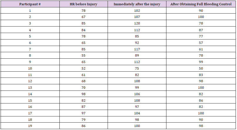
Altogether there were 19 episodes of iatrogenic injury in 10 animals controlled by 12 participants. One animal died after exsufflation due to relapse of bleeding after non-complete hemostasis. There were no conversions to open procedure. Temporary vessel control was obtained with a grasper, gauze, intraabdominal pressure elevation or temporary clip application. For final hemostatic purposes participants used Vicryl 2.0 or PDS II 3.0 suture. Heart rate of participants before the injury, during the repair and after obtaining a haemostasis is shown in Table 1. HR ranged from 52 to 97 per minute before the task and from 75 to 120 during the repair of injury. There was a tendency towards higher HR values before and during the task in experienced surgeons than in novices although this difference did not reach statistical significance. When evaluating this approach to training in disaster control eleven participants gave the exercise 5 points on a VAS scale and one participant gave it 3 points resulting in a total of 4.8 points for the whole group.
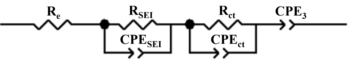
Table 1: Changes in participants’ heart rate before, during and after the vessel injury.
Discussion
With growing number of advanced laparoscopic HPB surgery worldwide there is a clear need for a structured laparoscopy training [3]. In order to prepare surgeons for these demanding procedures a variety of simulation models have been proposed so far. Advanced laparoscopy techniques can be taught in a simple boxtrainer. The box trainer however, apart from giving the opportunity to learn purely technical skills is much less effective in preparing for conditions in real life surgery [4]. A Virtual Reality (VR) training offers interesting approach to teaching without the need for the use of animal tissue and creating close to real life conditions. Unfortunately, at its current level of performance, it does not meet expectations. No additional benefit is observed from VR training in a multimodality laparoscopy training program [5]. A very interesting model with perfused pig liver can simulate almost lifelike conditions [7]. It is one of the few training modalities to offer trainees a highly simulated bleeding in order to acquire advanced laparoscopic suture skills and train under the pressure of bleeding [10]. The setting of such a training modality seems however too complex to be widely used for teaching laparoscopy. Also, contrary to the model described herein it does not offer the trainee the possibility to observe the effect of bleeding on a general status of the patients, concentrating only on the bleeding itself. In this sense, it seems closer to a box-trainer concentrating merely on a technical control of bleeding without the stress of observing worsening vital signs that clearly simulates real-life disaster. The closest to life experience can probably be achieved in live animal models [11]. It has been successfully used in creating a model for the intravascular treatment of IVC injury. In live anesthetized pigs after iatrogenic IVC injury a bleeding was controlled successfully by trainees using balloon insertion via femoral vein [12]. Live animal laparoscopy training using pigs has been shown to be useful in acquiring advanced liver laparoscopy skills [6]. While the benefits of this model over other approaches in teaching purely technical skills can be discussed it offers unique opportunity to create a simulation for a life-threatening intraoperative event. There are much less reports on the use of sheep as a model for advanced surgical training [12]. It is however known to be an interesting model for advanced colon resections [13]. During our study we have observed a higher level of stress measured as a rise in HR in more experienced trainees. While it was a bit surprising it can be explained by the fact that more senior surgeons are well aware of the potentially fatal complications of a major vessel injury during laparoscopy. Almost all participants including experienced and inexperienced surgeons agreed that this training modality was close to perfect in creating a stressful environment simulating reallife disastrous intraoperative event.
Conclusion
In-vivo pig and sheep models can be used for training in the management of major bleeding during HPB surgery. It is a modality that is highly appreciated by trainees. It seems that stress level during advanced exercises is higher in experienced surgeons than in newcomers.
Designing an Adjustable Head Frame for Surgery Using Mixed Reality Technology Hololens 2
Introduction
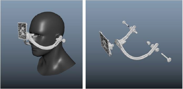
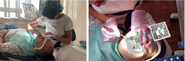
To position three-dimensional holograms to a strictly defined point in space, it is necessary to use special markers, which can be represented in the form of images [1], QR codes [2] or geometric objects [3]. In the case of using mixed reality technology in surgery, these markers must be rigidly linked to the patient’s anatomy in order to accurately position the 3D model of the anatomical structures. This can be achieved through the use of special frameholders of the marker [4], which are based on the individual anatomy of the patient and are made using the 3D printing method. The main disadvantage of such marker-holders is that for each patient it is necessary to design and manufacture a new marker-holder, which is time-consuming and expensive. To solve this problem, we have developed an adjustable frame (Figure 1), which is intended for performing operations on the head using mixed reality glasses [5]. This frame fits over the patient’s head and adjusts to his individual parameters. This device is entirely made of polyamide, which allows it to be sterilized before each procedure and used repeatedly in various operations related to neurosurgery and maxillofacial surgery [6].
Figure 1: Adjustable frame design.
Design and Basic Principle of Use
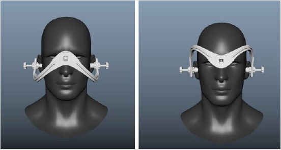
The design of the frame is designed in such a way that it rests on the fixed parts of the patient’s anatomy, namely the bridge of the nose and ear canals. All adjusting elements are near the ears. As a result of their adjustment, the frame is firmly adhered to the patient’s head due to the tension created between the support on the bridge of the nose and the ears. The original fitting position of the frame is designed neurosurgery, however it can be placed upside down in order to open access to the face for maxillofacial surgery (Figure 2). The frame also contains radiopaque markers, which can be used to compare the position of the frame relative to the CT scan and thus calculate the exact position and orientation of the hologram relative to the marker when using mixed reality glasses. The marker itself is inserted into a special slot in the frame, which allows you to set markers of various configurations depending on the surgical access and the position of the patient during surgery.
Figure 2: Two different fitting positions of the frame. For neurosurgery (left) and maxillofacial surgery(right).
Iterative Design Approach
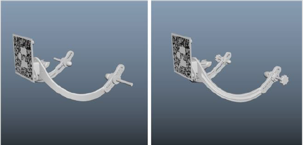
At the moment, the design of the frame has undergone 2 major development iterations. On each of them, various design changes were made to improve the ergonomics and quality of positioning of the holograms (Figure 3). The first version used plastic adjusting clips with metal rods. Despite the small length and size of the threads, they gave very strong interference in CT scans; as a result, the retainers were replaced with polyamide screws with nuts from the same material. In the second version, in addition to screws, polyamide plugs were made, which were put on over the screws and increased convenience in the process of adjusting the position of this frame. Also, the frame was reinforced with stiffening ribs to reduce possible deformation as a result of tightening the screws in the ears. In addition, the design of the installation of radiopaque markers has been revised. Now they are represented by small 2×2 mm set screws. This made it possible to significantly improve the quality of calibration and the positioning accuracy of the holograms. The result of using the frame during surgery with mixed reality glasses can be seen on the Figure 4.
Figure 3: Two versions of the frame. Old design (left) and new design (right).
Figure 4: Doctor wearing Hololens 2 glasses during the procedure (left) and picture from first point of view through glasses (right).
Conclusion
The adjustable frame allows surgeons to perform multiple surgeries using the same rig without creating custom system for each procedure. The latest version of the design is not final and requires some improvements. In particular, it is planned to revise the regulation mechanism to increase compactness in order to fit portable dental CT scans. We are also considering an option in which has an additional emphasis on the forehead to increase the rigidity of fixation.
From Traditional Braiding Methods to Additive Manufacturing for Fabricating Mckibben Artificial Muscles
Introduction
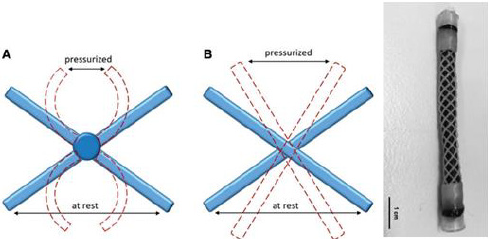
Mckibben artificial muscles [1-5] are of interest because of their practical engineering performances such as large contraction strains, high blocked forces and short response time. Since these performances are comparable to those of biological muscles, the demand for employing these muscles for robotic tools and medical devices is high. Mckibben artificial muscles are simply made of three essential parts: an inner elastomeric bladder, a braided sleeve and the fluid supply system [6]. The inner elastomeric bladder is surrounded by a braided sleeve which is connected to the fluid supply system. To activate the muscle, pressurized fluid is normally injected into the one end sealed inner bladder, once the inner bladder is fully pressurized, the volume of the inner bladder increases, and it produces force in radius direction against the braided sleeve. The braided sleeve subsequently transforms the generated radius force into the length direction along the braid axis [4]. The muscle, therefore, generates a length change or tensile blocked force depending on the experimental conditions. The magnitude of the generated force and length change significantly rely on the topology and mechanical properties of the braided sleeve. Previous literature described that the generated tensile blocked force normally decreases remarkably with increasing the initial angle of the braided sleeve up to the critical angle of 54.44 for a fixed input pressure. The amount of contraction strain also depends on the initial angle of the braided sleeve and for ideal systems is independent of internal pressure [3,7]. The amount of contraction strain usually declines with increasing initial braid angle and reaches zero contraction strain at the critical angle. Given the importance of the braided sleeve design to the performance of McKibben artificial muscles, here we review the trend of leading methods for manufacturing braided sleeves used in McKibben muscles and also suggest some design strategies for the future manufacturing.
Traditional Braided Sleeves
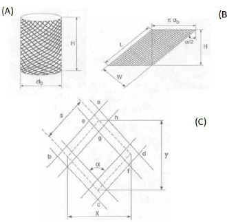
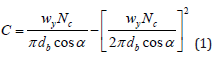
A braided sleeve is basically manufactured with several yarns interwoven with each other and fabricated around a mandrel [8,9]. There are important geometric variables that affect the final mechanical performance of the braided sleeve with an assumption that the braid is made of flat strip yarn. These variables include the braid angle, α, helical length. L, of one pitch of yarn, mandrel or braid diameter, db, yarn width, wy, and cover factor, C as shown in Figure 1. The cover actor is an essential property of the braid and is defined as the ratio of area occupied by yarn within a periodic pore unit to the total area of the pore unit, as shown in Figure 1. As derived by Zhang et al. [8], the cover factor is described by equation (1) and is a function of braid diameter, initial braid angle, yarn width, and the number of threads, Nc. Braided sleeves with the high cover factors are normally required in manufacturing McKibben artificial muscles due to the working conditions at high pressures. When fewer fibers are used in manufacturing of the braided sleeve this results in wider gaps between the fibers and consequently may result in muscle rupturing due to the internal bladder passing through the gaps at high pressures. Figure 2 illustrates the three different types of the braided sleeve with different cover factors.
Figure 1: Braided sleeve geometry (A) braided tube (B) Braid geometry of a helically slit tube of one pitch length (C) Unit-cell geometry used to determine cover factor; x and y are unit-cell height and width, respectively[8].
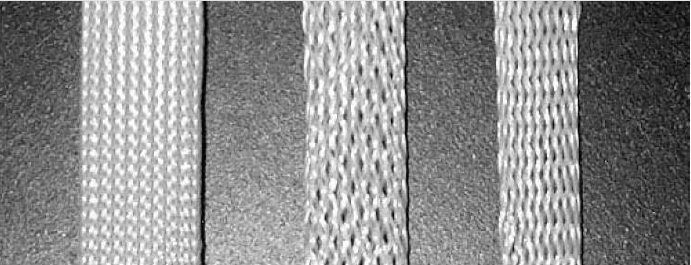
Figure 2: Three different types of braided sleeves designed with higher to less cover factor [9].
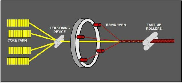
The traditionally two-dimensional made braided sleeves used in manufacturing conventional McKibben artificial muscles are sourced commercially and manufactured with industrial braiding machines [10]. As shown in Figure 3, the braiding machine assembles multiple individual fibers by using several rotary spools to produce a cylindrical hollow braided sleeve.
Figure 3: Typical set-up for a biaxial braid with core yarn.
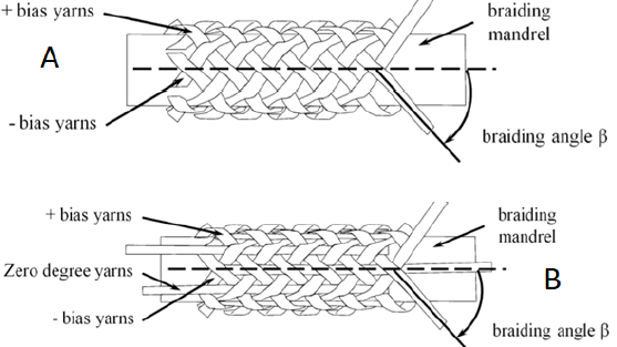
Two dimensional-braided sleeves are structurally divided into three categories. Biaxial-braided sleeve is the most widely used braid structure in industrial textiles, especially in the composite industry. A single yarn set are (generally orientated at an angle in the + θ and – θ directions) interlacing with each other around mandrel to form the braided fabric surface as shown schematically in Figure 2A. This structure, however, suffers from poor impact resistance because of crimp and low delamination strength due to the lack of binder fibers in the thickness. Triaxial-braided fabric normally consists of three sets of yarns and intertwine with each other around the axial yarns at about 45° angle as shown in Figure 2B. In this method, braiding very dense structure patterns is less feasible compared to biaxial-braided fabrics. Although the axial directional properties are improved in this method (Figure 4). Currently, various types of braids, made of nylon, polyester and carbon fibers are commercially available providing different advantages and disadvantages to the performance of the McKibben muscles.
Lab-scale braiding machines, however, suffer from several essential disadvantages. Firstly, the braiding machines are limited in generating only a narrow range of braid angles, where the braiding angle α is the angle between the longitudinal direction of the braided sleeve and the fibers that are helically wrapped to form the braid. Commercially available braids have a limited selection of braid angles typically in the range of 15o- 35o. Secondly, producing a consistent cover factor is limited due to the friction between fibers. The cover factor is defined as the ratio of area occupied by fibers to the total braid surface area and is a function of braid diameter, initial braid angle, fiber width, and the number of threads. Again, the variation in cover factor from commercially available braids is limited and most have a cover factor in excess of 85%. Third, long fiber lengths are needed to operate braiding machines, which limit the introduction of novel fiber materials for research-scale production especially when only short lengths of experimental fibers are available.
Smart Birded Sleeves for Contraction Sensing
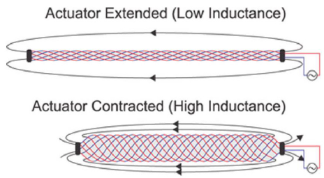
Using traditional or prismatic joints is generally required to precisely measure the motion of Mckibben muscles. Particularly in robotic applications using the Mckibben muscle with sensors is a normal practice to allow for closed-loop control of the generated motions. Research [11,12] has shown that measuring the motion is possible by using smart braided sleeves in manufacturing the McKibben muscles. The traditional braided sleeves of a pneumatic artificial muscle (PAM or McKibben muscle) were interlacing with conductive, insulated wires. Ultra-flexible wires with soft copper stranding and PVC insulations were utilized as conductive wires. This particular braid was assembled with 16 helices equally woven to the right and left directions. These wires acted as a solenoid-like circuit with an inductance that more than doubles over the PAM contraction. Following the actuator contraction, the direction of conductive fibers become more aligned therefore the inductance of the circuit increases. Figure 5 shows the schematic view of the smart braided sleeves used in manufacturing McKibben muscles.
Figure 4: Two dimensional-braided sleeves (A) Biaxial (B) Triaxial.
Figure 5: The smart braid sensors at (A) extended and (B) contracted motion[11].
In this study, three structurally different braids were assembled to match the mathematical models. As shown in Figure 6 the authors modeled the inductance of the smart braid with either a simple long solenoid
a) or by using the Neumann formula on 16 helices
b) that are radially distributed about the actuator and electrically connected in series. The results then were compared with measurements from a smart braid stretched over dowels of different diameter
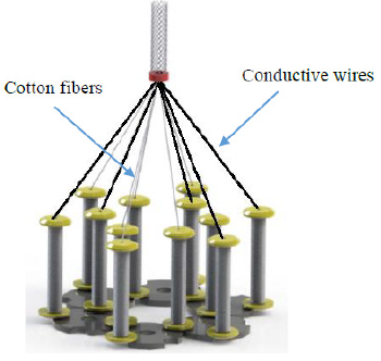
c) 4. Electrically conductive braided sleeves Conductive braided sleeves were used to manufactured miniature and bladderless McKibben artificial muscles [5]. As shown in Figure 7 the conductive braids were assembled with scale lab braiding machine using cotton fibers and steel wire in a parallel direction.
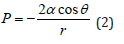
The resistivity of the conductive braid was reported to be ∼18 Ω. As mentioned earlier the cover factor of the braided sleeve is an important parameter and should be closely monitored the braid manufacturing process. In this study, the conductive braided sleeve was manufactured with different cover factors by independently decreasing the diameter of the braid yarn. The ultimate aim of this study was to keep the thermo-sensitive material (paraffin) inside the braided sleeve without using any inner bladder. The thermossensitive material was used to generate a sufficient pressure inside the conductive braided sleeve similar to air in pneumatic version. Adequate conductivity was required to electrically stimulate the thermos-sensitive material and consequently activate the muscle.
To manufacture the bladderless McKibben muscles, researches used the principles of breakthrough pressure [13]. The pressure needed to push a non-wetting liquid through the pores of a membrane is called the breakthrough pressure, P, and is related to the membrane and liquid properties by the following Young–Laplace equation where, r, is radius of the pores, σ and θ are the surface tension of the liquid and the contact angle, respectively. As shown in equation 2, for any pair of materials, the breakthrough pressure increase as the size of pores decreases. Pore sizes in a braid can be expressed in terms of the cover factor, C, which was defined earlier
Figure 6: Three structurally different types of smart braided sleeves[11].
Figure 7: The schematic illustration of the lab-scale braiding machine[5].
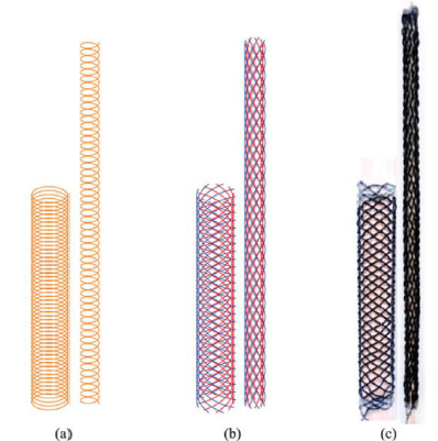
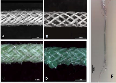
The muscle made of the conductive braided sleeve with the cover factor and average pore size of 0.73 and 0.27 mm was able to prevent the wax exuding through the pores during the actuation tests for many cycles. Figure 8 illustrates the microscopy images of two conductive braided sleeves with different cover factors packed with a thermos-sensitive material.
Figure 8: Microscopy images of conductive braided sleeves with high (A) and low (B) cover factors packed with thermossensitive materials (C and D). (E) the Entire conductive bladderless McKibben muscle[5].
Three-Dimensional Printed Braided Sleeve
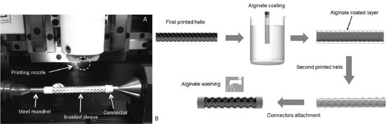
An alternative method was investigated to manufacture braided sleeves using a three-dimensional (3D) printing technique [14]. 3D printing method was chosen to achieve more versatility in controlling the geometry and the structure of the braids. This unique 3D printing technique is simple, fast, and accurate that can be easily modified to fabricate tools for small robotic systems where custom manufacturing is required. The braided sleeves in this study were made by employing an extrusion style threedimensional (3D) printing machine using a similar technique to that introduced recently. Each individual printed line was made of polycaprolactone (PCL) material and was precisely printed around a rotating cylindrical steel rod. An additional advantage of this method was the ability to incorporate the hydraulic end connectors directly into the braided sleeve structure. The end connectors are an integral part of the McKibben muscle system and achieving leak-free connection to the hydraulic fluid supply and robust mechanical connection to external loads is a challenge that can be uniquely addressed using 3D printing. As shown in Figure 9, the manufacturing process of 3D printed braids is a follow. The right to left printing direction was first performed as described above and then the entire mandrel with the printed helix was dip- coated in alginate solution and dried. The left to right printing direction was performed to form the second helical fiber on top of the dry alginate film. The mandrel was then immersed in the water bath to dissolve the alginate interlayer. By removing the alginate films from between the PCL helices the double-helix braids with disconnected fiber crossover points were successfully produced. The braided sleeves were then removed from the steel mandrel. The cover factor was constant at 0.47.
As shown in Figure 10C, the printed braids have integrated end connectors to simplify the assembly of the completed McKibben muscle. The effect of fiber connection in crossover points has been investigated. In this particular study, it has been found that the braided sleeves with connected fibers were unable to produce any actuation due to mechanical failure of the fibers (Figure 10A & 10B). Future directions in the future, it would be worthwhile to three-dimensionally print the braided sleeve using conductive materials. It would be then feasible to manufacture braided sleeves which contain conductive and non-conductive helices similar to those explained in sections 3 and 4 and pave the way for entirely printing the braided sleeves for in situ strain sensing applications. This task can be done via using conductive polymer composites with adequate viscosity for 3D printing applications.
Figure 9: (A) Photograph of printing set for manufacturing polymeric braided sleeve (B) Schematic illustration of the manufacturing process of 3D printed braids. All the manufacturing steps shown in the figure have been conducted around a mandrel[14].
Figure 10: Illustration of the deformed shape of one junction point unit (A) connected junction point before (blue ribbons) and after (red dotted lines) pressurization (B) disconnected junction point before (blue ribbons) and after (red dotted lines) pressurization (C) The entire McKibben muscles manufactured with 3D printed sleeve[14].
Analytical Platforms for Medical Diagnosis: A Study on the Performance and Recent Trends on Aptamer and Antibody Based Biosensors
Introduction
Advances in the field of molecular biology and chemistry have driven the studies in biosensing to an important and necessary level. The increasing attention of the population to healthcare summed to the alterations in their alimentary and social habits significantly changed the needs for personal health. Miotto, et al. [1] mentioned that the current context of healthcare demands to “ensure that the right treatment is delivered to the right patient at the right time”. In this scenario, the study of biosensors has provided sufficient tools, especially in the last decade, to advise the science of sensitive, rapid and accurate medical diagnostics. Clark and Lyons [2] were the pioneer in the field with the development of an enzymatic biosensor for detection of glucose. Their technology based on the oxidation of glucose by the enzyme glucose oxidase produced gluconic acid, hydrogen peroxide and electrons. This technology inspired unlimited researches up to nowadays and the more known commercial devices are still based on biosensing of glucose (being the first commercially available biosensor for glucose fabricated by the company Yellow Spring Instruments) [3].
Once biological molecules are irreplaceable agents in living beings to make humans and animals to perfectly function, not surprisingly, scientists and research companies devote maximum efforts to mimic the biochemical reactions that naturally occur in the nature. This is the basis of a biosensor. A biological element of recognition is attached to the surface of an electrode material to detect a target molecule by means of their specific sites. Changings in chemical and/or physical properties of the transducer system are thus monitored and associated to the presence or to the concentration of the molecule of interest. Regardless the numerous possibilities of substrate materials, transduction modes and kind of molecules of interest, possibly the study of bioreceptors is represents the golden effort to achieve the two most important characteristics of a tool for diagnosis: sensitivity and selectivity. In light of this context, this work proposes a critical review of the literature on biosensing technologies for medical diagnosis with respect to two of the most important bioreceptors employed in highperformance sensors: antibodies and aptamers. A discussion on the global features of biosensors, their importance and application in medical diagnoses, key aspects of antibodies and aptamers to be employed as bioreceptors are provided herein. This knowledge is illustrated with the most recent trends in current works available in the specialized literature in order to contribute to the field of biosensors and clinical bioassays.
Biosensors and Units of Biorecognition
Sensors are part of our daily lives, inserted in the most diverse equipment’s with the most different functionalities. In general, a sensor is a device that transforms a certain physical or chemical property into an analytically measurable signal. In this way we can classify sensors where the variation of a biochemical property generates any signal, these devices we call biosensors, which can be defined according to IUPAC as being “device that uses specific biochemical reactions mediated by isolated enzymes, immune systems, tissues, organelles or whole cells to detect chemical compounds usually by electrical, thermal or optical signals” [4] A biosensor consists of two parts, one formed by the biological recognition element (receiver) and the other by the transducer, which can be electrochemical, optical, thermal, piezoelectric, capacitive and field effect. We can classify them, by the different methods of transduction, as well as according to the element’s receptor. Here, we will classify them only this. Bioreceptors can be selective or not, but recognition element plays a crucial role in the overall biosensor performance and selectivity toward a particular analyte [5]. Temperature, pH, contaminants, ionic strength, type of solution (buffer solution, body fluids, water) are factors that determine the performance of the biosensors [6,7].
Aptamers / Aptasensors
Aptamers are short and single-stranded nucleic acids (DNA or RNA) with capacity to bind to target molecules with high affinity and specificity [8]. First introduced in 1990, the process of selecting an aptamer is called Systematic Evolution of Ligands by Exponential enrichment (SELEX), from a large oligonucleotide library [9,10]. Aptamers can be selected for a variety of targets, including small molecules, proteins, nucleic acids, microorganisms, cells, tissues, metal ions and chemical compounds [11-13]. With the advantages of small size, high binding affinity, good stability and easy synthesis, aptamers show potential for various applications, such as targeted therapy, detection and clinical diagnoses [14-17]. After selection and characterization, aptamers can be customized for developing sensors [18]. A large variety of aptamer-based biosensors (aptasensors) with various detection strategies have been developed and reported in the literature [19]. In comparison to antibodies, aptamers are smaller units containing oligonucleotides with sizes over 30 oligos [20]. They are similar to monoclonal antibodies in terms of binding affinities, being called synthetic antibodies [21] in addition to other advantages, such as chemical stability and regeneration of its threedimensional structure even after several cycles of denaturation/ renaturation [22]. Its small size allows a greater density of immobilized molecules. They are chemically synthesized, which allows the flexibility of the conformation of their two-dimensional structure, so it can be built for the detection of any antigen, from small molecules, heavy metals, protein, enzymes, microorganisms and cells, with the possibility of adjusting the sensitivity and selectivity [23-28].
Antibodies / Immunosensors
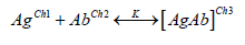
Antibodies (Abs) are proteins that can be employed as valuable tools in laboratory and clinics [29]. Antibodies include those secreted by a single clone of B lymphocytes, termed monoclonal antibodies (mAbs), and those produced by a mixture of various B lymphocyte clones, the polyclonal antibodies (pAbs) [30-32]. In 1975, Kohler and Milstein developed a system for the production of monoclonal antibodies. Abs demonstrate high affinity and specificity to target molecules and have been frequently selected for a wide variety of applications including immunodiagnoses, biomarker detection, immunological research and vaccine quality control [33-35]. Abs can be used to develop a variety of sensors (immunosensors) upon the formation of an antibody-antigen complex [36]. Immunosensors are based on antigen-antibody affinity, where an immunochemical reaction forms a very stable complex. Every protein has an isoelectric point (point where the global electrical charge is equal to zero) that varies according to the composition of the amino acids, thus determining the magnitude and polarity of that point at a specific pH [37]. One can assume that any protein (Ag), with charge Ch1, and its antibody pair with charge Ch2, the reaction of that system (AgAb) results in a global charge Ch3 which can be described by the following equation:
where K is the binding constant for this complex. This change in electrical charges can ideally be detected, depending on the antigen concentration and the transduction technique used. The ambivalence of this system still allows the use of a biosensor for the detection of an antigen, regarding the possibility of immobilizing an antigen and the antibody becomes the analyte. Abs possess a “Y” shaped structure consisting of two heavy and two light polypeptidic chains bound by S-S bonds with approximately 150 kDa and dimensions of 14 nm x 10 nm x 4 nm [38,39]. The base of this “Y” structure is called fragment crystallizable region (Fc) and is composed by the heavy chains. On the other two extremities, there are the antigen-binding sites, or epitopes, comprising the fragment antigen binding (Fab). The Fab branches exhibit different characteristics (such as the chemical composition, the physical structure and the isoelectric point) as a natural consequence of their properties to bind different analytes [39,40]. At the same time it is advantageous to orientate the immobilization of Abs by the Fc portion because it keep frees the active specific sites (Fab) to bind analytes, the extra protocol to allow this orientation makes the fabrication of oriented antibodies-based sensors more laborious and frequently more expensive.
Key Features on the Performance of Biosensors
The most important characteristics of a biosensor are its selectivity, reproducibility, stability, sensitivity and linearity. The combination of these parameters has been the focus of many researchers specially in the last decade to develop high performance devices for diagnosing molecules of medical interests. These features can be defined as follows: a. Selectivity: represents the ability of a sensor to present an analytical signal exclusively due to the recognition of the target analyte, not suffering the influence of interfering species at a significant level. Morales and Halpern [41] mention that selectivity is essential in the development of point-of-care biosensors. This is because the testing biological samples are typically very complex and can possess various interfering molecules capable to compete for the bioreceptor sites of the sensor; b. Limit of Detection (LOD): is the minimum amount of analyte able to generate an output signal distinguishable from the blank signal (analyte absence) [42]. Depending on the level of affinity between the biorecognition element and the analyte, the biosensor can achieve low LODs and meets a broader window of applications in the field of clinical diagnosis. This affinity is expressed in terms of the dissociation constant “KD” (reciprocal of the association constant “KA”), which relates the concentration of free and bound molecules in a solution to provide a sense of strength of these interactions. In this regard, the lower KD is, the higher is the affinity between the bioreceptor and the analyte and, consequently, the lowest concentrations can be detected by the biosensor. IUPAC recommends the use of the equation LOD = 3S/m to calculate LOD, where “S” corresponds to the standard deviation derived from the black measurements and “m” represents the slope of the calibration curve; c. Sensitivity: despite it is still very common to observe the misuse of this term to designate the LOD, the sensitivity actually refers to the variation of the analytical signal due to the variation of the target analyte. In other words, it is calculated as the slope of the calibration curve and has the unit of the transduction signal (e.g. Ampères, Ohms, Volts, degrees, Celsius degrees, Hertz, etc) divided by the unit of concentration [43]. Briefly, the higher is the sensitivity, the higher is the response of a biosensor when it binds an analyte; d. Stability: capability of keeping the analytical signal robust enough to not suffer the influence of extrinsic agents, such as environmental disturbances, loss of bioreceptors’ affinity to the target, molecules degradation over time, etc [38]; e. Linearity: corresponds to the obeyance of the calibration curve to a mathematical expression. Once the linearity is set known, the concentration of the molecule of interest in a certain medium can be predicted and this is the working principle of quantitative accurate biosensors; f. Reproducibility: can be defined as the ability to provide similar responses under similar conditions of detection. In addition to those basic analytical properties, some authors also defend the evaluation of the linear range of detection and the response time to validate the performance of a biosensor. The former represents the concentration range of the analyte at which the sensor generates linear output signals, which is important to define whether the working range meets the requirement for a certain application besides helping to calculate the LOD and the sensitivity. The latter is an important reference mainly in medical applications. The response time of a sensor is the time required by the device to generate the analytical output signal as a consequence of the recognition of the target molecule. It is also frequent in the literature to find this definition as the time required to obtain 95% of the data resulting from the detection [38]. In the context of clinical diagnoses, fast responses of biosensors allow doctors to manage the diseases at early stages, avoiding the spreading of infections and the worsening of the clinical picture of patients. Within the scenario of the ongoing pandemic of coronavirus disease (COVID-19) for instance, authors defend that the importance of a quick diagnosis lies on fact that SARS-CoV-2 has exhibited higher contagiousness and infection rate if compared to other coronaviruses infections [44]. Furthermore, early diagnosis contributes to fast decisions on medical treatments and quarantine strategies to slow down the spread of the transmission rate.
Traditional Analytical Techniques for Diseases Diagnosis
Diagnosis, detection and prognosis techniques have been studied for several years and many methods for fault detection and diagnosis have been developed [45]. Molecular diagnostics assays use in vitro biological techniques for detection. Polymerase chain reaction (PCR) and quantitative PCR are performed to detect and amplify a genetic material (DNA or RNA) from a specific organism, for instance, a virus [46,47]. The advantages of PCR include the high sensitivity, quick performance and the ability to detect lesscommon organisms. On the other hand, its disadvantages include the supply costs, machinery fees and training expenses [48,49]. At present, PCR assay is regarded worldwide to as the most accurate and reliable test to detect active COVID-19 infections [50,51]. Immunoassays, such as enzyme-linked immunoassays (ELISA) and point-of-care (POC) techniques can be used for detection of antigens or specific antibodies [52]. Currently, immunoassays play a prominent role in the analysis of many clinical laboratory analytes such as proteins [53]. A broad variety of tests detecting specific SARS-CoV-2 antigens and IgA, IgM and/or IgG antibodies were developed [54,55]. Although the classic immunoassays can provide very sensitive and accurate diagnoses, many of them possess some important limitations: high cost, they are time consuming, demand sophisticated equipment and high skilled staff [56].
Recent Trends in Biosensors for Detection of Analytes of Medical Interest
It is worthy notable that the field of biosensing through the design of assays to detect molecules of medical interest has attracted huge attention specially in the last year with the outbreak of COVID-19 around the world. Not exclusively due to the current pandemic, though, numerous researches have been devoted to some special improvements in the analytical sciences in order to ameliorate the performance of the already known technologies. Within the recent literature in this domain, one can easily recognize some trends in the newest biosensors of medical interest: the fabrication of point-of-care devices, the label-free detection, realtime measurements and the advance of electrochemical transducer mechanisms. Under all these trends, the use of antibodies and aptamers as bioreceptor agents seem to properly match the needs and expectations of current diagnoses.
Point-of-Care Biosensors
Point-of-care diagnoses collect several unquestionable advantages over traditional laboratory setups. Not surprisingly, the golden characteristic refers to the possibility of running the test wherever the patient is, on-demand and onsite [57]. It makes the sensors amenable for bedside monitoring, analysis in pharmacies or even by the user himself. Consequently, this kind of device tends to gain increasing visibility in the market. Eguilaz et al. [58] highlight that these devices are even more relevant in resourcelimited regions where the access to medical centers is difficult to the majority of the population. Nonetheless, point-of-care devices combine other interesting characteristics, such as (generally) the rapid detection, fewer steps for data/results acquisition, friendly interface, easy transport due to the reduced dimensions and light weight and demand for small sample volumes [57,59]. Concerning this last characteristic, though, there is a strategic point to be taken in account. Depending on the application, the target molecule is present at very low concentrations in the sample of analysis. Thus, a small volume for testing can contain insufficient quantity of analyte in such a manner that the biosensor would not be able to detect it [58]. In this regard, antibody- and aptamer-based biosensors are widely employed to overcome this drawback because of their high sensitivity resultant from the high affinity and selectivity of these molecules. Searching for overcoming the limitations of conventional diagnoses, Ferreira, et al. [60] worked on the development of an aptasensor for detection of breast cancer in undiluted human serum. This kind of cancer is unfortunately responsible for thousands of deaths annually. According to the authors, the diagnosis is mostly based on the detection of tumor markers present in blood or other corporal fluids at concentrations from 15 ng/mL to 75 ng/ mL (over the regular healthy range of 2- 15 ng/mL). Ferreira, et al. [60] exploited two functionalization methods to attach Human Epidermal Growth Factor Receptor 2 (HER2) aptamers to the surface of screen-printed electrodes (SPEs). These devices are widely recognized in the literature to serve as useful substrates for designing portable electrochemical sensors, mainly because of their good conductivity, electrical stability in typical electrolytes and reduced dimensions [61]. In a list of 110 recent articles reviewed by Ranjan, et al. [62] on point-of-care biosensors for breast cancer diagnosis, 23% were described the use of antibodies and 5% the use of aptamers as bioreceptors, which symbolically represents the large employment of these biomolecules in biosensors of medical interests. In this same work, other elements of recognition were described, e.g. enzymes, inorganic probes, DNA, proteins, receptorligand complexes and molecular imprinting polymers (MIP).
Label-Free Detection
The topic of label-free sensing in the context of bioassays generally converges to two points: the advantage of reducing the consumption of reagents and the consequent lower number of fabrication steps of biosensors in comparison to golden standard techniques (e.g. ELISA and PCR). Label-free sensing mechanisms consist in the direct detection of target molecules by the bioreceptor attached to the transducer substrates, i.g., without the needs for fluorescent chemicals, enzymes and so on [63]. Thus, since the label-free biosensors do not demand extra labels to run the detection, this characteristic nicely meets the requirement of point-of-care biosensors for the simplest incubation protocols and allows the use of unprepared samples at working environments. Among other interesting features, Andryukov, et al. [63] point out the following advantages over label-based similar analytical assays: simpler pattern of detection, lower response time, lower cost of analysis, opportunity to detect small molecules and possibility of multiplexing. Zhang and Liu [64] mentioned that the success of using DNA in label-free devices based on optical biosensors has inspired the same approach in the aptamer field. However, the authors explain that aptamers can fold DNA and hide its bases, providing slow kinetics of target binding, especially when the target is a small molecule. Therefore and since aptamers possess lower affinity to small molecules (KD around low micromolar units) than DNA (KD approximately in picomolar or low nanomolar), the detection of aptasensors tends to be more challenging, justifying the efforts on label-free sensing to enhance its analytical response. On the other hand, numerous works can be easily found in the field of labelfree immunosensors for detection of analytes for highly sensitive diagnoses [65-68].
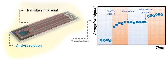
Real-Time Measurements
The key point of real-time biosensing is the necessity of the sensor to rapidly recognize the target molecule. If so, the output signal will be registered by the transducer source in short time intervals and a variation in its magnitude could be notable as illustrated in Figure 1. This need makes some important well recognized techniques such as ELISA and Luminex assay to fail as real-time methods for in vivo applications, since they require laborious and pre-defined longtime steps [69]. Cohen et al. [69] highlight that ELISA, for instance, depends on diffusion processes concerning the interaction between antibodies and antigens in a non-mixed solution, which is associated to a low binding equilibrium constant and makes the response time longer. Typically, this technique requires approximately 3 hours to be performed [70,71]. In this regard, Shengnan, et al. [72] reported the construction of an aptasensor for the real-time detection of vascular endothelial growth factor, one of the most important cytokines present in cancer patients (with average concentration of 434 pg/mL). The authors achieved a LOD of 0.1 pg/mL within a linear detection window from 2 pg/mL to 500 pg/mL. The mechanism of recognition was based on a Chronoamperometry test at the positive redox peak potential of ferrocene-labeled aptamer for 5,000 seconds.
Figure 1: Illustrative scheme referring to the fluctuations of the analytical signal of a biosensor as a consequence of rapid interaction between its bioreceptors and the target molecules.
Also taking advantages of the specificity of aptamers as bioreceptors, Soleimani, et al. [73] manufactured an aptasensor assisted by a computerized monitoring system to detect prostate specific antigen (PSA). To characterize their aptasensor and to construct the calibration curve towards PSA, the authors carried out Electrochemical Impedance Spectroscopy (EIS) and Cyclic Voltammetry (CV). Due to the steric hindrance of the analyte, the electrochemical signal of the transducer substrate increases over the time when aptamers bind the molecules of PSA. As a result, the findings showed that this kind of setup presented sensitive and rapid response fitting the real application aimed to the diagnosis of patients with prostate cancer.
Electrochemical Transducing
As per the examples of the previous sections, electrochemical mechanisms have progressively illustrated the transduction modes of many biosensors for medical applications. From 2017 to 2019, for example, these devices represented 45% of the published articles in the specialized literature of biosensors [74]. The reason is the collaboration that electrochemical reactions provide to enhance sensitivity, accuracy and response time. Briefly, in this kind of biosensor the electrical properties of biological molecules and their interaction with electro active surfaces are exploited for assessing the changes in current, potential, charge, impedance, conductivity, etc. Complementarily, depending even on the dimensions of target molecules, the distance from the electrode surface and needs for redox probes, the specific electrochemical technique can be chosen to achieve highest analytical performance [75]. The detection system consists of three (or two) electrodes, of which one is the sensing surface (named working electrode), one is the counter-electrode and the other is a reference. These electrodes must be immersed in a conductivity solution to allow redox processes to occur and charges transfer. When the detection of the analyte happens and the electrical properties of the surface is altered, an electronic system acts to amplify and manage the resultant data. Traditionally, this last step is performed by a potentiostat interfaced with a software for control of the required parameters. Mishra, et al. [75] pointed out details on the electrochemical aptasensors referring to design strategies and functionalization. The researchers reported that aptamers have been mostly immobilized to gold and carbon-based electrodes via chemical cross-linking with particular attention to ensure biochemical stability, surface coverture and optimal binding affinity. Most common electrochemical techniques used for fabrication of biosensors are CV [76,77], EIS [78-80], potentiometry [81,82] and amperometry [83,84]. When real-time performance is required, time-based assays (such as chronoamperometry, chronocoulometry and chronopotentiometry) well fits medical applications.
Efficiency of Immunosensors and Aptasensors
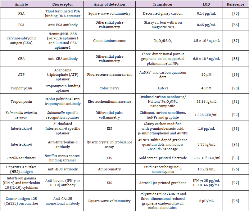
Cesewski and Johnson [85] point out that, in some cases, the high sensitivity of immunoassays are not enough to detect certain pathogens in the organism. In such circumstances, although these infectious agents are present, they do not generate enough available Abs in the blood, so the concentration of the Abs in the blood are lower than the LOD of the technique, failing the detection. According to the authors, this is a typical situation in which the employment of DNA-based systems is more useful. The biosensors consisting of nucleic acids, for instance, are usually able to recognize low concentrations of pathogens by themselves or through the indirect expression of toxins they release in the infected organism (e.g. toxins, other nucleic acids and raised cells. In this regard, Table 1 contains a list of recent researches in the literature of biosensors for medical applications using antibodies and aptamers as bioreceptors. It is worthy notable that these biomolecules facilitate the biosensing of analytes at concentrations as low as some femtograms per milliliter [86-90].
Table 1: Recent developments (from the last two years) in the field of aptasensors and immunosensor for assisting clinical diagnosis.
Note: *NPs = Nanoparticles
Regardless the obvious different protocols used to attach antibodies and aptamers to the different transducer substrates, it is worthy notable that the sensitivity of these devices are really high. Besides, in this recent literature is not rare to observe a trend in using label-free molecules to optimize the fabrication step and to allow accessible in-situ measurements [91-98]. Nonetheless, it is also evident that many authors have employed electrochemical techniques to ensure accuracy and high performance of biosensors, corroboration the previous discussion brought to this minireview in the section “Recent trends in biosensors for detection of analytes of medical interest”.
Conclusion
With the increasing humans needs for accurate, fast and friendly methods for health control, biosensors for medical applications have undergone important changes in the last decade. The immobilization of antibodies and aptamers on transducer substrates for high performance detection has been an exhaustive strategy for the production of biosensors, especially due to the high sensitivity of these bioreceptors. Articles published in the recent literature exhibit LODs in the order of femtograms per milliliter. To this end, added to the intrinsic advantages of antibodies and aptamers, there is a notable trend to search for label-free devices, with less functionalization steps, lower times for the formation of bioreceptor-analyte complexes, under selective and sensitive sensing modes. Thus, much is seen about the use of electrochemical techniques such as CV, EIS and amperometry, although optical and piezoelectric transduction techniques are also present in the field of biosensors for various applications including the ones for medical diagnostics. It is believed that this specific application demands advanced technologies, especially to shorten the detection time, since early diagnoses are essential for the administration of first aids and precise medications that can enhance the chances of cure and survival of patients (especially those who have less access to health centers). The main challenges in the area still seem to be related to the commercial viability of these devices. Likewise, quite possibly, the prospect of advances in technology is likely to be based on the study of alternative materials and methods to make immunosensors and aptensensors increasingly simple and inexpensive.
Empathy and Telepathy: Functional Imaging Psychiatric and Philosophic Correlates
Introduction
What is the Telepathy?
Objective data indicates that a significant percent of general population experience the feeling described as they had undefined communications with other persons who are not in contact with them. They define this exceptional perception in various visual or auditory modalities which are correlated with an altered consciousness state. ‘’Myers defined telepathy is as “the transmission of feelings, perceptions, experience and thoughts from one person to another, without using any of the recognized sensory channels or physical interaction’’ [1,2].
Are Telepathic Experiences Normal in Psychiatry?
Interestingly such perceptions are usually associated with stressful life situations that is a challenging situation for a psychiatrist to distinguish it from a real psychopathological situation. To make a precise diagnosis, the major psychiatric diagnostic algorithm necessitates to exclude all other possible organic causes, such as oxidative stress and internal and external traumatic conditions that predispose to mood and neurodegenerative disorders [3-7]. In this respect it is important to evaluate the functional or pathological correlates of such experience before we diagnose it as a psychiatric disorder. As in many psychiatric disorders the duration of symptoms and disease since the degree of the impairment of daily functionality is an important parameter that help us to make the psychiatric diagnosis [8,9]. This mean for example just as the diagnosis of a major depressive disorder where the depressive symptoms may result from normal, uncomplicated, situations which can be viewed as a simple reaction to stressful situation instead of evidence of mental disorder [9]. Interestingly many persons with telepathic experience are categorized as normal in psychiatry.
A Possible Link between Empathy and Telepathy
Telepathic communications usually occur between persons who share an emotional link instead of a physical, biological or genetic background. For example, such experiences have been reported between close friends although it can be not the case among members of the same biological family. There are also some interesting cases reporting that even physical symptoms especially pain have been experienced by telepathic communications. This has been occurred between twins, parents, and their children as well as some few psychiatrists who have reported telepathic experience with their patients. The well-known Swiss psychologist, Carl Gustav Jung also reported experiencing severe headache when his patient shot himself in the head [10]. All these above-mentioned data indicate that an emotional link rather than a physiological, biological or genetics play an important role in the communication of telepathic experience between emotionally linked persons. Regarding the background of emotional bound between person’s empathy has been reported as a significant indicator of their emotional dynamics that can be affected by various drugs [11]. Empathy is defined as the ability to sense other people’s emotions, and to imagine what someone else is thinking or feeling called also “Cognitive empathy” or “perspective taking”. If we look from this point of view, it can be hypothesized that there is a thin border between the empathic ability and telepathy [11]. Thus, it has been recently suggested that superior cognitive empathy is associated with special abilities, indicating that people with telepathy might be able to activate specific brain regions related to the empathy circuit. Studies have already shown that right hemispheric region of the brain plays an important role during the processes of both empathic and telepathic experience [1,12,13].
Non-Local Unconsciousness Theory and Real-Word Neural Correlates of Telepathy
In agreement with this, many studies in cognitive neuroscience indicate that the processing of symbolic and especially unconscious components are associated with the activity in the structures of the right hemisphere. This is especially important since both the telepathic experience and cognitive emphatic abilities are localized on the same hemispheric region suggesting that there might be a functional link between the activation of the unconscious part of the brain and these exceptional abilities [4]. Based on these data it can be assumed that activated specific brain regions might connect and collect the information from a common information network that is difficult to understand within the conventional time and space concept. This theory resembles us the theory of collective unconsciousness of Jung [10]. Jung’s collective unconscious theory is hypothesizing that human psyches are linked together via an unseen linkage that consists of basic shared perceptions, instincts, patterns of thinking, behavior and a pool of common knowledge which may function as a common information network enabling to connect and collect the information out of the time and space. Jung hypothesizes that each individual inherits a collective memory from past members of the species, that contributes to the collective memory and affects other members of the species in the future [10,14]. As opposed to the conservative functional neurobiology of the brain this window helps us to create a universal unconsciousness model including an enriched picture of causality. Despite these findings, Venkata Subramanian, et al. showed recently that successful telepathic session was associated with significant activation of the right limbic area in compared to the activated left frontal cortical region by the subject without telepathic ability [1,15] which is consistent with the multimodal role of fMRI. Moreover, other functional magnetic resonance imaging and magnetic field studies demonstrated that distant intentionality was related to the alterations of different brain functions in the isolated recipients which may be modulated with the artificial magnetic stimulation. Thus, there have been many studies suggesting the beneficial role of magnetic stimulation in various neuropsychiatric and neurodegenerative disorders characterized with empathy [8,9,16, 17].
Conclusion
Alternative paradigms including a universal and non-spatial nature of human consciousness could help us to understand the nature of telepathic experiences. Besides concrete rational findings where neurobiological and functional/metabolic correlations are still the only measurable macroscopic parameters in our real world, alternative consciousness models which seem out of space and time help us to expand the conservative consciousness model and understand the flexible cause-effect relationship in the human cognition.
Rare Case of Isolated Plasmodium Vivax Malaria Presenting with Pancytopenia: A Case Report
Plasmodium vivax is one of the most widely distributed specie of genus plasmodium causing infection in humans with approximately 80 million new cases annually. Although severe infection with P. vivax is extremely rare Kochar et al has reported a case of P. vivax presenting with severe infection leading to severe anemia, renal involvement and ARDS with multiorgan failure. Pancytopenia is an extremely rare complication of P. vivax malaria with various proposed mechanisms including macroangiopathic hemolytic anemia, hemophagocytic syndrome and direct bone marrow suppression [1]. This is one such case of isolated vivax malaria presenting with pancytopenia
Case Presentation
A 17-year-old girl with no previous co-morbids presented with high grade fever associated with rigors and chills, vomiting, loose motions and decrease appetite for 5 days. On examination patient had Bp 100/70, Pulse 92/min, Temperature 101F, Oxygen saturation 98% with room air. She was dehydrated and pale with bruises on arms and thighs. There was no jaundice or lymph nodes. On abdominal examination Abdomen was soft non-tender with palpable spleen with a span of about 2 fingers breath below the costal margin. Liver was not palpable. Cardiovascular and Respiratory examination was unremarkable. Complete blood picture showed TLC 2740/microliter, Hemoglobin 10.4 g/dl and Platelets 16000/microliter. Liver function tests, Renal function tests, Serum electrolytes, Urine routine examination and Erythrocyte sedimentation rate were normal. C-reactive protein was 48, LDH 443, Ferritin 1021 and D-dimers were 890. Peripheral film showed Retic count of 3%. Dengue serology and Covid-19 PCR was negative. Blood and Urine Cultures showed no growth. Ultrasound abdomen showed splenomegaly with size of 13 cm. Peripheral smear for Malarial Parasite showed early trophozoites of Plasmodium vivax. Patient was started on Intravenous Artesunate as she could not tolerate Oral anti-malarial. Her Hematologic parameters dropped further during her stay at the hospital with TLC to 2520/ microliter, Hb to 8.5 g/dl and platelets to 11000/ microliter. Patient responded well to Intravenous artesunate. She became afebrile and her blood parameters normalized after 5 days. She was discharged with follow up test for G6PD assay after one week.
Discussion
Malaria is a worldwide national health problem in many countries, and many occur in tropical and subtropical areas including sub-Saharan Africa, Asia and Latin America [2]. Worldwide approximately 3 billion people resides in areas which have high risk of malaria transmission. 1.1 to 2.7 million deaths occur due to severe malaria every year. Malaria is considered as a 5th leading cause of death due to infectious disease and 2nd leading causes of death in Africa with 1 million people dying every year in Africa. About 85% of all the malarial cases in the world are due to Plasmodium falciparum with Plasmodium vivax on 2nd number. 90% of all the malarial deaths occur in Africa [3]. Of all the plasmodium species, the deadliest and the most virulent is P. falciparum which can lead to life threating complications and death if not treated early. Species other than P. falciparum usually cause mild disease with minimal complications [2]. P. vivax malaria is a parasitic infection which is carried by Anopheles mosquito [4]. Life cycle of P. vivax is a unique phase known as hypnozoite stage in which the parasite remains dormant in the liver causing frequent relapses after acute infection is treated. Severe complications due to P. vivax are extremely rare compared to P. falciparum [5]. Hematological changes occurring in malaria include anemia, thrombocytopenia, leucopenia, neutropenia, leukocytosis, atypical leukocytosis and splenomegaly. One study has shown that lymphopenia, leucopenia and thrombocytopenia are the key predictors of malaria infection. Low Hb, High lymphocyte count, low platelets and monocytosis are more severe in chronic malaria compared to acute malaria [6]. Hematologic changes due to malaria depend on various factors including malaria endemicity, background hemoglobin disorders, demographic factors and level of malarial immunity. Thrombocytopenia and anemia are the main blood abnormalities occurring in malaria [3]. Pancytopenia is an uncommon manifestation of malaria and is mostly common with P. falciparum and occur due to both direct and indirect effect of infection on hematopoietic cells in the bone marrow [7]. Two main mechanisms involved in pancytopenia involve direct bone marrow suppression and hemophagocytosis, the latter is mostly reported with P. falciparum malaria. P. vivax has lesser pyrogenic threshold, marked inflammatory response and very high cytokine production as compared to P. falciparum. There have been case reports of severe disseminated infection with multi organ failure due to P. vivax. Studies have also shown that P. vivax can lead to acute tubular necrosis and acute interstitial nephritis termed as Malarial Nephropathy [1]. Pancytopenia secondary to P. vivax malaria is extremely rare and has only been reported in 0.9% of the confirmed P. vivax cases. To the best of author’s knowledge, only up to 5 cases of isolated P. vivax malaria with pancytopenia have been reported in literature without other associated comorbids [2,5]. Key mechanism involved in the development of pancytopenia in P. vivax is microangiopathic hemolytic anemia followed by hemophagocytic syndrome which has rarely been reported with P. vivax [5]. Hemophagocytic syndrome is an unusual clinicopathological syndrome that is characterized by overt immunological responses by T-cells producing high levels of interferon gamma, TNF alpha, IL-1, IL-2 and IL-18 leading to activation of macrophages which in turn causes phagocytosis of hematopoietic cells and bone marrow suppression. It can be fatal if misdiagnosed or diagnosis is delayed. Hemophagocytic syndrome mainly occurs secondary to many infections including viral, bacterial, fungal and parasitic infections [4]. Estimation of exact prevalence of malaria associated HLH is very difficult because bone marrow biopsy is not routinely performed to diagnose malarial infection and exact mortality rate is also not low. It can be fatal if not treated. Malaria associated phagocytic syndrome can lead to prolong hemophagocytosis which is a very rare complication and can lead to prolong anemia. It has been reported only with P. falciparum and not with P. vivax [2]. Various treatment modalities to treat HLH include treating the causative agent, immunosuppressant, steroids and IV immunoglobulins [8]. Various studies have proven that malaria associated HLH has an excellent response to antimalarials and supportive care without any need for immunosuppressants and steroids [2,8]. Malaria must always be kept in differential diagnosis as a cause of prolong fever with refractory anemia and pancytopenia particularly in endemic areas and asymptomatic patient despite of negative smear and rapid antigen test. Bone marrow biopsy is the key to diagnose P. falciparum malaria in such cases [9,10].
Conclusion
Malaria is one of many infections which can involve any organ of the body especially the bone marrow. Bone marrow involvement causes decrease in all the three cell lines leading to pancytopenia. It is important that malaria should always be included in the differential diagnosis whenever pancytopenia is worked up because it is one of the treatable causes of pancytopenia with excellent prognosis.
Impact of Air Pollution on Semen Quality: The Specific Situation of Terni (Central Italy)
Introduction
Infertility is a prevalent condition affecting an estimated 72, 4 million people globally that is well recognized by The World Health Organization (WHO). Although prevalence data are lacking, 9% of couples struggle with fertility issues and male factor contributes to 50% of the issues. Many genetic and lifestyle factors have been implicated in male infertility; however, about 30% of cases are still thought to be idiopathic [1]. The mechanism by which medical conditions affect fertility includes effects on hormonal levels, impairment of sexual function (including ejaculatory function), or impairment of testicular function/spermatogenesis. In the last 70 years, a decrease in sperm fertility and quality has been observed, including sperm count, ejaculate volume, alterations in sperm concentration and morphology [2]. Recent studies suggest that men with abnormal semen parameters have a higher risk of testicular malignancy [3]. Nowadays, environmental and lifestyle factors could be possible contributors to infertility conditions, such as use of smoke sigarettes, increasing of both parents age conception, abuse of alcohol and drugs, physical inactivity, obesity, social stress, exposure to environmental contaminants (polycyclic aromatic hydrocarbons-PAHs, or heavy metals, for examples) and air pollution [4,5]. In particular, epidemiological and experimental studies explained the link between air pollution and alterations of sperm parameters as the main risk factors for male infertility. Human activities such as transport, industrial and agricultural emission are considered the main causes of air pollution (solid particles, liquid droplets or gases), and people that living near these area, are more exposed to henanced emission source of carbon monoxide (CO), nitrous dioxide (NO2), sulfur dioxide (SO2), ozone and lead [6]. Ambient air pollution is associated with systemic increases in oxidative stress, to which sperm are particularly sensitive. In this contest, reactive oxidative species (ROS) have been related with a broad array of spermatogenensis effects, including the decrease of progressive motile sperm count, viability, abnormal sperm morphology, and fertilization rate and spermatogenic cell numbers [7]. In Italy, 12,482 areas with a high risk of environmental pollution, mainly due to industrial emission, were been identified. Some central provinces, such as Città di Castello, Foligno and Perugia exceeded the limit set for particulate matter with diameter less than 10 microns (PM10) and O3 emissions, in 2019. In particular, Terni is one of the most polluted urban and industrial area in Central Italy [8]. In fact, is situated in an intermountain depression, delimited by the Apennine mountain range. This area is characterized by the presence of typical urban PM10 emission sources such as vehicular traffic, domestic heating and industrial emission sources from a power plant for waste treatment. Peculiar geomorphological and meteorological conditions of Terni basin, limit the dispersion and augment the accumulation of the atmospheric pollutants. The “Thyssen Krupp AST”, a large steel factory founded at the end of 19th century and two more recent chemical industrial areas, are located close to the city center [9]. As a result of the intensive industrial activities and the geographical location, atmospheric pollution is the major local issue with high PM concentrations occurring throughout the year. According to European Commission Law, the daily maximum PM10 concentration allowed in cities is 50 μg-3 [9]. The threshold has not to exceede more than 35 times per year. In Terni, the atmospheric PM10 concentration exceede that daily limit on more than 70 days in 2012, as recorded by Regional Agency for Environmental Protection (ARPA), Umbria. The aim of the present study is to provide an association between ambient air pollution and sperm quality, analyzing seminal biofluid parameters of man living in the urban area of Terni- Papigno, with a high risk of pollution, comparated with those who live in rural areas with low risk of pollution.
Materials and Methods
Study Participants
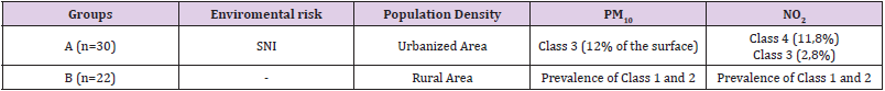
Signed written consent was obtained from all 52 participants (age of 20–40 years) enrolled in this study from January 2018 to December 2019. Patients referred to the Seminology Laboratory of the Division of Andrology and Urology Department for an infertility evaluation. Female partners of the infertile men were subjected to general gynecological evaluation and were reported to have normal reproductive health. Residence in the province of Terni was an inclusion criteria while, systemic and cronic disease, genetic abnormalities, alcohol or drug abuse, hormone treatment, varicocele infection microchidism and cryptorchidism, prostatitis and other factors that could affect semen quality (such as fever, medications, exposure to X rays etc.) were exclusion criteria. Men were divided in group A and B that includes 30 patients from the high pollution environmental risk area of Terni-Papigno and 22 subjects that live in neighboring areas, with a low risk of pollution, respectively (Table 1).
Table 1: Demographic and environmental patient’s classification.
Note: SNI: Sites of National Interest; PM10, Particulate Matter ≤ 10 μm; NO2, Nitrogen dioxide; OCSE Organization for Economic Cooperation and Development, class 1 and 2: rural area with population density <150 inhabitants/ km2 and PM10< 10 μg/m3; class 3 and 4: rural area with population density >150 inhabitants/ km2 and PM10> 10 μg/m3
Semen Analysis and Preparation of Samples
Semen samples were collected at the Andrology and Urology Laboratory by masturbation into a sterile container after 2–7 days of sexual abstinence and were analyzed immediately after liquefaction, according to the WHO guidelines [10]. Each sample was evaluated for seminal volume, pH, total sperm count, progressive motility, and morphology and leukocyte concentration. Semen volume was measured by graduated pipettes. Calibration strips were used to measure the seminal fluid pH. For the evaluation of the sperm concentration, following semen liquefaction, 10 μL of non-diluted, well-mixed semen sample was at first loaded in the middle of a clean Burker counting chamber, maintained at the temperature of 37 °C, gently covered with a cover glass, and examined using 200× or 400× magnification. The sample was diluted before proceeding with the sperm count. 1:2 dilution was used, strictly following the WHO 2010 manual recommendations [10]. The final concentration was calculated as: [(number of spermatozoa counted/the number of lines) x dilution factor] and expressed as 106 spermatozoa/mL. To evaluate the sperm motility, immediately after semen liquefaction, 10 μL of undiluted, well-mixed semen sample was loaded in the middle of a clean Neubauer counting chamber, maintained at the temperature of 37 °C, gently covered with a cover glass, and examined using 200× magnification. Sperm motility was assessed in 200 random spermatozoa and characterized as progressive and non-progressive motility. The total motility was calculated as the sum of progressive and non-progressive motility. Both progressive and total motility were expressed as percentages. Sperm morphology was evaluated in 200 spermatozoa and the value was expressed as percentages. Finally, the vitality was assessed using eosin staining according to the WHO recommendations [10].
Statistical Analysis
Data were analyzed with GraphPad Prism 6.0. Results were reported as mean ± standard deviation (SD) Analysis of variance and Kruskal-Wallis tests were performed. The significance threshold was set at 0.05.
Results
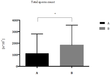
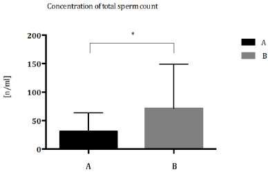
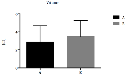
Patients were classified according to Demographic and Environmental characteristics (Table 1) Total sperm count (Figure 1) and sperm concentration (Figure 2) decreased significantly in seminal biofluid of patients living in urbanized area of Terni- Papigno (group A) when compared to subjects who lived in rural areas (group B). Seminal volume (mL) did not reach significant difference (Figure 3) between the groups considered.
Figure 1: Total sperm count (n*106) expressed as median with range and interquartile range in group A compared to group B. *P<0,05.
Figure 2: Concentration of total sperm concentration (n/ml) expressed as median with range and interquartile range in group A compared to group B. *P<0,05.
Figure 3: Seminal volume (ml) expressed as median with range and interquartile range in group A compared to group B. *P<0,05.
Discussion
In this study, conventional semen parameters (volume, appearance, pH, motility, and viability, and morphology, presence of aggregations, agglutination and leukocyte concentration) were analyzed for each sample according to the 2010 WHO criteria. Our results showed a significant decrease of total sperm count in group A patients respect to group B subjects (Figure 1). Nowadays, several studies showed how pollution can affect human fertility. Sperm count and sperm motility were reduced in men living in polluted cities. A decrease of sperm count was observed in America and Northern Europe during the last 50 years [2] and the reduction of semen quality and sperm count especially in industrialized countries supports the evidence that adverse environmental factors are key factors for men infertility [11]. Furthermore, different studies reported the enhancement of infertility, urogenital malformation and chronic disease (cancer, diabetes, etc.) in areas with a high environmental pressure [12]. In group A, a significant decrease of sperm concentration was demonstrated, respect to group B patients (Figure 2). In 2019, a longitudinal analysis on 8.945 semen samples, suggested the specific role of O3 pollution in sperm concentration [13]. The effect of air pollutants and ozone on sperm quality, was strongly elucidated by Sokol, et al. [14]; they reported that pollutants have no effect on sperm quality, however, ozone could make changes in sperms, including sperm DNA fragmentation via oxidative stress, resulting in decreased fertility [14]. The toxicity of O3 was largely demonstrated to be the major oxidant of photochemical smog and its exposure produces reactive ROS at respiratory level [15,16]. Extra pulmonary toxicity suggests that O3 or O3 reaction-products can cross blood-gas barrier and be absorbed into the circulating bloodstream, creating an environment caring to an inflammatory reaction [17,18]. It is unclear how O3 can negatively affect sperm quality, but O3-induced oxidative stress could be a possible mechanism, through which testicular and sperm function could be altered [19,20]. Under physiologic conditions, spermatozoa exist in a balanced environment of ROS and antioxidants, where ROS determine the biochemical steps required for normal fertilization (capacitation and the acrosome reaction). However, excessive amounts of ROS produced by leukocytes and immature spermatozoa can damage mature spermatozoa and the integrity of sperm DNA [21,22]. Concerning seminal volume (mL), our results did not reach significant difference between groups considered (Figure 3). Several studies suggested that alterations of semen parameters, including volume, progressive motility, total motility or morphology, may not relate to air pollution [23]. Several works observed a reduction of both sperm motility and morphology, associated with air heavy metals (lead, mercury and cadmium) [24,25], that determine alterations in sperm DNA. Rubes and colleagues showed that short-term exposure to pollutants caused serious damages in men and women’s reproductive system. Moreover, this study demonstrated that the incidence of sperm DNA alterations due to air pollution is higher in middle-aged men [26]. Also, exposition time of contaminants seem to be determinant, in fact a study of Hammoud showed that 3 months exposure to air pollution decreases motility levels, while removing contamination can restore normal parameters levels [27]. In an extensive cross-sectional study, Xu and co-workers, founded a strong correlation between motility, concentration, and morphology of semen biofluid of men exposed to air pollutants with the use of cigarette and alcohol [28]. On the other hand, Selevan, et al. [29] did not observed any relevant alerations in sperm count, motility and morphology of young men exposed to air pollutants, except for sperm DNA and chromatin [29]. Moreover, several authors specify that the morphological change of spermatozoa after exposure to pollutants is not an indicative diagnostic parameter. Association between air pollution and alteration of specific semen parameters is not clear and different relevant factors, such as geographic areas and lifestyle, should be considered for male infertility diagnosis.
Conclusion
The current study provides evidence of an association between ambient air pollution and sperm quality. Patient residents in areas with high environment exposure had a significantly decrease in sperm quality especially for sperm concentration and count but had no impact on the other sperm parameters of spermogram (motility, morphology, vitality). The individual role of specific pollutants is difficult to identify, since patient in this study are typically exposed to several pollutants simultaneously. The physiopathology leading to altered fertility is poorly understood. In the literature there are forward four mechanisms to explain the negative impact of air pollution on sperm, as hormonal disturbances, oxidative stress induction (ROS), cell DNA and epigenetic alterations, probably working in combination. Clearly, more research is needed to understand the detrimental effect of the pollutants on sperm and to characterize their action in more detail. Our results suggest the important role of human sperm as an early and sensitive biomarker of environmental pollution as it could represent an ideal tool for investigating and promoting health surveillance especially in environmental risk areas. Environmental contaminants and bad lifestyles can impair reproductive health and overall health, encouraging the development of chronic degenerative diseases affecting the adult and, through the sperm epigenome changes, future generations. Thus, identifying risk factors to improve the management of human wellness and health throughout standardized analysis, which correlates the toxic bioaccumulation of the seminal fluid with the multiple semen parameters, might be the main objective to be considered in public prevention policies.
Preparation of 3D-Porous Graphene Aerogel for High- Performance Anode of Lithium-Ion Batteries
Introduction
Energy is recognized as an important material basis as well as a guarantee for human survival and development all the time [1,2]. However, with the excessive use of coal, petroleum and natural gas, the ever-increasing energy shortage and environmental contamination had become two major challenging problems facing all global nations. Thus, to effectively address such critical issues, extensive effort has recently been devoted to exploring efficient and clean energy conversion applications, such as lithium ion, sodium ion and fuel batteries. As an excellent energy storage device, lithium ion battery (LIBs) has attracted worldwide concerned and has achieved commercialization. Due to the prestigious advantages of high power per unit mass, high cycle life and high safety performance, LIBs has been applied in a wide range of fields, such as in mobile phones, electric vehicles and aviation [3,4].
The structural configuration of LIBs mainly consists of positive and negative electrode materials, a diaphragm, electrolyte, and current collector [5]. In a typical discharging process, the Li+ generated by the negative electrode is embedded into the positive electrode through the electrolyte and the electrons are collected by the current collector and move in the opposite direction of the external circuit [6]. While for charging, it is indeed the reverse process as described above. Thus, during the charging and discharging period, the more Li+ ions moving between the positive and negative electrodes, the greater the activity being generated and the higher the charge-discharge specific capacity [7]. As a consequence, such principle of electrochemical conversion has confirmed that suitable anode materials are now playing pivotal role in improving electrochemical performance of LIBs.
As is well known, graphene, consisting of a single layer carbon atoms with sp2 bonding, is a typically two-dimensional carbon material with outstanding excellent electrical conductivity and mechanical strength [8, 9]. Its theoretical capacity can reach up to 744 mAh g-1, which is twice as much as that of graphite. This may be attributed to the fact that both sides of a graphene sheet can accommodate two Li+ ions, in each hexagonal loop of carbon (Li2C6) [10]. Therefore, graphene is regarded as a potential candidate to substitute commercial graphite anodes and has been widely used in LIBs.
However, the powerful π-π stacking interactions and Van Der Waals forces lead to graphene sheets easily self-aggregate, which significantly reduces the available specific surface area and increases Li+ ion transmission resistance [11]. In order to solve the aggregation problem, researchers have devised a great deal of advanced strategies. In particular, self-assembling graphene nanosheets into GA is one of the most effective methods. For example, our previous work demonstrated that PS nanospheres were used as sacrificial templates to fabricated nitrogen-rich graphene aerogel, where PS microsphere intercalation prevented the accumulation of layers in the GO sheet [12]. The obtained GA exhibited excellent electrochemical performance due to the layered porous structure. In addition, Zhang [13] et al. successfully fabricated free-standing nitrogen-doped graphene aerogel as the anode material for sodium ion batteries and it showed a high cycling performance (287.9 mA h g−1 after 200 cycles at a current density of 100 mA g-1). In another similar work, Albarqouni [14] et al. successfully prepared rGO aerogel as supercapacitor electrode materials by utilizing the reduction capability of carbonic acid in soft drinks and enhanced charge storage capacity (121 F g-1 at 0.4 A g-1). Thus, GA exhibit promising applications in electrochemistry field. However, to the best of our knowledge, works related to GA for LIBs are still limited.
In this work, GA used as the anode of LIBs were prepared by a facile hydrothermal strategy without any additives and templates. GA prevented the restacking of graphene nanosheets to enlarge the contact area between the electrolyte and the electrode, which simultaneously reduced ion and electron diffusion resistance [15]. Besides, it was feasible to tailor the oxygen-containing functional group and surface defects of GA through adjusting the hydrothermal synthesis time. Benefiting from their porous structure and large specific surface area, the prepared GA-based LIBs show outstanding cycling stability (664.8 mAh g−1 at 0.1 A g−1 after 100 cycles) and superior rate capability, which demonstrates their potential application for Li+ storage. The current research provides a valuable guidance for developing high-performance anode material for application in LIBs.
Experimental
Materials and Chemicals
All reagents were purchased commercially without further purification. GO was obtained by the method mentioned in our previously reported work [16].
Materials Characterization
To investigate the microstructure and morphology of the asprepared samples, they are examined by advanced characterization tools like X-ray diffraction (XRD, Bruker, Germany), ULTRA 55 scanning electron microscope (SEM, ZEISS, Germany) and Raman spectroscopy (LabRAM HR 800 UV), respectively. The functional groups and elemental analysis of all samples were evaluated by X-ray photoelectron spectra (XPS) on ESCALAB 250Xi (Thermo Scientific, USA).
Electrochemical Measurements
The mixture of active materials, carbon black and polyvinylidene fluoride (PVDF) (weight ratio = 8:1:1) was dissolved in N-methyl- 2-pyrrolidinone (NMP) solvent to prepare working electrodes. The assembling of 2016 coin-type cells was similar to that of reported previously. Cyclic voltammetry (CV) curves were recorded by a Shanghai Chenhua CHI 760e electrochemistry workstation in the range from 3.0 V to 0.01 V at 0.2 mV s-1. Furthermore, galvanostatic charge/discharge cycles were measured using a NEWARE CT-4008 battery testing system at a current density range from 0.05 to 2 A g-1 versus Li/Li+.
Results and Discussion
Structural Characterization
Fig. 1(a) presents the preparation and lithium storage process of the GA. Firstly, 10 mL of GO (3 mg mL-1) was transferred to 25 mL stainless-steel autoclave and then was maintained 180 ℃ for 2, 6, 10, and 14 h. Subsequently, the GA were obtained by freeze-drying and labeled as GA-2, GA-6, GA-10 and GA-14, respectively. The excellent Li+ storage performance of GA-X electrode was due to the porous and defective structure of graphene aerogels, which can provide more Li+ ions pathways and reduce diffusion resistance. In the process of hydrothermal reduction of graphene oxide, oxygen-containing functional groups and hydrogen bonds on the surface of graphene oxide promote cross-linking between the sheets, and the gaps are filled with water molecules [5,17]. As a result, the porous structure is preserved by a sublimation process in freeze drying.
Shown in Figure 1(b~k) are the SEM images of all samples in order to investigate their morphologies and microstructures. Through low magnification (Figure 1(b, d, h and j)) and high magnification (Figure 1(c, e, i, and k)), all samples exhibit the typically hierarchical and interconnected framework structure consisting of twisted and cross-linked graphene nano-sheets, which can provide more channels for Li+ ions transport and reduce electrons transport resistance, thereby enhancing the electrochemical performance of GA-X.
Figure 1: Schematic illustration of procedure to prepare GA anode and SEM images of GA-2(b, c), GA-6(d, e), GA-10(h,i) and GA-14(j,k).
To better clarify the structure variation of synthesized materials after the hydrogenation process, XRD, Raman and XPS measurements are performed. For comparison, Figure 2(a) shows the XRD patterns of graphite, GO, GA-2, GA-6, GA-10 and GA-14, respectively. It could be obviously observed that the characteristic peak of graphite appears at 26.3°, which corresponds to the interlamellar spacing of 0.348 nm [9,18]. After the Hummers method of oxidation process, the sharp peak shifted to 10.6°, which could be assigned to the (002) reflection [19], and the interlamellar spacing was expanded to 0.838 nm, which was caused by the presence of various oxygen-containing functional groups (hydroxyl, carboxyl, epoxy and carbonyl) and the introduction of H2O molecules [20]. More importantly, after hydrothermal treatment, the broad (002) diffraction angle and interlayer spacing of GA-2, GA-6, GA-10 and GA-14 gradually increases to 24.48° (0.372 nm), 24.80° (0.367 nm), 24.94° (0.365 nm) and 25.14° (0.362 nm), respectively, which is attributed to the fact that the surface functional groups and intercalated H2O molecules gradually decrease. This confirms that the hydrothermal time exerts different degrees of reduction impact on graphene oxide.
As can be seen from Raman spectra of all samples in Figure 2(b), two characteristic peaks at about 1348 and 1583 cm−1 appear [21]. G band represents the stretching vibration of sp2-bonded carbon atoms of graphitic layers, and D band corresponds to vibrations of sp3-bonded carbon atoms of the defective graphitic structure or disorders [22]. Generally, The D/G peak intensity ratio (ID/IG) value was used to assess the disorder of graphitic materials [23,24]. ID/IG of the GO (0.97), GA-2 (1.21), GA-6 (1.22), GA-10 (1.52) and GA-14 (1.61) increase with the increment of hydrothermal time, which was due to the partial removal of the oxygen-containing functional groups on the graphene surface, indicating the high degree of reduction in the process of crosslinking graphene into aerogels.
X-ray photoelectron spectroscopy (XPS), a significant characterization tool to give direct information about the elemental composition and surface functional groups of the obtained samples, has been carried out for further analysis. Two obvious peaks at 285 and 532 eV in Figure 2(c), which can be related to C and O elements, respectively [25]. More importantly, the atomic ratio of carbon and oxygen (C/O), is in the following order: GO (2.26) < GA-2 (4.52) < GA-6 (4.76) ~ (GA-10 4.73, GA-14 4.71), which indicated that the oxygen functional groups of GA successfully are decomposed with the hydrothermal reaction time and no significant change occurs after six hours (the detailed data was shown in Table 1). Based on in-depth analysis of the shark peak in high resolution C 1s XPS spectrum (Figure 2d), four types of C species can be detected in those samples. Furthermore, the peaks located at 290.9, 288.3, 285.8 and 284.3 eV correspond to carboxyl groups (-COOR), carbonyl groups (C=O), epoxy groups (C-O), sp3 and sp2 carbon, respectively [26-28]. Shown in Fig. 2(e) is the high resolution O1s spectra, from which it can be seen that GA exhibits four peaks at the binding energy of 536.1, 532.8, 531.3 and 530.1 eV, which are assigned as the H2O, HO-C=O, C-O-C and C=O, respectively [21, 29].
Table 1: Relative atomic percentages according to the XPS spectra.
Figure 2: (a) XRD patterns; (b)Raman spectra; (c) XPS spectra of GA-X. High-resolution XPS spectra of (d) C 1s, and (e) O 1s in GA-6.
To further investigate the electrochemical properties of GA-X in LIBs, the corresponding samples are tested by the standard half-cell configuration. Shown in Figure 3(a) are the cyclic voltammogram curves (CVs) of the GA-6 electrodes with the first three cycles being measured at 0.2 mV s−1. In the first cycle, a sharp reduction peak at 0.25 V appears, accounting for the irreversible decomposition of electrolyte and the subsequent formation of a solid electrolyte interface (SEI) film on the surface of carbonaceous structure [30-32]. In the subsequent cycles, CV curves are closely overlapped, which proves that the GA-6 anode has stable cyclic performance during the insertion and de-insertion process [33].
The charge-discharge curves of GA-6 composite materials in the 1st, 2st, 3st cycles are displayed in Figure 3(b). The GA-6 exhibits an initial charge and discharge capacity of 1744.4/976.8 mAh g-1 and the Coulombic efficiency is 56.0% for LIBs, which far outweighs that of others 1380.4/762.0 (55.2%), 1509.9/809.8 (53.6%), and 1411.8/828.1 (58.6%) mAh g-1. The massive irreversible capacity loss for LIBs is caused by the SEI layer decomposition, irreversible reaction between Li+ and surface functional groups and irreversible desorption of ultra-fine pores [31]. Shown in Figure 3(c) are the testing results about the rate performance of GA-X anode at diversified current densities from 50 mA g-1 to 2 A g-1. In addition, the corresponding specific capacities of GA-6 are 671.2, 453.1, 292.9, 238.4, 201.2, 168.9 and 113.8 mAh g-1, respectively, which can be observed in Figure 3(d). However, another three samples only exhibit specific capacities of 583.2, 384.3, 241.5, 188.4, 159.5, 133.8, and 89.7 mAh g-1 for GA-2; 572.5, 387.6, 240.3, 194.1, 170.9, 143.8 and 91.3 mAh g-1 for GA-10; 562.3, 366.8, 224.9, 187.8, 160.7, 133.8 and 83.8 mAh g-1 for GA-14, respectively. It should be noted that when returning to the current condition of 100 mA g-1, there is still a reversible capacity of 428.4 mAh g-1 in GA-6, higher than 355.8 (GA-2), 380.2 (GA-10) and 353.3 (GA-14) mAh g-1. The dramatically enhanced Li+ storage capacity of GA-6 is mainly because the three-dimensionally porous structure is capable of providing more Li+ ion transport channels and electrical conductivity [34-36].
Shown in Figure 3(e) is the information on the cycling stabilities of the four samples, and the corresponding test was performed at 100 mA g-1. As the hydrothermal reaction time increases, GA-6 (about 664.8 mAh g-1) exhibits a significantly enhanced cycling performance than GA-2 (around 499.9 mAh g-1) after 100 cycles. This may be ascribed to the reason that the hydrothermal time is too short, which leads to insufficient reduction and too many oxygen-containing functional groups. When the hydrothermal time continues to increase, the capacity decays again (around 560.7 and 577.6 mAh g-1) due to the excessive aging of graphene oxide. The results obviously indicates that the hydrothermal synthesis time exerts great impact on the electrochemical performance of the anode. Additionally, as shown in Figure 3(e), all GA anodes show nearly 100% coulombic efficiency, which further confirms that the cycling stability of the obtained GA materials is very high for LIBs (Figure 4).
Figure 3: (a) CV curves of GA-6 at a scan rate of 0.2 mV s-1. (b) Charging/discharging of GA-6 at current density of 50 mA g-1. (c) Rate performances of GA-X electrodes. (d) The rate capabilities of GA-6 electrodes cycled at different current densities. (e) The cycling performance of GA-X electrodes in 100 cycles at 100mA g-1. (f) Nyquist plots of the GA-X.
Figure 4: Nyquist plots of the electrodes.
The electrochemical impedance spectra (EIS) measurements are shown in Figure 3(f), which can further explain and verify the superior electrochemical performance. Nyquist plots of the composite illustrates an arc at the frequent regions corresponding to the interfacial charge-transfer resistance [37,38], and an approximate straight sloping line at the less frequent regions corresponding to Warburg impedance [39]. By contrast, GA-6 has a smaller semicircle and a higher slope, which is attributed to the porous structure preventing the self-aggregation of the GO sheets and providing more Li+ ion diffusion channels. In addition, the GA-X samples have the same equivalent circuit fitting values (Table 2) and the result affirms that the GA-6 composite possesses a smaller charge transfer impedance RSEI (100.3 Ω) and Rct (20.6 Ω) than the GA-2 (150.8 Ω and 38 Ω), GA-10 (225.1 Ω and 2.4 Ω) and GA-14 (135.7 Ω and 176.8 Ω), which demonstrated that porous structure could decrease charge-transfer resistance and improve the electrical conductivity. In addition, to better quantitatively analyze the testing data, an ideal equivalent circuit is utilized for fitting the impedance spectra, as is displayed in Figure 4. Re denotes the resistance caused by the electrolyte, RSEI represents the resistance of the electrode surface film, Rct is the charge-transfer resistance and CPESEI and CPEct are constant phase elements assigned as the surface film and double layer capacitance, respectively, and CPE3 is the constant phase element [40,41].
Table 2: The detailed values of the equivalent circuit components used for fitting the experimental curve.
Conclusions
In summary, a simple yet effective hydrothermal approach was reported to synthesize self-assembled porous carbon framework (GA) with prestigious advantages of large surface area, specific porous structure, superior electronic conductivity, excellent mechanical characteristic and ultrafast electron transport kinetic. Benefiting from their surface defects and abundant porosity, the 3D porous graphene exhibits highly stable cycling performance (664.8 mAh g−1 at 0.1 A g−1 after one hundred cycles). Moreover, it is convinced that by controlling the hydrothermal synthesis time, the surface defects of GA can be easily tailored. Correspondingly, GA-6 obtained by hydrothermal reaction for GA-6 exhibits the highest electrochemical performance. The present work provides a simple strategy to the design of electrodes with high performance for LIBs.
Dextromethorphan As A NOX2 Inhibitor Mitigates Endotoxemia-Induced Sickness Behavior and Brain Interleukin-1β Production in Mice
Introduction
Severe systemic infections (sepsis) can cause cytokine storm and lead to acute brain inflammation through the overproduction of proinflammatory factors, such as reactive oxygen species (ROS), nitric oxide (NO) and cytokines release by activated microglia [1]. Acute brain inflammatory responses might result in both short-term behavior alterations (sickness behavior) and chronic neuronal damage via sustained microglial activation [2-5]. Sickness behaviors associated with infections by bacteria, viruses or fungi are common [6]. Sickness or sickness behavior refers to a series of physiological and behavioral changes, which is thought as a malfunction due to systemic inflammatory response during infections or injury, including fever, fatigue, sleepiness, anorexia, cognitive dysfunction, social withdrawal [7,8]. Although it might be beneficial for individuals to fight the infection induced by pathogenic microorganisms [8], immoderate sickness could further aggravate its harm to the health, even death. For example, neuronal excitotoxicity would be exacerbation following persistence increased temperature in brain tissue during fever, even destroy the blood–brain-barrier permeability [9,10]. Multiple cytokines such as IL-1β, IL-6 and TNFα are associated with sickness behavior [7]. IL-1βseems to be the predominant mediator of sickness behavior in the brain, because blockade of its action by central administration of the IL-1 receptor antagonist significantly attenuates sickness behavior [11]. The propagation of neuroinflammatory cytokines during peripheral immune challenge not only induces sickness behavior but also triggers neuroinflammation characterized by the activation of microglia, the resident macrophage in the brain [12]. Moreover, prolonged neuroinflammation can cause long-term neuronal loss via the selfpropelling vicious cycle between injured neurons and overacted microglia [3,5]. Among various pro-inflammatory cytokines, IL-1β showed a high potent neurotoxicity [13,14]. Our recent study has revealed that the IL-1β signaling is essential for maintaining endotoxemia-associated microglial activation. Reducing brain IL-1β levels prevents chronic neuroinflammation and resultant neurodegeneration [15]. In this study, we investigated the role of NADPH oxidase (NOX2) in LPS-elicited sickness behaviors. NOX2, a major superoxide-producing enzyme, plays a critical role in the development of neuroinflammation [5- 18]. We hypothesized that NOX2 is essential for sickness behavior and brain IL-1β maturation during endotoxemia-mediated sepsis. Besides NOX2-/- mice, we also employed a NOX2 inhibitor dextromethorphan (DM), a widely used non-opioid cough suppressant, which exerts anti-inflammation and neuro-protection through the inhibition of microglial NOX2 [19,20]. In this study, we found that genetic knockout of NOX2 or pharmacological inhibition of NOX2 with dextromethorphan diminished sickness behavior in the LPS peritoneal injection mice model. Furthermore, the deficiency of NOX2 or the post-treatment of dextromethorphan significantly decreased IL-1β production in the brain and resultant microglial activation. This study demonstrated that NOX2 plays a critical role in modulating the brain level of IL- 1β and associated sickness behavior. Our data also suggests that dextromethorphan could be used in endotoxemia patients for relieving sickness behaviors and neuroinflammation.
Materials and Methods
Animals and Treatment
The C57BL/6J mice and NOX2-/- mice were obtained from the Jackson Laboratory (Bar Harbor, ME). Housing and breeding of animals were performed humanely with regard for alleviation of suffering following the National Institutes of Health’s Guide for the Care and Use of Laboratory Animals (Institute of Laboratory Animal Resources, 2011). LPS (2.5mg/kg) or saline (2.5 mg/kg) were intraperitoneally administrated to 8-week male C57BL/6J mice and NOX2-/- mice initially, and sickness behavior was observed every 3hs after LPS injection until 24h. Dextromethorphan (12.5mg/ kg) or saline (12.5mg/kg) were also intraperitoneally injected to C57BL/6J mice two times (3h and 9h after LPS injection) and sickness behavior was accessed every 3hs after LPS injection until 24h.
Sickness Behavior Assessment
We applied behavior test to evaluate the mice’s sickness behavior after treatment, including the response, the moving distance, ptosis, piloerection and excretion in eyes. a) Response. The toughing frequency until the mice moved. We have four degree: 0. Animal moved freely without stimulation. 1. The movement of mice under one- or two-times’ toughing stimulation. 2. Three to five toughing stimulation. 3. More than five stimulations. b) Moving distance. We also set four degree to assess moving distance. 0. The animal moved freely more than 20cm without any stimulation. 1. The moving distance is more than 20cm with stimulation. 2. The moving distance is 10cm to 20cm under toughing stimulation. 3. In case of mice moving a few steps with stimulation, they were regarded as grade 3. c) Appearance of ptosis, piloerection and excretion in eyes. 0. Negative. 1. Positive.
Immunohistochemistry
Mice were euthanized using fetal plus at the desired time points after injection. Brains were fixed in 4% paraformaldehyde and processed for immune staining as described previously [21,22]. We used the following primary antibodies for immunohistochemistry: antibodies against CD11b (1:400, AbDSerotec, Raleigh, NC). Immuno-staining was visualized by using 3,3’-diaminobenzidine (DAB) and urea-hydrogen peroxide tablets.
Quantification of the immunohistochemistry staining density
The quantification CD11b immunohistological staining in SN was performed by Image J software based on a protocol for quantifying western blots [23]. Briefly, the image was first converted into the grayscale picture, and the background was adjusted before the quantifying area was selected for the measurement of the total pixels. The relative density of the staining was compared based on the density of the total pixels of a certain brain region (total pixels/ area).
Cytokine Analysis
The brain tissues from WT and NOX2-/- mice were measured for mouse IL-1β (R & D Systems Inc.,Minneapolis,MN) according to the manufacturer’s instructions.
Real-time RT-PCR Analysis
Total RNA was extracted from the mouse brain by using a RNeasy Mini Kit from QIAGEN (Valencia, CA) to detect the level NOX2 (gp91) according to the previous description [4]. Total RNA was reversely transcribed with MuLV reverse transcriptase and oligo-dT primers. Then SYBR green PCR master mix was used for realtime PCR analysis. The primer sequences were as follow: GAPDH F (5′ TTC AAC GGC ACA GTC AAG GC 3′), GAPDH R (5′ GAC TCC ACG ACA TAC TCA GCACC 3′), NOX2 F (5′ GAT TCAAGA TGG AGG TGG GAC 3′), NOX2 R (5′ GGT CAG TGT GAA TGG GTG CC 3′). Real-time PCR amplification was performed using SYBR Green PCR Master Mix and QuantStudio 6 Flex Real-Time PCR System (Applied Biosystems, Foster City, CA, USA) according to manufacturer’s protocols. Amplifications were done at 95°C for 10 s, 55°C for 30 s, and 72°C for 30 s for 40 cycles. All samples were tested in duplicate and normalized with GAPDH using the 2-ΔΔCt method. Fold changes for each treatment were normalized to the control.
Statistical Analysis
All data were represented as mean ± SEM. Data were analysed using one-way or two-way ANOVA. Tukey’s post hoc tests were applied when comparing groups to only the control, or Sidak’s post hoc test when comparing between multiple treatment groups. Number of mice used is indicated in each figure legend.
Results
NOX2 Plays a Critical Role in Mediating LPS-Elicited Sickness Behavior
C57BL/6J and NOX2-/-mice received a single injection of LPS (2.5mg/kg; ip) for investigating the role of NOX2 in the development of LPS-induced sickness behavior. Sickness was assessed every 3hs after LPS administration up to 24h. In WT mice, the sickness behavior score showed a time-dependent increase over time, peaked around 9-15h following LPS injection, then declined gradually afterwards, but still remained higher than the base line values at 24h after LPS injection. A similar pattern of change in the sickness behavior score was observed in LPS-injected NOX2-/- mice. However, a significant reduction in sickness behavior score in NOX2-/- mice was observed at 9h,12h,15h, compared to WT mice (p<0.05; Figure 1).
Figure 1: The total scores of sickness behavior in different groups after LPS administration. NOX2-/- mice (N=9) displayed attenuated sickness behavior after LPS challenge compared to WT mice (N=9). Sickness behavior scores were measured every 3hs until 24h after LPS i.p. injection (2.5mg/kg) in WT and NOX2-/- mice.*p<0.05, the mean differences in total score between two groups was observed at 9h (p<0.0001), 12h (p=0.0405) and 15h (p=0.0382) after LPS treatment. Repeated-measure ANOVA and multi¬variate ANOVA to test the differences over time and differences between groups.
The absence of NOX2 Suppresses Brain Levels of IL-1β
As mentioned previously, increase in brain IL-1β plays a critical role in mediating sickness behavior. Our previous study indicated that the production of mature brain IL-1β in brain peaked around 9h after LPS injection [15]. Since the sickness behavior analysis showed high scores at 9h upon the peritoneal LPS stimulation (Figure 1), we measured mature IL-1β concentrations in brain tissues at 9h in both WT and NOX2-deficient mice. Results showed that level of IL-1β in brain tissues of WT mice was higher after LPS injection than that in vehicle-treated mice. Notably, the level of IL- 1β in brain tissues from NOX2-/- mice after LPS administration was significantly lower than that of LPS-treated WT mice (Figure 2). This result indicates that NOX2 is essential in mediating LPSinduced increase in the IL-1β production.
Figure 2: The brain mature IL-1β at 9h after LPS in WT and NOX2-/- mice. Mature IL-1β concentrations in brain tissue were measured by ELISA at 9h after LPS treatment and they were decreased in NOX2-/- mice compared to WT mice after LPS injection (2.5mg/kg) (n=4-9/group). **p<0.05, the differences of IL-1β concentrations between LPS-treated WT and NOX2 groups. Two-way ANOVA followed by Sidak’s multiple comparison test. (treatment×group, F(1,22)=5.152, p=0.0334; effect of groups, F(1,22)=4.434, p=0.0469; effect of treatment, F(1,22)=177.3, p<0.0001).
The Deficiency of NOX2 relieves LPS-Elicited Activation of Microglia in The Substantia Nigra
The reduction of brain mature IL-1β prevents sustained microglial activation induced by endotoxemia [15]. Therefore, we examined microglial activation in the absence of NOX2. Because sepsis-associated neuroinflammatory responses involve multiple brain regions and the substantia nigra (SN) was found a good area in our previous study to observe the persistence of microglial activation [3,4], we chose the SN in this study to evaluate the degree of microglial activation by immune-staining analysis. Typical morphological changes reflecting microglial activation were observed in WT mice 24h after LPS injection. Activated microglia exhibited significant enlargement of cell bodies, irregular shapes. This morphological activation was significantly reduced in NOX2 deficient mice, characterized by a small and round shape compared with WT mice. Analysis by gray quantification showed pattern changes similar to those of morphological observations (Figure 3a & 3b).
Figure 3: Microglia activation in SN was detected with CD11b IHC at 24h after LPS (2.5mg/kg) treatment. (a) Attenuated microglial activation in NOX2-/- mice compared to WT mice. Following a single injection of LPS (2.5 mg/kg; ip) or saline vehicle, mice were perfused 9 h thereafter for microglial CD-11b immunostaining (n = 3/group). Representative images at 9h were shown (Figure 3A). Scale bar = 300 μm. (b) CD-11b density was quantified. Results are expressed as folds of vehicle control. Histograms represent degree of microglial activation quantified by measuring the density of CD-11b staining with ImageJ. ****p<0.0001, the differences of gray quantification between LPS-treated WT and NOX2-/- groups. Two-way ANOVA followed by Sidak’s multiple comparison test. (treatment×group, F(1,68)=20.45, p<0.0001; effect of groups, F(1,68)=35.93, p<0.0001; effect of treatment, F(1,68)=56.21, p<0.0001).
Targeting NOX2 as a potential therapy for sepsis-related sickness behaviors
Results from NOX2-deficient mice suggest a potential target for developing drug therapy for sepsis-related sickness behaviors. As proof of principle, we employed dextromethorphan (DM), which displays potent anti-inflammatory property via inhibition of microglial NOX2 activity [20,24]. At 3h and 9h after mice received a LPS injection, DM was intraperitoneally injected and sickness behavior was accessed every 3hs for 24hrs. In LPS treated mice, the sickness behavior score increased gradually up to15h after LPS injection, then it decreased significantly until the end of observation. Analysis of ANOVA showed that DM significantly attenuated sickness behavior at 6h,9h, and 12h compared to mice with LPS treatment alone (p<0.05; Figure 4).
Figure 4: Post-treatment of dextromethorphan (12.5mg/kg) mitigatedLPS-induced sickness behavior in WT mice (N=5-10 for each group). Sickness behavior scores were measured every 3hs until 24h after LPS i.p. injection (2.5mg/kg) and indicated administration of dextromethorphan. *p<0.05, the mean differences in total score between two groups was observed at 6h (p=0.0459), 9h (p=0.0411) and 15h (p=0.0483) after initial LPS treatment. Repeated-measure ANOVA and multi¬variate ANOVA to test the differences over time and differences between groups.
DM reduces LPS-elicited the IL-1β production, NOX2 mRNA level and diminishes the microglial activation
LPS-elicited increase in IL-1β production from brain tissues and microglial activation were determined in WT mice. Similar to the results from NOX2-deficient mice, DM treatments effectively reduced LPS-induced IL-1β production (Figure 5a). NOX2 mRNA levels increased significantly following initial LPS treatment. While the NOX2 mRNA expression in DM post-treatment groups at different timepoints (6h and 12h)were lower than LPS-treated alone (Figure 5b). Pathologically, DM diminished the activation of nigral microglia as well (Figure 6a & 6b). Taken together, these results suggest a potential use of DM in ameliorating the severity of sickness behaviors and neuroinflammation in endotoxemia-related patients.
Figure 5: Post-treatment of dextromethorphan alleviated brain mature IL-1β levels and NOX2 mRNA expression. a) Post-treatment of dextromethorphan alleviated brain mature IL-1β levels. (LPS 2.5mg/kg) (n=5-10/group). *p<0.05, the differences of IL-1β concentrations between LPS-treated and post-treated dextromethorphan groups. One-way ANOVA followed by Tukey’s multiple comparison test. (F=32.6, p<0.0001). b) (NOX2 mRNA levels were measured by qPCR at 6h, and 12h after LPS treatment. DM was post-added to mice at 3h and 9h after LPS treatment, respectively. #P<0.05, ##P<0.01 for the differences of NOX2 mRNA level between LPS-treated and post-treated DM groups at 6h and 12h, NS for 9h. One-way ANOVA followed by Tukey’s multiple test (F(3,32)6h=31.42, F(3,32)9h=29.57, F(3,32)12h=32.66). **P<0.001, the differences of NOX2 mRNA level over time in both LPS -treated groups and post-treated DM groups. Repeated-measure ANOVA and multivariate ANOVA test the difference over time and differences between groups.
Figure 6: Post-treatment of dextromethorphan alleviated brain microglia activation in SN. a) The microglia activation in SN was detected with CD11b IHC at 24h after LPS treatment. (n = 3/group) (LPS 2.5mg/kg, DM 12.5mg/kg). Representative images at 9h were shown (Figure 6A). Scale bar = 300 μm. b) CD-11b density was quantified. Results are expressed as folds of vehicle control. Histograms represent degree of microglial activation quantified by measuring the density of CD-11b staining with ImageJ. One-way ANOVA followed by Tukey’s multiple comparison test. (F=62.73, p<0.0001). ***p<0.001, the differences of gray quantification between LPS-treated and posttreated dextromethorphan groups. ****p<0.0001, *p<0.05 and NS compared to vehicle group.
Discussion
This study provides the first evidence indicating that microglial NOX2 plays a critical role in endotoxemia-related sickness behaviors. Mechanistically, inhibition of microglial activation and subsequent reduction in IL-1β production through the inhibition of NOX2 underlie the amelioration of LPS-elicited sickness behaviors. This conclusion was based on both genetic knockout of NOX2 gene and pharmacological inhibition by DM. This study also suggests that microglial NOX2 can be a therapeutic target for developing anti sickness behaviors drug. In present study, we found that mice with NOX2 subunit knockout exhibited lower sickness behavior score than WT mice after LPS administration. Furthermore, compared to WT mice following LPS treatment, reduced activation of microglia (24h after LPS treatment) and decreased production of proinflammatory IL-1β (9h after LPS treatment) were both observed in this study. Mechanistically, enhanced production of NOX2- generated superoxide and its related ROS is regarded as the major source of pathological oxidative stress in CNS leading to neuron damage, inflammation, and dysfunction. In the current study, the NOX2 deficiency reduced acute production of IL-1β and activation of microglia, indicating that the NOX2 deficiency could attenuate the LPS-induced acute symptom via reducing acute neuroinflammation. After elucidating the role of microglial NOX2 in mediating LPS-induced sickness behaviors, as a proof of principle we further provide evidence showing high efficacy of DM in ameliorating sickness behaviors. In addition, post-treatment with DM following LPS also attenuated microglia activation, IL-1β production, as well as NOX2 mRNA in vivo. Over decades, numerous studies demonstrate that DM, a non-opioid cough suppressant, has neuroprotective effects, which is attributed to its antagonistic effect on NMDA receptor [25-27]. However, we previously reported that anti-inflammatory effect of DM is attributed by inhibiting preventing microglia over-activation [28] induced by LPS and MPTP–elicited neurotoxin in neuron-glia culture and in vivo studies [29]. Furthermore, these mechanistic studies revealed that DM reduced the NOX2-generated superoxide free radicals from microglia, which in turn reduced in production of iROS and cytokines. The ill patients always have representative characters in behavior and/or emotion, including fever, fatigue, anorexia, fragmented sleep, losing interesting in physical and social environment, even showing emotion disorders such as depression. Over past two decades, a term of “sickness behavior” is suggested to describe such changes mentioned above in ill individuals or animals. Many studies have clarified the underlying mechanism that pro-inflammatory cytokines, such as IL-1α, IL-1β, TNF-α, IL 6, play a critical role in sickness behavior and CNS dysfunction [30,31]. Despite moderate inflammation or sickness behavior is beneficial for coping with foreign microorganism-elicited infection, the uncontrolled cytokine storm or serious sickness behavior could impair the immunity system, interfere with inflammatory responses and produce further damage to patients. Therefore, from clinical perspective, it is desirable for ill patients to control or relieve excessive inflammation and sickness behavior after proper anti infection therapy. Our results suggested that DM and NOX2 deficiency not only inhibited activation of microglia, reduced sickness behavior, but also suppressed NOX2 gene expression and production of IL-1β, which is also a critical pro-inflammatory cytokine in neuroinflammation. IL-1β plays a significant role in the initiation of inflammatory response in CNS and transition to chronic neuroinflammation induced by various stimulation [32]. IL-1β could impair the integrity of BBB, leading to infiltration of peripheral immune cells into brain, and aggravating the neuroinflammation [33]. Meanwhile, IL-1β activated microglia and astrocytes, which in turn generate other pro-inflammation cytokines, such as IL-18, TNF-α, IL-6 in CNS. Furthermore, the IL- 1β indirectly recruits leukocytes by augmenting the expression of chemokines. These mechanistic studies indicated the IL-1β is a critical inflammatory cytokine in both acute and chronic inflammatory response in CNS. Thus, this study not only demonstrates a beneficial effect of inhibiting NOX2 in mitigating the sickness behavior symptoms during acute inflammation, but also can be beneficial in preventing the transition from acute to chronic neuroinflammation. Over the years, a number of studies have reported that DM has neuroprotective effects [25,34-36]. The mechanism of neuroprotective activity has been attributed to DM’s potential antagonistic effect on the NMDA receptor complex. Recently, studies indicated that DM also play neuroprotective roles in cognition deficit following systemic inflammation [37], together with previous reports [19], suggest that DM achieves these extraordinary protective effects through the inhibition of NADPH oxidase. Our study demonstrated DM and NOX2 deficiency attenuate the microglial activation during acute inflammation, meanwhile, the reduced IL-1β production and NOX2 gene expression in brain also was observed. Subsequently, the sickness behavior induced by LPS relieved following acute inflammation. This finding is similar with the protective effect of DM in chronic neuroinflammation [18,20]. Several studies have investigated the role of a variety of pro-inflammatory cytokines such as TNF-α, IL-1, IL-6, IL-8, IL-10 and IFN-γ and reactive oxygen species like superoxide and iROS in the pathogenesis of sepsis or infection. To relieve inflammation in septic or infected patients via blocking single cytokine (e.g., IL-1β receptor antagonist), while the crosstalk in cytokine network makes this effort uncertain and controversy [38-40]. The present study offers a novel approach by using DM to attenuate infection-related sickness behavior, which selectively targets NOX2. Furthermore, the findings from this study provide a potential therapeutic viewpoint: DM would be an alternative drug for septic or endotoxemia-related sickness behavior besides antibiotics and other life supportive therapy, such as ventilation, oxygen, fluid replacement therapy. In conclusion, DM attenuated sickness behavior in mice induced by LPS via inhibiting NOX2 activation and suppressing the production of IL-1β in brain. The present findings demonstrated that NOX2 is a novel therapeutic target for sickness behavior. Furthermore, this study also suggests that owing to its excellent safety record and high efficacy, DM has a great potential to be tested in clinical settings.
Anti-Radical Activity of Low Frequency and Low Amplitude PEMF
Introduction
If we expose biological tissue to Pulsed Electromagnetic Fields (PEMF) we can induce a measurable biological response and modulate inflammatory processes, promote bone repair, repair of skin wounds, even chronic ones, improve metabolic processes. Electromagnetic fields can interact with biological tissue inducing responses depending on their frequency and intensity. In the therapeutic field, the most promising results are obtained with the use of low intensity and very low frequency pulsed electromagnetic fields, or Extremely Low Frequency, indicated by the acronym PEMF LFLA. In fact, exposing cultures of fibroblasts and endothelial cells to PEMF LFLA, which vary during the stimulation process, we can quantitatively measure a reduction in the free radicals produced. Recent evidence has demonstrated that LPEMFs can prevent inflammation and oxidative stress as a non-invasive therapeutic method for various diseases. As an example, in 2019 a study [1] presented how, in an in vivo test, PEMF LFLA promote functional recovery following spinal cord injury, potentially by modulating inflammation, oxidative stress and HSP70. Furthermore, studies confirmed that PEMF LFLA can reduce ROS levels and enhance antioxidative stress responses in osteoblasts [2]. A polish clinical research [3] in 2018, attested that variable low frequency PEMF therapy meaningfully improved the overall condition of 57 patients through a decrease of oxidative stress markers while significantly affected positively their psychophysical abilities after stroke. In the present study it is presented a biophysical hypothesis of interaction between PEMF and biological working principle regarding the oxidative stress modulation PEMF derived, particularly in sport activities. Finally, we evidenced some results in vitro in fibroblast and endothelial cells and in vivo, in a case report on professional running athletes.
Biophysical Hypotheses Of Interaction between PEMF and Biological Tissues
There are several hypotheses of interaction between electromagnetic fields and cellular structures. Among the various hypotheses proposed in the scientific literature, the use of nonlinear physical descriptive models, seem to be the best tools for describing the induced biological phenomena. In particular, the use of solutions of Schrödinger equation, relating to waves with constant amplitude, called “solitons”, which will be better described below, allows to explain, as a theoretical model, the effects of PEMFs on the modulation of oxidative stress. The magnetic field can, in fact, induce variations in electrical charges through the insulating effect of the plasma membrane, acting on electrons, ions, radicals, molecules and macromolecules. The hypothesis of interaction of PEMF LFLA with cellular structures includes (1): 1. Action on RNA polymerase and stimulation of cell replication 2. Action on the synthesis and hydrolysis of ATP 3. Effectiveness of oxidation-reduction reactions Electro-magnetic fields can influence biological redox processes. The electron transport chain, involved in the synthesis of ATP and in respiration, is a process of oxygen reduction (by NADH and FADH2) through electron transfer in the mitochondria (it is the initial part of oxidative phosphorylation). The relatively small mass molecules involved in the chain, such as cytochrome-c, can move quite “easily” out of the mitochondrial membrane, carrying electrons from donor to acceptor [4]. This electron transport can be described using very complex physical theoretical modelling. Trying to simplify, the various models consider electron donor molecules loosely bound to “bridging” molecules, in turn loosely bound to acceptor molecules. But if we consider high molecular weight molecules such as the NADH-ubiquinone oxidoreductase proteins and cytochrome c-oxidase, which are also involved in the electron transport chain, these result to be practically fixed in the inner mitochondrial membrane. Most of these proteins have an alpha-helix conformation. When the electrons are released from the donor molecule, they are transferred into the protein and accompanied by a deformation of the endoplasmic reticulum which forms a potential barrier that attracts the electron itself. A relationship between protein conformation and formation of the electron-lattice deformation bond is hypothesized. The charges then propagate, accompanied by a deformation of the lattice. Mathematically, through the solutions of non-linear equations, it is possible to describe this propagation which assumes a waveform with particular characteristics, such as the amplitude that remains constant. We will see that this wave has been called “soliton”. To demonstrate this result, the ion-resonance frequency relationship can be inserted into the solution of the non-linear Schrödinger equation, defined in quantum mechanics [5]. If we consider the presence of potential U(x,t) the formula of the equation can be written as follows:
Cyclotron ion resonance is a phenomenon related to moving ions immersed in a magnetic field and is based on the Lorentz force. The magnetic field acts on electronically charged elements such as ions (eg Na +, K +, Ca ++) and electrons, certainly available in biological systems. The magnetic field can act on the movement of these electric charges through a force called Lorentz and defined by
where the contribution of the electric field E which acts on the charge q, and the contribution of the magnetic field of intensity B, which acts on the same charge as a function of its speed v, determines the intensity of the force F. Thanks to the Lorentz force, a free ion immersed in a magnetic field of intensity B, moves on a circular trajectory with an angular velocity ω, dependent not only on the intensity of the magnetic field but also on its own charge and mass. Using the information of the Lorentz force and the circular trajectory, the following formula can be obtained:
If we insert this frequency value into Schrödinger’s equation and solve it, the solution obtained is the wave function of the electron ψ (x,t ) . More correctly, its square provides information on the probability of finding the electron when trying to measure its position. Applied to the electron transport chain, the wave function provides information on the position of the particle along the protein lattice. Considering an alpha polypeptide chain, the solution of this equation is a particular wave, which indefinitely maintains its amplitude. In the literature this wave has been descripted through the shape of a “soliton”.
The soliton is a solitary wave that keeps its amplitude constant and represents the “module” for transporting energy and information at the cellular level. With the same solution we can calculate the deformation of the lattice (chain) in the “soliton” state, which is proportional to the probability of the presence of the electron (x). From the mathematical demonstration we obtain that the electron and the deformation of the lattice together, propagate along the polypeptide chain, with constant speed, from a donor molecule to an acceptor molecule, as a localized wave. This wave is exceptionally stable, able to propagate along macroscopic distances, thanks to the combination of low energy dispersion and non-linear phenomena (non-linear nature of its formation). The oscillatory character of soliton propagation with frequency given by Equation 1 is accompanied by the propagation of the local deformation of the polypeptide chain. At a “macroscopic” level, solitons are well known and used in some fields of application. For example, in optical fibre transport networks, the non-monochromatic light, due to the Kerr effect, can propagate without changing over time. Tsunamis are common in seismology, generated by submarine earthquakes, capable of spreading for kilometres without dispersing energy, before impacting the coasts, with all their destructive force. They are therefore waves that do not lose their energy and their intrinsic characteristics by propagating and this is due to the combination of dispersion and non-linear effects, which interact by cancelling each other. The name “soliton” is due to their characteristic of remaining unchanged in the event of reciprocal interaction: two soliton waves that collide do not add up their effects. The hypothesis that some biological phenomena are based on the presence of solitons is due to Alexander Davydov who in 1979 and then in 1985 hypothesized their existence in alpha polypeptide chains [4]. Davidov demonstrated, mathematically, that the energy from ATP hydrolysis could be stored as a vibration of the amide group of the alpha helix protein, through a non-linear interaction that traps, the energy into a soliton [6,7]. The energy comes from the hydrolysis of ATP, as widely known in biology, but this is stored in the form of mechanical vibration of the amide, with constant amplitude. The subsequent propagation of this energy can explain the muscle contraction [8] (Figure 1). Over the years, scientific literature has shown, mathematically, how the solitons mechanism was a good solution to explain biological phenomena. It therefore becomes clear how PEMF LFLA can interact with these mechanisms, promoting biological phenomena.
Figure 1: Alexander Davydov was the first to hypothesize the action of solitons in biological systems.
Biological Working Principle of ROS Regulation with PEMF Exposure
Both in the therapeutic and sports fields, an important effect of PEMF LFLA is the reduction or, more correctly, the regulation of oxidative stress. This plays an important role in numerous pathological conditions on an inflammatory basis, involving the cardiovascular, neurological, metabolic and respiratory compartments. In the respiratory tree, free radicals cause epithelial and vascular necrosis, connective tissue degradation and activation of pro-inflammatory factors [9]. Sports-related oxidative stress slows post-workout recovery, increases fatigue and the risk of myocyte injury. ROS free radicals are produced during physiological chemical reactions that use oxygen. These molecules are particularly reactive and unstable due to at least one unpaired electron in their outer orbital. To reach an electromagnetic equilibrium state, they try to “recover” that electron binding other atoms and leading to new and further unstable molecules. Modulated PEMF LF LA can reduce the average “duration” of the oxidative free radical, limiting its damage action, in particular during inflammatory processes, both acute and chronic, which limit the physiological healing processes or sports recovery. The “spin chemistry”, through the “radical pair mechanism”, can describe the mode of action of the low intensity magnetic field with chemical reactions [10]. During an oxidative reaction, radical pairs are formed, therefore, as known, with “unpaired” electrons. These electrons can be “parallel” or “antiparallel”. In the first case the radical couple will be in the electronic “triplet” state, while in the second case the couple will be in the “singlet” state [11]. During the chemical reaction, with the formation of the radical pair, there is an interconversion between singlet and triplet states. This reaction influences the speed and the course of the chemical process and can be influenced by applied magnetic fields, in particular if they have low energy and variable frequency. The PEMF LFLA fields therefore promote the transition to a more stable state, that is the “triplet” state, reducing the “average life”, that is the duration, of the free radical [12].
In vitro tests
In order to demonstrate the ROS-reducing action, PEMF LF LA was applied to in vitro cultures of fibroblasts and endothelial cells [5]. By means of a solenoid with 54 coils and a generator of sinusoidal electrical signals that allowed the combination through DDS of several sinusoids at different frequencies and above all, to make the frequencies vary during the treatment, impulsive e.m. fields have been generated. at frequencies that can be varied in the range 0-300Hz and with average intensities of 100 μT. In conditions of oxidative stress, free radicals accumulate, but by exposing cell cultures to modulated pulsed electromagnetic fields, it is possible to limit their production, or, more correctly, reduce the duration of activity of ROS by reducing their quantity overall. The following figure (Figure 2) shows the comparison of the quantities of ROS, measured by the Oxyselect kit in cultures of fibroblasts and endothelial cells [5], exposed to the described LF LAPEMF 20 minutes/day for 21 days [13]. The curves show the progressive variations of ROS species on a daily basis: in fibroblasts a reduction is observed, compared to the control, starting from the 8th day, while in endothelial cells the reduction of ROS is evident from the first exposure to PEMF LFLA 0-300Hz, 100uT.
Figure 2: Quantity of ROS species in fibroblast (left) and endothelial (right) cells vs treatment days (20min / d): green line with squares represents the treated cells, while the red line with rhombuses the control cultures.
Clinical Test Report on Athletes
An explorative clinical test report was performed in order to evaluate the ROS influence as a systemic effect after low intensity and low frequency PEMF exposure. The PEMF emitting systems was composed by a mattress with three pairs of solenoids at increasing numbers of coils (18, 36, 50). The coils are connected to a generator which supplies the therapy as per in vitro tests (variable frequencies 0-300 Hz and variable intensity 100 μT mean). We measured systemic ROS levels of n.5 professional athletes with the use of a portable system (FRAS 5, H&D S.r.l. – Italy) before and after exposure with PEMF at different timepoints during their training session. A small amount of capillary blood was taken from the fingertip and analysed through photometer which determines the blood concentration of ROM (Reactive Oxygen Metabolites = Free radicals) in U CARR (10). Patients undergo a 30-minute PEMF exposure lying on the mattress with solenoids once a day. At time 0, 7, 14 and end time (21 days) the analysis of the values of free radicals (d-ROMs) were carried out, before and after a treatment session with PEMF, and then athletes continued the treatment at home. The points in the above figure (Figure 3) showed a decrease of the measurement of systemic ROS after 30 minutes of exposure with PEMF in every session resulting in an overall decrease at the end of the study. This is confirmed by the mean between the measurement before and after PEMF treatment throughout the study in five professional running athletes (Figure 4).
Figure 3: Mean of d-ROMs in U CARR in capillary blood of 5 athletes before (pre-PEMF) and after (post-PEMF) exposure with PEMF treatment at start of treatment (t0) and after 7, 14 and 21 days (tend). In green the standard deviation.
Figure 4: Overall Mean of d-ROMs in U CARR in 5 athletes before (pre-PEMF) and after (post-PEMF) exposure with PEMF treatment of the 4 sessions.
Discussion
LFLA PEMFs are able to interact with biological tissue and modify its responses, for example by reducing free radicals and consequently their action in inflammatory processes. The theoretical hypothesis is based on the modulation of soliton waves capable of acting on cellular elements with an electric charge, in the processes of cellular respiration, realizing the transport of electrons, and promoting the formation of stable states and reducing the duration of action of the free radical. Initial evidence has been found in vitro in fibroblast and endothelial cells, where the exposure to PEMF resulted in a decrease of ROS levels. Clinical exploratory test report on professional athletes showed a decrease of systemic ROS levels after PEMF exposure. LFLA PEMF showed capability to reduce ROS level in vitro and in vivo, but further studies need to be implemented in a wide population in order to confirm this first experimental results.