Journals on Plastic Surgery

Use of Barbed Sutures for Fixation of Spreader Grafts in Rhinoplasty: A Novel Technique

Introduction

Dr. Gregory Ruff first developed the concept of placing microscopic barbs along the length of suture in order to provide a more stable fixation for soft tissues in plastic and reconstructive surgery [1]. His technology advancements were further developed by Angiotech Pharmaceuticals Inc., with the development of the Quill™ suture retention system for wound closure. In 2007, absorbable barbed sutures became available for commercial use and these have been used in all surgical specialties for soft tissue fixation over the past decade [2]. Covidien™ and Ethicon™ have both developed their own barbed suture technologies. These are all variations of a similar theme. They alter a smooth suture with microscopic barbs to allow improved fixation. Quill™ SRS utilizes bidirectional barbed sutures along the length of a monofilament fibre suture that changes direction at its midpoint. Surgical Specialties, ™Inc., took over production and distribution of Quill™, and also offers unidirectional suture products.

This fixates the tissues and eliminates the need for knot tying. The numerous barbs along the length of the suture distribute tension across the wound more evenly than comparable smooth suture products of the same material. In addition, by eliminating the need for knots, the speed at which wound closure can occur is improved. And better scar cosmesis can be achieved. Scar widening and suture extrusion due to the presence of knots along the suture line is eliminated. This timesaving also reduces costs and eliminates the need for additional surgeons for assistance [3]. I have personally been using barbed suture, Quill™, STRATAFIX™, and V-Loc™ suture since 2007 in both facial aesthetics and body contouring. Several investigators have reported use of the material in progressive tension sutures for drain less abdominoplasty [4,5] for periareolar breast wound closure [6] and rectus fascia repair [7]. In a report on new and emerging uses of barbed suture I referenced the potential use of barbed suture in rhinoplasty and describe the technique here in greater detail [8].

Technique

The technique for spreader graft placement, preparing the recipient site and harvesting donor cartilage has been described in the literature extensively. Oftentimes, a 3mm to 5mm wide piece of graft material is utilized and placed on both sides of the dorsal septal cartilage. A 3-0 Monoderm Bidirectional Quill™ suture is placed in the mid-portion of the graft site following placement of the grafts (Figure1). On one side, the suture is brought through the upper lateral cartilage, spreader graft, dorsal septal cartilage and then out through the spreader graft and upper lateral cartilage on the contralateral side. Gently straightening the curved needle to a more linear, Keith-like, needle is often necessary to facilitate the passage of the suture. In a serpentine fashion, the suture material is passed from side to side, in the cephalic and caudal, direction from the midpoint fixation where the bidirectional barbs converge (Figure 1). No portion of the suture material is present along the dorsal contour and additional dorsal contouring is possible following placement of the suture. When completed, the suture is cut at each end without the need for knot tying.

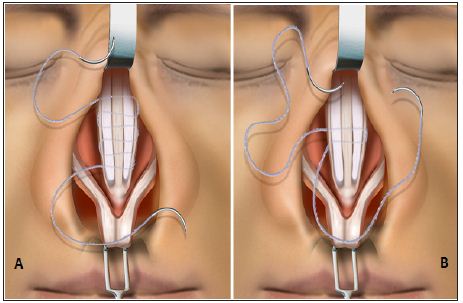
Figure 1: (A) Securing septal cartilage spreader grafts for internal valve repair during rhinoplasty. The first 2 passes with the barbed suture secure and unitize the spreader grafts to the dorsal septum at the transition zone of the bidirectional barbed suture. (B) The spreader grafts are shown completely secured by multiple back-and-forth passes with the bidirectional barbed suture. Reprinted with permission of the manufacturer. ©2013 Angiotech Pharmaceuticals,™Inc.

Discussion

Internal valve collapse in rhinoplasty often requires septal or conchal cartilage for reconstruction by placement of the cartilage graft between the dorsal septum and the upper lateral cartilages. This improves internal valve patency and assists with airflow in patients suffering from internal valve collapse. Following cosmetic rhinoplasty, it also allows for improvement in dorsal aesthetic lines and can be used to straighten a deformed dorsal septum. It can also before provide lengthening of short noses via tongue-in-groove septal extension techniques. The fixation of spreader grafts has often been performed with interrupted horizontal mattress sutures of monofilament, permanent or absorbable materials. It has always been critical to avoid placement of suture material over the dorsal aspect to minimize the palpability or visibility of sutures and to allow additional dorsal contouring at the conclusion of the procedure. The placement of multiple interrupted sutures along the length of the graft is oftentimes made technically more difficult with knot tying in the limited space, even in open rhinoplasty techniques. It is for this reason that I began using absorbable barbed suture for fixation of cartilage spreader grafts for internal valve reconstruction.

Conclusion

The use of barbed suture for cartilage graft fixation in internal valve reconstruction provides a more secure, unitized fixation with continuous suture distributing tension forces throughout the entire graft site. The process is quick, with an easy learning curve. It reduces the time needed to knot interrupted sutures. In addition, it has eliminated mobility of cartilage grafts and, therefore graft migration or distortion. It has become my exclusive technique for spreader graft fixation in reconstructive and cosmetic rhinoplasty. I believe additional novel uses of barb suture technologies will help evolve additional new techniques to help surgeons reduce operative time, reduce cost, improve soft tissue fixation, and achieve better results.

For More Articles: Biomedical Journal Impact Factor: https://biomedres.us

Journals on Plant Pathology

Survey of Chilli Leaf Curl Complex Disease in Eastern Part of Uttar Pradesh

Introduction

Chilli (Capsicum annuum L) is one of the most valuable cash crops of India. It is a common and widely cultivated spices crop almost all over the world. Chilli is a richest source of vitamin C and A Howard [1].The chilly fruits are small in size and known for their sharp acidic flavor and colour. At present, chilly is produced in India about 1260.1thousands metric ton from an area of 792.1 thousands hectare, Anonymous [2]. Andhra Pradesh is the largest producing state of chilly. Indian chillies are mostly exported to Sri Lanka, USA, Nepal, Mexico, Malaysia and Bangladesh. Chilli suffers from a large number of viral, fungal, bacterial, nematode and phytoplasma diseases. Viruses is known to cause different symptoms like mosaic, ring spot, curling, yellowing etc. on chilly and these symptoms result heavy economic losses of about 15billion US Dollar per annum worldwide Van Fanbing [3].

Among them, chilly leaf curl is very common and affected to entire plants in the field with variable symptoms. It has been observed to cause high disease incidence with showing the symptoms of leaf curling, puckering and reduced size of leaves, closely set internodes and dwarfing of plants. These symptoms produce witch broom appearance and causes to failure of fruits setting. The fruit sets usually small and deformed. However, there are no published reports on the distribution of leaf curl viral diseases in major chilly growing areas of eastern Uttar Pradesh viz; Faizabad and sultanpur districts although, it is important disease of chilly crop.

Material and Methods

The design adopted for the survey was stratified multistage sampling in two districts namely Faizabad and Sultanpur. The districts were classified into three blocks based on number of village surveyed. The survey was confined to five village of each block and villages were selected at random. These survey villages were visited for two consecutive years 2014 and 2015 from planting stage to harvesting stage i.e. 3rd week of March, April and May. Data on the total number of plants, number of virus infected plants in per square meter. The days after disease appearance and visual disease incidence scoring method was adopted 0-9 point scale, Percentage of disease incidence was obtained by standard methods Joshi and Chaudhry [4] Other viral disease symptoms on chilly plant in the surveyed area were seen and collected separately for further confirmation through visual observation with consultation of standard literature McRae et al. [5-7] Per cent disease incidence was calculated given by Joshi and Chaudhry (1981) [4] as under:

Equation 1:

1. Results and Discussion and Discussion

Field survey were conducted from 2014 and 2015 in 30 villages and total 150 fields in major chilly growing area of Faizabad and Sultanpur districts of eastern Uttar Pradesh. The 1. Results and Discussion of survey revealed that most of the fields were found more than one viral infected symptoms. The virus infected leaf sample were collected and visual identified as leaf curl, mosaic mottle, puckering, yellowing, leaf rolling and distortion mosaic with the standard literature cited by Paul et al. [8], Puttarudriah [9] and Muniyappa [10]. The infection of chilly leaf curl virus was found in almost all fields with 36.86 to 67.70 % in 2014 and 25.53 to 67.39 % average disease intensity in 2015. The highest leaf curl incidence was noticed in Sewra (67.39 %) followed by Hasuimukundpur (65.22%) and Etwara (64.07%) in 2014, whereas in 2015, it was highest in Sewra (61.44%), Hasuimukundpur (59.79%) and Isawli (57.07%).

The lowest leaf curl incidence was noticed in Ahran (25.53- 30.30%) Sholapur (36.83%-36.04%) and Isawlibhari (37.33%- 31.89%) in 2014 and 2015, respectively (Table 1) When the leaf curl disease incidence data was analyzed at block wise. The maximum incidence was found 59.53, 50.79% at Amaniganj and 54.70, 52.91% at Kurwar in 2014 and 2015, respectively. While the least incidence was observed at Milkipur 47.46 % in 2014 and 41.48% in 2015 (Table 2) The average leaf curl disease incidence was observed highest in 2014 at both districts viz., Sultanpur (54.76%) and Faizabad (51.72%) due to favorable environmental condition was found for the growth of white fly, which is a key vector for the transmission virus. The similar finding was reported by Gupta et al. [11] and Meena et al. [12] The leaf curl disease incidence was recorded lowest in March and highest in May at both the year because of less inoculums were persist in the field during the March, when plants are in growing phase but inoculums were consequently perpetuate due to build up of white fly population during April and May and transmit to another Plants for its infection. The observations have been supported by Iqbal et al. [13] and Navot et al. [14] (Table 3).

Table 1: Survey of chilli leaf curl disease incidence during the growing period of 2014 and 2015 in different villages of eastern UP.

Table 2: Incidence of chilly leaf curl virus in various block of Sultanpur and Faizabad districts during 2014 and 2015.

Table 3: Incidence of chilli leaf curl virus at district level during the different growing periods of 2014-2015.

During the survey, different types of viral symptoms were observed in all blocks, the natural of symptoms on chilly plants were collected and grouped. The grouped symptoms were identified and calculated per cent disease severity separately. The symptoms of the viral infected chilly plants were found clearing of veins of the apical leaves, followed by dark green to light green mottling, mild to severe mosaic, necrotic streaks on the vein and petiole with stunted and bushy appearance were identified as necrotic leaves Talukdar et al. [15]. The less number of branches with reduced length of root was considered as mottle virus Yadav et al. [16]. Wavy midrib, upward curling, reduced leaf area with vein clearing and banding was grouped in mosaic mottling. The characteristic field symptoms were considered for leaf curl is upward curling; puckering and reduced size of leaves with severely affected plants was stunted and produced no fruit Senanayake [17,18] The viral severity was estimated based on the number of plants found characteristics symptoms of each virus (Table 4). Infection of leaf curling was found severe in all blocks. Whereas as mosaic mottling virus was severe in Dhanpatganj and leaf rolling was in Baldirai. Other disease symptoms were recorded moderate to mild infection.

Table 4: Types of viral symptoms in different blocks of eastern Uttar Pradesh during crop periods of 2014 and 2015.

***Severe incidence, **Moderate Incidence and *Mild Incidence.

Conclusion

An overall survey revealed that chilly leaf curl virus was found all surveyed field in continuous growing period. It is due to growing of susceptible local cultivars prevailing in the districts as for multiplication and spread of virus.

For More Articles: Biomedical Journal Impact Factor: https://biomedres.us

Journals on Agriculture

An Overview on the Use of Enhanced Efficiency Nitrogen Fertilizers in Irrigated Mediterranean Agriculture

Abstract

In Mediterranean regions climate change has led to greater inter and intra-annual rainfall irregularity, causing an increase in the demand for irrigation water, inevitably accompanied by an increase in the use of others resources like nitrogen fertilizers. Nitrogen is associated with negative economic and environmental impacts, which requires particularly important decision-making in terms of its application to meet crop needs. The agronomic efficiencies of water and nitro genvary widely among different environments and can be modified due to management practices. However, some studies suggest that agronomic practices alone are not sufficient to entirely avoid nitrogen losses. In irrigated cropping systems, where classical nitrogen fertilizers are partially applied through irrigation water, Enhanced Efficiency Fertilizers (EEF), namely slow release and controlled release products, and nitrification inhibitors, have the potential to reduce nitrogen losses, contributing to higher resource-use efficiencies. This work aims to provide an overview of the possibilities of successfully using this type of fertilizers in irrigated agriculture, especially in Mediterranean climate regions, and the need for research in this field.

Keywords: Irrigation; Fertilization; EEF; WUE; NUE

Abbreviations: Cs: Temperate climate with dry Summer or Mediterranean climate (according to Köppen classification); EEF: Enhanced Efficiency Fertilizers; NUE: Nitrogen Use Efficiency; WUE: Water Use Efficiency

Agriculture faces two great challenges:

i. The need to meet the growing world food demand,

ii. While decreasing agriculture’s global environmental footprint [1,2]. At the same time, the agricultural sector must strength itself to face the risks associated with climate change, particularly in susceptible regions where water irregularity and scarcity is a key issue [3-7].

In regions with a Mediterranean climate there is typically a large variability and irregularity in the distribution of annual and inter-annual rainfall with a mild wet season trough autumn and winter months and a dry season in the summer months. The balance between the key climate variables that characterize Mediterranean or Temperate climates with a dry Summer (Cs, according to Köppen classification)and the development period of a large number of agricultural plant species implies that the success of the agricultural sector in Mediterranean regions and other regions with water availability constraints depends to a very large degree on proper water management, suitable fertilizer applications, as well as on the success of irrigation implementation [8,9]. With the development of large irrigation schemes in these regions farmers are gradually resorting to irrigation, resource-use is growing and a profound change of the agricultural landscape is taking place within new irrigation areas. In fact, as a result of agriculture intensification, a growing demand for irrigation water has been inevitably accompanied by an increase in the use of others resources, like nitrogen fertilizers. In addition, there is a growing pressure on farmers to increase the cropping systems efficiency, and often doing it without the knowledge and the necessary assistance for the adoption of the best strategies and practices in a changing agriculture [10].

Nitrogen is the fourth most abundant nutrient in plants and nitrogen fertilization is needed because soils normally have insufficient nitrogen to meet the crops needs. The requirements of nitrogen by crops vary throughout their development cycle. The effect of the application of nitrogen depends very much on the crop development stage. If nitrogen is supplied to the crops in a fractional and gradual way, it will be more completely metabolized, minimizing the occurrence of soluble forms such as the nitrate ion (NO3-) [11]. Water availability and nitrogen supply to crops are factors in close interaction, affecting plant growth and their productive responses. Suitable nitrogen fertilization promotes leaf area and vegetation cover expansion, thus increasing the evapotranspiration efficiency of plants [12].For the large majority of cultivated plants, adequate nitrogen supply is required to achieve high yields, but negative effects from improper nitrogen fertilizer use threaten environmental quality and human health at both local and global scales as a result of water pollution from nitrate leaching or runoff, air pollution and greenhouse gas emissions [13].

Two of the major losses of nitrogen in the soil are due to gaseous losses denitrification and leaching. Denitrification losses contribute to global warming and the destruction of the ozone layer. Leaching losses affect the quality of soil, water and can interfere with human and animal health. Agriculture is considered to be the main source of contamination of the waters with nitrogen, accounting for more than half of the nitrate ion losses to groundwater [12,13]. It is suspected that the ingestion of high levels of nitrate, both in water and in food products rich in nitrates, may be associated with the formation of carcinogens of the nitrosamines type, and lead to the onset of cancer in the digestive tract. In young children and in ruminants, the consumption of water or foods rich in nitrates may also lead to their conversion into nitrite ion by bacteria in the stomach [12]. Thus, although there are doubts about the real risks to human health of nitrate ion intake, legislation in European Union countries, like Portugal, imposes a maximum nitrate ion content in drinking water of 50 mg of NO3-/dm3 [14].

Crop yield response to water and nitrogen vary widely among different environments, and they can be shifted due to technological, environmental, or economic factors [9,13]. In order to meet the needs of crops and improve water use efficiency (WUE) as well as nitrogen use efficiency (NUE) of the cropping systems it is necessary to find a compromise in management between the quantitative and the qualitative aspects [15]. A wide range of fertilizers, known as Enhanced Efficiency Fertilizers (EEFs), are commercially advertised as improvers of nitrogen use by crops. These kinds of fertilizers, that delay the bioavailability of nitrogen in the soil, matching its release with the crops higher needs periods, are classified as:

i. Slow-release fertilizers (obtained as condensation products of urea and urea aldehydes);

ii. Controlled-release fertilizers (products containing a conventional fertilizer whose nutrient release in the soil is regulated by sulphur or/and polymer coatings);

iii. Stabilized fertilizers (which are modified during the production process with a nitrification inhibitor). Several studies have shown that the use of such fertilizers has been successful in conditions of high rainfall and in sandy soils [16].

Irrigation is a major factor that influences N leaching [17]. In irrigated crops, where classic nitrogen fertilizers are partially applied through the irrigation water, EFFs have the potential to reduce leaching risks, thereby contributing to the increase of resource-use efficiency, both by promoting higher yields and reducing water and fertilizer inputs. For example, higher yields were found in irrigated wheat with a controlled release nitrogen fertilizer when compared to conventional split applied urea throughout the crop growing cycle [16]. Despite its large apparent advantages, the use of EEFs in agriculture has been lower due to doubts regarding the cost / benefit ratio and the insufficient demonstration of its advantages over conventional fertilizers [18,19]. Taking all this in consideration, finding both optimal irrigation and nitrogen application strategies, that is, finding a dynamic balance between these factors, adapting them to the very specific needs of each crop, it’s a significant contribution not only to maintain soil and groundwater quality but also to preserve food safety and agriculture sustainability. For this purpose, more research on this topic should be carried out in order to study the best management options in the use of EEFs, especially in the most important Mediterranean irrigated crops.

For More Articles: Biomedical Journal Impact Factor: https://biomedres.us

Open Access Journals on Medicine

Brown tumour revealed: A literature review with a case study

Introduction

This disorder, called brown tumour, has its determinant a metabolic bone disarray caused by the excess production of parathyroid hormone, which is produced by the four parathyroid glands located posterior to the thyroid gland, which are responsible for the control of phosphorus (P), calcium (Ca) and vitamin D; The level of calcium present in the blood is considered a triggering factor of the disorder by the release and production of this hormone. Shetty “the hyperparatiroidism HPT is a disease in which there may be a complex, of biochemical anatomic and clinical abnormalities”. Therefore the Brown Tumor has its histopathological complexity described as multiple adenomas, numerous osteoclastic cells, of cystic format, and separated by a highly vascularized tissue which will confer the lesion a dark red or brownish coloration which characterizes its nickname. Some of the signs and symptoms observed in this disorder are fatigue, nausea, weakness, anorexia, excessive thirst, polyuria, constipation, pain, swelling and frequent urination. In severe cases, may present kidney stones, loss of bone mass and fractures, mental confusion and consequent depression.

The systemic scenario begins when the body presents in hypocalcemia, and in the other hand will be inhibited in hyperalcemia, it will be stimulated the uptake of calcium to the extracellular environment, which leads to the increase of serum calcium concentration and decrease of the phosphate ion which is responsible to transport the phosphorous. Two sites of action in bone metabolic disorder are recognized, which are described respectively as actions on kidneys that reduce phosphate absorption and increase calcium absorption and the action on bones that stimulates bone mobilization with increased serum calcium concentration. Another important aspect and the issue of vitamin D which is a regulator of osteo mineral calcium physiology, vitamin D is used in the treatment of the secondary form of the disorder as supplementation. The disorder may be divided into primary, where the treatment will be surgical removing the lesion, and secondary where the basic disorder that will affect the normal functioning of the parathyroid glands and vitamin D supplementation should be treated.

Literature Review

Brown tumor of Hyperparathyroidism it is a metabolic disorder that can affect the entire skeleton. This disorder is named due to macroscopic aspect to the intraossic lesions, presenting reddishbrownish hue, due to the intense bleeding that occurs in these lesions and the deposit of hemosiderin. Commonly, this disorder is a result of a framework of primary hyperparathyroidism, secondary rarely and tertiary. Brown tumor arises only in chronic cases of non treated Hyperparathyroidism (PTH). The lesions are caused by the increased production and release of PTH, leading to increased re absorption of calcium in bones. With the constant calcium sequestration l, it will start to grow a repair tissue, which increases gradually in size [1,2]. This disorder usually rise in individuals who are in third, fourth and sixth decade of life, especially in females. The most affected is in the head and neck area especially in mandibular area [1,2]. Clinical manifestations vary according to the location and extent of the Tumour. In the mandibular bone, there may be pain and hard tumefaction. When extensive it can lead to deformation of the region, impairing the function of the bone [1,2].

HT’s symptoms are observed including fatigue, weakness, nausea, anorexia, excessive thirst, polyuria, constipation and depression [1,2]. The diagnosis are achieved through radiological exams, computerized tomography scans and Histopathological examination of the Tumour. Histopathologically the Brown Tumor is characterized by a soft tissue mass composed of giant cells inside of a fibro vascularstrome, like cystic spaces have queued for connective tissues. The focus of hemorrhage and hemosiderin deposition appears as a reliable reddish-brown mass, a characteristic that gave rise to your name [3,4]. It resembles, yet histopathologically to other inside giant cells, as the central giant cell lesion and cherubism. It is important to analyze the history of the disease and assessment and laboratory tests for determination of definitive diagnosis. The identification of the lesion, through laboratory findings (Hyperparathyroidism) rule out the existence of central giant cell lesion, featuring, in its way the presence of Brown Tumour [4]. Radiographically, the Brown Tumor is characterized as a lytic lesion, expansive, well defined, involves the bone cortical and may present a fine mesh of intralesional steroid septa, which gives the condition a multi side appearance, and may also be uniside.

The Brown Tumour generally shows multiple occurrences, although it can also occur as a single one. When present in the mandibular bone, it usually appears at the hard blade [5,6]. In addition one could observe bone trabeculae neo formed with osteoblasts tumefactions on its edges. Blood tests indicate the increased levels of calcium and alkaline phosphates, as well as the reduction in the level of phosphorus is useful for finding the correct diagnosis [1,2]. The Brown Tumor does not demand specific treatment in most cases because the correction of Hyperparathyroidism leads to the lesion disappearement. However, it can cause pathological fractures and bone marrow compression even when it involves the spine. When it reaches the face can cause breathing difficulties and facial deformities. In such cases, it is recommended that the patient undergoes surgical treatment [7-9].

According to radiographs and Tomography scans: it shows a radiolucent / hypodense diffuse, irregular image without bulging of the vestibular and / or lingual bone cortices was verified. Radiographic and CT scans did not show radiopaque halus, that is, descorticalized, involving the region of the element 36. Bone resurfacing in the furcation region in the same element. In computed tomography (CT), the medullary bone presents a granular aspect with bone and lingual cortical thinning, vertical bone loss located in the mesial and distal part of the 36 element, and increased ligament space with absence of hard lamina, apparent mobility, initial external root resorption in the Element mentioned above. Hypothesis: Bone tumor, endo-periodontal envelopment or giant cell lesion. According to the laboratory test of Parathyroid hormone (PTH), by the enzyme immunoassay method by chemiluminescence with a result of 502.8 pg/ml (normal 12 to 65 pg/ ml), creatinine level is high with 2.94 mg/dl (Normal for male patients is 0.70 to 1.30 mg/dl). Histopathological examination by Hematoxylin and Eosin staining method with diagnosis of Brown Tumour (osteoblast matrix with prominent osteoblasts and osteoclasts, amidst fibrous stromal fibroblastic proliferation: proliferated, ecstatic and congestive vessels, haemorrhage and hemostasis).

The parathyroid glands were increased in cervical ecography. The findings of hyperparathyroidism were confirmed together with other exams to the diagnosis of Brown Tumour of Hyperparathyroidism. Patient was submitted to surgical intervention under general anaesthesia, naso-tracheal intubation, vestibule access in the region of the left jaw body, enucleation of the lesion, removal of the element 36, curettage and synthesis of the region. All material was collected and sent to the pathology center of the State University of Rio de Janeiro (UERJ) for histopathological analysis where the diagnosis of Brown Tumour of Hyperparathyroidism was confirmed. Post surgery evolution of the patient in question was compatible with the surgical procedure and without intercurrences.

Case Report

Patient male, 35 years W.L.M. R, Brazilian, born in Rio de Janeiro, attended the Oral clinic and traumatology maxillofacial surgery at Municipal Lourenço Jorge Hospital, Barra da Tijuca, capital of Rio de Janeiro. During the anamnesis reported as main complaint “volume increase in unilateral mandibular region being the left side, painful. Current history of the disease (HDA) with account of start approximately 2 (two) years, as a minor injury to the gums, which evolved gradually, resulting in lumps region mentioned above. Still reported that does not have habits like smoking and alcoholism, drug hyper sensibilities, morbid antecedents denies personal and family. Clinical and physical examination showed, increased mandibular unilateral left significant volume, pain and mobility the palpation of the 46 element associated with bulging Protuberance in the abovementioned causing bone, with absence of fungi signs and infectious in the oral cavity (Figure 1).

Figure 1: Clinical and physical examination of patient.

In the dental restorations, specific resin systems are applied to the damaged tooth area to form a cast restoration that is then heat-treated using special ovens under controlled laboratory conditions. The ideal temperature for heat treatment application depends on the thermal behavior of each composite, such as glass transition temperature (Tg) analysis and initial degradation temperature [7]. The Tg can successfully be used as a reference to sign the ideal heat treatment for photo-irradiated resin composites. Above Tg, the secondary molecular interactions are weakened and, as a consequence, material properties are optimized once trapped radicals are given the opportunity to react [8-9]. In addition, the maximum temperature for heating without damaging, i.e., initial degradation temperature, needs to be determined to avoid weight loss [10]. In the present investigation, Conventional TG-DTA is a powerful and convenient thermal analysis technique which allows various important physical and chemical transformations such as glass transition and degradations are examined (Figures 1-4). Glass transition temperature (Tg) and number of phase transitions are evaluated for different dental materials are shown in Table 1.

Anatomical assessment: Preoperatively, the median radial tilt was 29 degrees of dorsal angulation in the group and 32 in the Non Spanning Syringe Distractor group. Postoperatively, the median tilt was 8 degrees of volar angulation in the Hoffman II group and 2 degrees volar in the Non Spanning Syringe Distractor group (p = 0.002). At the time of removal of the fixators, there was still a statistically significant difference in radial tilt: 9 degrees of volar angulation in the Hoffman II group and 4 degrees in the. Non Spanning Syringe Distractor group (p= 0.04). At 1 year, the difference was no longer statistically significant. For the other anatomical variables, no statistically significant differences were found (Table 2).

Figure 2: Computed tomography scan.

Figure 3: Diagnostic hypothesis: Bone tumor.

Figure 4: Diagnostic hypothesis: Endo-or giant cell lesion periodontal.

Figure 5: Result of presence of parathyroid harome, by the method of chemiluminescence enzyme Immuno assay.

Figure 6: Brown Tumor diagnosis.

Histopathological examination, by coloring method by hematoxylin and eosin with Brown Tumor diagnosis (with prominent osteoid matrix osteoblasts and osteoclasts in fibrous stroma with fibroblastic proliferation: vases proliferated, entranced and congestion, pockets of deposition of hemosiderin and hemorrhage). Parathyroid glands were increased in cervical ultrasound. The findings of hyperparathyroidism confirmed together with the other tests the diagnosis of Hyperparathyroidism Brown Tumor (Figure 7). Patient underwent surgery under general anesthesia, naso-tracheal intubation, accomplished access in lobby Fund in the region of left mandibular body, enucleation of the lesion, dental extraction of 36 element, curettage and synthesis of the region. All material was collected and forwarded to the pathology Center of Rio de Janeiro State University (UERJ) for histopathological analysis where it was confirmed the diagnosis of Hyperparathyroidism Brown Tumor. Postoperative evolution of the patient in question was compatible with the surgical procedure and without complications (Figure 8).

Figure 7: Result of microscopic examination.

Figure 8: Patient underwent surgery under anesthesia.

Discussion

The Brown tumour is a lesion associated with the hyperparathyroidism and can be divided into primary or secondary. Affects more the mandible than the maxilla and your prevalence is by the feminine gender above 50 years. We can clinically identify a Brown tumour of hyperparathyroidism because of an increase volume in the region causing pain and mobility, presenting as an extroverted mass of slow growth and destructive character. Depending on where the tumour is located, they can cause: diplopia, deformity, bleeding, chewing trauma, among other adversities. Radiographically, we identified this Brown tumour of hyperparathyroidism, due to the presence of a uni or multiocular radiolucent image, descorticalized, besides that when the Computed Tomography was performed, described in the above case, absence of the hard blade was revealed.

Histologically, the Brown tumour of hyperparathyroidism manifests as a mass of soft tissue composed of a giant cells inside the fibro vascular stroma, presenting focus of bleeding and hemosiderin deposition as a friable red-brown mass.

Diagnostic errors can occur if we only evaluate their histological characteristics, since other lesions such as the giant cell lesion present similar aspects to this lesion, thus causing unnecessary radical therapeutics and severe sequels if the diagnosis is incorrectly concluded. . Therefore, it is extremely important to analyze the history of the disease and laboratory tests. On laboratory examination, elevated creatinine level revealed parathyroid gland hyperplasia confirming the diagnosis, in addition with the other exams, of Brown tumour of hyperparathyroidism. The treatment accomplished in the relate of case was surgical with the objective was remove all the tumour and for this reason a naso-tracheal intubation was performed, with your purpose was allow a good surgical access, giving better tranquillity to the surgeons work, besides ensuring that the patient was treated in a single surgical time. Posteriorly, was done the enucleation of the tumour, which was the purpose of the surgery, the element 36 removal, curettage and suture of the region.

Final Consideration

The literature review on the mechanism of pathological action of the brown tumour associated with a clinical case study, it was possible to reaffirm that dentistry is on the correct path both to diagnose the disorder through the diagnostic exams, establishment of differential diagnostic and laboratory exams as long as long-term treatment, not only through aspects closely related to the dental surgeon, but also in the supplementation of vitamins and minerals, it is worth emphasizing that all cases should maintain long-term treatments booking appointments regularly, although cases of Brown Tumour are rare, the treatment was performed with total efficiency.

For More Articles: Biomedical Journal Impact Factor: https://biomedres.us

Medical Journals

We Need To Look At the Comorbidities of Obesity during Childhood and Adolescence

Opinion

Nowadays, almost 50% of boys and girls with ages between 5 and 9 years old are overweight or obese. And these children frequently have one or more comorbidities. Unfortunately, there is a wrong belief that the great problem of obese children is the risk of this child to be an obese adult. In fact, there are a lot of comorbidities that already appears during infancy and, if treated, probably will not progress to adulthood. We have been studying some of these comorbidities from the last 5 years. We studied dyslipidemia and insulin resistance and we found that 69.4% of the children had high cholesterol, 45.2% high LDL, 54.8% low HDL and 53.2% high triglycerides [1]. It is consensual that we have a vicious circle including obesity and insulin resistance [2] and we evaluated 383 children with ages between 7 and 18 years using fasting insulinemia and Homa as indicators of insulin resistance. The prevalence was very high, independently of the method: 33.1% using fasting insulin > 15 and 37.8% using HOMA> 3.5 [1]. We also looked at blood pressure and we showed high blood pressure values among obese children, when compared to eutrophic children [3-5].

Regarding the heart, we investigated if obese children, with ages between 6 and 9 years, has detectable modifications of the heart anatomy, especially regarding to the left ventricle, and we found that, among 5 index evaluated, two of them (the LV mass and the LV3 index) showed differences, with greater thickness of the ventricle among obese children [4]. The intima-media complex of common carotid artery can be accessed using ultrasonography. This is a very safe and accurate method to evaluate the onset appearance of atherosclerosis. We evaluated 59 children of both genders, between 7 and 10 years old [5]. The average thickness of the intima-media complex in the group overweight / obese was 0.49mm; in the non-obese group, the measurement was 0.41mm. There was a significant difference between groups (p <0.01).And, also, we showed a correlation between the increase of the z-score of BMI and the increase of the intima-media complex [6]. The waist circumference is nowadays considered very important for children health [7]. We evaluated the abdominal adiposity distribution [8]. Results from 59 children of both genders, between 7 and 10 years old showed high correlation of fat deposits between each other and the two compartments of abdominal fat deposition increased together. And, even more important, both subcutaneous fat and visceral fat showed almost the same correlation with abdominal circumference.

This means that, at this age, when we measure the abdomen, we are measuring both visceral and subcutaneous fat and this two measurements increase together with the increase of the abdominal circumference [8]. At this same study, we evaluated the liver and a hiperechoic image is an indicator of the presence of Non Alcoholic fat Liver Disease (NAFLD). Children with normal liver tend to have less subcutaneous fat then children with more echoic liver. And, similarly, children with normal liver tend to have less visceral fat then children with more echoic liver [8]. In conclusion, even among school children, we could find a lot of obesity comorbidities, as: dyslipidemia, insulin resistance, high blood pressure, left ventricle hypertrophy, carotid intima hypertrophy, visceral fat accumulation and NAFLD. And we need to treat these children early, to prevent the evolution to adult diseases and to give them a better quality of life.

For More Articles: Biomedical Journal Impact Factor: https://biomedres.us

Open Access Journals on Chemical Sciences

Review on Application of Agro-Waste Biomass Biochar for Adsorption and Bioremediation Dye

Introduction

Biomass-based fuels dominated the world energy market until the middle of the 19th century, when coal and other fossil fuels become increasingly popular. Since the 20th century, just over 100 years, mass production and utilization of fossil energy have caused a series of worldwide energy supply and global environmental and climate problems. Hence the use of sustainable energy sources has been attracting research attention over the last several decades. Compared to the use of other sustainable fuel resources, biomass has the advantage of wide availability and it can be developed everywhere in the world as a local fuel resource. Due to a rapid increase of global population and the fast development of urbanization, about 1 billion metric tons of organic solid wastes are annually generated from world cities, causing great challenges on the management of these wastes Lim, 2016. The major negative effect of burning agricultural wastes is the release of carbon dioxide gas (CO2), which is the most important factor of greenhouse gas production by the human. Carbonization of woody or cellulosic wastes to produce biochar which is a charcoal-like product. It has been suggested by several researchers to avoid negative impacts of the direct burning of woody matter. Biochar is resistant to biological decay and hence preserved in the terrestrial systems for the much longer time. For example, woody residues or compost, therefore, the beneficial effects are extend [1,2].

Carbonization through pyrolysis is to produce biochar is a successful mean to avoid the negative impacts on human health and environment. Biochar is the black solid containing recalcitrant organic carbon (OC), which helps in global warming mitigation [3]. Recently, attention has been paid to the utilization and application of biochar, which is derived from the biomass via incomplete combustion operations such as pyrolysis and carbonization [4,5]. Pyrolysis could be considered one of the fastest ways for depolymerization of biomass macromolecules to smaller molecular fragments. As a stable carbon-rich material, the application of biochar for solving environmental problems can span several categories including: carbon sequestration, soil management, pollution remediation, and agricultural by-product/ waste recycling. Among these uses, biochar as an adsorbent for the removal of environmental pollutants has emerged as a promising technology [6]. Currently, low-temperature pyrolysis is usually applied to convert biomass, typically agricultural biomass, into biochar [7], which helps to avoid organic leaching and secondary pollution from the raw biomass. Meanwhile, the surface area of the biochar usually increases with an increase in pyrolytic temperature at up to 700oF [4].

As such, it was deemed both reasonable, and feasible, to promote the application of biochar for practical decontamination purposes. The residual dyes in the effluent stream from different sources (e.g., textile industries, paper and pulp industries, dye and dye intermediates industries, pharmaceutical industries, tannery, and Kraft bleaching industries, etc.) contains wide variety of organic pollutants is one of the important source of water pollution, ecological problems and health effects of all over the world [8]. The presence of lowest concentrations of dyes in the effluent is highly visible and undesirable in the environment. The effluent also contains the residues of reactive dyes and harmful chemicals. Therefore, such type of wastewater needs to be properly treated before release into the environment. There are several decolorization techniques is reported in which physical, chemical, and biological decolorization technologies available. The physical sorption technique is a most effective and attractive process for the decolorization of dye-containing wastewater [9]. In general, adsorption processes are using a commercial activated carbon which is effective for the decolorization of dyes from contaminated wastewater. The consumption of activated carbon is steadily increasing because of its application in pollution control in the word [10,11]. Adsorption has been considered to be one of the most effective physical techniques for removing contaminants from water because using adsorption methods is inexpensive and easy to manage [12]. Biochar is a carbon-rich product which is produced by combusting biomass, such as wood, manure, wheat straw, wicker, sewage sludge, or leaves, at between 350 and 7000C [13,14] in a closed chamber with insufficient air or no air. Biochar is currently being used in a number of fields, including energy production, waste management, climate change mitigation, water treatment, and soil amelioration [4]. 2 Using biochar as an adsorbent has been found to offer great potential for removing organic contaminants from water [15]. Recently, works have been done on the methylene blue adsorption by biochar. Delwiche, 2014 showed that the use of 1 kg/m2 pine chip biochar could decrease the leaching of methylene blue from a homogenized soil column by 52% [16]. Found that the adsorption of methylene blue on Biochar of Casuarina seeds involved the mechanism of the weak H-bond with hydroxyl groups and also concluded that p-p electron donoracceptor and steric effects play important roles in adsorption. [17] Revealed that the biochar prepared from agro wastes when added into soil could increase the sorption of Casuarina seeds; however, the reaction mechanism relating to the soil characteristics was not well interpreted. Biochar from different sources have different properties and different adsorption characteristics. The mechanism of adsorption was examined by characterizing biochar prepared from Casuarina seeds agro wastes. The choice of these feed stocks depended on the availability and ease of access in India [18]. The feed stocks were selected with the aim of utilizing droppings and vegetable wastes produced in agricultural areas. The physicochemical properties of these biochar were systemically characterized in relating to their adsorption behavior. The effects of key parameters (initial methylene blue concentration, temperature, and pH) on the adsorptive removal of methylene blue by the biochar mentioned above were evaluated using batch experiments and compared. Although a number of low-cost adsorbents such as: natural materials, agricultural, and industrial, wastes are capable of removing methylene blue efficiently [19-21].

Acknowledgement

The authors are thankful for Department of Chemical Engineering and Technology, IIT (BHU), Varanasi for his valuable guidance through the experiments. Authors also acknowledge Project Varanasi for the financial support.

For More Articles: Biomedical Journal Impact Factor: https://biomedres.us

Open Access Journals on Orthopedic

Non-Spanning Syringe Distractor: An Alternative Technique for Fracture Distal End Radius Fixation for Rural Areas of Developing Countries with Limited Resources

Introduction

Since their description by Abraham Colles, Prof of anatomy and surgery of Trinity college of Dublin in 1814, distal radial fractures remain a therapeutic enigma [1]. In describing patient outcomes of traditional non operative treatment, Colles stated “one consolation only remains, that the limb will at some remote period again enjoy perfect freedom in all its motions and be completely exempt from pain [2].” However, as recent studies emphasize outcomes using patient functional outcome measures, it has been shown that not all patients with distal radius fractures that have been treated non-operatively have had the excellent outcomes as first described by Colles. Unstable distal radius fractures are those which are mechanically prone to re-displacement after closed manipulation resulting in collapse and articular incongruity. Several factors have been associated with the instability [3].

a. The initial displacement of the fracture especially radial shortening.

b. The age of the patient due to osteoporosis.

c. The extent of metaphyseal comminution.

d. The amount of intra-articular comminution and steps.

So if treated with K-wires incorporating cast only, often results in permanent deformity, pain and loss of function [4]. There are various external fixation devices described for this. Often orthopedic surgeons from rural areas of developing countries are found hesitant to use it for the various reasons described below.

Cost factor

a. More operative time.

b. Limited anesthetic facilities.

c. Elder age with comorbid conditions.

d. Low socio economic class with low functional requirements.

e. Iatrogenic complication like infection and stiffness.

f. Associated other injury or fracture requiring priority treatment.

However, Non-Spanning Syringe Distractor provides an alternative option with comparable functional outcome.

Material and Methods

A total number of 32 cases of distal radius fracture treated by Non-Spanning Syringe Distractor from 2015 to 2016 admitted in Primary Level Hospital, India. Ethical committee approval was obtained prior to initiation of the study. All fractures were classified as per the AO classification [5]. Presence of distal ulnar fracture was recorded separately. Our study group was compared with a historical control group of 30 (mean age 61) patients who were operated for Hoffman II type of non-bridging external fixator (Figure 1) between 2015 to 2016 in India and followed up for 1 year by the same author but at the Tertiary Level center.

Inclusion criteria

a. Displaced unstable comminuted fracture of the distal radius, which was defined as any distal radial fracture with more than 20° of dorsal angulation, metaphyseal comminution with or without intra-articular extension, and more than 10 mm loss of radial height

b. Fresh fracture (reported within 7 days of injury)

c. Age >50 years (elder population)

d. Informed consent for operative care.

Exclusion criteria

a. Open fracture

b. Pathological fracture

c. Fractures where adequate reduction was not achieved on operative table

d. Injury severity score (ISS) of >17

e. Ipsilateral upper limb pathology which would affect the functional outcome e.g. arthritis, scaphoid fracture.

Non Spanning Syringe Distractor Technique

All surgeries were performed under regional anesthesia. Once reduction was achieved by ligament taxis, usually 2-3 K-wires (size 2 or 2.5 mm) were used for fixation. Then maintaining the stability at fracture, K-wires were bended carefully by wire bender up to an angle perpendicular to radial shaft. Proximally 1 or 2 threaded K-wires (size 3mm) inserted in distal radial shaft in dorso-lateral plane 4-5 cm proximal to fracture level depending upon fracture pattern and quality of bone. Distal ulno radial trans-fixation k wire if used for distal radio ulnar joint instability can also be used for distraction purpose if provides enough length. Length of K-wires were cut approximately 4-5 cm away from the skin making them blunt for fixation inside syringe. Under the image intensifier, the required distraction was applied across the wires, and approximate length judged by scale for hole making in syringe. A 5 or 10 cc plastic syringe used as distracter after drilling holes in one plane by similar sized K-wire over T handle. This makes opposite surface of holes as shield covering blunt cut ends of K wires. All the patients were given below elbow plaster (slab) applied meticulously by cotton bandage and soft paddings (Figure 1).

Figure 1: Technical diagram of (a) Hoffman II construct (B) Non Spanning Syringe Distractor.

An active range of motion at hand, elbow and shoulder was encouraged. Most of the patients could hold a cup of tea within next day of surgery and felt comfortable with the fixator. Oral antibiotics were given for 5 days. Slab was substituted by crape support at 4 weeks in all the patients to enhance early active wrist mobilization. Syringe distracter was removed at 6th to 7th week of surgery depending on the clinical and radiological signs of the union. Check X-rays were taken on 2nd week of the fixation, at the time of removal of the fixator and on subsequent follow-ups at 3 and 6 months and 1 year. Radiological and clinical outcomes were compared with control group of Hoffman II fixator. The total upper extremity function was assessed by self-evaluation, using an American translation of the original German DASH ranging from 0 (no disability) to 100 points (full disability).We recorded the degree of pain using VAS (Visual Analogue Score). Our study group outcome were accessed by Gartland and Werley score and compared with various standard case series of different modality for similar fracture [6] (Figures 2 & 3).

Figure 2: Case 1 of Non Spanning Syringe Distractor.

Figure 3: Case 2 of Non Spanning Syringe Distractor.

A sample size calculation showed that in order to show a difference with a 5% significance level and 80% power, 30 individuals in each group would be needed for radial tilt as outcome, while more than 8,000 individuals would be needed with inclination as the outcome. The mean differences, 95% confidence intervals and p-values for comparisons of the mean values at pre and postoperatively, at time of removal at 6 week and at 1 year follow-up when evaluating anatomical measures and at 6 week, 3 month, 6 month and 1 year follow-ups when evaluating functional measures. We considered p-values less than 0.05 to be statistically significant.

Results

Out of 32 one patient died and one lost follow up reducing sample size to 30 same as comparison group. The mean age of patients treated was 62 years as compared to 61 years of control group. The patients were predominantly males (60%) in both groups. The dominant hand was injured in 21 (70%). Domestic falls followed by road traffic accidents were the predominant (>80%) modes of injury. Fractures belong to A3 or C1-3 class according to AO classification (Table 1).All the fractures were united within 3 months. 4 patients had pain mainly due to prominent ulnar styloid secondary to malunion or DRUJ instability. Wrist and finger pain and stiffness significantly improved after physiotherapy except in one due to Reflex Sympathetic Neuropathy (RSN). One out of two diabetic patients had developed pin track infection, which was healed subsequently. No one developed radial neuritis.

In the dental restorations, specific resin systems are applied to the damaged tooth area to form a cast restoration that is then heat-treated using special ovens under controlled laboratory conditions. The ideal temperature for heat treatment application depends on the thermal behavior of each composite, such as glass transition temperature (Tg) analysis and initial degradation temperature [7]. The Tg can successfully be used as a reference to sign the ideal heat treatment for photo-irradiated resin composites. Above Tg, the secondary molecular interactions are weakened and, as a consequence, material properties are optimized once trapped radicals are given the opportunity to react [8-9]. In addition, the maximum temperature for heating without damaging, i.e., initial degradation temperature, needs to be determined to avoid weight loss [10]. In the present investigation, Conventional TG-DTA is a powerful and convenient thermal analysis technique which allows various important physical and chemical transformations such as glass transition and degradations are examined (Figures 1-4). Glass transition temperature (Tg) and number of phase transitions are evaluated for different dental materials are shown in Table 1.

Table 1: AO classification of fractures.


Anatomical assessment: Preoperatively, the median radial tilt was 29 degrees of dorsal angulation in the group and 32 in the Non Spanning Syringe Distractor group. Postoperatively, the median tilt was 8 degrees of volar angulation in the Hoffman II group and 2 degrees volar in the Non Spanning Syringe Distractor group (p = 0.002). At the time of removal of the fixators, there was still a statistically significant difference in radial tilt: 9 degrees of volar angulation in the Hoffman II group and 4 degrees in the. Non Spanning Syringe Distractor group (p= 0.04). At 1 year, the difference was no longer statistically significant. For the other anatomical variables, no statistically significant differences were found (Table 2).

Table 2: Comparison of radiological assessments, mean (95% CI).

Functional assessment: At 6 weeks, the mean loss of flexion was 24 degrees in the Hoffman II group and 34 degrees in the Non Spanning Syringe Distractor group (p = 0.001). At the other times, the differences between the groups were not statistically significant. There were no statistically significant differences between the groups concerning loss of extension, radial and ulnar deviation, supination, or pronation at the different times (Table 3). There were no statistically significant differences in mean values of the VAS score between the groups at any time (data not shown). At 1 year, the mean (CI 95%) DASH score was 9 (3–14) in the Hoffman II group and 13 (8–20) in the Non Spanning Syringe Distractor group. According to Gartland and Werley score, 24(80%) excellent to good and 6 (20%) fair to poor results were achieved by Non Spanning Syringe Distractor. These results are also compared with different other landmark studies (Table 4).

Table 3: Comparison of functional assessments by mean (95% CI) loss of movement (in degrees) in the injured wrist compared to the uninjured wrist.

Table 4: Comparison of various study results (functional).

Discussion

The fracture of distal end radius is the most common fracture we treat. Management of fracture distal end of radius is still a challenge for orthopedic surgeon and pose therapeutic problem in term of reduction of fracture, maintenance of reduction till the fracture unites mobility of the joint after fracture union. Moreover outcome of these fractures is not uniformly good regardless of treatment instituted. We agree with D.L. Fernandez et al that a good functional result usually accompanies a good anatomical reduction [7]. Collapse, loss of palmar tilt, radial shortening, and articular incongruity is frequent after closed treatment of unstable and comminuted intra-articular fractures of the distal radius and these often results in permanent deformity, pain, and loss of function [8]. Hence, skeletal fixation to maintain the reduction has been recommended [9]. The incorporation of transfixing Kirschner wires (K-wires) within the plaster or use of external fixation is being used for comminuted fractures. Many external fixation devices are described to achieve reduction and fixation of the fragments without loss of position and acceptable functional results [10]. Although the first description of an external fixator in the treatment of forearm fractures by Ombrédannein 1929 was of a non-bridging device, standard technique involves and current texts describe bridging constructs that cross and necessarily immobilize the wrist during the bony healing process [11]. The ligamentotaxis is the basic principle used by external fixation [12]. Prolonged rigid immobilization of the wrist in spanning external fixator leads to decreased blood supply to bone and soft tissues and causes periarticular fibrosis. This leads to osteoporosis, poor motion, and compromised functional outcome.

The early mobilization of the wrist leads to normalization of blood supply, hastened functional recovery, earlier resolution of wrist swelling, and decreased joint stiffness [2,4,8]. The dynamic external fixators have also been developed to provide mobilization of the wrist while reduction and fixation are maintained [13]. One such fixator was first designed by Penning (1990) [14]. The device allows wrist flexion by a hinge joint, with the center of motion being at the capito-lunate joint. This is based on several anatomic and biomechanical studies by Short WH et al. [15]. Our case series attributes to 24(80%) excellent to good and 6(20%) fair to poor results, which are quite comparable with all other landmark studies. No statistically significant difference found in radiological and clinical variables or DASH scores between control Hoffman II fixator group and study group of Non Spanning Syringe Distractor. Thus we believe that Non-Spanning Syringe Distractor is an alternative mechanical device especially for those who are prompted to not to use external fixator because abovementioned reasons. It combines the advantages of pins only and wrist spanning external fixator preventing their disadvantages at the same time.

Conclusion

Finally we would like to conclude that Non-Spanning Syringe Distractor is an easy, cost effective and reliable treatment in treating intra articular and unstable extra articular distal end radial fractures where limited resources in rural part of developing country prompt the orthopedic surgeons to opt for more conservative modality of plaster +/-pins and more frequently complicated by collapse and mal-union. We recommend this device as an alternative method to the currently used modalities and not as a superior substitute .We cannot pretend to present well-objectified functional result unless controlled multicenter trial in larger patient group, but can confirm the feasibility of the Non Spanning Syringe Distractor technique.

For More Articles: Biomedical Journal Impact Factor: https://biomedres.us

Journals on Physical Sciences

Thermal Analysis on Acrylic Based Denture Materials

Introduction

Acrylic resins were first utilized as denture base materials. Poly (methyl methacrylate) is the primary base resin used today. Polymerization of poly (methyl methacrylate) may be effected by heating the polymer–monomer mixture in a water bath, by chemical activation at room temperature using a material such as dimethyl-p-toluidine, or by activating the reaction using microwave energy or visible light [1]. Addition polymerization requires the activation of the initiator (benzoyl peroxide) to provide free radicals. Polymerization takes place as the free radicals open the double bonds of the methyl methacrylate, creating a chain reaction where the monomer attaches to polymer free radicals. Barron, Rueggeberg & Schuster [2] stated that the degree of monomer conversion of resin materials is a measure of the carbon double bonds (C=C) converted into carbon single bonds (C–C).

The goal of conventional/ rapid curing of acrylic resins is to completely polymerize the resin without porosity. In the conventional/ rapid curing methods, the monomer molecules are moved by thermal shocks from other molecules, and passively moved due to external heat. In the microwave method, the monomer molecules are moved by internal heat produced by a high-frequency electro-magnetic field [3-6]. This investigation involves structural and chemical changes in different resins by using thermal analysis.

Material and Methods

Three denture base resins namely Acralyn-H, RR Cold Cure material and Quick Ashvin were polymerized using casting method, then samples are used characterized by x-Ray diffraction technique polymerization changes are analyzed by using TG-DTA.

Results and Discussion

The typical X-ray diffraction patterns were obtained in the present investigations for the various compositions as shown in the Fig.1 represents partially crystalline nature with peaks of three phases .The broad peak in the XRD pattern corresponds to the amorphous region of the material. The intensity of the crystalline peak same in all acrylic based resins. It clearly indicates that the crystallinity of acrylic based resin is due to thermoplastic phase. The acrylic based resin exhibits higher percentage shows more amorphous.

In the dental restorations, specific resin systems are applied to the damaged tooth area to form a cast restoration that is then heat-treated using special ovens under controlled laboratory conditions. The ideal temperature for heat treatment application depends on the thermal behavior of each composite, such as glass transition temperature (Tg) analysis and initial degradation temperature [7]. The Tg can successfully be used as a reference to sign the ideal heat treatment for photo-irradiated resin composites. Above Tg, the secondary molecular interactions are weakened and, as a consequence, material properties are optimized once trapped radicals are given the opportunity to react [8-9]. In addition, the maximum temperature for heating without damaging, i.e., initial degradation temperature, needs to be determined to avoid weight loss [10]. In the present investigation, Conventional TG-DTA is a powerful and convenient thermal analysis technique which allows various important physical and chemical transformations such as glass transition and degradations are examined (Figures 1-4). Glass transition temperature (Tg) and number of phase transitions are evaluated for different dental materials are shown in Table 1.

Figure 1

Figure 2: XRD patterns of Acrylic resins.

Figure 3: TG-DTA patterns of DPI_RR Cold cure resin.

Figure 4: TG-DTA patterns of Quick Aswin resin.

Conclusion

a) This method of quantifying degree of polymerization represents a repeatable and expedient analysis of monomer conversion without the use of an internal standard.

b) All resins obtained similar degrees of peaks determined by XRD.

c) Acrylic resin such as Acralyn-H, RR Cold Cure material and Quick Ashvin values for the casting technique, though statistically significantly variation in weight percent with variable glass transition temperature (Tg).

For More Articles: Biomedical Journal Impact Factor: https://biomedres.us

Open Access Journals on Biological Sciences

Synthesis and screening of some novel 1-((5-phenyl-1,3,4- oxadiazol-2-yl)methyl)-3-((piperazin-1-yl)methyl)-1Hindazole

Introduction

Recent drug discovery studies have focused on the design and synthesis of small molecules that have a 1H-indazole nucleus as the core structure and that act as tubulin inhibitors [1]. Drugs that bind to tubulin act by interfering with the mitosis of cells during the M-phase, resulting in mitotic arrest and eventually lead in to apoptosis [2]. Therefore, microtubules are a sensitive target for the development of anticancer drugs. Due to the introduction of vinca alkaloids such as vincristine and vinblastine for the clinical therapy of cancer, 1H-indazole carrying compounds have generated considerable interest [3-8]. A large numbers of synthetic 1H-indazole-containing drugs and clinical candidates have been identified over the past few years Chang and co-workers reported a large number of compounds with 1H-indazole core structure. In addition to the synthesis and evaluation of the anticancer activity of these compounds, they have revealed some SAR and pharmacophore modeling data [4,5,9-13]. Research on 1- and 3-aroylindoles9 showed that 3-substituted 1H-indazole derivatives exhibited significant activity compared with 1-aroyl1H-indazoles and the electronic effects on the 1H-indazole ring were important for activity potency [11].

The oxdiazole chemistry has been developed extensively and is still developing. Presently there are a number of drugs used clinically, which comprise oxadiazole moiety in association with various heterocyclic rings. 1, 3,4-oxadiazoles are biologically active, synthetically useful and important heterocyclic compounds. The synthesis of novel oxadiazole derivatives and investigation of their chemical and biological behavior have gained more importance in recent decades for biological, medicinal and agricultural reasons. Different classes of oxadiazole compounds possess an extensive spectrum of pharmacological activities. Differently substituted oxadiazole moiety has also been found to have other important activities such as antibacterial [12], antimalarial [13], antiinflammatory [14], antifungal [15], anticonvulsant [16], analgesic [17], antimicrobial [18], antimycobacterial [19], anticonvulsant [20], antitumor [21], antimalarial [22], herbicidal [23], vasodialatory [24], cytotoxic [25], hypolipidemic [26] ulcerogenic [27] (Figure 1) and (Table 1).

Figure 1:

Table 1

Experimental Section

Chemistry

Chemicals and reagents used in the current study were of analytical grade. The reactions were monitored by thin layer chromatography (TLC) on Merck pre-coated silica GF254 plates. Melting points were determined using a Mettler Toledo FP62 capillary melting point apparatus (Mettler-Toledo, Greifensee, Switzerland) and were uncorrected. Infrared spectra were recorded on a Perkin-Elmer Spectrum One series FT-IR apparatus (Version5.0.1) (Perkin Elmer, Norwalk, CT, USA), using potassium bromide pellets; the frequencies were expressed in cm-1. The 1 H- and 13C-NMR spectra were recorded with a Varian Mercury-400 FT-NMR spectrometer(Varian, Palo Alto, CA, USA), using tetramethylsilane as the internal reference, with chloroform- CDCl 3 as solvent, the chemical shifts were reported in parts per million (ppm) and coupling constants (J) were given in hertz (Hz). Elemental analyses were performed on a LECO 932 CHNS instrument (Leco-932, St. Joseph, MI,USA) and analyses for C, H, and N were within } 0.4% of the theoretical values.

General procedure for the synthesis of compounds (3)

1H-indazole (1) (2 mmol, 235 mg) was dissolved in 20 ml of ethanol-water (1:1) solution, and formaldehyde 37% (3mmol) and substituted piperazine (2) (2 mmol) were added. The mixture was stirred at room temperature and the reaction was controlled by TLC in benzene: methanol (9:1) and toluene: ethyl acetate: diethylamine (75:25:1).At the end of the reaction, the precipitate was filtrated, dried, and recrystallized using an appropriate solvent. Yield: 45%: mp 179.7 ◦C. IR (KBr) cm-1: ν 3130 (N-H), 3095-2756 (C-H). 1H-NMR (CDCl3): δ 8.10 (bs,1H, 1H-indazole N-H), 7.77 (d, 1H, indole H4 , J = 7.6), 7.36 (d, 1H, 1H-indazole H7 , J = 8), ,6.92- 6.82 (m, 3H, 1H-indazole H2, H5, H6), 3.79 (s, 2H, C-CH2 -N), 3.20 (t, 4H, piperazine H3, H5 , J = 4.8), 2.68(t, 4H, piperazine H2, H6 , J = 4.8). Anal Calc.: C, 77.35; H, 7.35; N, 14.42%, found: C, 78.16; H, 6.94; N, 14.25%

Ethyl2-(3-piperazin-1-yl)methyl)-1H-indazole) acetate(4)

An equimolar mixture of 3-(piperzin-1-yl) methyl)-1H-indazole (3) and chloro ethyl acetate were dissolved in dimethyl formamide solvent and to this reaction mixture anhydrous K2CO3 was added and the reaction mixture was stirred at room temperature (350C) for 8 hours and the progress of the reaction was monitored by TLC using cyclohexane and ethyl acetate solvent mixture (7:3) as eluent the reaction mixture was kept overnight. After completion of the reaction the solvent was evaporated on rota-evaporater. The gummy solid was separated and it was recrystallized from -2-propanolpetrolium ether (800c) solvent mixture. The crystalline solid was found to be -2-(3-formyl-1H- 1H-indazole) acetate. With a yield of 75% and mp 143-1450C.The indole-3-carbaldehyde used in the present studies was purchased from Aldrich Company and was used without any for their purification. Yield 75%, m.p.:143-1450C.

Yield: 55%: mp 185.7 ◦C. IR (KBr) cm-1: ν 3150 (N-H), 3095- 2782 (C-H). 1H-NMR (CDCl3): δ 7.60 (d, 1H, 1H-indazole H4 , J = 7.6), 7.20 (d, 1H, 1H-indazole H7, J = 8), ,6.95-6.85 (m, 3H, 1H-indazole H2, H5, H6), 3.85 (s, 2H, C-CH2 -N), 3.25 (t, 4H, piperazine H3, H5 , J = 4.8), 2.70(t, 4H, piperazine H2, H6 , J = 4.8), 1.29 (t,3H, J=13.2Hz, CH3 of ethyl group), 4.13 (q, 2H, J=13.2Hz, CH2 of ethyl group),. Anal. Calc. for: C, 78.32; H, 7.26; N, 14.42%, found: C, 78.18; H, 6.70; N, 14.15%,

2-(3-((piperazin-1-yl)methyl)-1H-indazole-1-yl) acetohydrazide(5):

A solution of 4 (0.01mol) and hydrazine hydrate (0.015) in ethanol (20ml) was refluxed for 5 hours. The reaction mixtures was cooled and poured in to ice-cold water with stirring. The separated solid was filtered, washed with water and recrystallized from ethanol.

Yield: 50%: mp 180.7 ◦C. IR (KBr) cm-1: ν 3160 (N-H), 3070- 2780 (C-H). 1H-NMR (CDCl3): δ, 7.65 (d, 1H, 1H-indazole H4 , J = 7.6), 7.35 (d, 1H, 1H-indazole H7 , J = 8), ,6.80-6.85 (m, 3H, 1H-indazole H2, H5, H6), 3.80 (s, 2H, C-CH2 -N), 3.25 (t, 4H, piperazine H3, H5 , J = 4.8), 2.70(t, 4H, piperazine H2, H6 , J = 4.8), ,4.28(s,2H,-NH2), ). 4.36 (s, 2H N-CH2-C =O), 4.98 (s,1 H,-N-NH), Anal. Calc. for: C, 78.32; H, 7.26; N, 14.42%, found: C, 78.18; H, 6.94; N, 14.25%.

1-(( phenyl (1,3,4-oxadiazol-2-yl)methyl)-3-(piperazine- 1-yl)methyl)- 1H-indazole 6(a)

A mixture of 2-(3-((piperazin-1-yl) methyl)-1H-indol-1- yl) acetohydrazide (5) (0.01 mol) and substituted carboxylic acid (0.01 mol) was heated at 100-120 oC in presence of excess polyphosphoric acid (PPA) for 4-5 h. After cooling, the mixture was poured into crushed ice, and neutralized with 5% aq.NaHCO3 solution. The precipitated solid was filtered and purified using column chromatography (petroleum ether: ethyl acetate, 9:1).

Yield: 60%: mp 190.7 ◦C. IR (KBr) cf-1: ν 3150 (N-H), 3050- 2750 (C-H). 1H-NMR (CDCl3): δ, 7.65 (d, 1H, 1H-indazole H4 , J= 7.6), 7.35 (d, 1H, indole H7 , J = 8), ,6.80-6.85 (m, 3H, indole H2, H5, H6), 7.35-7.45(m,5H,phenyl group),3.80 (s, 2H, C-CH2 -N), 3.25 (t, 4H, piperazine H3, H5 , J = 4.8), 2.70(t, 4H, piperazine H2, H6 , J = 4.8),Anal. Calc. for: C, 78.32; H, 7.26; N, 14.42%, found: C, 78.18; H, 6.94; N, 14.25%.

1-(( tollyl (1,3,4-oxadiazol-2-yl)methyl))-3-(piperazine- 1-yl)methyl)- 1H-indazole 6(b)

Yield: 58%: mp 195.0 ◦C. IR (KBr) cm-1: ν 3100 (N-H), 3020- 2720 (C-H). 1H-NMR (CDCl3): δ, 7.60 (d, 1H, 1H-indazole H4 , J = 7.6), 7.30 (d, 1H, indole H7 , J = 8), 7.40-7.55(m,4H,phenyl group) ,6.60-6.65 (m, 3H, 1H-indazole H2, H5, H6), 3.60 (s, 2H, C-CH2 -N), 3.75 (t, 4H, piperazine H3, H5 , J = 4.8), 2.50(t, 4H, piperazine H2, H6 , J = 4.8), 2.43(s,3H,-CH3 ),2.40(s,3H,phenyl attached CH3 group) , Anal. Calc. for C, 70.32; H, 7.15; N, 14.20%, found: C, 70.18; H, 6.94; N, 14.10%

1-((chlorophenyl(1,3,4-oxadiazol-2-yl)methyl))-3- (piperazine-1-yl)methyl)-1H-indazole (d)

Yield: 53%: mp 160.0 ◦C. IR (KBr) cm-1: ν 3020 (N-H), 3090- 2710 (C-H). 1H-NMR (CDCl3): δ, 7.10 (d, 1H, 1H-indazole H4 , J = 7.6), 7.20 (d, 1H, 1H-indazole H7 , J = 8), 7.15-7.40(m,4H,phenyl group),6.20-6.15 (m, 3H, 1H-indazole H2, H5, H6), 3.20(s, 2H, C-CH2 -N), 3.10 (t, 4H, piperazine H3, H5 , J = 4.8), 2.20(t, 4H, piperazine H2, H6 , J = 4.8),, Anal. Calc. for: C, 65.32; H, 6.15; N, 12.20%, found: C, 65.18; H, 6.24; N, 12.10%.

1-((Bromophenyl(1,3,4-oxadiazol-2-yl)methyl))-3- (piperazine-1-yl)methyl)-1H-indazole 6(e)

Yield: 51%: mp 165.0 ◦C. IR (KBr) cm-1: ν 3000 (N-H), 3010- 2710 (C-H). 1H-NMR (CDCl3): δ, 7.15 (d, 1H, 1H-indazole H4 , J = 7.6), 7.40 (d, 1H, 1H-indazole H7 , J = 8), 7.05-7.25(m,4H,phenyl group), ,6.10-6.15 (m, 3H, 1H-indazole H2, H5, H6), 3.10(s, 2H, C-CH2 -N), 3.15(t, 4H, piperazine H3, H5 , J = 4.8), 2.20(t, 4H, piperazine H2, H6 , J = 4.8), Anal. Calc. for: C, 65.15; H, 6.15; N, 12.20%, found: C, 65.10; H, 6.04; N, 12.05%.

1-((nitrophenyl(1,3,4-oxadiazol-2-yl)methyl))-3- (piperazine-1-yl)methyl)-1H- indazole 6(f)

Yield: 49%: mp 175.0 ◦C. IR (KBr) cm-1: ν 3020 (N-H), 3020- 2750 (C-H). 1H-NMR (CDCl3): δ, 7.20 (d, 1H, 1H-indazole H4 , J = 7.6), 7.00 (d, 1H, 1H-indazole H7 , J = 8), 7.30-7.40(m,4H,phenyl group),6.20-6.25 (m, 3H, 1H-indazole H2, H5, H6), 3.15(s, 2H, C-CH2 -N), 3.20(t, 4H, piperazine H3, H5 , J = 4.8), 2.25(t, 4H, piperazine H2, H6 , J = 4.8), Anal. Calc. for C, 64.15; H, 4.15; N, 8.20%, found: C, 64.10; H, 4.04; N, 8.05%.

Anti-Bacterial Activity

The anti bacterial activity of synthesized compounds was studied by the disc diffusion method against the following pathogenic organisms. The gram-positive bacteria screened were staphylococcus aureus NCCS 2079. The gram negative bacteria screened were Escherichia coli NCCS 2065 and pseudomonas aeruginosa NCCS 2200. The synthesized compounds were used at the concentration of250 μglml and 500μglml using DMSO as a solvent the Cefaclor 10μglml disc was used as a standard. (Himedia, Laboratories Ltd, Mumbai).The test results presented in the (Table 1), suggest that 4b,4d,4e exhibit high activity against the teased bacteria, the rest of the compounds were found to be moderate active against the tested microorganisms.

Antifungal activity

The antifungal activity of synthesized compounds was studied by disc diffusion method against the organisms of Penicillium and Trichophton. Compounds were treated at the concentrations of 500μglm and 1000μglml using DMSO as solvent. The standard used was Clotrimazole 50 μglml against both organisms. The test results were presented in the (Tables 2 & 3).

Table 2: Antibacterial activity by disc diffusion method of indazole linked 1, 3,4 oxadiazole 4(af).

Table 3: Antifungalactivity.

Acknowledgement

a) It’s my pleasure to express my thanks to Department of Chemistry for giving an opportunity to do research.

b) I express my sincere thanks to P.RAVISANKARA REDDY (Sr. Excutive in Biological E Ltd company, shameerpet, Hyderabad), who is giving valuable guidance during my research.

For more Articles: Biomedical Journal Impact Factor: https://biomedres.us

Open access Journals on Surgery

Difficult Laparoscopic Cholecystectomy

Mini Review

Laparoscopiccolecystectomy( L C ) hasdramaticallyreplacedconventional open cholecystectomy [1]. LC was introduced by Mouret in 1987. It is one of the most common procedures being performed by the general surgeons all over the world. With more and more endeavors being made in the field of laparoscopy, more and more complicatedcases, which were relatively contraindicated a few years ago, are now being performed laparoscopically. Advantages of LC are a better cosmesis, shorter hospital stay, shorter recovery time and decreased morbidity [2]. Now a days LC has become the gold standard not only for routine gall bladder removal due to asymptomatic or symptomatic gall stones but also for the treatment of acute gall bladder inflammation [3]. One of the most important aspects of safe LC is a meticulous dissection of the structures in the triangle of Calot. Cystic ductan dcysticartery must be recognised and carefully dissected to prevent intraoperative injury of major bile ductsandrigh the paticartery or excessive intraoperative bleeding. Rate of conversion from laparoscopic to open technique can be minimized by adhering to the basic principles of surgery and keeping in mind the critical anatomy while doing the safe and meticulous dissection [4]. In certain circumstances difficult LC can be expected. Difficult LC can be anticipated based on the pre-operative and intra-operative factors such as old age, male sex, historyof multiple attack so frecent origin or repeated admissions, diabetes mellitus, previous upper abdominal surgery, liver cirrhosis and patients presenting with acute cholecystitis, pancreatitis or cholangitis [1-4].

Difficult LC is considered in cases of dense adhesions at the triangle of Calot, contracted and fibrotic gall bladder, previous upper abdominal surgery, acutely inflammed and gangrenous gall bladder, empyema of the gall bladder, Mirizzi ̍s syndrome, previous cholecystostomy and cholecystogastric or cholecystoduodenal fistula [4,5]. Every case should be considered as difficult until completed successfully. Level of difficulty may vary with the skill and experience of the surgeon but conditions, mentioned above, are really difficult, irrespective of the experience and skill of the surgeon. Bile duct injury is the most catastrophic event that can happen to the patient during LC, leaving the patient with high morbidity and high treatment cost. Measures taken to prevent bile duct injury are careful dissection of the triangle of Calot, keeping in mind the anatomy of the hapato biliary system and possible an atomic variations and step-by-step progression until removal of the gall bladder [6]. In cases of unclear anatomy and failure to progress in laparoscopic dissection, conversion to the open procedure should be done and it should not be takenas a failure or a complication on the part of the surgeon [6,7].

Conclusion

Difficult LC can be expected in certain circumstances. Some scoring and validation systems have been recommended to predict difficult cases. Step-by-step meticulous dissection of the triangle of Calot should be performed to avoid bile ductinjury. Conversion to open procedure should be done in cases of unclear anatomy or failure to progress in laparoscopic dissection.

For more Articles: Biomedical Journal Impact Factor: https://biomedres.us