Journals on Radiology

A Comprehensive Review on Pemphigus Vulgaris

Abstract

Pemphigus vulgaris is a life threatening chronic autoimmune disease characterized by the formation of intraepithelial blisters on the skin and mucous membranes. Pemphigus vulgaris initially manifests in the form of intraoral lesions which spread to other mucous membranes and the skin. Pemphigus vulgaris (PV), the most common and important variant, is an autoimmune blistering disease characterized by circulating pathogenic IgG (immunoglobin) antibodies against desmoglein 3 (Dsg3), about half the patients also having Dsg1 autoantibody. Oral lesions are initially vesiculobullous but readily rupture, new bullae developing as the older ones rupture and ulcerate. Biopsy of perilesional tissue, with histological and immune staining examinations, is essential to the diagnosis. The diagnosis is based on clinical findings and laboratory analyses, and it is usually treated by the combined administration of corticosteroids and immune suppressants. Detection of the oral lesions can result in an earlier diagnosis. The current review focuses on the etiopathogenesis, diagnosis and current treatment of pemphigus vulgaris.

Key words: Pemphigus; Oral mucosa; Autoimmune bullous disease

Introduction

Pemphigus vulgaris (PV) is a chronic autoimmune mucocutaneous diseases characterized by the formation of intraepithelial blisters. It is a rare disease (0.1-0.5 cases/100,000 inhabitants/yr), with onset usually in the fifth or sixth decade of life [1]. Various research have considered pemphigus as idiopathic in nature but strong evidences suggest it to be a disorder which is autoimmune in nature and is due to the presence of production of antibodies PV results from an autoimmune process in which auto antibodies are produced against the desmoglein protein which his adhesive protein and is responsible for holding the cells of epithelium together [2]. Dsg 3 is mainly present in the oral cavity while the desmoglin 1 is present over the skin region. The serum antibodies responsible for PV are always IgG type and these antibodies act against the desmoglein adhesion protein and lead to formation of bulla [1] (Table 1). The diagnosis of PV is based on three independent set of criteria: clinical features, histology, and immunological tests [3]. Research advances have expanded the therapeutic arsenal against PV, which now includes treatments with: pulse therapy (intravenous infusion of very high doses of immunosuppressant for a short time period); high doses of intravenous immunoglobulin; plasmapheresis; immunospecific immunoadsorption; extracorporeal photopheresis with exposure of serum to psoralens and UVA; antagonists of tumor necrosis factor a (TNFa); cholinergic antagonists; and anti- CD20 monoclonal antibodies (e.g., rituximab) [4].

However, no treatment has demonstrated superiority over the others Beutner [5]. The separation of cells, called acantholysis, takes place in the lower layers of the stratum spinosum, whereas according to Jordan et al it was noted that IgG auto antibodies are present in the sera which acts against the desmoglein protein which is responsible for cell to cell adhesion. Mahoney M et al. [6] said that the underlying mechanism responsible for causing the intraepithelial lesion of PV is the binding of IgG auto antibodies to desmoglein 3, a transmembrane glycoprotein adhesion molecule present on desmosomes. Some investigators believe that binding of the PV antibody activates proteases, whereas more recent evidence supports the theory that the PV antibodies directly block the adhesion function of the desmogleins. The separation of cells, called acantholysis, takes place in the lower layers of the stratum spinosum. Electron microscopic observations show the earliest epithelial changes as a loss of intercellular cement substance; this is followed by a widening of intercellular spaces, destruction of desmosomes, and finally cellular degeneration with ulceration. In a non immunologic reaction, a drug directly stimulates monocytes or lymphocytes to release cytotoxic chemical mediators. No immune response is involved in this situation, and these reactions are not antibody dependent.

Investigation and Diagnosis

Davenport S et al. [7] mentioned that diagnosis of pemphigus depends on biopsy confirmation of intraepithelial vesicle formation, acantholysis and the presence of Tzanck cells. Ettlin DA [8] discussed that demonstration of immunoglobulins especially IgG and complement in the intercellular space by direct immunofluorescence (DIF) is a very reliable test for pemphigus vulgaris. Scully [9] said that Indirect Immunofluorescence studies enable a search for circulating auto antibodies in the patient’s serum and are usually performed after direct immunofluorescence studies reveal antibody deposits in the mucosa or skin. Bhol K et al. [10] reported that ELISA is a quantitative method for measuring antibody levels and a useful test for the diagnosis of pemphigus. This method is more sensitive and specific than IIF. It is also superior to sophisticated immunoblotting techniques. Davenport S et al. [7] entioned that diagnosis of pemphigus depends on biopsy confirmation of intraepithelial vesicle formation, acantholysis and the presence of Tzanck cells. Ettlin DA [8] discussed that demonstration of immunoglobulins especially IgG and complement in the intercellular space by direct immunofluorescence (DIF) is a very reliable test for pemphigus vulgaris. Scully [9] said that Indirect Immunofluorescence studies enable a search for circulating auto antibodies in the patient’s serum and are usually performed after direct immunofluorescence studies reveal antibody deposits in the mucosa or skin. Bhol K et al. [10] reported that ELISA (enzyme linked immunosorbant assay) is a quantitative method for measuring antibody levels and a useful test for the diagnosis of pemphigus. This method is more sensitive and specific than indirect immunoflouroscence. It is also superior to sophisticated immunoblotting techniques.

Management of Pemphigus Vulgaris

Silverman S L et al. [11] proposed that treatment protocol for the initial/induction phase for PV typically involves the use of moderate to high dose(60 to 80 mg/day in a single morning dose) corticosteroids and an adjunctive immunosuppressive drug, azathioprine, 100 to 150 mg/day, for steroid-sparing effect, thereby reducing steroid-induced side effects. Moderate to high doses of corticosteroids administered daily, as a single dose, for 2 to 3 weeks are generally safe and well tolerated. Potential side effects of longterm steroid therapy have been extensively reviewed Tan-Lim R and Bystryn [12] stated that Plasmapheresis is the process by which plasma is removed from blood using a cell separator as antibodies are contained within plasma, plasmapheresis results in the removal of the pathogenic PV auto antibodies. Multiple case series have evaluated the efficacy of plasmapheresis in treating PV.20-23 Of the 28 patients evaluated in these studies, 18 (64%) experienced complete remission, 6 (33%) experienced partial remission and 4 (22%) had no clinical improvement. Adverse effects encountered included systemic infections, acute hepatitis, thrombocytopenia, anemia, hypocalcemia, nausea, dizziness, urticaria, fever, and hypotension. In a multicentric study (n=40) were randomized to receive prednisolone alone or prednisolone plus large-volume plasma exchange. While plasmapheresis failed to demonstrate a therapeutic benefit in this study, it has been suggested that an additional immunosuppressive (i.e, cyclophosphamide) or immunomodulatory (i.e., IVIg) therapy may be required to prevent the rebound production of pathogenic auto antibodies associated with disease flares. High dose intravenous immunoglobulin (IVIg) has been reported to be of benefit as a steroid-sparing agent in the management of patients with PV. In a review of eight reports high dose IVIg was effective in 17 of 18 patients .Cyclophosphamide is an alkylating cytotoxic agent that is a highly effective steroid sparing agent. In a review of five studies with a total of 51 patients, complete remission was achieved in 32% of patients. The rate of remission for each of the five studies that were reviewed was very wide (0%-80%), reflecting the small number of patients in each study. Cyclophosphamide has been used as pulse therapy (1 g intravenously every four weeks) [12].

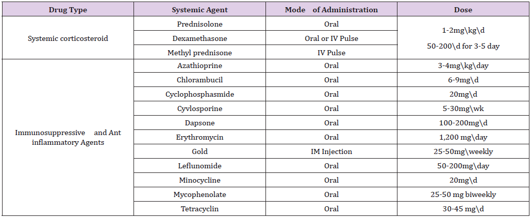
Rook AH et al. [13] mentioned that Extracorporeal photo chemotherapy [ECP] is a new form of immunotherapy which involves the extracorporeal photo inactivation of peripheral blood cells by 8-methoxypsoralen in the presence of ultraviolet A irradiation, followed by re administration of the cells. There are only 2 small case series and 2 case reports in the literature that document the use of ECP for refractory PV. Of the 9 PV patients treated with ECP in these studies, all experienced significant clinical improvement, while no adverse effects from ECP were noted. Shimanovich I et al. [14] said that Immunoadsorption resulted in a dramatic clinical response and a rapid decline in desmoglein-specific IgG auto antibodies. Recently, a small case series demonstrated that IA, administered in combination with rituximab, may result in long-term remission. In all studies, IA was safe and well tolerated. Harman KE et al. [15] concluded that systemic corticosteroids are still the most frequently used drugs and best-established therapy in pemphigus. The guidelines for managementof PV by the British Association of Dermatologists recommend patients with mild disease to receive an initial prednisolone dosage of 40 to 60 mg/d and in more severe cases, 60 to 100 mg/d. If no response is achieved within 1 week, the dosage is increased by 50% to 100% until disease control. Yeh SW et al. [16] reported that adjuvant drugs are usually administered in combination with systemic corticosteroids so as to reduce corticosteroid related side effects and increase efficacy. Conventional adjuvants include various immunosuppressive agents such as azathioprine, mycophenolate mofetil, methotrexate, cyclophosphamide, chlorambucil and cyclosporine, as well as antiinflammatory agents such as gold, dapsone, colchicine and a variety of tetracycline antibiotics (Table 1).

Table 1: Drug Regimes in Management of Pemphigus Vulgaris.

Bystryn JC et al. [17] discussed that Intravenous Immunoglobulin (IVIg) is a fractionated and purified blood product derived from the plasma of between 1,000 and 15,000 healthy donors per batch. It contains a high concentration of IgG and has a broad range of antibodies directed against pathogens, foreign antigens, and selfantigens. Although its exact mechanism of action remains unclear, IV IgG is associated with a rapid and selective decline in the serum levels of pathogenic PV auto antibodies. Jacobi A et al. [18] stated that TNF-á antagonists, tumor necrosis factor may be beneficial for the treatment of PV as experimental studies have demonstrated that TNF-á plays a role in the acantholytic process. Two case reports document the successful use of Infliximab for refractory PV. Anhalt G et al. [19] concluded that selective therapy using Intravenous Desmoglein 3 peptides was developed to suppress the production of anti-desmoglein 3 antibodies through inactivation and/or deletion of disease-associated CD4+ T lymphocytes. Joly P et al. [20] mentioned that Rituximab is a chimeric murine/human IgG1 anti-CD20 monoclonal antibody that targets pre-B and mature B lymphocytes, resulting in complement and antibody-dependent cytotoxicity and apoptosis. Multiple case reports suggest that rituximab is an effective treatment option for PV. The largest clinical study evaluating the use of rituximab in PV has been a case series of 14 patients with refractory PV in which 12 (86%) experienced a complete remission at 3 months after a single cycle of rituximab.

Schmidt E et al. [21] concluded that Rituximab is a chimeric murine/human IgG1 anti-CD20 monoclonal antibody that targets pre-B and mature B lymphocytes, resulting in complement and antibody-dependent cytotoxicity and apoptosis. Rituximab reduces circulating B cells, thereby preventing their maturation into antibody-producing plasma cells. Of the 18 patients with refractory PV reviewed, 3 (17%) experienced complete remission, 4 (22%) experienced clinical remission with further therapy required and 11 (61%) experienced partial remission.

Conclusion

The diagnosis of PV at an early stage when it is limited to oral cavity requires careful assessment and correlation of the clinical appearance, histological features and immunofluroscence findings. Their exist various treatment modality for treating Pemphigus which may range from topical and systemic steroid to current immunomodulators. The incidence of remissions in pemphigus is unclear, because these are usually reported at a single point in the evolution of the disease. Thus, it is uncertain whether treatment simply suppresses the manifestations of the disease and consequently must be continuously administered, or induces complete and long-lasting remissions that permit therapy to be discontinued. The prognosis of untreated oral lesions is a progression that involves other mucosa, including the skin. When treated, the prognosis depends on the age of the patient, the initial severity, the extent of lesions, the interval between symptom onset and start of treatment, and the drug dose required to control the disease, among other factors.

For More Articles: Biomedical Journal Impact Factor: https://biomedres.us

Journals on Medical Sciences

Nodular Fascitis of Zygoma- A Case Report

Abstract

Nodular Fasciitis is a benign reactive lesion of fibroblast that presents as a solitary, well circumscribed, rapidly growing soft tissue mass. It is most commonly located in the upper extremities, chest and trunk. Nodular fasciitis is rarely found in the Head & Neck region. Although benign it can be associated with a malignant process because of its rapid growth. Hence accurate diagnosis is important to avoid unnecessary and often mutilating surgery. We present a case of nodular fasciitis over zygoma in a 10 yr old child and its management.

Introduction

Nodular Fasciitis was first described by Konwaler, who named it pseudo sarcomatous fasciitis because of its resemblance to sarcoma. Nodular Fasciitis is also known as psedosarcomatous fibromatosis, proliferative fasciitis and infiltrative fasciitis [1-4]. It is an idiopathic, reactive, self limited proliferation of fibroblast and myofibroblast most commonly found in subcutaneous tissue. It commonly presents as a solitary, well circumscribed, rapidly growing soft tissue mass that may be painful of tender. Lesions most commonly occurs on upper extremities (43%), followed by trunk (25%), lower extremities (43%) and head & neck (10%). Although head & neck region is rare in adults, it is the most common site in pediatric population. Has an approximately equal sex distribution. Nodular Fasciitis is a benign lesion that can often be confused with myofibromatosis or a sarcoma due to its rapid growth, rich cellularity and mitotic activity. Hence it is important to distinguish the lesion from a more aggressive condition [5-10].

Case Presentation

A 10 year old child was referred to our college from a child hospital with a provisional diagnosis of osteoma of zygomatic bone. History reveled that there was presence of a small painless progressively enlarging swelling over right zygoma region for past 2months [11-13]. On examination, a single solitary nodule of size 1.5 x 1.5 cm, spherical in shape was seen over the area of right zygoma (Figure 1). The lesion was firm; surface was smooth and fixed to the underlying zygomatic bone. On applying pressure firmly in an upward and downward direction, slight mobility can be appreciated. Conventional x-ray was taken and it revealed a soft tissue density mass [14-17]. FNAC was performed and it was suggestive of an osteoma. Following the FNAC, an ultrasonography was done which revealed a well-defined homogenous hypoechoic mass with slight internal echo lateral to right zygomatic arch and features were suggestive of a benign soft tissue lesion (Figure 2). There was evidence of tissue intervening the lesion and the outer surface of zygomatic arch [17-21]. On doppler ultrasonography, no evidence of internal vascularity was found. Following the findings in ultrasonography, another FNAC was repeated for the second time and report was suggestive of a spindle cell neoplasm probably neural or fibrohistocytic lesion. As the patient was only 10 years old and a soft tissue lesion was suspected, an MRI was planned. Urea and creatinine test was done and it was within the normal range. An MRI was taken and it reveled 15x10x10mm well-defined T1 isointense and T2 hyper intense lesion in subcutaneous plane over the right zygomatic arch (Figure 3). The lesion is not suppressed on fat-saturation sequence. The lesion is not eroding the underlying bone.

Figure 1: Spherical in shape was seen over the area of right zygoma.

Figure 2: Homogenous Hypoechoic Mass with Slight Internal Echo Lateral to Right Zygomatic arch and Features were Suggestive of a Benign Soft Tissue Lesion.

Figure 3: T1 Isointense and T2 hyperintense lesion in subcutaneous plane over the right zygomatic arch.

Surgical procedure

The patient was taken up for local surgical excision. Through a transfacial approach an incision was made along the direction of the relaxed skin tension lines. Layer wise dissection done to reach the plane of the lesion. The mass was easily dissected out from the surrounding tissues and was submitted for histopathology.

Gross examination of the specimen showed a well circumscribed 1×1.5cm yellowish white mass. Microscopically, bland spindle cells arranged in short fascicles and set in a myxoid to fibrous stroma with extravasations of red cells. Mitosis were evident but no gross atypical were noticed (Figure 4). Hence it was interrupted as nodular fasciitis. Post operatively recovery was uneventful. Three months follow up revealed; there was no evidence of facial nerve paresis and no residual facial contour deformity.

Figure 4: Mitosis was evident but no gross atypia were noticed.

Discussion

Nodular Fasciitis can occur anywhere in the body. Most common location in adults being upper extremities (39-54%), especially in the volvar aspect of the elbow. They can also be found on the trunk (15-20%), lower extremities (16-18%) and head & neck region (7-20%). In children however head and neck region is the most common site. Approximately 10% of all lesions are found in children. Nodular Fasciitis most commonly presents as a rapidly growing firm nodule of one month duration or less. Mostly as solitary, round to oval subcutaneous nodules with involvement of deep fascia and muscle in small percentage of patients. The cause is unknown but has often been linked to local injury or trauma. Radiographic evaluation with MRI renders an accurate assessment of the degree of soft tissue extension. Proper diagnosis requires histological confirmation. Nodular Fasciitis typically shows well circumscribed, encapsulated nodules composed of spindle cells. Treatment is most commonly by local surgical excision. However in some cases recurrence has been associated with incomplete excision of the lesion. When lesions recur alternate diagnosis of malignant process should be considered.

Summary

Nodular Fasciitis is an uncommon benign, idiopathic, proliferation of fibrous tissues. It is important to distinguish it from a malignant process because it can be successfully treated with complete local excision.

The importance of the nature of fascitid relates to the potential for overtreatment on the basis of microscopic features and its rapid growth. It should be emphasized that nodular fascitis is most common in the head & neck region of children and should be considered in the differential diagnosis of all fibrous lesions of that region.

For More Articles: Biomedical Journal Impact Factor: https://biomedres.us

Journals on Chemical Science

Rare Earth Elements Resources and Different Recovery Techniques from Egyptian Ores

Introduction

In fact, bastnaesite, monazite and xenotime are the most important rare earth-bearing minerals known as sources for extraction of rare earths by hydrometallurgical approaches to produce intermediate products in the form of rare earth-chlorides, fluorides, halides, and oxides. In the following paragraphs I will summarize some of the rare earth elements extraction using variety of their extraction techniques from some treated Egyptian ores. Leachability of lanthanides from the radioactive pegmatite pockets found in Gabal Abu Furad area, southwest Safaga city, limited by latitudes 26°37′ and 26°43′ and longitudes 33°36′ and 33°43′E. Central Eastern Desert, Egypt. It was investigated using different acids at different concentrations, liquid-to-solid ratios, contact times and temperatures in order to determine the optimum recovery conditions. The best leaching efficiency was obtained with 40g/l HCl solution; 8hrs contact time, 90°C temperature and 1:5 (g/ml) solid: liquid ratio. Afterwards, lanthanide stock solutions were prepared by applying the optimum leaching conditions. Lanthanides were then precipitated by either 10% oxalic acid or 20% sodium hydroxide solutions. Electron scanning microscope (ESM) analysis was utilized to determine the qualitative percentage of the existed lanthanides. It was found that thorium percentage was around 10.05%. High percentages of light lanthanides were detected, e.g., La (11.16%), cerium (39.97%), Pr (6.21%), Nd (13.99%), Sm (2.00%), and Gd (1.06%) [1].

The study area of El-Garra El-Hamra is one of the igneous masses located in the southern part of the Western Desert of Egypt. This work has been carried out to investigate the process of gadolinium separation from REEs cake obtained from El-Garra El-Hamra ore concentrate. It is considered as a new approach in the Nuclear Materials Authority of Egypt. Firstly, the optimum leaching conditions achieved are 200 gm/L H2SO4, 1/2 solid/liquid ratio, 4 hours agitation time, 125 mesh size and at 90OC temperature. Then by solvent extraction method using 1mol/L D2EHPA middle REEs were extracted (Figures 1 & 2). The organic extractant was stripped by using 2mol/L HCl and Gd pregnant solution was obtained. Then, the relevant optimum factors were 0.3 mol/L D2EHPA diluted by kerosene, 10 minutes contact time, and 4/1 organic/aqueous ratio giving 87% Gd extraction. Then, the scrubbing of the loaded extractant was by 1mol/L HCL followed by Gd stripping using 5M HCl, contact time of 30 min. and 1/3 organic/aqueous ratio. The strip solution was subjected to evaporation and little amount was dried to obtain GdCl3 powder having a purity of about 81% associated with YCl3 4% [2].

Figure 1: Geologic map of Gabal Abu Furad, Central Eastern Desert, Egypt (modified after Mahmoud, 1995 – Dardier and El Wakeel 1998).

Figure 2: Map of Egypt showing location of the study area.

The study area of El-Garra El-Hamra is one of the igneous masses located in the southern part of the Western Desert of Egypt. In this study, it is emphasized upon the Y extraction because as far as the author is aware, it is the first time to prepare Y concentrate from the local ores. Thus, the prepared sample is enriched in U, Th, REE and Y and hence it is suitable for such study. In the Y extraction process the REE cake would first be dissolved in 8 N HNO3 and then contacted by the organic solvent TBP (100% v/v for 15min. while using an O/A ratio of 1:1). By applying the counter current solvent extraction system, it is expected that 2 stages are sufficient to achieve almost complete Y extraction. Scrubbing step would be performed upon the loaded solvent to remove most of the undesired elements (Figure 3 & 4). This is done by using distilled water for 2cycles. Y loaded TBP solution would then be subjected to the stripping step by 6 N H2SO4 acids when using A/O ratio of 1:2 for 60min. Also, it is expected that 5 stripping stages are sufficient to strip out most of the Y content while the depleted TBP will be recycled to the regeneration tank. Y could be precipitated by adjusting the stripped solution at pH 7 by using ammonia solution and then by adding 12 % oxalic acid drop by drop during stirring until almost complete precipitation of Y.

Figure 3: The Geographical map of EI-Hamra area, South western Desert Egypt.

Figure 4: Location map of Qattar and prospect area Gabal Qattar, Northern Eastern desert, Egypt.

The obtained Y oxalate was then claimed at 550OC for 2hours to obtain yttrium oxide Y2O3 [3]. Phosphogypsum (PG), a residue of the phosphate fertilizer industry that has relatively harmful concentrations of rare earth elements (as lanthanides) is accumulated in large stockpiles and occupies vast areas of land. The present work was oriented to decrease the concentration of lanthanides by leaching of PG using the synergism of methyl iso-butyl ketone (MIBK) and Tri-octyl amine (TOA) in kerosene modified by 1-octanol. The factors which affect the leaching process such as agitation time, concentration of the solvent, liquid/solid ratio, and temperature were optimized. Based on the experimental results, about 92.1% of lanthanides were successfully transferred from the PG into the solvent phase. Nitric acid has been used to strip the lanthanides from the organic solvent. The precipitation of 93.4% of lanthanides was carried out using oxalic acid under the desirable conditions. A decontaminated product that can be safely used in many industrial applications was obtained [4].

Gebel Gattar area is considered one of the most promising uranium mineralization occurrence located in the northern Eastern Desert of Egypt, located at the intersection of latitude 27O 7/ 30// N and longitude 33O 17/ 5// E covering about 2 km2. Hydro thermal alterations of Gabal Gattar granitic mass at GII occurrence exhibited an increase in Ni, Cu, Zn, Pb, Mo, Nb, Rb beside HREEs. A technological sample from GII occurrence was subjected to extraction of REEs through both leaching and precipitation processes. The optimum leaching conditions achieved were as follows: 40 g/l HNO3 acid, 12 hours contact time, 1/2 S/L ore/acid ratio, 60 mesh grain size and room temperature (25OC) giving REEs leaching efficiency of about 97%. REEs were directly precipitated using sodium hydroxide (20%) where REEs hydroxide was obtained at pH 8.5 [5]. The above text through a light on some applications of extracting rare earth elements performed in my dear country Egypt, I hope it may be benefit for the interested readers.

For More Articles: Biomedical Journal Impact Factor: https://biomedres.us

Journals on Chemistry

Silica Chloride Catalyzed Efficient Synthesis of 2,3-Dihydroquinazoline-4(1h)-Ones Derivatives In Water

Abstract

Describe method consist of eco-friendly procedure for the preparation of 2,3-dihydroquinazoline-4(1H)-ones from equimolar 2-aminobenzamide and substituted aromatic aldehydes in presence of 20mol% of silica chloride. Green impact of reaction significantly enhanced due to use of water as solvent and recyclable silica-chloride as catalyst. Good to excellent yield of products, simple working strategy and easy purification are the advantage of present methodology

Keywords: Quinazoline; Silica-chloride catalyst; Water mediated; Green methodology

Introduction

Search of expeditious and cost-effective methodologies to replace tedious, low productive traditional methodologies gains its own importance. Now a day’s green methodologies has attract significant attention and environmentally benign, recyclable, chap solid catalysts get ultimate reputation. Such methodology offers to obtained complex pharmaceutically important molecules or intermediate by possibly viable ways. Such methodologies shine with imminent light when water incorporates as solvent, due to its non-toxic, green, cheap nature and biochemical consequence [1,2].

Quinazoline has been occupied distinct position in nitrogen containing heterocycles due to its spectacular wide spectrum of pharmaceutical properties. Various reports of quinazoline underline its widely biopharmaceutical activity like, anticancer [3- 5], antibacterial [6-8], antiinflammation [9,10], antituberculosis [11], antihypertension [12] and antidiabeties [13]. Such wide spectrum of quinazoline strongly demands possible derivatisation to test out for further pharmaceutical possibilities. Various methods have been proposed to obtained quinazoline analogues using catalysts like ammonium bromide[14], Zirconyl chloride [15] Heteropoly acids [16], Gallium (III) triflate [17], Titanium oxide nano-particles [18], Starch solution [19], cyanuric chloride [20] and Cyclodextrin sulphonic acid [21].Most of these methodologies are suffers from long reaction time, high temperature, use of expensive catalyst and tedious work procedures. ‘On water’ reports [22] of quinazoline synthesis by using expensive catalyst increase cost of reaction.

Readily accessible solid support catalyst like Silica chloride for the synthesis one pot quinazoline has not attempted. Such solid supported catalyst simplifies the reaction procedure and do not pass on unpleasant toxic residue to environment. Silica, perhaps most easily available substance and extensively used as support in reactions [23]. Much organic reactions and used silica chloride as efficient catalyst [24-28]. In continuation of our previous research work [29] to develop fast, naturally benign, productive methodology for small and fused heterocyclic compounds, we intended to developed facile, efficient, cost-effective and easy workup method for the synthesis of quinazoline derivatives. Here, introduce facile methodology as shown in scheme 1 for syntheses of quinazoline derivatives (Figure 1).

Figure 1: 1: Syntheses of Quinazoline Derivatives.

Experimental

The reagents and solvents were purchased from Alfa aesar and Aldrich Chemical companies and used without further purification. All compounds obtained were describe for open head capillary tube for their melting point and are uncorrected. The samples were analyzed by FT-IR spectroscopy (JASCO FT/IR-460 plus spectrometer). 1NMR and 13CNMR spectra of compounds were recorded on a Bruker DRX-400 Avance instrument in DMSO-d6.

Preparation of Silica-chloride

To a well-stirred silica gel (20 g) in DCM (50 mL) was added to this slow drop wise SOCl2 (20 g) introduced at room temperature. After stirring for 1-2 hour, the solvent was removed under reduced pressure to dryness. The silica chloride thus obtained was used in the describe experiments as catalyst [28].

General Procedure

In a RBF containing 20 ml of water was added 2-aminobezamide (0.01mol 1.36gm), substituted aldehyde (0.01mol) and portion wise SiO2-Cl (2 gm 20 mol%) with stirring. The reaction mixture was stir at room temperature. Progress of reaction was monitor by thin layer chromatography (TLC) using Ethyl acetate-Hexane. After completion of reaction, reaction mixture was filter off and filtrate neutralized by saturated solution of sodium bicarbonate, brine and extracted with ethyl acetate. Organic layer dried on anhydrous sodium sulphate and evaporated in reduced pressure to afford pure product after recrystallization from ethanol. Representative compounds were scan for spectral data and found satisfactory agreement with reported.

Silica Chloride as Reusable Catalyst

Catalyst obtained after filter off from first reaction cycle, and dried at 110°C for 2 hours and used for second cycles. Process was repeated until trace product formation takes place during the course of reaction (Table 2).

Spectral data of representative compounds

A. 2-phenyl-2,3-dihydroquinazolin-4(1H)-one; (1) m.p.= 219°C, 1HNMR (400 MHz, DMSO-d6): δ= 8.27 (s, 1H), 7.61 (d, 1H), 7.50 (d, 2H), 7.31-7.41 (m, 3H), 7.22 (t, 1H), 7.06 (s, 1H), 6.72 (d, 1H), 6.69 (t, 1H,), 5.75 (s, 1H) ppm; IR (KBr): 3310, 3014, 1671, 1630, 1523 cm-1.

B. 2-(4-methoxyphenyl)-2,3-dihydroquinazolin-4(1H)-one; (2) m.p. 184°C, 1HNMR (400 MHz, DMSO-d6,) δ: 8.28 (s, 1H), 7.70 (d, 1H), 7.51 (d, 2H), 7.30-7.35 (m, 1H), 7.10 (s, 1H), 7.04 (d, 2H), 6.83 (d, J=7.6 Hz, 1H), 6.76 (d, 1H), 5.79 (s, 1H), 3.83 (s, 3H); IR (KBr): 3292, 3161, 2830, 1655, 1614, 1543, 1480, 1393, cm-1

C. 2-(p-tolyl)-2,3-dihydroquinazolin-4(1H)-one; (4) m.p.= 224°C. 1HNMR (400 MHz, DMSO-d6): δ= 8.17 (s, 1H), 7.60 (d, 1H), 7.41 (d, 2H), 7.26−7.17 (m, 3H), 7.02 (s, 1H), 6.75 (d, 1H), 6.71 (t, 1H), 5.70 (s, 1H), 2.31 (s, 3H) ppm. IR (KBr): 3312, 3065, 1656, 1611, 1542, 1487 cm-1.

D. 2-(4-Bromophenyl)-2,3-dihydroquinazolin-4(1H)- one; (5) m.p. 1950C; 1HNMR (DMSO-d6, 400 MHz) δ: 8.28 (s, 1H), 7.58-7.65 (m, 3H), 7.43 (d, 2H), 7.22 (d, 1H), 7.14 (s, 1H), 6.71 (d, 1H), 6.65 (d, 1H), 5.77 (s, 1H); IR (KBr): 3300, 3174, 3029, 1672, 1656, 1600, 1482, 1430.38, 1290, 1133 cm-1.

Results and Discussions

Series of reactions were performed to optimized reaction condition including amount of catalyst with respect to yield of product. Room temperature and ‘on’ water was kept as fix reaction parameters. 2-aminobenzamide and p-methoxy benzaldehyde were taken for model reaction and various reaction condition were applied. At began, model reaction was done without catalyst to ensure necessity of catalyst and was found little productive (Table 1). When silica without surface modification introduces as catalyst (Table 1) reaction was fruitful, but yield of reaction was not satisfactory. Good to excellent results obtained after introduction of silica chloride as catalyst. Amount of silica chloride were vary in search of optimum amount of catalyst from 5 mol % to 25 mol %. It was observed that (Table 1) 20 mol% of catalyst offers optimum product.

Silica chloride catalyst reusability study was performed with model reaction (Table 2). First two cycles offers excellent yield of product and productivity decreases after third cycles. It was observed that when reused at fifth time gave 18% of product, as this reading matches with no-catalyst (Table 1) assumed complete neutralization of efficiency of catalyst and no further reusability were checked. By keeping 20mol% of silica chloride as optimum amount of catalyst further derivatisation were performed. Study of effect of various substituents on the yield of reaction was performed (Table 3). It was observed that electron releasing groups present on aromatic aldehydes offers more conversion of product. p-OCH3, p-Br, p-CH3 were found highly productive, whereas, p-NO2 benzaldehyde was the less productive substituent. The representative products structures were substantial from their spectral data and found satisfactory agreements with reports in literature [20-30] (Figure 2).

Figure 2: Hypothetical pathway of progress of reaction.

Table 1: Optimization of amount of Silica-chloride using model reaction strategy.

Isolated yields Reaction condition: 2-aminobenzamide (0.01 mol), p-methoxy benzaldehyde (0.01 mol) stirred in water (20 ml) at rt. for 60 minutes.

Table 2: Optimization of amount of Silica-chloride using model reaction strategy.

aIsolated yields,

Table 3: Showing Quinazoline derivatisation with respect to yield of reaction, time and physical constant of obtained products.

aIsolated yields; Reaction condition: 2-aminobenzamide (0.01 mol), p-methoxy benzaldehyde (0.01 mol), silica-chloride (20mol%), stirred in water (20 ml) at rt.

Conclusion

In conclusion, an efficient and simple method for the synthesis of quinazoline analogues has been described using readily available and cost effective silica chloride as a heterogeneous catalyst. The green reaction profile and mild reaction conditions are main advantage of this method. Reaction takes place at room temperature by simply stirring method, with operational simplicity offers excellent yields.

For More Articles: Biomedical Journal Impact Factor: https://biomedres.us

Journals on Pharmacy

Patterns of International Coauthor Collaboration in Bioinformatics

Abstract

Objective: To investigate journal features by collecting data from Medline and to visualize the journal characteristics of Bioinformatics.

Method: Selecting 11,411 abstracts, author names, countries and MESH (medical subject heading) terms by a keyword “Bioinformatics”[Journal] on October 31, 2017 from the Medline, we applied social network analysis (SNA) and Google Maps to report following features:

a. Nation distribution and coauthor collaboration,

b. Journal features represented by paper MESH terms.

We found that

a. The most number of papers are from nations of U.S. (4175, 36.58%) and Germany (1010, 8.85%)

b. The most linked MESH terms are algorithms and software.

Keywords: MESH terms; Authorship Collaboration; Google Maps; Social Network Analysis; Medline

Introduction

An interdisciplinary field of science developing schemes/ methods and software tools for understanding and utilizing biological data for health care is popular in recent years [1]. By searching keyword Bioinformatics from Medline library on October 31, 2017, we found 228,865 published papers in which 3,928 with bioinformatics in title. Bioinformatics combines computer science knowledge, statistics and engineering to analyze and interpret the biological data using mathematical and statistical techniques has become an important part of many areas of biology in a short span of time. However, the pattern of international coauthor collaboration as well as the main MESH (medical subject heading) term [2,3] is still unclear.

An apocryphal story often told to illustrate the concept of cooccurrence is about beer and diapers sales. It usually goes along with both beer and diapers sales which were strongly correlated [4-6] in a market place. As such, all possible pairs of our observable phenomena can be combined and analyzed using computer techniques. However, we have not seen any computer algorithms that help us select the most possible pairs co-occurred with each other till now.

Social network analysis (SNA) [7-9] has applied to authorship collaboration in recent years. It is because co-authorship among researchers that forms a type of social network, called co-author network [10]. We are thus interested in using SNA and Google Maps to display the most pair relations for a journal in international author collaboration and MESH terms.

Aims of the Study

Our aims are to investigate journal features by collecting data from Medline and to visualize the journal characteristics of Bioinformatics in following representations:

a) Nation distribution and coauthor collaborations,

b) Journal features represented by paper MESH terms.

Methods

Data Sources

We programed Microsoft Excel VBA (visual basic for applications) modules for extracting abstracts and their corresponding coauthor names as well as MESH terms on October 31, 2017 from the US National Library of Medicine National Institutes of Health (Medline) by a keyword “Bioinformatics”[Journal]. Only those abstracts published by Bioinformatics and labelled with Journal Article were included. Others like those labelled with Published Erratum, Editorial or without author name(s) were excluded from this study. A total of 11,411 abstracts were retrieved from Medline since 1999.

Data Arrangement to Fit SNA Requirement

We analyzed 11,411 papers with complete data including authors’ countries, names, and MESH terms. Prior to visualized representations of research findings using SNA, we organized data in compliance with the SNA format and guidelines using Pajek software [11]. Microsoft Excel VBA was used to arrange data fitting the SNA requirement.

Graphical Representations to Report

We combined SNA and Google Maps to present the distribution of nations and their corresponding collaborations by separating isolated and clustered nodes (e.g., nations). The bigger bubble means the more number of authors (including their coauthors) in papers. The wider line indicates the stronger relations between two nodes. Community clusters are filled with different colors in bubbles. Similarly, keywords of MESH terms represent the research domain for Bioinformatics, the stronger relations between two MESH terms can be highlighted through the SNA, like the concept of co-occurrence about beer and diapers sales. The presentation for the bubble and line is interpreted in results.

Statistical Tools and Data Analyses

Google Maps [12] and SNA Pajek software [11] were used to display visualized representations for Bioinformatics. Author-made Excel VBA modules were applied to organize data. Gini coefficient [13] is used to measure the strength of a role in a network: the higher is the Gini, the stronger is the role in the network.

Result

Authors’ Nations and their Relations

A total of 11,411 papers with complete authors’ nations based on journal article since 1999 are collected. The most number of papers are from nations of U.S. (4175, 36.58%) and Germany (1010, 8.85%). The distribution of coauthor nations is present in Figure 1. The closest relation is linked by U.S. and Taiwan, see the widest line in (Figure 2). All coauthors connected to Taiwan can be shown in Figure 3. After we click the bubble and the diagram. Interested readers are recommended to practice it by clink the link in reference [14].

Figure 1: International author collaborations in bioinformatics.

Figure 2: International author collaborations in bioinformatics with links.

Figure 3: International author collaborations in bioinformatics focused on a specific nation/region.

Keywords to Present the Journal Research Domain

The most linked Keywords denoted by MESH terms are algorithms, software, *algorithms sequence analysis, dna/*methods, information storage and netrieval/*methods, and sequence analysis/instrument/Methods, see (Figure 4). The closest relation is between algorithms and software with a highest frequency of 848. Two terms of algorithms and sequence alignment/*methods (760) follow [15].

Figure 4: Main keywords using Mesh terms to describe the Journal of Bioinformatics dispersed in clusters.

Discussion

In this study, we found that

a. The most number of papers are from nations of U.S. (4175,36.58%) and Germany (1010,8.85%);

b. The most linked MESH terms are algorithms and software.

Using Google Maps to show the relations of author collaboration and MESH term to represent the features of a Journal that is never seen in previous published papers.

Many previous researches [7-9] have investigated coauthor collaboration using SNA. However, the results have not been incorporated with Google Maps to clearly show the international author pattern. An apocryphal story often told to discover the cooccurrence about beer and diapers sales [4-6]. However, we have not seen any that demonstrates a concrete way to show how to conduct this exploration and to present informative messages to readership. Furthermore, what are the most popular terms that present in journals of Bioinformatics have been investigated in [Figure 4].

Incorporating Google Earth, Google Maps and/or network visualization with Pajek software, one can overlay the network of relations among addresses in scientific publications on the geographic map. We demonstrated and provided illustrations with hyperlinks [14,15] for interested authors to practice in their own ways. There are several limitations that should be concerned in future. First, the interpretation and generalization of the conclusions of this study should be carried out with caution because the data were merely extracted from a single journal. It is worth noting that any attempt to generalize the findings of this study should be made in the similar journal domain with similar topic and scope contexts.

Second, although the data were extracted from Medline and carefully dealt with every linkage as correct as possible, the original downloaded text file including some errors in symbols which are hard to deal with and might lead to some bias in the resulting nation distribution. Third, the social network analysis is not subject to the Pajeck software we used in this study. Others such as Ucinet [16] and Gephi [17] are suggested for readers to use in future.

For More Articles: Biomedical Journal Impact Factor: https://biomedres.us

Journals on Orthopedics

Reduction of Distal Radius Fractures under Haematoma Block

Abstract

Introduction: Most common fracture in elderly patients is distal radius fracture. The most common method of management is closed reduction and immobilization. The aim of the study is to evaluate the analgesic effects of hematoma block for closed reduction of distal radius fracture.

Materials and Methods: A prospective study was carried out among 50 patients of age >60 years of either sex who had fracture distal radius between 2015-2016. The patients having multiple fractures, pathological fractures were excluded from the study. After taking informed written consent, reduction of fracture was done following after infiltration with 2% lignocaine into fracture hematoma site. Pain score was compared by VAS before, during and after manipulation. Time taken from presentation at emergency department to reduction and discharge from hospital was also recorded.

Results: 50 patients of mean age 65.1 years, male: female 22:28 with fracture distal radius were studied. Mean time from admission to fracture reduction was 2.05 hours. Discharge time from hospital after reduction of fracture 2.0 hours .Average VAS during reduction was 0.94. 10 minutes after reduction VAS was 0.20.

Conclusion: For closed reduction of distal radius fracture, hematoma block with lignocaine is safe and effective alternative to other form of anaesthesia.

Keywords: Eldely Patient; Distal Radius Fracture; Haematoma Block; Close Reduction

Abbreviations: PR: Pulse Rate; RR: Respiratory Rate; BP: Blood Pressure; SO: Oxygen Saturation

Introduction

Distal radius fracture is most common fracture of musculoskeletal system. It accounts for about 16% of all fractures treated at emergency department [1-4]. Fracture distal radius is most common in elderly and more in females than males [5- 7]. It usually occurs due to minor fall than severe trauma [8,9]. Various methods are used to decrease the pain during closed reduction of fracture radius like brachial plexus block, intravenous regional anaesthesia, general anaesthesia, conscious sedation and haematoma block. Each of these methods has certain merits and demerits [7]. The aim of the study was to evaluate the result of hematoma block for closed reduction of distal radius fracture.

Materials and Methods

This was a prospective study done in 50 patients of age >60 years of either sex at PCMS&RC, Bhopal, M.p in department of orthopaedics and Anesthesia, between 2015-2016.cases were posted for closed reduction of distal radius fracture at orthopaedic OT. After taking and informed written consent, the patients were taken for manipulation under hematoma block. The following parameters were recorded for study:

a. Demographic data

b. Pain score by VAS before, during and 10 minutes after reduction (VAS on scale of 0-10, zero no pain and ten being intense pain).

c. Time from presentation to hospital to reduction of fracture and time taken for discharge from hospital following reduction.

Patients having multiple fractures and pathological fractures were excluded from the study.

Preanaesthetic evaluation was done in all the patients of study. Inside OT, suitable intravenous line was secured and multipart monitor connected for continuous monitoring of Pulse Rate (PR), Respiratory Rate (RR), Blood Pressure (BP) and Oxygen saturation (SpO2). Following proper sterilization of the affected part, hematoma was confirmed by aspiration of 1-2mL of old hematoma blood. 10mL of 2% lignocaine was injected into the hematoma at the dorsal aspect of wrist and also into the adjacent periosteum in an aseptic manner. Massaging was not done after lignocaine injection. Reduction of fracture was allowed 10-15 minutes after injection of the drug. Immobilization was done following reduction by plaster of paris cast. Patients were not given any other analgesic before the procedure. Pain score by VAS was recorded before, during and 10 minutes after reduction of fracture. Total time in presentation at emergency department to reduction and discharge from hospital after reduction of fracture was recorded.

Result

Demographic data like age and sex were comparable in both the groups in Table 1. The above table shows demographic characteristics of the study population. The mean age in years was 65.01 years. The ratio of male: female was 22:28. Table 2 shows time taken from admission to reduction and also reduction to discharge from hospital. Average time taken for admission to reduction was in our study was 2.05 hrs, and average time taken from reduction to discharge from hospital is 2.0 hrs. Table 3 shows the VAS Score 10 minutes before reduction, during reduction and 10 minutes after reduction. The average VAS score 10 minutes before reduction was 7.68, during reduction was 0.94 and 10 minutes after reduction was 0.2 recorded. The graphical representations VAS score with tme given in Figure 1.

Table 1: Demographic data like age and sex were comparable in both the groups.

Table 2: Shows time taken from admission to reduction and also reduction to discharge from hospital.

Table 3: Shows the VAS Score 10 minutes before reduction, during reduction and 10 minutes after reduction.

Figure 1: The graphical representation of VAS scores with time.

Discussion

There are different methods to relieve pain during reduction of fracture of distal radius as described by different authors each having its own merits and demerits. Haematoma block has been studied as a procedure for fracture reduction by various authors. Kendal et al. [5] studied hematoma block in 1995. The increasing cost and time taken for general anaesthesia in comparison to hematoma block for reduction of distal radius made hematoma block more popular. Singh et al. [4] did a comparative study between haematoma block and conventional sedation in 1992 and found that pain score in hematoma block was significantly low in comparison to sedation group [10]. Compared results of brachial plexus block and haematoma block for reduction of fracture radius in 2008. He found no difference in analgesic effect between the study groups. Funk [7] in a study compared the VAS during reduction of fracture radius between IV general anaesthesia and hematoma block and found that VAS in general anaesthesia group was zero and hematoma block was 3.7.

Demographic data in our study is comparable to other studies showing mostly elderly and females affected by this fracture [5]. In our study, VAS during reduction was 0.94. This is different from the result obtained by Funk. This might be due to waiting for 10 minutes after giving lignocaine into the hematoma for the block to be effective – the procedure followed in our study.

Post reduction pain score in our study is comparable to study by Funk. We obtained a score of 0.20. This could be explained by the fact drug administered was lignocaine- a local anesthetic, which also contributed to post procedure analgesia. In our study, we found a significant reduction in mean time from admission to reduction of fracture and discharge from hospital in compare to other modality of anesthesia; this is similar to the study of Funk. Usually, other modality of anaesthesia requires an operation theatre, anaesthesia machine, a source of oxygen, fasting protocol of at least 6 hours and continuous monitoring, which is time consuming and costlier. Haematoma block for reduction of distal fracture radius can be done as Non-Operating Room Anaesthesia – NORA, at emergency department itself. Moreover, it can be done in a set up where a skilled anesthetist is absent or proper OT set up is not available.

Limitations

During hematoma block by infiltration, the close fracture maybe converted to an open fracture giving way for the entry of microorganisms thereby causing infection [11]. However, in our study, we did not witness any such complication, which could be because of strict aseptic protocol being followed.

Conclusion

Thus, we conclude that hematoma block for reduction of distal fracture radius is a safe, simple and effective alternative to other type of anesthesia. It is also safe procedure in compare to other modality of anesthesia but should be given in the presence of qualified anesthetic.

For More Articles: Biomedical Journal Impact Factor: https://biomedres.us

Chemistry Journal

Marine Drugs Research in Saudi Arabia

Opinion

Recently drug discovery and development program based on the sustainable use of marine biodiversity have attracted much attention, because many scientists believe that its integration with recent advances in biotechnology not only promises economic benefits but also promotes the protection and conservation of marine biodiversity. Some biotechnological innovations have enabled to generate ecologically and environmentally sound approaches, which contribute greatly to the sustainable use of marine biodiversity. This emerging multidiscipline is especially interesting to be developed in Saudi Arabia as a wide country with highly diverse marine resources. Therefore here we will deal with developing a concept about how drug discovery based on marine natural products can be implemented to promote the sustainable use, protection and conservation of marine biodiversity as well as to secure economic benefits. Marine Organisms such as sponges and corals are broadly known as rich sources of novel and useful bioactive marine natural products as marine cancer drugs. However, it is not only elaborate pharmaceutically useful compounds but also produce a lot of toxic substances.

One of the most important societal contributions of applied chemists has been the isolation and identification of toxins responsible for seafood poisoning. Outbreaks of seafood poisoning are usually sporadic and unpredictable because toxic fish or shellfish do not produce the toxins themselves, but concentrate them from organisms that they eat. Most marine toxins are produced by microorganisms such as dinoflagellates or marine bacteria and may pass through several levels of the food chain. The identification of marine toxins has been one of the most challenging areas of marine natural products chemistry. The major occupation of marine natural products chemists for the past two decades has been the search for potential pharmaceuticals. It is difficult to single out a particular bioactive molecule that is destined to find a place in medicine. However, many compounds have shown promise. Marine organisms produce some of the most cytotoxic compounds ever discovered, but the yields of these compounds are invariably so small that natural sources are unlikely to provide enough material for drug development studies. These organisms are frequently colonized by bacteria. Some of these bacteria can be pathogenic or serve as beneficial symbionts. Therefore, these organisms need to regulate the bacteria they encounter and resist microbial pathogens.

Many of the natural products isolated from marine invertebrates share structural homology with compounds of microbial origin, leading to the hypothesis that the marine compounds are actually produced by microorganisms living in association with the invertebrates. Marine invertebrates are indeed largely sessile, filter-feeding organisms that contain a complex assemblage of symbiotic microorganism. Thus, the real producer of biologically active compounds from marine invertebrates is always uncertain. Detailed investigation of bioactive metabolite symbioses is a field that is still in its infancy. Whilst marine compounds are considered in current pharmacopoeia, it is anticipated that the aquatic environment will become an invaluable source of novel bioactive compounds in the nearest future. It was known that many of these natural products act as regulators of specific biological functions. Some of them have pharmacological activity due to their specific interactions with receptors and enzymes.

The development of marine compounds as therapeutic agents is still in its early stage due to the lack of an analogous ethno-medical history as compared with terrestrial habitats, together with the relative technical difficulties in collecting marine organisms. The systematic investigation of marine environments is reflected in the large number of novel compounds reported in the literature over the past decade. Some of these agents have entered preclinical and clinical trials, and it may be expected that this number will increase in the future. It was known that the isolation of new anticancer agents derived from marine sources has been based on the collection of marine macroorganisms. The progress in scuba-diving techniques and deep-water collection instruments has been pivotal in the collection programs implemented by academic and pharmaceutical groups. Recently, a lot of research programs are emerging to exploit marine microorganisms and the results are promising. These studies have demonstrated the capability of marine bacteria to produce compounds not available from terrestrial sources. They also have led to an increase in knowledge of the many bioactive compounds produced by these microorganisms. Nowadays, the compounds are systematically tested for relevant biomedical properties including antiproliferative effects. The major screening system is carried out by the National Cancer Institute of the USA. This system looks for selective activity in a panel of more than 60 human tumor cell lines. Alternative strategies employ a more mechanistic-based approach, with systems designed to screen for substances with inhibitory properties towards specific enzymatic reactions. This type of assay offers specificity and can focus on a number of discrete drug targets. The potentially confounding effects of toxic components are also avoided, permitting the screening of crude extracts from marine organisms. Thus, the search for new and useful bioactive natural products from marine soft corals and sponges has been one of the main research subjects and that research will be performed within several projects funded by King Abdulaziz City for Science and Technology in Riyadh, Saudi Arabia, and by many Saudi Arabia universities.

For More Articles: Biomedical Journal Impact Factor: https://biomedres.us

Orthopaedics Open Access Journal

Triflange Acetabular Cup for Severe Acetabular Bone Defect

Introduction

Acetabular bone defects in revision hip arthroplasty are a challenging problem. Its treatment relies on preoperative and intraoperative assessment of the size of the defect, presumed quality of the bone, integrity of the acetabular columns and presence of pelvic discontinuity. Several surgical techniques advocated for treating pelvic discontinuity are ilio-ischial cages, plate fixation of structural allograft, triflange cups, Steinmann pin fixation, acetabular revision with additional pelvic screws, acetabular reinforcement rings, oblong cups and cup-cage reconstruction [1- 5]. Severe cases of acetabular bone loss often require an implant specifically made to match the patient’s individual anatomy. Triflange acetabular component is a patient-matched implant designed in partnership with the surgeon, using the patient’s own computed tomogram (CT) scan data. We present a case with severe acetabular bone loss treated with a custom triflange acetabular cup.

Case Report

A 59-years old lady presented with gradual worsening pain in her right groin and thigh for a year. More recently, she has noticed progressive shortening of her right leg. The pain with the limb length discrepancy was affecting her function including activities of daily living. She had undergone a primary uncomplicated right total hip arthroplasty in 1986 at age 39 years which was subsequently revised in 1993 for aseptic loosening. She had a past history of Hepatitis C viral infection which she supposedly contracted from a blood transfusion. Other medical illnesses of note were hypothyroidism, osteoporosis and chronic back pain. On examination, she had an obvious limb length discrepancy of 10 mm, a positive Trendelenberg’s test, and painful hip range of movements. She was a lean built lady with a BMI of 18. Her preoperative Oxford hip score was 11/48. The plain radiographs (Figure 1) showed loosening of the acetabular component with extensive osteolysis of the periacetabular bone and ballooning of the medial wall. A CT scan was also obtained to establish the extent of bone loss (Figure 2). The 3D reformat images highlighted the significant bone loss further and were utilized to design the patient specific triflange acetabular cup. The custom made triflange acetabular cup was used as it allows for screws to be inserted in to the ilium, ischium and pubic bones to allow fixation of the cup in to the pelvis (Figure 3). A liner was then cemented in the triflange acetabular cup which articulates with the head of the femoral component. Patient has been recently reviewed in the clinic, twelve-months postoperatively, the hip was pain-free and stable (Figure 4).

Figure 1: Preoperative radiographs- The right hip acetabular component has loosened and penetrated through the wall of the acetabulum. a. AP view of the pelvis b. Lateral view of the hip joint.

Figure 2: Preoperative CT scan- This is a computed tomography scan of the same patient, demonstrating extensive bone loss, medial acetabular wall fracture with migration of the implant. a. Axial reformat, b. Coronal reformat.

Figure 3: Clinical photographs showing the plastic model for trial and the final custom triflange acetabular cup.

Figure 4: Postoperative radiograph-Six-month postoperative radiographs showing the triflange custom acetabular implant in situ. a. AP view b. Lateral view.

Discussion

Patients with large acetabular defects and pelvic discontinuity pose a difficult problem for surgeons. The goal of treatment in these cases is fixation with a stable acetabular construct, permitting healing of the discontinuity, and ultimately biological fixation of the acetabular component through osseointegration. If the healing potential of the bone stock is considered to be sufficient, but the defect too large to allow sufficient anchoring with a standard hemispherical uncemented cup, alternative methods of fixation may be required. Extensive bone grafting, porous metal augments, cage or cup cage or triflange acetabular cup may be necessary to restore the pelvic bone stock and augment the reduced acetabular rim. Large defects associated with reduced bone quality should be treated with distraction. Distraction is achieved by a highly porous metal component with metal augments or with the use of custom triflange acetabular components [6]. A cup-cage construct might help to augment construct stability. A polyethylene cup is then cemented into the reconstruction cage [7-8]. The custom designed triflange acetabular components are titanium plasma sprayed, porous or hydroxyapatite coated acetabular components with flanges to aid fixation on to ilium, ischium and pubic bones.

The construct provides the advantage of initial rigid stability until further stability is achieved through the biological fixation of the implant. The use of this implant however requires recognition of the bone loss or pelvic discontinuity in advance to facilitate design and production of the implant and surgeon’s co-operation with the manufacturers to check the images, planning and plastic model before the final product is produced using reverse engineering techniques. A prototype of the implant is created using reconstruction of the patient’s CT scan data into a 3D bone model (Figure 3). The pelvic model informs the surgeon of the areas of bone to be removed to implant the prosthesis and the trajectory of the screws. In addition, the custom design allows a relatively uncomplicated surgical technique without the need to shape, fit or fix allograft or to bend and fix cages, or augments [9]. Published studies report a high complication rates, particularly instability with an incidence of up to 30% with the use of these implants [10]. The costs of these custom implants however, were found to be comparable to the trabecular metal cup-cage construct [11]. In summary, the triflangeacetabular cup provides a viable solution for difficult acetabular reconstructions involving severe bone loss or pelvic discontinuity by achieving stable and rigid initial implant fixation on host bone, while it achieves osseointegration.

For More Articles: Biomedical Journal Impact Factor: https://biomedres.us

Journals on Chemistry

A New Look at the Origin of the Immune System and New Immune Theory of Aging: Lymphocyte Regulation of Cellular Growth of Somatic Tissues: (History and Modern Concepts)

Abstract

Specialized cell populations should exist to carry out intercellular regulation of the growth of various types somatic tissues (“cellular hyper cycle”). Based on the data of immunology and cell biology, it is suggested that such a system should be represented by specialized T-lymphoid cells. The function of the regulation of growth of various types of somatic tissues may be more primary in phylogenies in the formation of the immune system (new theory of the origin of the immune system). The age-related immune decrease in the function of such regulatory cells may be the main mechanism of aging of the organism’s self-renewing somatic tissues and may determine an age-related decrease in the growth potential of tissues of an aged organism (new immune theory of aging).

Keywords: Cell Growth Regulation; Immunity; Theory of Immunity; Aging; Theory of Aging

Introduction

The problem of regulating cell division and cell growth is one of the most fundamental problems of both theory and practice of contemporary biology and medicine. Modern theories of aging suggest also a close relationship of aging and the processes of growth and development, also consider the age-related decrease in cellular self-renewal of tissues due to their decreased tissues growth potential [1-4]. The most important level of regulation of cell growth is the interaction between growing cell populations, in order the unity of the whole organism. To describe this process, we have proposed a theory of self-organizing “cellular hyper cycle” based on the general theory of hyper cycle [5]. “Hyper cycle” in general is defined as “a concept of intrinsic self-organization that determines the integration and coordinated evolution of a system of functionally related self-replicating units” [6]. The hyper cycle theory is based on the evident inevitability of the generation during evolution of higher order regulatory relations between selfreplicating units and systems of lower order (e.g., between cells or cell populations), which form a new unified super system of a higher level. In terms of immunology and theories of “regulatory nets,” this can be presented as positive selection specific clones, e.g., lymphocytes, during lymphocytes-organ interactions.

Here we briefly set out our main ideas about that:

i. Part of immune cells directly affect cellular growth of somatic cells of different types,

ii. This function is primary for the immune system and defines the primary development of regulatory cells of the immune system (new theory of the origin of the immune system),

iii. Age-related deficiency of these lymphocytes leads to a reduction of cellular growth of somatic tissues and determines tissue degeneration during aging (new immune theory of aging).

T-Lymphocytes as Regulators of Somatic Cell Growth

The importance of lymphocytes for morphostasis and regeneration processes has long been known, even before the formation of modern immunology [7,8], as a stimulating effect of immuno-tropic agents. Both theoretical concepts and experimental data clearly indicate that the main role in regulation of somatic cell growth belongs to T-lymphocytes-regulators (helpers and suppressors), namely, to their nearest precursors (presumably, nonspecific cells-regulators of somatic cell growth – CRP) and to T-cells, which participate in the “syngenic mixed lymphocyte culture” (sMLC), and react to own cells of organism, unlike immune cells, which react to “alien” (for the specific CRP). Funds on regeneration processes [9]. The greatest attention was paid to studies concerned with the lymphocyte transfer of “regeneration information”: lymphocytes taken from animals with regenerating tissue of any type and transferred to intact syngenic animals were found to induce mytoses and cell growth of the appropriate type of tissue [10-12].

The transfer of the hyper plastic reaction by lymphocytes is possible for any tissue and any process, e.g., isoproterenol-induced hypertrophy of the salivary gland of rodents, functional heart hyperplasia etc. [13-15]. It was shown also that signs of bone tissue growth-osteoporosis are corrected by transfer of lymphocytes from healthy animals; moreover, parathyroid hormone, which is specific for osteoblast, produces the effect through T-lymphocytes, which have receptors to this hormone unlike the osteoblast [16]. The regulation of fibroblast proliferation by T-lymphocytes, including the release of fibroblast-specific lymphokines, is well known. General growth inhibition, e.g., dwarfism in mice, can be prevented by the transfer of lymphocytes from healthy animals; and T-lymphocytes have receptors to the growth hormone and their number increases in the period of the animals’ growth. There is a known paradox of “nude” mice, which contradicts the theory of tumor supervision (tumor supervision was thought to be the main evolutionary factor in lymphoid system development): the incidence of spontaneous and induced tumors in thymus-free mice is decreased rather than increased; moreover, the transfer of lymphocytes from normal mice restored the usual incidence of tumors in them; the decreased regeneration and tissue renewal (the dystrophy syndrome) in these animals cannot be explained only by decreased immunity and bacterial infection [17,18]. The “graft-versus-host” reaction leading to the inactivation of the host’s own lymphocytes are accompanied by typical “nonimmune” symptoms a decrease in the induced regeneration, tissue degeneration, etc.

Facts are known of cases where lymphocytes stimulate and inhibit tumor growth, and these effects are not reduced to the typical “killer” or other purely immune phenomena [19,20]. Thus, lymphocytes are involved in the objectives a number of processes of regulation of cellular growth of different types of somatic cells and may not only be responsible for the immune processes of defense against infection or rejection of “alien” and the tumor tissue.

New theory of the Origin of the Immune system

The system of T-cell-regulators is much more complex than the systems of T- and B-effectors of immunity, and moreover, T-cells of the immune system recognize a foreign antigen in а complex with antigens of the major histocompatibility complex. The science of immunology presents thorough studies and detailed descriptions of the so-called “Syngenic Mixed Lymphocyte Culture” (sMLC), when T-cells, mainly of the T-helper and T-suppressor type, show a vigorous proliferation in response to the organism’s own cells, including nonimmune ones; these are “surplus” cells in classical immunology and they can be removed by antisera without changes in the ordinary immune reactions [13,21,22].

Therefore, we have suggested that the function о regulation of cell growth of somatic cells is phylogenetically more ancient and more important, then immunity itself. T- And B- effectors of immunity are phylogenetically younger [5,23-26]. In this case, the immune system is part of more general and complex system – of cell Growth Regulation System (CGRS):

i. A special system regulating somatic tissue growth (CGRS) must exist on the intercellular level, which originated, developed, and became complicated very early during evolution;

ii. The CGRS which include certain populations of T-lymphocytes (for contemporary mammals) are units of this system;

iii. Other types of cells can also form units of this system, and this permits us from following its development during evolution (first and foremost, these are macrophages and large granular lymphocytes);

iv. This system mediates many regulatory influences of hierarchically higher systems (e.g., hormonal influences);

v. During evolution, it necessary to regulate cell growth in the multicellular organism and not supervise tumors that caused a generation of the complicated T-lymphoid system, which is considered in immunology to be a regulatory system only for immunocytes:

vi. CGRS can be isolated and studied, their activity can be influenced, and they can be used to obtain specific and nonspecific regulatory factors, which affect various physiological and pathological processes;

vii. The CGRS is a special system separate from the immune system of the organism and responsible for more general and important tasks than the immune system;

viii. The immune system can be considered to be a specialized part of these system.

This function (somatic cell growth regulation) is primary for the immune system and defines the primary development of regulatory cells of the immune system in phylogenesis (new theory of the origin of the immune system in phylogenesis).

A new immune-Regulatory theory of Aging

After the appearance of the immune theory of formulated aging by F. Bernet etc., functions of the immune system during aging were comprehensively studied and their definite relation to the aging process was shown: age-related progressive atrophy of the thymus and lymphoid tissue as a whole, a decrease in expected lifetime with a decrease in the number of circulating T-lymphocytes, the similarity of senile changes and the consequences of early thymectomy and other immuno-deficiencies, immune disorders, and damaged expression of histocompatibility antigens in progeria, etc. Age-related changes are found in all functions of the immune system, especially those of the immune T-system: atrophy of the thymus, spleen and lymph nodes; a decreased number of peripheral T-cells and increased number of immature lymphocytes due to delay in their differentiation; a decreased number of T-cell precursors; a pronounced decrease in the production of thymus hormones associated with the activation of T-suppressor mechanisms and nonspecific T-helpers and T-suppressors to lead to the release of autoimmune processes; a decreased variety of lymphocyte antigen decreased production of interleukin-2 by T-helpers pronounced decrease in lymphocyte activation by organism’s own cells, i.e., a decrease in syngeneic lymphocyte culture; decreased anti-tumor resistance of the organism etc.

However, attempts to theoretical link two processes: agerelated increased lymphoid dystrophy all decreased self-renewal of other tissues, which is the main mechanism of aging in selfrenewing tissues have failed. This age-related decrease in selfrenewal of varies tissues is known to be accompanied by the development of a generalized G1/S block of proliferation: the cells prepared for proliferation is increased, but they are not stimulated for proliferation-there is a lack of growth-stimulating external factors. However, the reason for this block is well known in immunology for lymphocyte-effectors of senile animals; produced by an imbalance in the functions of T-lymphocyte-regulators. This is manifested by a reduction of the total number of T-regulators and an increased fraction of T-inhibitors. These data, along with the known sharply decreased sMLC in senility, correlate with above-mentioned concepts of the CGRS existence in organism. We have proposed a new lymphoid theory aging: a decrease in cell renewal (physiological regeneration) during aging is preceded by degeneration of the thymus and T-lymphocytes of CGRS; were proposed the immune theory of aging [23-26]. It is also known also that symptoms of ageing can be transferred to syngeneic transfer of lymphocytes from old mice [27]. Effects on thymus mediated by through the pituitary gland and the hypothalamus [28]. Age-related changes of the thymus could be reversed by transplantation of the hypothalamus from young mice to old animals, accompanied by the symptoms of their rejuvenation [29]. This is consistent with the known immune theory of aging develops and clarifies the specific mechanism of the influence of lymphocytes on tissue ageing [30- 31].

With regard to significance of the decreased cell growth potential of somatic tissues in the aging process of the multi-cellular organisms, a new immune theory of aging has been developed, which emphasizes the important role of age-related deficiency of the lymphocyte-dependent regulation of somatic tissue growth during aging: age-related deficiency of lymphocyte-dependent regulation of somatic tissue growth is a crucial mechanism of aging in multi-cellular organisms, that determines the decrease of selfrenewal and growth of tissues in old age and therefore age-related tissues degeneration [25,26]:

a) A decrease in the self-renewal of cells is a leading mechanism somatic tissue aging.

b) The decrease of cell growth of somatic tissues during aging is determinated by changes in the system of lymphoid regulation of somatic cell proliferation.

c) The essence of this changes in the CGRS in senility is an increase in the fraction of CGRS inhibitors and an absolute decrease in total number of CGRS.

d) The disrupted ratio between the stimulating and inhibiting types of CGRS results in a decreased rate of somatic cell passage from G1 phase and generation of the G1/S block in tissues of aged animals.

e) The declining proliferative activity somatic tissue cells increases the “aged” cell fraction and “senile” changes are caused by the manifestation normal properties of such cells with a prolonged life time, which are in a permanent state of differentiation.

f) Since there are continuous processes of cell renovation going on in tissues, processes of adaptation, hypertrophy etc. take place along with aging processes.

g) Change in the CGRS result from the continued influence of regulators, which limit the growth of the organism after growth termination, with the involvement of the hypothalamic – pituitary system and thymus.

Thus, the function of T-lymphocytes is suggested decrease sharply during aging due to changes in the organism’s regulatory systems. We have discovered a variety of experimental evidence and have shown that it is possible to reactivate and rapidly regenerate the growth potential of somatic cells under the influence of the immunity and to reverse the symptoms of aging in the experiment [32,33]. Thus, the proposed new immune-age theory is not only of theoretical interest but also makes it possible to use the whole potential of immuno-pharmacology to counteract one of the crucial mechanisms of aging: the age-related decrease in cell self-renewal in the multi-cellular organisms, including mammals and humans.

For More Articles: Biomedical Journal Impact Factor: https://biomedres.us

Journals on Medicine

College Students Attitude toward Elderly Persons after Aging Simulation Experience

Abstract

Objective: To evaluate the impact of the Aging simulation experience on students’ attitudes towards the elderly.

Method: One hundred fifty six college students (mean age: 22.74) were recruited and were assigned either experiment group (EG) or control group (CG). Students in EG were dressed in an aging simulation suits(SAKAMOTO) and wore Aging simulation glasses (GERT, Germany), which transformed students into a frail senior citizen, suffering from weaken muscle, impaired vision, hearing loss, kyphosis and joint stiffness. With aging simulation suits, students performed daily activities for 2 hours in three sessions in 5 areas which include a bedroom, living room, bathroom, kitchen and dining room. Then they went outside to experience real life such as taking public transportations, going for grocery shopping, watching movies, etc. for another two sessions. Student’s attitudes toward older adults were measured with the Ageing Semantic Differential before and after the intervention.

Results: There was a statistically significant improved in attitude in EG towards the elderly following four weeks of the aging simulation program. Attitude toward seniors in EG was significantly positively changed (t=2.47, p<.05) while that of CG was negatively changed. All three subscales–“Instrument-ineffective”, “Autonomous-dependent” and “Personal acceptability-unacceptability turned significantly positive after the experience.

Key words: Aging simulation; Attitude; Students; ASD

Introduction

The aging speed of South Korea is apparently faster than that of developed countries; The percentage of individuals over 65 years is predicted 38% in 2050(National Statistical Office, 2017). As this rapid aging phenomenon, one of the critical challenges is to prepare our future workforce to care for seniors in this enlargement and unique group. Attitudes have long been associated with behavior. The theory of planned behavior Ajzen [1] suggests a causal relationship between attitudes, intention and behavior. According to the theory, attitude towards particular population, such as older adults, will affect intentions, which eventually behavior toward them. Positive attitude toward older adults leads a favorable behavior and actions. A more personalized learning may allow students to develop their own beliefs, values, and attitudes based on their own experience and reflections.

There has been considerable research examining student attitudes to aging, the majority focusing on nursing and medical students Jeong, Lucchetti, Lucchetti, Douglass [2-4]. Researchers have reported significant improvement in participants’ empathy and attitude towards in the health care system after integrating an aging simulation game. However, only limited number of studies applies this on students from other service professions [5-9]. Therefore, the purpose of the present study was to examine whether the Aging simulation experience would cause more positive attitudes toward the aged in the general college students.

Materials and Methods

Subjects: One hundred fifty-six students were assigned either the experimental group (EG) or the control group (CG). EG participated in aging simulation program for six times, while CG attended the basic introduction of social work.

Aging simulation program: The aging simulation program was conducted for four times a month at the KN University Aging simulation center. Students wore aging simulation suits and glasses and performed daily activities for 3 hours in three sessions at the five areas of the center including a bedroom, living room, bathroom, kitchen and dining room. Then they performed the two-hour particular activity at the community level such as using a public transportation either bus or subway, going to grocery shopping, visiting the public library, and going to movie theater, etc. Aging simulation suit (Sakamoto Co., Japan) (Figure 1) used weights and straps to resists movement to mimic the effect of muscle loss and limit dexterity of the students to make everyday tasks difficult. Aging simulation glasses (GERT, Germany) simulate six different eye disease including macular degeneration, unilateral retinal detachment, cataract, diabetic retinopathy, glaucoma and retinitis pimentos (Figure 1). Students had the aging simulation experience guided by a principal investigator and six trained staff members. The contents and the order of the aging simulation program were presented as in (Table 1).

Table 1: Aging simulation program content and procedure.

Figure 1: Aging experience.

Measures: The Ageing Semantic Differential (ASD) were administered to measure if aging simulation experience change students attitudes toward older adult. The ASD was developed by Sanders, Montgomery, Pittman and Balk well (1984) and had permission for utilizing for a research by the author. The ASD consists of 20-item that assess the attitudes or perceptual predisposition of students towards older adults. Each item consisted of bipolar adjective reflecting extremes of words that are used to described elderly persons with a response range of 1-7, for example wise (1) to foolish (7). A score between one and three represents a more positive attitude toward older adults (they are wiser than foolish), a score of four represents a neutral response (neither wise nor foolish), and a score five to seven represents a negative attitude towards older adults (they are more foolish than wise). The lower ASD score indicates a more positive view of older adults. The ASD can be broken down into the subgroup “Instrument-Ineffective,” “Autonomous Dependent,” and “Personal Acceptability-Unacceptability.” The validity and reliability of the original ASD was Cronbach’s alpha 0.96, and the Cronbach’s alpha in the present study was 0.931 indicating a high degree of internal consistency for the overall scale.

Statistical analysis: Data analysis was conducted using SPSS version 21.0 (IBM, Chicago, IL, USA). Students provide a unique ID code that enables their pre- and post-scores to be matched for analysis. Students who completed both pre- and post- scores to be matched for analysis were included. Descriptive statistics were used to summarize the quantitative data. Homogeneity between the groups was analyzed using a t-test, and Fisher’s exact test. Paired t-test and t-test was adopted for testing changes of attitudes toward the elderly.

Results

Demographics: In total 156 students completed pre- and post- ASD survey and emphasize survey (response rate 93%) allowing them to be matched. Sixty-seven percent of students were female and 33% male with mean age of 22.74. Twenty-eight percent of students were the freshman, 33% sophomore, 25.6% junior and last of them are senior. The majority (62.8%) of the respondents had ever lived with elderly persons, and only 8.3% of the respondents live with parents and grandparents in the present. 62.8% reported they had felt and experienced that the society turned to the aging community.

Ageing Semantic Differences: The mean total score of 20 items on the ASD survey pre-test in EG was 78.17, which decreased significantly post-test to 74.81(t= 2.467, p=.016), indicating on average students’ attitudes become increasingly positive toward older adults (Table 2). In contrast, a total score of ASD in CG was increased (Table 2). Three subscales on the ASD changed significantly from pre- to post-test. “Instrument-ineffective” scores in EG significantly decreased from 44.62 to 42.86 (p<.048) in contrast to significantly increase in CG. “Autonomous-dependent” scores in both EG and CG significantly decreased (p<.001), and “Personal acceptability-unacceptability” scores in EG decreased from 14.60 to 13.45 (p=0.007) in contrast to unchanged in CG.

Table 2: ASD scores pre- and post-aging simulation experience.

*p<.05, **p<.001.

Discussion

The quasi-experimental study investigated the effect of aging simulation experience on college students’ attitude towards the older adults. In results, aging simulation experience positively influences college student’s attitude and gave a meaningful learning opportunity to understand the physiological changes with aging. These opportunities provide the students with a greater understanding of each professional’s role and their role in the aged community. With the super-aging society’s approaches, there is need to develop the Aging simulation experience as the 101 curricula in the university.

For More Articles: Biomedical Journal Impact Factor: https://biomedres.us