Open Access Journal on Gastronomy

Evaluation of the Habitual Amaranth (Amaranthus Spp) Consumption Effect on Biochemical and Anthropometric Indicators of a Women’s Group with Diabetes Mellitus 2 in Michoacán, México

Introduction

Diabetes mellitus 2 is a metabolic, chronic, and systemic disease of multifactorial origin, caused by an alteration in insulin production, use, or action. The diabetes risk factors cover advanced age, ethnic minorities, generalized obesity, adiposity, and socioeconomic status [1]. Type 2 Diabetes mellitus (DM2) occurs at any age, generally over 40 years of age, increasing its risk through obesity, lack of physical activity, dyslipidemias, family history of gestational diabetes, hypertension, and age. In most cases, modifying the lifestyle can avoid the disease. The predominance of diabetes is higher in developed countries. However, its proportional increase is higher in developing countries [2]. DM2 is a chronic disease of great epidemiological interest due to its strong relationship with society’s progress. The disease prevalence and incidence have been rising parallel with obesity, sedentary lifestyle, and age [3]. Its primary manifestation is chronic hyperglycemia, which affects the intermediate metabolism of carbohydrates, proteins, and fats [4]. Diabetes is one of the world’s leading health problems, and it is considerably affected by excessive body weight and physical inactivity [5]. This pathology has positioned itself in recent decades as a fundamental cause of premature morbidity and mortality worldwide [4]. It is essential to notice that the pathology mentioned above without adequate control leads to a series of micro and macrovascular comorbidities that significantly affect the patient’s quality of life and life expectancy. On the other hand, specialists diagnose more frequently DM2 and, in most cases, under advanced stages of late complications [6].

It is worth noting that a component of the treatment of this pathology involves lifestyle modifications, including diet and physical activity, and medication, considering diet modification as the fundamental pillar of its treatment [7]. Several studies have shown that interventions based on diet and exercise reduce the risk of developing DM, are helpful for its treatment, and support the delay of its complications by reducing the abdominal circumference, improving insulin sensitivity, decreasing the plasma glucose and triglyceride concentrations, as well as increased HDL8 values [8]. Over more than three decades, DM2 has gradually formed a lousy panorama in Mexico’s public health, frequently associated with obesity and a sedentary lifestyle. The Ministry of Health considers diabetes as the most significant health challenge. This statement represents a clear example of the epidemiological and health care transition that the country is experiencing nowadays. Our country ranks worldwide sixth with people with diabetes [9] (IFD, 2013). During the past few decades, the number of people who have diabetes in Mexico has increased, and it is currently among the leading causes of death in the country [9]. The data identify 6.4 million Mexican adults with diabetes, signifying 9.2% of adults in Mexico have already received a diabetes diagnosis [10]. The national strategy for preventing and controlling diabetes mellitus is creating indicators to improve eating habits and exercise. However, mortality has increased by 17% since its implementation [11]. In Mexico, the average age of people who died from diabetes in 2010 was 66.7 years, suggesting a reduction of 10 years of life expectancy; thus, the seriousness of the diabetes epidemic and the fact that it is a preventable disease call for strengthening strategies to deal with it. Its impact on the quality of life of people who suffer from it makes this disease a priority for the health sector [12].

In Michoacán, according to the Ministry of Health’s data [13], the problems faced by the State in terms of health are characterized by uncontrolled growth of chronic-degenerative diseases. Such as hypertension, obesity, and DM2, for the last one, up to December 2012, was reported 17603 diabetics under treatment in the first level care units. Concerning amaranth, its cultivation antecedents are ancient. Due to its origin and use, it can be considered a native crop of Mesoamerica. Its cultivation dates to approximately 5000 years BC; amaranth or huauhtli was a cereal with high social, religious, and economic importance before the Conquest. Back then, it was as valuable as the cultivation of corn and beans. Amaranth has a high agri-food potential since its seed, plant, and leaves have nutritional values that surpass some commonly used cereals [14]. The analyzes indicate that the contents of crude proteins, lipids, fiber, and ashes of amaranth, in general, are higher than those of grains, while the proportion of carbohydrates is lower. Amaranth seeds contain around 13 to 18% crude protein, although some authors have found 15 to 22% values. Amaranth proteins are mainly found in the embryo (65%), unlike corn, rice, or soybeans, with 80% of their proteins in the endosperm [15]. Amaranth proteins meet the requirements of essential amino acids, according to the standards for adults established in 1985 in the joint report of the Food and Agriculture Organization of the United Nations (FAO), the World Health Organization (WHO), and the United Nations University (UNU). Several studies have focused on evaluating and studying the ability of amaranth to complement the protein quality of other grains, such as corn, wheat, and beans. In combination with roasted amaranth flour, corn constitutes a source of protein that can satisfy the requirements of children and teenagers.

Amaranth, known as kiwicha in some Central and South American countries, has already achieved a renowned and marked interest along with other grains considered “super grains,” such as quinoa, chia, millet, and sorghum, these as an alternative in combating food security and rural development problems [14]. From the perspective of its potential as a functional food, amaranth contains several potentially essential agents in reducing blood cholesterol, such as dietary fiber, squalene, tocotrienols -which are forms of vitamin E- and isoprenoids [16]. Dietary fiber contains indigestible polysaccharides, oligosaccharides, lignin, and substances associated with the plant. The calcium, magnesium boron, iron, and aluminum content are higher than in the seeds of other cereals. Among the most representative bioactive peptides are the Angiotensin-Converting Enzyme (ACE) and Dipeptidyl Peptidase IV Enzyme (DPP IV) inhibitors. DPP IV is responsible for the degradation of insulinotropic hormones related to stimulating the synthesis and promoting insulin secretion by the pancreas’ beta cells, thus lowering blood glucose levels [17]. In the last decades, amaranth has been of great importance due to the functional properties of its macromolecules, mainly proteins. A bioinformatic study showed the existence of encrypted sequences in the reserve proteins of the amaranth grain. Through enzymatic hydrolysis, can be released peptides with various biological functions such as inhibitors of Angiotensin-Converting Enzyme I (iECA-I) and DPP-IV, which are – in that order – the activities of the peptides that occur with the highest frequency of appearance in the proteins of this pseudocereal [18].

The use of DPP-IV inhibitors increases the time of action of these hormones and enhances their effects. There are several types of DPPIV inhibitors that have shown promising results as antidiabetic agents. Besides the hypoglycemic effect, people treated with methanolic extract of amaranth or glibenclamide presented similar cholesterol levels, triglycerides, HDL, LDL, VLDL, and serum phospholipids [19]. Studies in rats fed with diets high in cholesterol demonstrate the positive effect of amaranth consumption on plasma lipid profiles [20]. In contrast, the cholesterol-lowering effects in hamsters produced by consuming protein, whole grain, and amaranth oil isolates [21]. To summarize, apart from its high nutritional value, amaranth is a source of bioactive peptides and a series of phytochemical compounds with beneficial properties for human health and with potential biotechnological development [22]. Moreover, the food product’s multiple nutritional and health-benefit properties from the culinary perspective have great versatility for agribusiness and culinary preparations at the domestic level.

Methodology

The research was conducted among 32 women with DM2, with an average age of 68 years, belonging to the Grupo de Diabéticos of Centro de Salud-Secretaría de Salud of Cojumatlán de Régules, Michoacán. The municipality choice was because DM2 is among the three leading causes of morbidity and mortality between 2000 and 2012, according to the Sanitary Jurisdiction Number 2 of Zamora’s data. Indicating an epidemiological profile in which this pathology represents a significant public health problem, surpassing these data the rest of the municipalities in the Ciénega de Chapala region. The patients received a breakfast enriched with 30 grams of amaranth in grain and 12 grams of amaranth leaf, from Monday to Friday, calculated this mealtime according to a diet that provided 1800 kilocalories. The study ran from March to November 2015. The patients did not present any complications derived from DM2 nor protein restriction by their treating physician. Furthermore, they did not modify the schedule of their pharmacological treatment or their diet at home. Every four months, the patients obtained an evaluation of their biochemical indicators (glucose, cholesterol, and triglycerides) and body weight and body mass index (BMI). Between the second and third quarters, the patients completed a lipid profile. Moreover, a food and nutritional orientation program supplemented the food intervention. Initially, the patients received a diet of 1800 kilocalories according to the patients’ requirements. Since all the patients had some degree of obesity or were overweight, the study restricted to people with not present kidney failure to avoid protein restriction on a patient’s diet. Amaranth enriched their diet (popped grain -30g- and leaf -12g-), considering the regional food culture [23], and given the versatility of this food, local preparations used it at a low cost.’ The study performed a descriptive statistical analysis from the data of 27 remaining patients. It evaluated the differences through non-parametric techniques: Friedman test, followed by Wilcoxon matched-pairs signed rank test (analogous to the analysis of variance and parametric post hoc tests), given the sample size and the non-corroboration of the assumption of normality of the distribution of the variables of interest. The research followed the established in the Reglamento de la Ley General de Salud en Materia de Investigación para la Salud, and it was approved by the Comisión de Bioética del Estado de Michoacán.

Results and Discussion

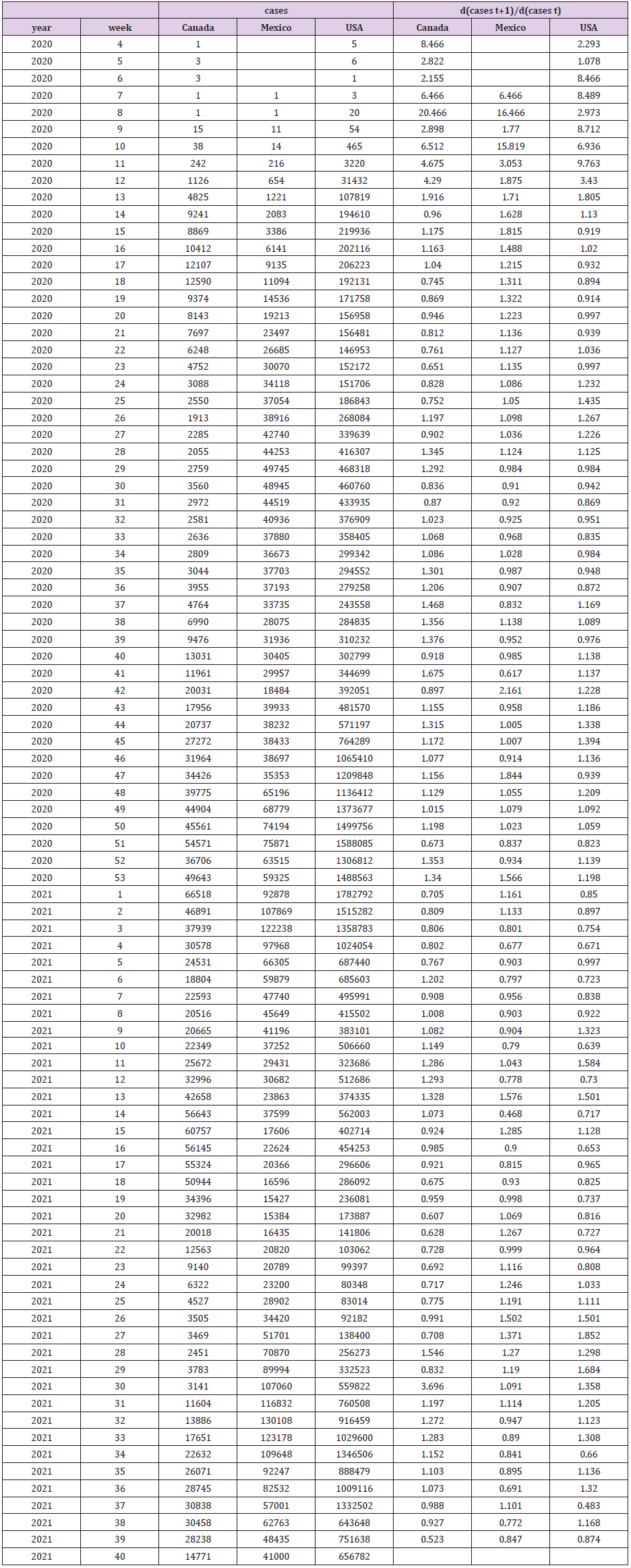
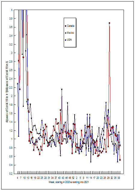
Depicted below are some general results observed during the study. It is important to remark that there was a shortage of amaranth during December, which impacted the biochemical data of the patients (Chart 1). The decrease in glucose levels is observed in the patients, being more significant during the blood glucose level measurements and the first four months of the amaranth intake regularly (Chart 2). This biochemical indicator increases throughout the study, attributed to the character of amaranth as a cereal, remembering that the usual diet of the patients did not undergo any modification (Chart 3). This indicator behavior in the patients significantly decreased between blood cholesterol samples at the beginning of the study and the first monitoring four months later, with total cholesterol increasing in the following two evaluation periods. However, to better understand this indicator was closely monitored with a lipid profile to characterize the cholesterol type (Chart 4). Throughout characterization of the cholesterol type found in the patients, the study showed a significant increase in HDL between six months and one year after the dietary intervention (Chart 5). Between six months and 12 months after the intervention with amaranth, the research registered an increase in LDL cholesterol levels. Although there was no significant weight loss, this component of body composition remained stable throughout the entire study group. Therefore the Body Mass Index prevailed stable (Table 1). About this indicator, the women in the study had a slight decrease in their hip circumference (Table 2). According to the results, the patients’ blood levels over a year evidenced a decrease in glucose between the initial and final states. Regarding triglycerides, these had an upward behavior in the patients. Concerning total cholesterol, this indicator presented fluctuations, and the initial and final evaluations were practically the same. The determinations of the lipid profile, the HDL of the patients rose significantly; LDL also showed an increase, although in a lesser proportion than HDL. About the levels of VLDL cholesterol, they remained without considerable change. Finally, the patients’ body weight did not increase. To summarize, Table 3 depicts the different variables that were analyzed using the Friedman test. This table shows the initial and final behavior of the parameters that were significant throughout the study: glucose, triglyceride, and cholesterol levels, as well as the HDL, LDL, and VLDL levels of the patients. Meanwhile, Table 4 shows that both cholesterol and VLDL did not change their levels between the initial and final states (Tables 5 & 6). On the other hand, the triglycerides, HDL, and LDL increased significantly compared to the decreased glucose level.

biomedres-openaccess-journal-bjstr

Table 1: Body mass index of the patients over a year.

biomedres-openaccess-journal-bjstr

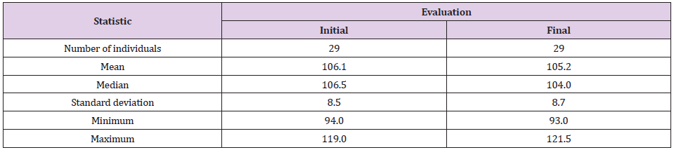
Table 2: Hip circumference (cm) of patients over a year.

biomedres-openaccess-journal-bjstr

Chart 1: Glucose levels behavior.

biomedres-openaccess-journal-bjstr

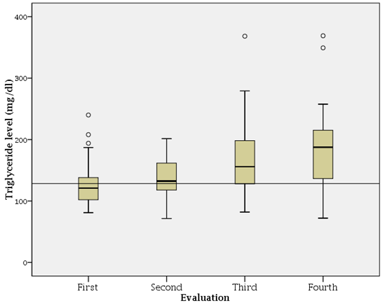
Chart 2: Triglyceride levels.

Conclusion

The study evidenced that the habitual consumption of amaranth has a positive effect determined from the amaranth active protein fraction with its inhibitory capacity of DPP-IV related to insulin secretion and the soluble fiber present in amaranth for glycemic control. The presence of polyunsaturated fats allowed an increase in HDL levels. Although the study identified that the amaranth’s behavior could be considered a functional food of habitual consumption for the DM2 patients, further studies among a more significant number of individuals will allow emitting that this cereal contributes to improving their quality of life.

Acknowledgement

This project was funded by por Fundación Mujer Banorte (2015-2016) and México Tierra de Amaranto AC.

For More Articles: Biomedical Journal Impact Factor: https://biomedres.us

Open Access Journals on Physiotherapy

Radial Extracorporeal Shockwave Therapy and A Supervised Exercise Programme in Patients with Lateral Elbow Tendinopathy? A Research Protocol

Background

Lateral elbow tendinopathy (LET), one of the most common lesions of the arm work-related or sport-related pain disorder, is the most appropriate term to use in clinical practice because all the other terms such as lateral epicondylalgia, lateral epicondylosis, lateral epicondylitis and/or tennis elbow make reference to inappropriate pathophysiological, aetiological, and anatomical terms [1]. LET is a syndrome of pain in the area of the lateral epicondyle [2] that may be failed healing tendon response or degenerative rather than inflammatory [3]. Hence, vascular hyperplasia, proteoglycans and glycosaminoglycans, the increased presence of fibroblasts together with disorganized and immature collagen may all take place in the absence of inflammatory cells [4]. The origin of the extensor carpi radialis brevis (ECRB) is the most commonly affected structure is [4]. The dominant arm is commonly affected, between 30 and 60 years of age is the peak prevalence of LET [2,5] and the disorder appears to be severed and of longer duration in women [3,6]. The main complaints of patients with LET are decreased function and pain [2,3]. A therapist should be able to reproduce the symptoms by:
a) Resisted wrist extension and/or resisted middle-finger extension with the elbow in extension,
b) Getting the patient to grip an object and
c) Digital palpation on the facet of the lateral epicondyle [2,3,5].

No ideal treatment has emerged for LET management. A conservative approach is advocated by many clinicians as the treatment of choice for LET [2,3,7,8]. Physiotherapy is usually recommended for LET patients [2-9]. A wide array of physiotherapy treatments has been recommended for the management of LET [10-14]. Such a variety of treatment techniques suggests that the optimal treatment management is not known, and more research is needed to find out the most effective treatment approach in patients with LET [10-14]. An exercise programme is the most common physiotherapy treatment for LET [2-14]. There are two types of exercise programs: home exercise programs and exercise programs carried out in a clinical setting. A home exercise program is commonly advocated for patients with LET because it can be performed any time during the day without requiring supervision by a physiotherapist. Our clinical experience, however, has shown that patients fail to comply with the regimen of home exercise programs [15]. This problem can be solved by exercise programs performed in a clinical setting under the supervision of a physiotherapist. For the purposes of this report, ‘‘supervised exercise program’’ will refer to such programs. Therefore, such a supervised exercise program will be used in the present trial.

Although a supervised exercise program is an effective treatment approach, a supplement to the exercise program should be found to reduce the treatment period. One such modality is Extracorporeal shockwave therapy (ESWT). ESWT is a safe noninvasive procedure [16] in which a device delivers acoustic energy (shockwaves) through the skin surface onto the affected area. Focused shockwaves are typically generated by electromagnetic or piezoelectric techniques [17]. Radial shockwave (RESWT) is nonfocused and generated by a ballistic source [18]. Recently research has started to be published on RESWT, which is relatively new and a promising modality to treat LET. ESWT may promote the release of angiogenetic growth and proliferating factors (e.g. vascular endothelial growth factor and endothelial nitric oxide synthase) that induces neovascularization for tissue regeneration [17]. Furthermore, ESWT may down-regulate inflammatory mediators and directly suppress nociceptors by hyperstimulation analgesia [19].

RESWT is applied only in the area of pain, in the case of LET in the ECRB. I wonder if it can be applied in a different way, like scanning the whole area for example muscles of the forearm or parallel to the elbow joint line. Perhaps, the combination of the above two ways of application, scanning and painful (sensitive) point, leads to a better clinical result. To our knowledge, the effectiveness of the above combination of RESWT in the management of LET has not been investigated. It is possible to combine a supervised exercise programme with RESWT, scanning and painful (sensitive) point, to see if the combination of the above reported therapeutic approaches offers superior results to supervised exercise programme and RESWT in the painful point in LET patients. Therefore, the aim of the present article will be to make a comparison of the effects of an exercise programme consisting of Tendon Neuroplastic Training (TNT) of wrist extensors, static stretching exercises of wrist extensors, isometric of wrist extensors, concentric – eccentric training of wrist extensors and strengthening of supinator, rotator cuff and scapula muscles exercises with RESWT, scanning and painful (sensitive) point and an exercise programme consisting of consisting of Tendon Neuroplastic Training (TNT) of wrist extensors, static stretching exercises of wrist extensors, isometric of wrist extensors, concentric – eccentric training of wrist extensors and strengthening of supinator, rotator cuff and scapula muscles exercises and RESWT in the painful point only for the treatment of LET.

Methods

To assess the effectiveness of an exercise programme with RESWT in the management of LET, a randomized controlled, monocentre trial will be conducted in the in the Laboratory of Neuromuscular and Cardiovascular Study of Motion (LANECASM) for 24 months. Crossover designs are limited in situations where patients are cured by the intervention and do not have the opportunity to receive the other treatments after crossover, therefore a parallel group design will be used [20]. Three investigators will participate in the study: (1) a physiotherapist (DB) who will perform all baseline and follow-up assessments, and will gain informed consent, (2) a physiotherapist, (MS), who will administer the treatments. and (3) the primary investigator, (DS) who will evaluate the patients to confirm the LET diagnosis and will allocate patients to groups. DB will interview each patient to ascertain clinical characteristics, including patient name, sex, age, duration of symptoms, previous treatment, occupation, affected arm and dominant arm and baseline demographic. To demonstrate statistical clinical significance for all outcome measures on LET a sample size of 25 subjects per group is sufficient. Measuring pain relief and functional outcomes in response to physiotherapeutic interventions such as low-power laser light, clinical effects of 20% had been reported as clinically meaningful in placebo-controlled studies. Baseline variance for pain and functional outcomes will be set at 25% in this study. Power calculations will recommend that 25 patients per group is sufficient to detect a 20% change in outcome measures, assuming that variance will be equivalent to 25% with a 5% significant level and 80% of power. The formula that will be used to determine the proper sample size will be:

N=16σ2/d2

where d2 is the effect size and σ2 is the variability of the data. For example, in our trial σ=25 and d=20. Therefore, the above formula is N=16(252)/ (202) =16χ625/400=25

Patients over 18 years old with lateral elbow pain will been evaluated and examined in the LANECASM in Athens between October 2021 and April 2023. In addition, persons with paraplegia, wheeled chair users and athletes with paraplegia as well as, can participate in the study. All patients will live in Greece, will speak Greek and will be either referred by their physician or physiotherapist or self-referred. Patients will participate in the study if, at the time of presentation, they will have been clinically diagnosed LET for at least 4 weeks. Patients will participate in the trial if they report (a) less pain during resistance supination with the elbow in 90∘ of flexion rather than in full extension and (b) pain on the facet of the lateral epicondyle when palpated and (c) pain in at least two of the following four tests [7]:
a) Handgrip dynamometer test.
b) Mill’s test (full passive flexion of the wrist)
c) Tomsen test (resisted wrist extension)
d) Resisted middle finger test
Patients will not participate in the study if they have one or more of the following conditions:
a. Radial nerve entrapment
b. Dysfunction in the shoulder, neck (radiculopathy) and/or thoracic region
c. Neurological deficit
d. Had received any conservative treatment for the management of LET in the 4 weeks before entering the study
e. The affected elbow had been operated on
f. Limitations in arm functions; and
g. Local or generalized arthritis [21-24].

A written explanation of the trial prior to entry into the study will be received by all patients. All patients will sign an informed consent to take part in the study. The study will be approved by the Ethics Committee of the University of West Attica. The allocation of the patients to two groups will be done randomly by drawing lots. Patients in Group A will be treated with a supervised exercise programme and RESWT in the painful point and patients in Group B will be treated with the supervised exercise programme and RESWT, scanning the relative area and painful (sensitive) point. All patients will be instructed to use their arm during the course of the study but to avoid activities that will irritate the elbow such as knitting, lifting, driving a car, using a screwdriver, grasping and handwriting. They will also be informed to refrain from taking pain killer drugs or other conservative treatment throughout the course of the study. A treatment diary will be used to monitor Patient compliance.

Interaction (verbal and non-verbal) and communication between the patient and therapist will be kept to a minimum, and behaviours sometimes used by physiotherapists to facilitate positive treatment outcomes will be purposefully avoided. For example, patients will be given no feedback on their performance in the pre-application and post-application measurements indication or any of the potentially beneficial effects of the treatments [25]. In both groups the elbow will be on the bed extended, the forearm pronated, the wrist in extension (and the hand hanging at the edge of the table. From this position, subjects will flex their wrists and then return to the extension (starting position). In the starting position, subjects will carry out an isometric contraction of wrist extensors. When the isometric contraction finishes the subjects will carry out the eccentric – concentric contraction and so on. The exercise will involve isolated wrist extension and flexion paced to an external audio/visual cue on the patients’ smartphone (PR Metronome; http://eumlab.com/pro-metronome/ ). Subjects will be to track the movement and listen to the sound of the metronome with their eyes. Each beat will be ten seconds apart, so that the pace of the metronome will be setted to 6 beats per minute. This will allow a ten second eccentric, concentric and isometric phase [26]. Both groups will perform three sets of 15 repetitions of slow progressive exercises of the wrist extensors at each treatment, with 1-min rest interval between each set. Subjects will be informed to continue with the exercise even if they complain of mild pain. However, subjects will be informed to stop the exercise if the pain becomes disabling. The disabling and mild pain will be monitored asking the subjects to rate the pain on VAS before and after treatment period. The definition of mild pain will be below 4 on VAS whereas the definition of disabling pain will be above 8 on VAS [21,23]. When subjects are able to carry out the exercise programme without experiencing any discomfort or pain, free weights will be used to increase the load.

Both treatment groups will perform static stretching exercises of the wrist extensors. Three times before and three times after the exercises six times totally will be repeated the static stretching exercises at each treatment session, with a 30 second rest interval between each repetition. The other hand will help to be performed the static stretching exercises of the wrist extensors. The patient’s elbow will be placed in extended position, the forearm in pronated position, and the wrist in ulnar deviation and flexion according to the patient’s tolerance. 30–45 seconds will be holding this position each time and then releases [21-24]. Furthermore, the scapular and rotator cuff muscles will be strengthened. The strengthening exercises will be included
(i) Shoulder lateral rotation and medial with the elbow in 900 and 00 of abduction
(ii) (Shoulder abduction to 900 with flexed elbow:
(iii) Scaption and
(iv) Diagonal pattern from flexion to extension [27].

Upper trapezius, rhomboids, serratus anterior and levator scapulae will be also strengthened [28]. Each exercise will be carried out twice at each treatment with 12 repetitions in each set and 1 min rest interval between each set [29]. Subjects will be informed to continue with the exercise even if they will complain of mild pain. However, subjects will be informed to stop the exercise if the pain became disabling. When subjects will be able to carry out the strengthening exercises without experiencing any discomfort or pain, the load will be increased using therabands or free weights.

Finally, the supinator will be strengthened. Strengthening exercises of the supinator will be carried out with the elbow extended on the table, the forearm pronated, the wrist in mid – position and the hand hanging over the edge of the table. From this position, the patient will supinate their arm slowly while counting to 15 using chronometer, then return to the starting position (pronation) [30]. The above reported exercise programme will be followed five times per week for 4 weeks and will be individualized on the basis of the patient’s description of pain experienced during the process. RESWT will be administered without anaesthesia in the most sensitive point on patient’s lateral epicondyle in Group A. Each patient will receive a total of 2000 shocks per session (8 Hz; 2,5 bars). Each subject will participate in 3 treatment sessions held at weekly intervals. RESWT will be also administered without anaesthesia in Group B. First 2000 shocks (8 Hz, 2.5 bars) will be applied to the most sensitive point of the lateral epicondyle and then 2000 shocks to the dorsal part of the forearm using the same frequency and pressure variables. Each patient will participate in 3 treatment sessions held at weekly intervals. Function, drop-out rate and pain will be measured in the present trial. Each subject will be evaluated at the beginning of the treatment (week 0), at the end of treatment (week 4), at 3 months after the end of the treatment (week 16) and at 6 months (28weeks) after the end of treatment.

A visual analogue scale (VAS) will be used to measure pain. 0 (cm) on VAS means “least pain imaginable” and 10 (cm) on VAS means “worst pain imaginable”. The pain VAS will measure the patient’s worst level of pain over the previous 24 h before each evaluation, and this approach has been shown to be sensitive and valid of the VAS [31]. A visual analogue scale (VAS) will be used to measure function. 0 (cm) on VAS means “no function” and 10 (cm) on VAS means “full function”. Subjects will be informed to report their overall level of elbow function over the previous 24 h before each measurement, and this approach has been shown to be sensitive and valid of the VAS [31]. The Patient-Rated Tennis Elbow Evaluation (PRTEE) Questionnaire will be used to assess pain and function. Patients rate their pain and functional limitation on a scale of 0e10, with 0 being no difficulty and 10 being unable to perform. The scores for the various items are used to calculate an overall scale score ranging from 0 (best score) to 100 (worst score). The scale is scored such that 50% of the score is obtained by summing the five pain items and the remaining 50% is by obtained by summing the 10 functional (specific and usual activity) items and then divided that subtotal by 2. This creates a score out of 100 points where 100 is the highest level of pain and disability. The PRTEE questionnaire, provides a very quick (it takes less than 5 min to complete), easy, and standardized quantitative description of pain and functional disability in patients with LET. The Greek version of PRTEE Questionnaire is a reliable and valid measure when administered to patients with LET [32].

Moreover, pain-free grip strength will be used to measure function. The definition of Pain-free grip strength is the amount of force each patient is able to generate with an isometric gripping action before monitoring pain [25]. A Jamar hand dynamometer that had adjustable handles to accommodate different hand sizes will be used to measure force in pounds. The elbow will be placed in extension, forearm in pronation and internal rotation such that the palmar aspect of the hand faced posteriorly with the arm placed by the patient’s side. Subjects will be then informed to squeeze the dynamometer handles until they will first experience pain and then to release their grip [25]. The attained grip force will be subsequently recorded. The reading will not be visible to the subjects. Three measures of pain-free grip strength will be determined with a 30 seconds rest interval between each effort, and the mean value of these repetitions will be calculated. The rate of drop-out will also be used as an indicator of treatment outcome. Reasons for patient drop out will be categorised as follows:
a) Not returned for follow-up
b) Request for an alternative treatment and
c) A withdraw without reason.

The change from the beginning of the treatment will be calculated for each follow-up. The independent t test will be used to determine the differences between groups. A paired t test will be used to analyze the difference within groups between beginning of the treatment and end of treatment. The level for statistical significance will be adopted as a 5% level of probability. The statistical analysis will be carried out using the SPSS 21.00 statistical software.

Discussion

The main aim of this RCT is to investigate the effectiveness of two physiotherapy treatments in improving function, strength, and pain in LET patients at the end of treatment, at 3 months follow-up and at 6 months follow up. It is expected to examine the following null hypothesis: “there is no difference in function and pain for subjects undergoing physiotherapy intervention with RESWT in the painful point and RESWT, scanning the relative area and painful (sensitive) point”. Many treatments have been recommended for the management of LET. However, there is not the gold standard treatment. Eccentric contraction is recommended [33,34] over other types of contractions for the management of LET. On the other hand, Malliaras and his colleagues [35] proposed that therapists should consider eccentric-concentric loading alongside or instead of eccentric loading. Martinez-Silvestrini et al. [36] concluded that, unlike Achilles’ tendinopathy, LET is often related to forceful grip activities requiring isometric contraction, which would be more beneficial than eccentric contraction in LET. Recently, isometric exercises have been recommended to manage and reduce tendon pain improving the strength at the angle of contraction without producing inflammatory signs [37]. The exercise program in LET should include strengthening exercises for supinator, rotator cuff and scapular muscles [38,39]. Moreover, proprioception is also reduced in LET patients [40]. Techniques to improve the reduced proprioception is also recommended. Furthermore, tendon neuroplastic training (TNT) is needed combining isometric or isotonic strength training with an externally paced audio or visual cue [41]. Finally, stretching has positive effects in the management of tendon injuries such as LET. Τhe aim of stretching is to orientate the new collagen fibres, experience consequently less strain during joint motion and lengthen the muscle-tendon unit, [42-44].

Although a supervised exercise program is an effective treatment approach, a supplement to the exercise program should be found to reduce the treatment period. One such modality is RESWT which is a relatively new treatment approach, but it is reported to be used by clinicians worldwide [45]. Karanasios et al. [46] found in their systematic review that ESWT (radial and focused) offers no benefit in the management of LET patients. On the other hand, two published meta-analyses in 2020 [47,48] reported superior outcomes of the ESWT (radial and focused) compared with sham or other passive treatments in pain reduction and function improvement. The above recommendations should be treated with caution because the three reported mentioned studies examined the effectiveness of ESWT as monotherapy in the management of LET. However, ESWT is rarely applied as a monotherapy in clinical practice. ESWT does not use as a substitute for exercise but as a supplement to exercise program. The present study will be the first trial to examine the effectiveness of RESWT, scanning the relative area and painful (sensitive) point, on chronic LET. A radial shock wave stimulates a much larger area of tissue than a focused shock wave. The effective focal zone of the latter is very small [49], thus, the area of affected tissue that can be treated is also small. The radial shock wave allows treating the original site of the “problem” (e.g. the lateral epicondyle), as well as other affected areas.

One previous pilot study assessed the effectiveness of this RESWT approach on chronic LET [50]. However, the purpose of this article was to evaluate and compare the efficacy of radial and focused shock wave therapies applied to treat LET and no to compare two applications of RESWT in the management of LET, as in our study. The findings of these two trials will encourage the design of future well-designed trials that might produce strong evidence for the effectiveness of RESWT on LET management. RESWT is a dose-response modality and the optimal treatment dose has obviously not yet been discovered. Future studies are needed to standardize RESWR parameters in the management of LET (acute, chronic and calcific).

Conclusion

This is the first trial to examine the efficacy of RESWT, scanning the relative area and painful (sensitive) point, in the treatment of LET. It is expected, the conclusion of the trial to improve the scientific knowledge providing evidence that using RESWT is an effective and safe tool in the treatment of LET symptoms, specifically function and pain.

For More Articles: Biomedical Journal Impact Factor: https://biomedres.us

Open Access Journals on Medicine

Radial Extracorporeal Shockwave Therapy and A Supervised Exercise Programme in Patients with Lateral Elbow Tendinopathy? A Research Protocol

Background

Lateral elbow tendinopathy (LET), one of the most common lesions of the arm work-related or sport-related pain disorder, is the most appropriate term to use in clinical practice because all the other terms such as lateral epicondylalgia, lateral epicondylosis, lateral epicondylitis and/or tennis elbow make reference to inappropriate pathophysiological, aetiological, and anatomical terms [1]. LET is a syndrome of pain in the area of the lateral epicondyle [2] that may be failed healing tendon response or degenerative rather than inflammatory [3]. Hence, vascular hyperplasia, proteoglycans and glycosaminoglycans, the increased presence of fibroblasts together with disorganized and immature collagen may all take place in the absence of inflammatory cells [4]. The origin of the extensor carpi radialis brevis (ECRB) is the most commonly affected structure is [4]. The dominant arm is commonly affected, between 30 and 60 years of age is the peak prevalence of LET [2,5] and the disorder appears to be severed and of longer duration in women [3,6]. The main complaints of patients with LET are decreased function and pain [2,3]. A therapist should be able to reproduce the symptoms by:
a) Resisted wrist extension and/or resisted middle-finger extension with the elbow in extension,
b) Getting the patient to grip an object and
c) Digital palpation on the facet of the lateral epicondyle [2,3,5].

No ideal treatment has emerged for LET management. A conservative approach is advocated by many clinicians as the treatment of choice for LET [2,3,7,8]. Physiotherapy is usually recommended for LET patients [2-9]. A wide array of physiotherapy treatments has been recommended for the management of LET [10-14]. Such a variety of treatment techniques suggests that the optimal treatment management is not known, and more research is needed to find out the most effective treatment approach in patients with LET [10-14]. An exercise programme is the most common physiotherapy treatment for LET [2-14]. There are two types of exercise programs: home exercise programs and exercise programs carried out in a clinical setting. A home exercise program is commonly advocated for patients with LET because it can be performed any time during the day without requiring supervision by a physiotherapist. Our clinical experience, however, has shown that patients fail to comply with the regimen of home exercise programs [15]. This problem can be solved by exercise programs performed in a clinical setting under the supervision of a physiotherapist. For the purposes of this report, ‘‘supervised exercise program’’ will refer to such programs. Therefore, such a supervised exercise program will be used in the present trial.

Although a supervised exercise program is an effective treatment approach, a supplement to the exercise program should be found to reduce the treatment period. One such modality is Extracorporeal shockwave therapy (ESWT). ESWT is a safe noninvasive procedure [16] in which a device delivers acoustic energy (shockwaves) through the skin surface onto the affected area. Focused shockwaves are typically generated by electromagnetic or piezoelectric techniques [17]. Radial shockwave (RESWT) is nonfocused and generated by a ballistic source [18]. Recently research has started to be published on RESWT, which is relatively new and a promising modality to treat LET. ESWT may promote the release of angiogenetic growth and proliferating factors (e.g. vascular endothelial growth factor and endothelial nitric oxide synthase) that induces neovascularization for tissue regeneration [17]. Furthermore, ESWT may down-regulate inflammatory mediators and directly suppress nociceptors by hyperstimulation analgesia [19].

RESWT is applied only in the area of pain, in the case of LET in the ECRB. I wonder if it can be applied in a different way, like scanning the whole area for example muscles of the forearm or parallel to the elbow joint line. Perhaps, the combination of the above two ways of application, scanning and painful (sensitive) point, leads to a better clinical result. To our knowledge, the effectiveness of the above combination of RESWT in the management of LET has not been investigated. It is possible to combine a supervised exercise programme with RESWT, scanning and painful (sensitive) point, to see if the combination of the above reported therapeutic approaches offers superior results to supervised exercise programme and RESWT in the painful point in LET patients. Therefore, the aim of the present article will be to make a comparison of the effects of an exercise programme consisting of Tendon Neuroplastic Training (TNT) of wrist extensors, static stretching exercises of wrist extensors, isometric of wrist extensors, concentric – eccentric training of wrist extensors and strengthening of supinator, rotator cuff and scapula muscles exercises with RESWT, scanning and painful (sensitive) point and an exercise programme consisting of consisting of Tendon Neuroplastic Training (TNT) of wrist extensors, static stretching exercises of wrist extensors, isometric of wrist extensors, concentric – eccentric training of wrist extensors and strengthening of supinator, rotator cuff and scapula muscles exercises and RESWT in the painful point only for the treatment of LET.

Methods

To assess the effectiveness of an exercise programme with RESWT in the management of LET, a randomized controlled, monocentre trial will be conducted in the in the Laboratory of Neuromuscular and Cardiovascular Study of Motion (LANECASM) for 24 months. Crossover designs are limited in situations where patients are cured by the intervention and do not have the opportunity to receive the other treatments after crossover, therefore a parallel group design will be used [20]. Three investigators will participate in the study: (1) a physiotherapist (DB) who will perform all baseline and follow-up assessments, and will gain informed consent, (2) a physiotherapist, (MS), who will administer the treatments. and (3) the primary investigator, (DS) who will evaluate the patients to confirm the LET diagnosis and will allocate patients to groups. DB will interview each patient to ascertain clinical characteristics, including patient name, sex, age, duration of symptoms, previous treatment, occupation, affected arm and dominant arm and baseline demographic. To demonstrate statistical clinical significance for all outcome measures on LET a sample size of 25 subjects per group is sufficient. Measuring pain relief and functional outcomes in response to physiotherapeutic interventions such as low-power laser light, clinical effects of 20% had been reported as clinically meaningful in placebo-controlled studies. Baseline variance for pain and functional outcomes will be set at 25% in this study. Power calculations will recommend that 25 patients per group is sufficient to detect a 20% change in outcome measures, assuming that variance will be equivalent to 25% with a 5% significant level and 80% of power. The formula that will be used to determine the proper sample size will be:

N=16σ2/d2

where d2 is the effect size and σ2 is the variability of the data. For example, in our trial σ=25 and d=20. Therefore, the above formula is N=16(252)/ (202) =16χ625/400=25

Patients over 18 years old with lateral elbow pain will been evaluated and examined in the LANECASM in Athens between October 2021 and April 2023. In addition, persons with paraplegia, wheeled chair users and athletes with paraplegia as well as, can participate in the study. All patients will live in Greece, will speak Greek and will be either referred by their physician or physiotherapist or self-referred. Patients will participate in the study if, at the time of presentation, they will have been clinically diagnosed LET for at least 4 weeks. Patients will participate in the trial if they report (a) less pain during resistance supination with the elbow in 90∘ of flexion rather than in full extension and (b) pain on the facet of the lateral epicondyle when palpated and (c) pain in at least two of the following four tests [7]:
a) Handgrip dynamometer test.
b) Mill’s test (full passive flexion of the wrist)
c) Tomsen test (resisted wrist extension)
d) Resisted middle finger test
Patients will not participate in the study if they have one or more of the following conditions:
a. Radial nerve entrapment
b. Dysfunction in the shoulder, neck (radiculopathy) and/or thoracic region
c. Neurological deficit
d. Had received any conservative treatment for the management of LET in the 4 weeks before entering the study
e. The affected elbow had been operated on
f. Limitations in arm functions; and
g. Local or generalized arthritis [21-24].

A written explanation of the trial prior to entry into the study will be received by all patients. All patients will sign an informed consent to take part in the study. The study will be approved by the Ethics Committee of the University of West Attica. The allocation of the patients to two groups will be done randomly by drawing lots. Patients in Group A will be treated with a supervised exercise programme and RESWT in the painful point and patients in Group B will be treated with the supervised exercise programme and RESWT, scanning the relative area and painful (sensitive) point. All patients will be instructed to use their arm during the course of the study but to avoid activities that will irritate the elbow such as knitting, lifting, driving a car, using a screwdriver, grasping and handwriting. They will also be informed to refrain from taking pain killer drugs or other conservative treatment throughout the course of the study. A treatment diary will be used to monitor Patient compliance.

Interaction (verbal and non-verbal) and communication between the patient and therapist will be kept to a minimum, and behaviours sometimes used by physiotherapists to facilitate positive treatment outcomes will be purposefully avoided. For example, patients will be given no feedback on their performance in the pre-application and post-application measurements indication or any of the potentially beneficial effects of the treatments [25]. In both groups the elbow will be on the bed extended, the forearm pronated, the wrist in extension (and the hand hanging at the edge of the table. From this position, subjects will flex their wrists and then return to the extension (starting position). In the starting position, subjects will carry out an isometric contraction of wrist extensors. When the isometric contraction finishes the subjects will carry out the eccentric – concentric contraction and so on. The exercise will involve isolated wrist extension and flexion paced to an external audio/visual cue on the patients’ smartphone (PR Metronome; http://eumlab.com/pro-metronome/ ). Subjects will be to track the movement and listen to the sound of the metronome with their eyes. Each beat will be ten seconds apart, so that the pace of the metronome will be setted to 6 beats per minute. This will allow a ten second eccentric, concentric and isometric phase [26]. Both groups will perform three sets of 15 repetitions of slow progressive exercises of the wrist extensors at each treatment, with 1-min rest interval between each set. Subjects will be informed to continue with the exercise even if they complain of mild pain. However, subjects will be informed to stop the exercise if the pain becomes disabling. The disabling and mild pain will be monitored asking the subjects to rate the pain on VAS before and after treatment period. The definition of mild pain will be below 4 on VAS whereas the definition of disabling pain will be above 8 on VAS [21,23]. When subjects are able to carry out the exercise programme without experiencing any discomfort or pain, free weights will be used to increase the load.

Both treatment groups will perform static stretching exercises of the wrist extensors. Three times before and three times after the exercises six times totally will be repeated the static stretching exercises at each treatment session, with a 30 second rest interval between each repetition. The other hand will help to be performed the static stretching exercises of the wrist extensors. The patient’s elbow will be placed in extended position, the forearm in pronated position, and the wrist in ulnar deviation and flexion according to the patient’s tolerance. 30–45 seconds will be holding this position each time and then releases [21-24]. Furthermore, the scapular and rotator cuff muscles will be strengthened. The strengthening exercises will be included
(i) Shoulder lateral rotation and medial with the elbow in 900 and 00 of abduction
(ii) (Shoulder abduction to 900 with flexed elbow:
(iii) Scaption and
(iv) Diagonal pattern from flexion to extension [27].

Upper trapezius, rhomboids, serratus anterior and levator scapulae will be also strengthened [28]. Each exercise will be carried out twice at each treatment with 12 repetitions in each set and 1 min rest interval between each set [29]. Subjects will be informed to continue with the exercise even if they will complain of mild pain. However, subjects will be informed to stop the exercise if the pain became disabling. When subjects will be able to carry out the strengthening exercises without experiencing any discomfort or pain, the load will be increased using therabands or free weights.

Finally, the supinator will be strengthened. Strengthening exercises of the supinator will be carried out with the elbow extended on the table, the forearm pronated, the wrist in mid – position and the hand hanging over the edge of the table. From this position, the patient will supinate their arm slowly while counting to 15 using chronometer, then return to the starting position (pronation) [30]. The above reported exercise programme will be followed five times per week for 4 weeks and will be individualized on the basis of the patient’s description of pain experienced during the process. RESWT will be administered without anaesthesia in the most sensitive point on patient’s lateral epicondyle in Group A. Each patient will receive a total of 2000 shocks per session (8 Hz; 2,5 bars). Each subject will participate in 3 treatment sessions held at weekly intervals. RESWT will be also administered without anaesthesia in Group B. First 2000 shocks (8 Hz, 2.5 bars) will be applied to the most sensitive point of the lateral epicondyle and then 2000 shocks to the dorsal part of the forearm using the same frequency and pressure variables. Each patient will participate in 3 treatment sessions held at weekly intervals. Function, drop-out rate and pain will be measured in the present trial. Each subject will be evaluated at the beginning of the treatment (week 0), at the end of treatment (week 4), at 3 months after the end of the treatment (week 16) and at 6 months (28weeks) after the end of treatment.

A visual analogue scale (VAS) will be used to measure pain. 0 (cm) on VAS means “least pain imaginable” and 10 (cm) on VAS means “worst pain imaginable”. The pain VAS will measure the patient’s worst level of pain over the previous 24 h before each evaluation, and this approach has been shown to be sensitive and valid of the VAS [31]. A visual analogue scale (VAS) will be used to measure function. 0 (cm) on VAS means “no function” and 10 (cm) on VAS means “full function”. Subjects will be informed to report their overall level of elbow function over the previous 24 h before each measurement, and this approach has been shown to be sensitive and valid of the VAS [31]. The Patient-Rated Tennis Elbow Evaluation (PRTEE) Questionnaire will be used to assess pain and function. Patients rate their pain and functional limitation on a scale of 0e10, with 0 being no difficulty and 10 being unable to perform. The scores for the various items are used to calculate an overall scale score ranging from 0 (best score) to 100 (worst score). The scale is scored such that 50% of the score is obtained by summing the five pain items and the remaining 50% is by obtained by summing the 10 functional (specific and usual activity) items and then divided that subtotal by 2. This creates a score out of 100 points where 100 is the highest level of pain and disability. The PRTEE questionnaire, provides a very quick (it takes less than 5 min to complete), easy, and standardized quantitative description of pain and functional disability in patients with LET. The Greek version of PRTEE Questionnaire is a reliable and valid measure when administered to patients with LET [32].

Moreover, pain-free grip strength will be used to measure function. The definition of Pain-free grip strength is the amount of force each patient is able to generate with an isometric gripping action before monitoring pain [25]. A Jamar hand dynamometer that had adjustable handles to accommodate different hand sizes will be used to measure force in pounds. The elbow will be placed in extension, forearm in pronation and internal rotation such that the palmar aspect of the hand faced posteriorly with the arm placed by the patient’s side. Subjects will be then informed to squeeze the dynamometer handles until they will first experience pain and then to release their grip [25]. The attained grip force will be subsequently recorded. The reading will not be visible to the subjects. Three measures of pain-free grip strength will be determined with a 30 seconds rest interval between each effort, and the mean value of these repetitions will be calculated. The rate of drop-out will also be used as an indicator of treatment outcome. Reasons for patient drop out will be categorised as follows:
a) Not returned for follow-up
b) Request for an alternative treatment and
c) A withdraw without reason.

The change from the beginning of the treatment will be calculated for each follow-up. The independent t test will be used to determine the differences between groups. A paired t test will be used to analyze the difference within groups between beginning of the treatment and end of treatment. The level for statistical significance will be adopted as a 5% level of probability. The statistical analysis will be carried out using the SPSS 21.00 statistical software.

Discussion

The main aim of this RCT is to investigate the effectiveness of two physiotherapy treatments in improving function, strength, and pain in LET patients at the end of treatment, at 3 months follow-up and at 6 months follow up. It is expected to examine the following null hypothesis: “there is no difference in function and pain for subjects undergoing physiotherapy intervention with RESWT in the painful point and RESWT, scanning the relative area and painful (sensitive) point”. Many treatments have been recommended for the management of LET. However, there is not the gold standard treatment. Eccentric contraction is recommended [33,34] over other types of contractions for the management of LET. On the other hand, Malliaras and his colleagues [35] proposed that therapists should consider eccentric-concentric loading alongside or instead of eccentric loading. Martinez-Silvestrini et al. [36] concluded that, unlike Achilles’ tendinopathy, LET is often related to forceful grip activities requiring isometric contraction, which would be more beneficial than eccentric contraction in LET. Recently, isometric exercises have been recommended to manage and reduce tendon pain improving the strength at the angle of contraction without producing inflammatory signs [37]. The exercise program in LET should include strengthening exercises for supinator, rotator cuff and scapular muscles [38,39]. Moreover, proprioception is also reduced in LET patients [40]. Techniques to improve the reduced proprioception is also recommended. Furthermore, tendon neuroplastic training (TNT) is needed combining isometric or isotonic strength training with an externally paced audio or visual cue [41]. Finally, stretching has positive effects in the management of tendon injuries such as LET. Τhe aim of stretching is to orientate the new collagen fibres, experience consequently less strain during joint motion and lengthen the muscle-tendon unit, [42-44].

Although a supervised exercise program is an effective treatment approach, a supplement to the exercise program should be found to reduce the treatment period. One such modality is RESWT which is a relatively new treatment approach, but it is reported to be used by clinicians worldwide [45]. Karanasios et al. [46] found in their systematic review that ESWT (radial and focused) offers no benefit in the management of LET patients. On the other hand, two published meta-analyses in 2020 [47,48] reported superior outcomes of the ESWT (radial and focused) compared with sham or other passive treatments in pain reduction and function improvement. The above recommendations should be treated with caution because the three reported mentioned studies examined the effectiveness of ESWT as monotherapy in the management of LET. However, ESWT is rarely applied as a monotherapy in clinical practice. ESWT does not use as a substitute for exercise but as a supplement to exercise program. The present study will be the first trial to examine the effectiveness of RESWT, scanning the relative area and painful (sensitive) point, on chronic LET. A radial shock wave stimulates a much larger area of tissue than a focused shock wave. The effective focal zone of the latter is very small [49], thus, the area of affected tissue that can be treated is also small. The radial shock wave allows treating the original site of the “problem” (e.g. the lateral epicondyle), as well as other affected areas.

One previous pilot study assessed the effectiveness of this RESWT approach on chronic LET [50]. However, the purpose of this article was to evaluate and compare the efficacy of radial and focused shock wave therapies applied to treat LET and no to compare two applications of RESWT in the management of LET, as in our study. The findings of these two trials will encourage the design of future well-designed trials that might produce strong evidence for the effectiveness of RESWT on LET management. RESWT is a dose-response modality and the optimal treatment dose has obviously not yet been discovered. Future studies are needed to standardize RESWR parameters in the management of LET (acute, chronic and calcific).

Conclusion

This is the first trial to examine the efficacy of RESWT, scanning the relative area and painful (sensitive) point, in the treatment of LET. It is expected, the conclusion of the trial to improve the scientific knowledge providing evidence that using RESWT is an effective and safe tool in the treatment of LET symptoms, specifically function and pain.

For More Articles: Biomedical Journal Impact Factor: https://biomedres.us

Open Access Journals on Ophthalmology

Intraoperative Anterior Segment Ocular Coherence Tomography in Ophthalmic Surgery

Introduction

Optical Coherence Tomography (OCT) is a non-contact imaging technology that produces detailed cross-sectional images, using low-coherence interferometry in biological tissues. [1] Anterior Segment OCT (AS-OCT) is an innovative tool for evaluation of the cornea, conjunctiva, sclera, anterior chamber (AC), anterior chamber angle, intraocular lens, and adjacent anterior segment structures. It has been helpful for the diagnosis and management of conjunctival diseases, anterior segment tumors, Corneal diseases, and AC inflammation. [2] Standard clinical OCT systems are large and stationary. In the past few years, the OCT became an important tool for selected ophthalmic surgeries, and adaptations had to be made to convert the OCT to a tabletop system. Today there are several commercial systems of microscope-integrated intraoperative OCT (iOCT), which focuses on OCT video visualization in highresolution, maximum integration with the microscope, and some of them integrate a three-dimensional heads-up display system for maximum convenience of the surgeon while visualizing the surgical field. [3]

Intra-Operative Oct

The iOCT is a rather new technology and it is currently applied in a wide variety of ophthalmic surgeries. The AS-OCT is used in lamellar keratoplasty, Penetrating keratoplasty (PKP), cataract surgeries, glaucoma surgeries, and other corneal procedures (post-trauma, epithelial/fibrous growth). The posterior segment OCT aids in cases of optic pit-related maculopathy, retinopathy of premature, macular hole, retinal detachment, proliferative diabetic retinopathy, epiretinal membrane, and posterior uveitis. [3] Over the years, two major studies were conducted to test the benefits of the intra-operative OCT (iOCT)- the PIONEER study and the DISCOVER study. Both studies collected Clinical characteristics and iOCT imaging was obtained during surgical milestones as directed by the operating surgeon. A surgeon questionnaire was issued to each surgeon and completed after each case. The PIONEER study was published in 2014 and enrolled 531 eyes- 275 anterior segment cases and 256 posterior segment cases. In the anterior segment surgeries, the most common procedure was Descemet stripping automated endothelial keratoplasty (DSAEK, n=135), and the second most common was cataract extraction with an intraocular lens implant.
Immediately after the surgery, the surgeon was required to fill a feedback form. Overall following iOCT, 48% of the eyes revealed persistent interface fluid requiring additional manipulations. In deep anterior lamellar keratoplasty (DALK), 1 of 3 cases where the surgeon did not believe the trephination was deep enough, iOCT revealed the depth was optimal and did not require further deepening. In 56% of the cases of DALK, iOCT prompted further manual dissection to deepen the initial trephination. [4] The DISCOVER study was published in 2015, enrolled 227 eyes, 91 of them were anterior segment cases. The most common procedure was DSAEK (43%), following DALK (9%). In this study, 8% of the cases were Descemet endothelial keratoplasty (DMEK) cases (in the PIONEER study there were no DMEK cases at all). According to surgeons in the study, 44% of the total anterior segment cases were changed or modified due to the iOCT findings. [5] Regarding the influence of iOCT on surgical time, the DISCOVER study concluded that in 47% of the cases, the iOCT minimized the surgical time by eliminating unnecessary manipulations but did not measure the minutes that were spared. Recent studies of iOCT use in anterior segment procedures (Figure 1).

biomedres-openaccess-journal-bjstr

Figure 1: Corneal staff in Hadassah Hospital during DMEK procedure, using iOCT, ARTEVO 800 of produced by ZIESS.

DALK

The DALK procedure gained popularity as an alternative to PKP. The “big-bubble” technique uses a forceful injection of air into the deep stroma, to create cleavage and to separate the Descemet membrane (DM) from the overlying stroma. The reported rate of successfully achieving a big bubble is 66% to 90%. For novice surgeons, the step of big-bubble generation is the most common surgical step at which perforation of DM occurs. [6] According to Myerscough et al, there are two main advantages of iOCT in DALK procedure – the first is assessing the depth of the cannula tip before performing pneumatic dissection, and the second is confirming that pneumatic dissection has indeed occurred. [7] Liu et al presented a new approach for DALK surgery in which a low-energy FSL created an anterior stromal dissection and a pre-Descemet intrastromal tunnel for the air injection in one step, to a preprogrammed depth, with the guidance of the iOCT and to facilitate big-bubble creation. 14 eyes were included: 11 eyes with keratoconus and no evidence of a history of acute hydrops and 3 eyes with corneal scars. In all cases, a big bubble was successfully achieved without intraoperative complications related to lasers (Figure 2).

biomedres-openaccess-journal-bjstr

Figure 2: A 30-year-old woman with advanced keratoconus, in DALK procedure using iOCT.
1. Picture number 1: separating the stroma layer from the DM, with the cannula corneal plane clearly visible.
2. Picture number 2: During air injection (Big bubble), the air reached the stroma, and caused an emphysema.
3. Picture number 3: The emphysema in the stroma is seen, and there is an additional air bubble underneath the stroma, separating it from the pre-Descemet. This separation succeeded after six attempted injections of air. The iOCT was a crucial factor is the success of this procedure.
4. Picture number 4: Removing the stroma. The pre-Descemet is exposed.
5. Picture number 5: after full removal of the stroma.
6. Picture number 6: After suturing the graft- it is visible in the OCT that it is attached properly to the DM

DSAEK

Most of the data about the DSAEK procedure still relies on the data drawn from the PIONEER study, due to its relatively large cohort. The two main surgical techniques of DSAEK differ in the air infused to the anterior chamber- one uses an active air infusion system while the other manually introduce air into the anterior chamber for graft positioning. Hallahan, et al analysed the fluid dynamics and clinical outcomes for iOCT-assisted DSAEK from the PIONEER study. They used a few features measured with the iOCT and discovered that high amounts of interface fluid significantly correlated with graft non-adherence rates within the first postoperative week, following placement and optimization of intraoperative lenticule adherence. Also, the iOCT revealed a significant difference between the area, volume, and thickness of maximum fluid pockets between the two surgical techniques- the manual technique had higher values, but in both techniques, there was a significant reduction of interface fluid during the procedure [8].

DMEK

DMEK is currently the preferred procedure to replace diseased endothelium and is superior to DSAEK and PKP for visual recovery and a lower graft rejection rate. On the other hand, the surgeon must be skilled to perform this procedure. The difficult steps are preparation of the donor lamella, transfer of the graft into the AC, unfolding and orientation of the graft, and successful attachment after air filling. Another major challenge is operating through a hazy and/or scarred cornea, which makes the visibility obscure. [9-10] Muijzer, et al. examined 38 eyes which underwent DMEK procedure. iOCT was used during the surgery and its main steps. In 42% of the cases, the iOCT altered the surgical decision-making process. In 21% of the surgeries, OCT revealed interface fluid or a minor detachment of the graft, findings that were not noted using the en face surgical microscope view; In 32% of surgeries, the iOCT image provided crucial information regarding the graft orientation [10].

Traumatic/Post-Operative Damage

Epithelial downgrowth is a rare complication of intraocular surgery or trauma characterized by the invasion of surface epithelial cells into the AC of the eye. Fibrous downgrowth is a similar but somewhat less aggressive condition characterized by fibrovascular connective tissue invading into the eye. Those pathologies may have a devastating sequela. There are a few case reports published that emphasized the utility of iOCT in those uneventful cases. Shazly, et al. discussed a case of a woman that underwent a DSAEK procedure in an eye with fibrous ingrowth and 2 glaucoma shunt devices. The iOCT provided a clear dissection plane of the fibrous membranes and a clear view of their relation to the iris and corneal endothelium. This proved to be valuable given poor visibility through the opacified cornea. In addition, it allowed determining the extent and location of the interface fluid gap between the DSAEK graft and the host cornea. [11] Ruland, et al. published a case of a woman with primary open angle glaucoma and corneal decompensation of the right eye secondary to tube shunt presented for a 3-month followup of PKP. The patient had a membrane connecting the iris to the host cornea and underwent a biopsy and excision of the membrane assisted by iOCT. The poor view of the peripheral anterior chamber secondary to recent corneal transplantation was aided using iOCT, especially in the manipulation and acquisition of material for biopsy [12] (Figure 3).

biomedres-openaccess-journal-bjstr

Figure 3: A 23-year-old patient who suffered an eye trauma 10 years ago. He underwent vitrectomy with intraocular lens implantation successfully. After 10 stable years, his intra-ocular pressure increased uncontrollably, and an Ahmed valve was eventually implanted with a suspicion of epithelial downgrowth. Later, the patient underwent an ingrowth membrane removal with 5-Fluorouracyl treatment and followed by eight Methotrexate injections to the AC twice a month. Then he was referred to remove the remained tissue, opening the Ahmed valve and go through a DMEK procedure.
1. Picture number 1: During the insertion of the maintainer the surgeon came across a tissue which cover the whole corneal endothelium and the Ahmed valve opening.
2. Picture number 2: The tissue is demonstrated in the iOCT
3. Picture number 3: Excision of the tissue by pulling it through a limbal incision.
4. Picture number 4: The epithelial downgrowth is demonstrated in the iOCT- a bright finding appears in the center of the cornea from the outer layer to the innermost layer and into the AC.
5. Picture number 5: Removing the hidden epithelial downgrowth tissue with a vitrectome.
6. Picture number 6: An additional cryotherapy was conducted in the area to prevent further growth of the epithelium. 7. Picture number 7: A DMEK procedure was conducted, the graft seals the limbal opening.
A year and a half after the surgery, there is no return of the epithelial growth, and the patient best corrected visual acuity is 0.5 (decimal).

Conclusion

Anterior segment IOCtl in ophthalmic surgeries is proven to be an efficient and important tool, which can impact surgeon decision in many cases, and perhaps surgical outcomes. Cases of opaque corneas which were almost impossible to operate are given hope due to this technology. Further prospective studies should be conducted on this issue.

For More Articles: Biomedical Journal Impact Factor: https://biomedres.us

Open Access Journals on Biotechnology

Health Informatics: A Vital Strategy to Tackle Pandemic Diseases

Mini Review

Medical records for a patient’s medical information were written on paper with the details of their medical history and care, but they were not in widespread use until 1900-1920 [1]. Physicians have to go through all paper charts to search for relevant information for the patients. They may end up not finding any information linked to the disease because these records on paper have restrictions on retrieving information and being limited with information. The writings of medical records have evolved to be maintained in a computer system to make it convenient for physicians [2]. Data is a valuable asset in the calibration, validation, and evaluation of any condition, and it plays a critical role in comprehending the disease. As of now, we are aware of the critical significance of health informatics, particularly in the maintenance of medical records. Medical records and health-related data play an important part in many disease outbreaks, the secondary disease approaching after these diseases, one of them the whole world faced recently is COVID-19.
According to statistics, the rising number of people diagnosed with COVID-19 as a tragedy can provide a wealth of information for measuring and studying these types of ailments in the future, allowing for early detection and treatment. The term “health informatics” refers to the use of information technology and modern computer software to maintain medical records that contain not only episodic medical interactions but also health and lifestyle data with information on the effectiveness of drugs and therapeutic strategies in the form of Electronic Health Records (EHR), has become popular in recent years [1]. When we talk about Health Informatics (HI), the discussion is about the multidisciplinary field encompassing a wide range of disciplines that one conceptualizes, constructs, develops, implements, and evaluates. The assessment is based on related methods, tools, and concepts for clinical care and research support [3]. Due to the virulence and transmissibility of the causative virus, SARS-CoV-2, the pandemic coronavirus outbreak of 2019 has piqued the interest of many researchers and medics throughout the world [2]. This pandemic has had an impact on the global economy and healthcare system. Even in an era when information technology reigns supreme, exact information about the number of cases, the severity of disease, mortality rate, and clinical predictions lags [4]. Applying digital technologies such as big data analytics, next-generation communications networks, and artificial intelligence could solve this fundamental difficulty connected to pandemic management and containment. Collaborative data infrastructures, databases, and digital technologies are some of the existing health informatics solutions that have the potential to speed up COVID-19 epidemiology, pathophysiology, and healthcare system dynamics discoveries. There are issues with data sharing and governance and the near-term directions for improving and supporting clinical research in the COVID-19 pandemic [5].
Public health authorities must be able to access the data shared globally to monitor the COVID-19 outbreak. The ‘Worldometer,’ which offers a real-time update of the actual number of individuals suffering from the covid-19 disease worldwide, daily new cases of the disease [6], disease distribution by nations, and disease severity, are just a few of the initiatives taken by the organizations [7]. Artificial Intelligence (AI) and Deep Learning techniques can help to improve COVID-19 detection and diagnosis. These algorithms can be used as a primary screening tool for suspected infections, and those who are at a higher risk of disease can be tested for confirmation or quarantined. Although most patients with coronavirus infection exhibit minor symptoms, clinicians are using the same amount of isolation, treatment, and monitoring techniques on all of them [7]. By automating various processes such as determining the role of treatment and care by analyzing clinical data with the use of pattern recognition approaches, and digitalization of patient’s reports in terms of medical records for future reference, AI and machine learning-based systems can be used to reduce the burden of work for health care professionals and medical staff [8]. Furthermore, this massive data can be utilized to train multiple machine learning algorithms to classify patients as patients with mild, moderate, or severe disease, particularly those at high risk of mortality, based on the severity of the infection, to treat the patient most effectively accordingly [7]. The patient data can be utilized as a training dataset for predicting other patients’ mortality risks using a prognostic prediction algorithm based on machine learning approaches [8].

For More Articles: Biomedical Journal Impact Factor: https://biomedres.us

Open Access Journals on Gynecology

Gynaecological Management in Siddha System of Medicine

Obstetrics and Gynaecology in Siddha Medicine

In India, the prevalence of gynecological disorders are alarming in situation at present. Due to the social stigma, the women do not disclose their gynecological disorders even to their closest neighbors. They disclose their problems to the local herbal healers or practitioners only. Noticeable rise in the gynecological disorders in India during the last few decades has drawn interest of all medical sciences to search for an effective alternative in the treatment of female reproductive tract disorders with minimal untoward effects. Women of contemporary era seek guiding principle towards sustaining her wellbeing. The practitioners are performing an excellent task in this regard. It is the responsibility of consultants to adhere to this commitment and principles, explore the knowledge and provide healing to her ailments. The health status of a girl /women is the backbone of the family and society. The health issues during childhood, adolescent period, pregnancy and postmenopausal stages are preventable and can be managed well with Siddha system of Medicine. The branch dealing with the health of the female reproductive organs (Gynaecology) [1] in Siddha system is called as “Magalir Maruthuvam” and the branch dealing with healthy maintenance of pregnancy, labour and puerperium (Obstetrics) in Siddha system called is called as “Sool Maruthuvam”. Pre and post-natal care in Siddha system for attaining a healthy offspring is essential for future generation. Also, the higher statistics of caesarean section in this advanced technical days, make the people to get the inputs of traditional system for their safe parturition. Hence Sool, Magalir Maruthuvam is the branch of Medicine dealing with the diseases of female reproductive organs in female children, adolescent girls, post-menopausal women, female infertility, pregnancy, antenatal, natal & post-natal care and their management and treatment through Siddha system of Medicine [2]. Siddha aims at preservation and maintenance of health and hence there is a tremendous scope of Siddha practitioners in Gynaecology to face the challenge of making Siddha as primary and only care for many benign disorders.

Common Gynecological Diseases

The common gynaecological disorders are infertility, amenorrhea, menorrhea, Poly Cystic Ovarian Syndrome (PCOS), Pelvic Inflammatory Disease (PID), Leucorrhoea etc., These are termed as mentioned below:
a) Sinaipaineerkatti (PCOS)
b) Soothagavali (Dysmenorrhea)
c) Perumpadu (menorrhagia)
d) Vellainoi (leucorrhoea)
e) Maladu (infertility) [2] etc.,
The treatment for female gynaecological diseases with present day modern allopathic drugs that may be prescribed for a long period of time, have significant side effects such as nausea, vomiting and other digestive problems; liver and kidney disorders, involvement of heart related problems [3]. Various Siddha formulations and available herbal-polyherbal- herbo mineral practices for gynaecological disorders, which are depicted in the Siddha literatures possessing potential therapeutic values, with scientific validations are being practiced by Physicians across the state of Tamilnadu, India. The therapeutics in Siddha enunciates the treatment to the core of the ailments [4].

Infertility in Females

Infertility is a common clinical problem and is defined as inability of a couple to conceive naturally after one year of regular unprotected sexual intercourse. In Siddha System of Medicine, Female Infertility is generally called as Karpa Rogam (i.e diseases which prevents pregnancy) or Pen Maladu. The patients were treated according to everyone’s complaints and conditions. Siddha system of medicine provides a complete and trusted line of treatment for female infertility.

Antenatal Care

Makapperu kaalam (Pregnancy period) is a crucial period in the life of a woman during which she has to adopt the healthy behaviours for the development of the embryo in healthy environment. Some major food regimens are mentioned in Siddha literatures for women that have to be followed throughout the gestational period for combating the possible occurrence of common ailments for the women during the entire gestational period. Another food regimen is dealt for the wellness of both the mother and the growing foetus to have normal labour process. This has to be adhered during every month of pregnancy for a healthier antenatal period (under the guidance of an expert) and for a normal delivery [5].

Postnatal Care

Most of the deaths of either mother or newborn after delivery are preventable if utmost care of both is available. The mother who had experienced the stress and strain during labour requires time to recover completely. In addition to this, she has to take care of her baby too as the newborn baby is completely dependent on her. Accordingly, Siddha system of Medicine has scientifically designed the post-natal care of both mother and the baby with diet and customs that are aimed to have normal puerperium for mothers and the normal health status of the newborn. Many complications may occur leading to delayed developmental milestones and sometimes death also. The regimen described in the literatures are planned in order to bring the woman to her pre pregnancy state and to make adaptation of the baby to the external environment. The Siddha post-natal regimen is formulated based on the food and nutrition, the physical and the mental activities of mother and child. This includes daily food pattern for the feeding mother with the ingredients of galactagogue and uterine tonics. The feeding of colostrum’s to the baby is stressed to boost the immunity of the newborn [6].

Care of Women in Postmenopausal Period

Menopause is the permanent cessation of menstruation resulting in the loss of ovarian follicle development [7]. Irregular periods, hot flushes, night sweats, vaginal dryness and itching, and mood swings– all these are typical symptoms of menopause. Siddha system of Medicine view menopause as a natural transition of life process in a female. Health problems arising during this period indicate imbalances in the humoral factors of the body and diet of the women plays a vital role in balancing hormones during premenopause and menopause periods [8]. Siddha system emphasizes to take a varied diet which contains fruits, vegetables, whole grains that are rich sources of phyto estrogen. Lifestyle management is recommended during the time of menopause [9]. The treatise of Siddhar Theraiyar on “Prevention of diseases” which insists every individual to follow certain codes and regulations in daily and seasonal regimens is the actual method of combating menopausal disorders in a women. Siddha system of Medicine is the traditional system which takes care of a women even from the day one of the conception of the zygote which is going to be the futuristic women.

For More Articles: Biomedical Journal Impact Factor: https://biomedres.us

Open Access Journals on Medicine

The Spread Rate of Covid-19 in North America

Introduction

Leightner, Inoue, and Lafaye de Micheaux [1] are the first researchers to apply a variable slope estimation procedure to the spread of an infectious disease – Covid-19 [2]. They applied this technique to data from Brazil, Europe, South Africa, the United Kingdom, and the USA for early 2020 to the end of March 2021. They used weekly data on the number of “new” Covid-19 cases to estimate the change in “new” Covid-19 cases next week due to one more “new” Covid-19 case this week. I follow what they did in some ways and deviate in others. Specifically, I continue to use a one week lag because the Center for Disease Control [3] says that the medium incubation period for Covid-19 is four to five days and the data available for North America is weekly. However, I deviate from what Leightner, et al. [1] did by using the number of cases of Covid-19 in time periods t and t + 1 instead of the number of “new” Covid-19 cases. I made this change because all current cases of Covid-19 can spread the disease, not just “new” cases. Leightner, et al. [1] data ended in March of 2021, but the data for this paper extends through October 9, 2021.This extension of the data is important because the Delta variant of Covid-19 hit North America after March of 2021, and the Delta variant is much more infectious. Finally, I find estimates for the spread rate of Covid-19 in Mexico and Canada – two countries not examined by Leightner, et al. [1]. I use the same variable slope estimation technique employed by Leightner, et al. [1] which is Reiterative Truncated Projected Least Squares (RTPLS).

Materials and Methods

The data was downloaded from the webpage of the European Centre for Disease Prevention and Control (2021) [4]. I began the data series for each country at the point when that country’s number of Covid-19 cases remained above one for the rest of the data set. This resulted in Canada’s and the USA’s data starting in the fourth week of 2020 and Mexico’s data starting in the seventh week of 2020. The data is presented on the left side of Table 1 and depicted in Figure 1. Leightner, et al. [1] use simulation tests to compare and contrast three different variable slope estimation methods – variable slope ordinary least squares (VSOLS), variable slope generalized least squares (VSGLS), and reiterative truncated projected least squats (RTPLS). Although VSGLS is theoretically the best linear unbiased estimate (BLUE), Leightner, et al. [1] used RTPLS because simulations show that RTPLS produces noticeably less error than VSGLS when all the variation in the dependent variable is due to omitted variables and RTPLS captures non-linear relationships better than VSGLS and the spread of a disease is nonlinear [5]. Simulations show that VSOLS was always far inferior to both VSGLS and RTPLS.

biomedres-openaccess-journal-bjstr

Figure 1: Number of Cases of Covid-19.

All three variable slope estimation methods are built upon the following ideas:
(1) Omitted variables destroy the reliability of estimated coefficients and statistics when they affect the estimated slope (if an omitted variable does not affect the slope in the sample or population, then it merely adds more random noise to the estimation, and does not bias the estimates),
(2) If omitted variables affect the estimated slope and they are ignored, then the estimation procedure produces a constant slope, when in truth the slope varies due to the omitted variables (thus the resulting estimates are hopelessly biased), and
(3) Omitted variables will affect the relative vertical position of observations [6].

All three variable slope estimation procedures use the relative vertical position of observations to capture the influence of omitted variables; all three produce a separate slope estimate for every observation where differences in these slope estimates are due to omitted variables. Variable slope estimation produces estimates that show all the ways that the dependent and independent variables are related (i.e. total derivatives); in contrast to more traditional estimation techniques which produce estimates holding all other included variables constant (thus, partial derivatives). Leightner, et al. [1] explain all three variable slope estimation procedures, test all three using simulations, and then uses RTPLS to estimate the spread rate of Covid-19 as described above. Leightner, et al. [7] and Leightner [8] explain RTPLS and present the mathematical equations used in it. Both Leightner, et al. [1] and Leightner, et al. [7] are open access articles. Published applications of RTPLS include estimates of the inflation/unemployment tradeoff, pollution abatement, effectiveness of monetary, fiscal, exchange rate, and trade policies, and the effects of China accumulating US dollars on the value of the US dollar and on the US trade deficit.

Results

Figure 1 plots the data over time. The two upward spikes in the number of Covid-19 cases in the USA correspond to new variants of the virus. Variant B.1.1.7 hit the world in September 2020 (≈week 38) and the Delta variant hit North America in June of 2021 (≈week 25) (Center of Disease Control and Prevention, 2021) [9]. Figure 1 shows that Mexico and Canada also experienced these new variant spikes (although Canada’s Delta spike occurred with a 6 week lag). The noticeable declines in Covid-19 cases in early 2021 in all three countries corresponds to when vaccines were first available. The estimates for d(cases of Covid-19 in t+1)/d(cases of Covid-19 in t) are given in the right half of Table 1 and depicted over time in Figure 2. The d(cases of Covid-19 in t+1)/d(cases of Covid-19 in t) estimates are highest and most unstable when Covid-19 first emerged in each country. The d(cases of Covid-19 in t+1)/d(cases of Covid-19 in t) value for Canada in the fourth week of 2020 means that an additional case in that week would be correlated with 8.466 additional cases the next week. A close examination of Figure 2 reveals some common trends for all of North America: (1) from 2020 week 13 through the end of 2020 d(cases of Covid-19 in t+1)/ d(cases of Covid-19 in t) estimates were on an upward trend, (2) d(cases of Covid-19 in t+1)/d(cases of Covid-19 in t) fell in early 2021 as vaccinations become available, (3) d(cases of Covid-19 in t+1)/d(cases of Covid-19 in t) again rose between weeks 4 and 13 of 2021, then fell between weeks 14 and 22, rose again between weeks 21 and 30, and then fell after week 30 of 2021.

biomedres-openaccess-journal-bjstr

Table 1: The Data and Empirical Estimates.

Discussion

It should be noted that the estimates of d(cases of Covid-19 in t+1)/d(cases of Covid-19 in t) given in Table 1 and depicted in Figure 2 are for both increases and decreases in the number of cases in time t. However, every time the estimate of d(cases of Covid-19 in t+1)/d(cases of Covid-19 in t) was less than one, Covid-19 cases declined the next time period. This means that if Canada, Mexico, and/or the USA could reduce the number of cases in time t by one [perhaps by stricter social distancing laws, mandating vaccines, or rapid testing and quarantining [10], then the number of cases in time t+1 would decline by less than one. However, if something (like a new Covid-19 mutation or more humid, cooler weather) would increase the number of Covid-19 cases in time t, then the number of cases in time t+1 would increase by more than one. Thus it is harder to kill this virus than it is for it to spread. Governments and people need to do all that they can to beat this virus, because it is easier to spread than it is to defeat.

biomedres-openaccess-journal-bjstr

Figure 2: d(Cases of Covid-19 in t+1)/d(Cases of Covid-19 in t).

Declarations of Interests

None.
• This research did not receive any specific grant from funding agencies in the public, commercial, or not-for-profit sectors.

For More Articles: Biomedical Journal Impact Factor: https://biomedres.us

Open Access Journals on Pharmacology

Relationship Between Vitamin D Levels and Covid-19 Severity Due to Circulating SARS-COV-2 Variants in Argentina: A Presentation of Clinical Cases

Introduction

During the current pandemic of atypical pneumonia caused by the SARS-CoV-2 coronavirus, it has been observed that vitamin D (VD) deficiency would represent a significant risk factor in the severity and prognosis of COVID-19 with a higher prevalence of hypertension and cardiovascular diseases. As of August 2021, Argentina was the second Latin American country with the highest number of confirmed cases and the fifth with the highest number of deaths from COVID-19, according to official statistics [1], despite having been subjected to one of the longest quarantines of the world, which justifies and strengthens the selection of this country to carry out research works such as the one presented here. Previous studies consider that the weighted average prevalence of VD deficiency in the Argentine adult population is around 43.3% [2]. In this context, the present study aimed to investigate the possible relationship between the evolution of the disease concerning serum VD levels in a series of clinical cases of patients with COVID-19. They were admitted to the critical care area of Hospital Luis Carlos Lagomaggiore, Mendoza, Argentina.

Presentation of the Series of Clinical Cases

We present a series of 5 patients, 2 men and 3 women in an age range of 25 to 86 years, with a positive diagnosis by polymerase chain reaction (PCR) to detect SARS-CoV-2. They all required admission to the critical care unit due to acute respiratory failure and received the standard care recommended to manage this pathology (invasive hemodynamic monitoring, mechanical ventilation, and other procedures). Table 1 shows the clinical cases and the most outstanding variables analyzed. Biochemical/ inflammatory parameters were requested (vitamin D, D-Dimer, ferritin, ultrasensitive C-reactive protein, blood count, among others). Likewise, ventilatory mechanics measurements were performed at the time of linkage to mechanical ventilation (MRA). As observed in the series of cases presented, the serum VD levels in all the patients analyzed are below what is established as normal or sufficient levels of VD (> 30 ng/mL), reaching not only levels of insufficiency (<30 ng/mL), but even VD deficiency (<20 ng/mL). Regarding mortality, of the total sample (100%), 2 (40%) patients died. The deceased patients were identified as 3 and 4, and they also had the lowest values in the VD dosage (7.98 ng/mL and 17.3 ng/mL, respectively).

biomedres-openaccess-journal-bjstr

Table 1: Variables were analyzed in the 5 patients belonging to the series of clinical cases studied. Abbreviations: acute respiratory distress syndrome (ARDS), HT (hypertension), BMI (body mass index), TB (smoking), DBT (diabetes), ICU (intensive care unit), PEEP (end-expiratory pressure), MRA (mechanical ventilation), CRP (C-reactive protein), and Mean ± SD (mean ± standard deviation of the 5 patients) for the quantitative variables.

Discussion

The 5 patients studied showed a significant increase in inflammatory parameters, accounting for the severity of the COVID-19 developed, starring the characteristic cytokine storm. Consistent with our results, some previous studies have suggested the existence of an inverse relationship between serum VD levels and the degree of severity due to COVID-19 [3-8]. However, none of them has specifically evaluated this relationship in patients affected by the variants of SARS-CoV-2 Gamma (Manaus) and Lambda (Andina), with a majority presence in Argentina at the date of this study [9].Therefore, the importance of this research is fundamentally in that its results would contribute significantly to establish an inverse relationship between serum levels of VD and severity of COVID-19 in patients infected by the variants mentioned above, representing an essential contribution to the genomic surveillance process [10], not only in Argentina, if not in all those countries in the world that are mainly affected by the presence of the Lambda and Gamma variants. This contribution would significantly improve the prevention and treatment of COVID-19, mainly when any of these variants develops the infection. Likewise, additional studies should be performed in populations with another viral circulation profile to evaluate this relationship (VD levels vs COVID-19 severity) versus other SARS-CoV-2 variants.

Authors’ Contribution

All authors contributed in the same way in the conception and design of the review, with a substantial contribution on the data, analysis and interpretation of the contents, writing and critical review of the article for its intellectual content.

Declaration of Conflict of Interest

The authors declare no potential conflicts of interest concerning the research, authorship and/or publication of this article.

Financial Support

The authors declare that they have not received financial support for the research, authorship and/or publication of this article.

For More Articles: Biomedical Journal Impact Factor: https://biomedres.us

Open Access Journals on Medicine

Immunogenicity and Safety of Sinopharm COVID-19 vaccine in young mice

Introduction

The severe acute respiratory syndrome caused by coronavirus 2 (SARS-CoV-2), also called COVID-19, has quickly spread over the whole world and raised severe public health distresses. The scientific society is intensely requested investigating treatments that would potentially be effective in fighting COVID-19 [1,2]. During July 2021 WHO revealed that the COVID-19 pandemic caused more than four million deaths worldwide. Vaccination has been established to limit the further spread of SARS-CoV-2 virus. Children are also vulnerable to SARS-CoV-2 infection, although they display milder clinical symptoms of the disease [3]. This susceptibility raises the possibility of transmission between family members and risk to elderly members who are more prone to the disease [4]. There are four categories of vaccines in use: whole virus, protein subunits, viral vectors and nucleic acid (RNA and DNA). There are additional vaccine candidates currently in the pipeline for COVID-19. All vaccines are trying to attain immunity to the virus, and some may be capable to stop transmission. By finding a proper molecule on the virus they are initiating an immune response to the antigen. In the case of COVID-19 the antigen is usually a characteristic spike protein found on the surface of the virus which assist attacks of human cells [5-7]. In case of using the entire virus (such as Sinovac and Sinopharm) it produces an immune response with the help of antigen presenting cells (APCs) such as dendritic cells (DCs) [8].
In particular, DCs have essential functions in capturing molecules, fragmenting them into smaller peptides and presenting the antigenic peptides on their major histocompatibility complex (MHC) I and II to prime T cells for the start of cellular and humoral immunity against the virus [9]. The study aims to evaluate the immune response of Sinopharm COVID-19 vaccine and its safety in young mice aged two weeks.

Materials and methods

Experimental Animals

Young Swiss Albino male mice (10 ± 2 g) with 14 days old were used for experiments. In order to reduce the contact caused by environmental alterations and handling during behavioral studies, mice were acclimatized to the Laboratory Animal Holding Center and laboratory surroundings for three days and at least one hour before experimentation, respectively. Mice were kept under standard conditions with food (low protein diet) and water available ad libitum. The animals were housed six per cage in a light-controlled room (12 h light/dark cycle, light on 07:00 h) at 27°C and 65% relative humidity. All experiments were carried out between 09:30 and 15:00 h. Each test group consisted of 12 mice, and each mouse was used only once. All animal experiments were conducted according to guidelines set by the Institutional Animal Ethics Committee of the National Medical Research Centre (NMRC35/2009).

Clinical and Necropsy Observations

This study represents one constituent of the safety evaluation program for using Sinopharm COVID-19 vaccine for very young mice to assess efficacy and toxicity. The aim was to evaluate these parameters following the administration of the proposed human vaccine dose. The mice were divided into three groups of 12. Group one received a single dose of 0.5 ml Sinopharm COVID-19 vaccine, Group two received two doses of 0.5 ml Sinopharm COVID-19 vaccine and the second dose was given after 21 days, and group three(control) received two doses of 0.5 ml of 0.9 % NaCl. Mice were examined every day for 40 days. Any signs of ill health were recorded daily. Blood samples for IgM and IgG were taken from animals on day 14 and day 30 after first vaccine application. At necropsy a full macroscopic examination was performed on each animal. Organs macroscopically examined were the spleen, lungs, liver, kidney, heart, brain, testes, and ovaries.

Statistical Analysis

The difference among various treated groups and the control groups were analyzed using one-way-ANOVA followed by using unpaired Student’s t-test. The results were expressed as the mean ± SEM of the number of experiments, with p< 0.05 indicating a significant difference between groups. All p values reported are for a one-tailed test. The significance level was chosen at α = 0.05.

Results and Discussion

Mice have been the most generally used animals in scientific research [10,11]. This could be attributed to the fact that the mouse genome is 99% identical to the human genome, and mice have similar patterns with respect to human organs and systemic physiology. The Sinopharm Beijing Covid-19 vaccine is produced by Beijing Institute of Biological Products (BIBP), subsidiary of China National Biotec Group (CNBG), they use inactive or weakened virus (19nCoV-CDC-Tan-HB02) strain as antigen which based on a form of the virus that has been inactivated or weakened so that it does not cause disease, but is still able to produce an immune response. It has been reported that the effectiveness of the vaccine is approximately 87.5% for the prevention of hospitalizations of Covid-19 patients, 65.9% for prevention of Covid-19, 90.3% for the prevention of intensive care unit admissions, and 86.3% for the prevention of Covid-19-related deaths [12]. Furthermore, children younger than 12 years old are at their crucial phase of growth and development; concern must be taken to assess the long-term effect of COVID-19 vaccine on their growth and development. In addition, children who are going to be vaccinated should have enough immunity and safety against COVID-19 vaccine [13]. It has been reported that Pfizer and Moderna messenger RNA (mRNA) vaccines studied in children older than 12 years and were found safe and effective. In addition, Pfizer and Moderna vaccines were also tested in children under 12, with the aim of involving babies from just six months old. Although teenagers only seldom get badly sick with Covid-19, they are able to spread the infection. Hence, vaccination will be able to assist stopping the pandemic [14].
Sinopharm’s institute in Wuhan approved for emergency utilization on children aged between three and seventeen by the China National Biotec Group in August 2021. China began to permit people aged between three and seventeen to obtain a dose of COVID-19 vaccines in early June 2021, making it the first country to declare the endorsement of vaccines for such a young age group [15]. None of the mice used in the study showed any sign of abnormality or ill health throughout the 42 days postimmunization observation for the three groups after the first dose of immunization. At necropsies no macroscopic treatment related changes were observed. Antibody binding the SARS-CoV-2 spike protein was induced by vaccination, and as expected, the temporal induction of anti-spike IgM was faster than that of IgG. The mice injected with Sinopharm COVID-19 vaccine, or 0.9% sodium chloride solutions were generally in good condition, no obvious clinical unusual symptoms were observed, and no death occurred during the observation period. The mice body weights in control and vaccine groups increased but the increase was more with the vaccinated group by around 1.5 times compared with the initial weights. There were significant differences in body weights and food intakes noted between the vaccine and negative control groups throughout the study period. Furthermore, no abnormal changes were found in the gross autopsy results of all mice investigated.

Conclusion

Our study shows that the Sinopharm COVID-19 vaccine given to 14 days old mice produces an immune response with no side effects ascertain its safety and protection efficacy against COVID-19. We highly recommend post-marketing surveillance of the vaccine safety when given for children for a longer period than that in adults.

For More Articles: Biomedical Journal Impact Factor: https://biomedres.us

Open Access Journals on Biology

Contributions from the Autonomous University of Zacatecas in the Epidemiology, Diagnosis and Treatment of Trichinellosis, 1986-2021

Introduction

In Mexico, the first reports of Trichinellosis were by Dr. Olvera in 1896 [1], later by Dr. Perrin in 1939, and Dr. Mazzotti carried out the first epidemiological studies in 1943 [2]. The latest reports from the national health secretary are those obtained from 1990- 1994, 2000-2002, 2014, 2015 and 2016, with a total of 1122 cases, the states with the highest number of cases from 1990-2016 were: Hidalgo 216, Chihuahua 113, Veracruz 81, Jalisco 76 and Oaxaca 55, Zacatecas no cases have been reported since 1994, and from 1990- 1994 40 cases were reported [3].
Zacatecas is a state of the Mexican Republic located in the north of the country, it has an area of 75,275.3 Km2, a population density of 21.5 inhabitants / Km2 that represents 3.8 of the national territory and with a population of 1,622,138 inhabitants. distributed in 58 municipalities being the most populated Guadalupe, Zacatecas and Fresnillo, which represents 1.3% of the national population. There are 95 men for every 100 women. 59.4% have basic education, 77.5% have piped water, 96.3% have drainage, 99.3% electricity, 79.7% have some medical safety regime. The economically active population is dedicated to agriculture, livestock, mining, services and industry [4]. The Autonomous University of Zacatecas (UAZ) is considered the civilizer of the north, it was founded in 1832, and its autonomy was in 1968, which currently has an approximate population of 40 thousand students and 6 thousand teachers and administrative personnel UAZ 2020.
Trichinellosis was detected for the first time in Zacatecas in 1975, and 4 outbreaks were reported in 1978, the most significant being that of Laguna de Carretero (Municipality of Villanueva), with a fatality of 33%, from 1979-1988 17 outbreaks were reported. The most important being Valparaíso (with one death), and Pozo de Gamboa where a 20-year-old pregnant woman lost the product. Trichinellosis was found to be more frequent in urban than rural areas, the age group from 15-44 years being more affected (49% of cases) and referring to sex 1: 1.8 male / female. Zacatecas currently has 58 municipalities and Zacatecas, Villanueva, Valparaíso, Panucho, Jerez, Jalpa and Guadalupe were affected at that time, and the transmission route was due to the consumption of poorly sewn chorizo, the diagnosis was by direct plate compression techniques, that received the name of trichinoscopy, and by the indirect technique of micro immuno-diffusion-double [5]. The objective of the present work is to make a report of the work carried out in this parasitosis during the period of 1986-2021 in the Autonomous University of Zacatecas.

Materials and Methods

a) Implementation of experimental models, murine (Balc / C mouse, Long Evans rat), domestic dog, rabbit, York pig [6-13]. b) Characterization of the life cycle of Trichinella spiralis [11,14,15].
c) Establishment of direct techniques (plate compression, artificial digestion, and hematoxylin / eosin) and indirect (double microimmunodiffusion, Dot-ELISA, IFI, Western Blot, intradermal reaction) for the diagnosis of Trichinella spiralis [16-23].
d) Evaluation of albendazole in a murine and pig experimental model, its evaluation with 3, 5, 7, 10, 14 days of treatment. In the initial phase of the infection, in the intestinal and muscular phase [11,23,25,26].
e) Evaluation of albendazole in pregnant rats, with the same treatment days [27].
f) Evaluation of the total antigen of Trichinella spiralis in murine and pig models, and of the 45 KDa band in murine models. In the intestinal, muscular phase and after 30, 60, 90 and 120 days of immunogen application. The purpose was to evaluate the modifications of the T. spiralis nurse cell in Long Evans rats immunized with total soluble antigen of T. spiralis and sacrificed at different times. We worked with 25 male rats of 2 and a half months of age, immunizing 20. Subsequently, the 25 rats were challenged with infected T. spiralis meat, sacrificing 5 rats every month, plus a control rat per 4 months, when sacrificing them, direct plate compression techniques, artificial digestion and the hematoxylin-eosin technique were performed; indirect MIDD and Western Blot techniques were performed on the sera [11,26,28-32].
g) To evaluate the protective effect of Trichinella spiralis Total Soluble Antigen combined with bacterial vaccine and VITS via sublingual route in Long Evans rats and to evaluate Lactobacillus casei and VITS in intestinal infection by T. spiralis in a murine model [11].
h) In all treatment models, groups of 5 animals were used, the healthy control, Trichinella spiralis infection control, and the different groups according to the treatment, and direct and indirect techniques were performed. And the results were analyzed by statistical analysis of ANOVA or Student’s t test. i) Epidemiological studies in humans, pigs, dogs, and domestic rats for the detection of Trichinella spiralis, by indirect techniques of double microimmunodiffusion, Dot-ELISA and Western Blot [33-35].
j) Evaluation of a Diagnostic Kit by Dot-ELISA for detection in the field.

Methodology

Used in the experiments were obtained from the animal facility of the Academic Unit of Biological Sciences of the Autonomous University of Zacatecas.
The parasite (Mexican strain) was identified with Edoardo Pozio PhD, in the Istituto Superiore di Sanita in Rome, Italy 2000, and has been maintained by serial passage in mice and rats since 1986 at the Laboratory of Cell Biology and Microbiology at the Academic Unit of Biological Sciences from the Autonomous University of Zacatecas, Mexico. All the animals were maintained in controlledtemperature rooms and fed with rodent balanced food [6,13].

Ethical Approval

This study was reviewed and approved by the Bioethics Committee of the Biology Faculty of the Autonomous University of Zacatecas, in accordance with the Official Mexican Norm (NOM-062- ZOO-1999), published by the Secretariat of Agriculture, Livestock, Rural Development, Fisheries and Food (SAGARPA) in the Official Gazette of the Federation (Mexico) on June 28, 2001 [13].

Direct Techniques

Plate Compression Technique

For the plate compression technique, approximately 5 mg of tissue were used (diaphragm, masseter, tongue, intercostals, leg), each sample was placed between 2 lamellae and compressed, occupying an area of 1 x 5 mm, it was observed to the optical microscope, with the 10x and 40x lens [10,11,33,36, 37].

Artificial Digestion Technique

30 g samples were used of homogenized tissue, and they were incubated at 37 °C, in a sack-shaped tulle sieve, suspended in a 0.3% solution of pepsin (10,000 U) and 37% HCl (0.2M) in 500 ml of distilled water, inside a separating funnel; 24 hours later, the larval package was separated with the ILs, which were deposited at the bottom of the funnel, observed in a newbawer camera under an optical microscope with a 10X lens, and the larval package was quantified [7,25,36, 38].

Hematoxylin-Eosin Staining

Hematoxylin-Eosin (H-E) stain. Leg, tongue, and diaphragm samples were taken from each experimental model. The tissues were fixed in 10% buffered formalin for 24-48 hours. Subsequently, they were transferred to 70% ethyl alcohol for automatic processing for approximately 12 hours in two steps of 70% OH, 80% OH, 96% OH, 100% OH and xylol. From this last step, the samples were removed and embedded in paraffin, forming support blocks for making 5-8 μm thick histological sections on a Leica Model 820 microtome. The sections were placed in a float bath at 50 °C and they were lifted on a slide to dry, deparaffinize for an hour in an oven and then go on to the staining process with the Hematoxylin and Eosin technique according to the criteria of Viloria [28].

Indirect Techniques

Double Microimmunodiffusion Technique (MIDD)

For double immunodiffusion, a 1% agar gel was made in distilled water with sodium azide, to avoid contamination; It was placed in an amount of 4.5 mL at 55 °C on a glass slide, once in solid form, the rosette was formed with a hole-hole, ensuring an equidistance of 0.5 cm between well and well; the comparison was carried out by always placing the antigen in an amount of 10 μL (10 μg) in the center and, around it, a serum of known reactivity, (in the same proportion by undiluted volume), leaving at room temperature environment in a humid chamber for 24 to 48 hours, until precipitation lines are observed between the positive serum and the antigen; then the gel was stained with Coomassie brilliant blue G 250, 25% by volume [10].

Dot-ELISA

Several nitrocellulose papers were squared, depending on the number of samples to be used, each 1 cm2 square a 10×5 cm paper. The antigen was placed on the undiluted paper placing the equivalent of 10 μg / μL, then it was allowed to dry at room temperature and once dry, it was proceeded to block with 3% fat-free milk in PBS this for 18 hours. Once the blocking time had concluded, it was washed once with PBS 0.5% Tween 20 for 10 minutes and 2 consecutive times with PBS for 10 minutes each time, then, 10 μL of each test serum was placed in each square and incubated again with 3% fat-free milk in PBS for an hour and a half, at the end of this time we proceeded to wash again with PBS 0.5% Tween 20 for 10 minutes and 2 consecutive times with PBS for 10 minutes, then the second antibody, Anti-IgG, Anti-IgM or Anti-IgA at a concentration of 1: 2000 in PBS conjugated with peroxidase, 10 μL of the second antibody and allowed to dry at room temperature, once dry it was incubated once more with Milk 3% fat free in PBS for an hour and a half and the container containing the paper was completely covered with aluminum foil. At the end of the hour and a half, the paper was washed with PBS Tween 20 at 0.5% for 10 minutes and 2 consecutive times with PBS for 10 minutes. The paper was then developed with 3,3-diaminobenzidine DAB, using 37% hydrogen peroxide as substrate [11,19,27].

Indirect Immunofluoresence (IFI)

The infective larvae (IL) were obtained from the rat muscle infected with T. spiralis, 20 μL of IL were taken and washed for three periods with PBS for 5 min in magnetic stirring, they were incubated with 20 μL of the first Ab with a 1: 100 dilution for 45 min with PBS, they were washed with PBS on 3 occasions for 5 min in magnetic stirring, the liquid was extracted with care to avoid absorbing the LI, 200 μL of the monovalent anti-gamma-fluorescein conjugate was added (IgG dilution 1: 1000), it was incubated for 45 min at 37 °C, the liquid phase was extracted, 3 washes were carried out with PBS in magnetic stirring for 5 min each, lamellae were mounted with the IL, covering them with cover objects and sealed with resin, they were observed under a confocal microscope [38].

Western Blot (WB)

The product obtained from the polyacrylamide gel run was transferred to NC paper [17], using the Transblot-Cell camera (Bio- Rad) at 35 volts, overnight at 4 °C. The NC paper was dyed with fast green for 5 min. With constant stirring, the dye was removed and decolorized in distilled water, to verify protein transfer, it was allowed to dry and the strips of the approximate width of each lane (0.5 cm) were cut. After the above, each strip was covered with a solution of PBS-3% milk powder and 0.15% sodium azide at 4 °C, with constant stirring overnight. They were then washed 3 times for 10 min. with PBS, incubation was continued for 1.5 h. with the sera of the rats in a dilution of 1: 100 in PBS-3% milk powder at 37 °C with constant agitation, subsequently they were washed twice with PBS-0.3% Tween 20 for 10 min and three more with PBS for another 10 minutes. Then, they were incubated with the second anti-rat IgG antibody, conjugated with peroxidase 1: 2000 PBS-3% milk powder for 1 h., At room temperature, with shaking, then they were washed 2 times with PBS-Tween 20 at 3% and rinsed with PBS, for 10 min. The Banding pattern of each strip was developed with 3,3´ di amino-benzidine (DAB), 50 mg in 100 mL of PBS, using, as a substrate, 37% hydrogen peroxide [39].

Intradermal Reaction (IDR)

Intradermal reaction was applied with Total Soluble Antigen of T. spiralis which were 10 units (10 μg of protein), the area of application of the AST was observed at 2, 24 and 48 hours [17,11].

Obtaining Immunogens

The antigen of T. spiralis was obtained, by means of extraction with liquid nitrogen, the infecting larvae were obtained by the artificial digestion technique, liquid nitrogen was added in sufficient quantity to cover the IL and by bursting the exit of antigenic components were centrifuged at 3500 rpm for 1.5 hrs. The supernatant was the soluble antigen of T. spiralis, whose protein concentration was determined and used in the different immunological tests, and as an immunogen (secretion / excretion antigens) in protection studies [6,7, 18,19,11,36].To determine that the antigen had the adequate protein concentration, a standard curve was obtained using bovine serum albumin, according to the methodology of Bradford, 1976, adjusting the concentration of proteins obtained to an optical density of 610 nm using Coomassie blue at 0 .06 % prepared in 2.2% HCl. The value of the optical density of the antigen was interpolated, to that of the standard curve of albumin, the concentration of proteins contained in the two types of antigenic extract was obtained [18].To obtain the immunogenic protein of 45kDa, the AST was subjected to polyacrylamide gel electrophoresis to separate by molecular weight (MW) the proteins that were identified by a PM marker of the 45kDa protein and by elution of bands the required protein was obtained.

Results

The experimental models of infection were implemented in a murine model, Balb / C mouse, Long Evans rat, domestic dog, rabbit, York pig (Figure 1), in all there was the reproduction of the infection and the life cycle, handling an IL per gram of weight. It was in pigs that we observed the clinical picture of the disease: diarrhea in the intestinal phase, and in the systemic and muscular phase: eyelid edema, joint injury, increased temperature. At sacrifice, IL was detected in muscle and brain tissue.

Characterization of the Life Cycle of Trichinella Spiralis

The 3 stages of Infective Larvae (IL) were observed in muscle, male and female adults in intestine and Newborn Larvae (NL) in intestine and muscle. Male and female adults (Figure 4), technical observation of plaque compression and the small intestine was performed with the Fernandez Balls Technique

Direct Diagnose Techniques

In all the experimental models by plaque compression (Figure 7), artificial digestion (Figure 8) and the Hematoxylin-Eosin technique, IL from Trichinella spiralis (Figure 9) were observed. Indirect Techniques: Indirect techniques, double microimmunodiffusion (MIDD, Figure 9.), Dot-ELISA, Immunofluorescence (IFI, Figure 10), Wester Blot (WB, Figure 11) and Intradermoreaction were positive, in WB a triplet of 42,45 and 48 kDa. The intradermal reaction is effective from the first month of infection with T spiralis, which is a very timely diagnosis for the pig without the need for a specialized infrastructure, as well as being inexpensive and can easily be carried out to the field with simple animal handling and not very aggressive, with a presumptive diagnosis after 2 hours of application of the intradermal reaction and confirmatory after 24 and 48 hours, which can prevent infection in man and even other animals in a timely manner. Evaluation of treatment with albendazole, it was effective from day 7, 10 and 14, both in intestinal and muscular phase (Figure 12), murine and pig models, being statistically significant compared to the infected control without treatment with a -P <0.001, by ANOVA, when evaluating the parasite load by artificial digestion.When performing the trypan blue technique to see viability of the IL obtained from the digestion, they were dead, there was penetration of the dye.

Evaluation of the Total Soluble Antigen of Trichinella spiralis in Murine and Pig Models

A statistically significant protection effect was observed in muscle phase, by analysis of variance p <0.0001. (Figure 14), in relation to the infection control group.When evaluating the T. spiralis implant at 30, 60, 90 and 120 days it is observed how the recovery of the tissue occurs, The modifications in the nurse cell of T. spiralis in tissues of Long Evans rats immunized with AST and sacrificed in Different times were evident with the direct techniques of C / P, D / A and H / E staining (Figure 15), it is observed how the encystment is lost and the spiral is no longer viable, being statistically significant with a value of P < than 0.01. By ANOVA.

Evaluation of the Soluble Antigen of T. Spiralis, VITS, Bacterial Vaccine and Lactobacillus Casei

The administration of commercial Lactobacillus casei confers protection at the intestinal mucosa level in T. spiralis infection. Furthermore, based on the results obtained, administering a treatment in the early stages of the infection protects the host against infection by this parasite. The administration of an immunomodulator (VITS) and treatment with commercial Lactobacillus casei promotes a potentiated immune response by producing a greater degree of intestinal mucosa. Immunization with VITS in the experimental model prepares the organism to act against the parasite with a greater degree of effectiveness, thus observing an effective immune response against T. spiralis. An ANOVA statistical analysis was performed with the GraphPad Prism 6 program using the parasite loads obtained from the artificial digestion of each individual and with 95% confidence, resulting in that the groups treated with VITS, Bacterial Vaccine, Lactobacillus casei and soluble antigen of Trichinella spiralis compared to the infection control had a P <0.0001.

Epidemiological Studies

From the sampling of 1096 sera from backyard and technical farm pigs from various municipalities of the state in 1998, an 8% incidence of Trichinellosis was found, the Dot ELISA technique for its diagnosis being reliable and affordable .A study was carried out in pigs in the municipal slaughterhouse of Zacatecas, Jerez, and Ojocaliente in 2006, where 85 samples were obtained from each of them, taking 15 grams of masseter which was analyzed by compression and artificial digestion, resulting in 2 positives.51 languages of dogs collected in the Department of Pharmacology of the Academic Unit of Human Medicine and Health Sciences, of the UAZ, 3 (5.82%) were positive with infection with T. spiralis, which allows defining that the parasite is found in domestic dogs, more often than you might think. In 2002.Analysis of 100 diaphragms of domestic rats and serum from the municipal garbage dump of Zacatecas. Diagnosis by direct techniques of compression and artificial digestion and indirect by MIDD, WB, Results: Trichinella spiralis was detected in 3 diaphragms of domestic rats by direct compression and artificial digestion technique, and by indirect MIDD and WB techniques. In 2006.A study was carried out on 3490 open population sera, of which 640 correspond to sera from children under 15 years of age and 2850 from adults, which were analyzed using MIDD techniques, presenting. In human sera from the Zacatecas Health Center, Zacatecas in 2006, 3 positives were detected by MIDD and WB with a predominance of the triplet of 42,45 and 48 kDa, From the 2008 Guadalupe Zacatecas Health Center, 12 were positive by MIDD, Dot-ELISA and IET techniques with a predominance of the 42,45 and 48 kDa triplet, examining 1209 human serum samples.
In human sera from the open child population of the Calera Zacatecas Health Center, from 2011, 3 positives were detected by MIDD techniques, and 12 by Dot-ELISA and IET with a predominance of the triplet of 42,45 and 48 kDa. Ninety-two sera were collected, which were evaluated using the indirect techniques of MIDD, Dot- ELISA and WB, from a technical high school in Fresnillo and 2 were positive by the three techniques in 2017.

Diagnostic Kit

Diagnostic kit based on the Dot-ELISA technique for which a murine model infected with different parasite loads from five to 1500 infective larvae (IL) of T. spiralis was used. The Dot-ELISA technique shows sensitivity from the second week after infection with T. spiralis IL. When making modifications to this technique, a non-significant difference was observed compared to the standardized technique. The objective of making the Trichinellosis Diagnostic Kit in the field was met. Which decreased time and steps. The statistical analysis was carried out in the GraphPad Prism 6 program applying an ANOVA between the results obtained with the standardized technique and after making the modifications. Resulting a non-significant variation with a value of P = 0.5382.

Discussion

The present study is an analysis of the work carried out in the Department of Cell Biology Microbiology of the UAZ, in the line of research of Trichinella spiralis, from 1986-2021.Which has allowed to carry out research work, teaching, training of undergraduate and postgraduate human resources, 80 theses, which have contributed results to the study of Trichinella spiralis., In implementation of experimental models, characterization of the life cycle of T. spiralis, establishment of direct and indirect techniques, evaluation of treatment with drugs and immunogens, epidemiological studies for the detection of the parasite and the proposal of a low-cost and accessible diagnostic kit for the field. Our results coincide with other authors, the investigations have been published. For us it is very important to continue with this line of research, because unfortunately the infection in many cases is not diagnostic, hence our interest in having effective treatments, diagnostic techniques that are preventive against this zoonosis that is increasingly distributed worldwide. climate change favors their presence, the loss of ecosystems, and all this contributes to diminishing and impacting the quality of life not only of humans but also of animals and ecosystems.

Conclusions

The Autonomous University of Zacatecas has a research line in Trichinella spiralis, which has allowed to carry substantive functions, including teaching, research, extension, dissemination, and having an impact on the training of human resources. The findings reported in this research include: a reproducible experimental model for the study of Trichinellosis, reliable diagnostic techniques and confirmatory WB, an effective treatment using albendazole (which must be used under medical prescription), and the study of products such as the bacterial vaccine, the VITS immodulator, the Lactobacillus casei strain, which decrease the parasite load as well as the immunogen of the total soluble antigen of Trichinella spiralis. The contribution of this study has been to provide deeper insights in a disease which is present in the state of Zacatecas, Mexico and is commonly not properly diagnosed or confused with other diseases, we consider very important to continue promoting having a proper diagnosis and prevention of the disease.

For More Articles: Biomedical Journal Impact Factor: https://biomedres.us