Medical Journals

We Need To Look At the Comorbidities of Obesity during Childhood and Adolescence

Opinion

Nowadays, almost 50% of boys and girls with ages between 5 and 9 years old are overweight or obese. And these children frequently have one or more comorbidities. Unfortunately, there is a wrong belief that the great problem of obese children is the risk of this child to be an obese adult. In fact, there are a lot of comorbidities that already appears during infancy and, if treated, probably will not progress to adulthood. We have been studying some of these comorbidities from the last 5 years. We studied dyslipidemia and insulin resistance and we found that 69.4% of the children had high cholesterol, 45.2% high LDL, 54.8% low HDL and 53.2% high triglycerides [1]. It is consensual that we have a vicious circle including obesity and insulin resistance [2] and we evaluated 383 children with ages between 7 and 18 years using fasting insulinemia and Homa as indicators of insulin resistance. The prevalence was very high, independently of the method: 33.1% using fasting insulin > 15 and 37.8% using HOMA> 3.5 [1]. We also looked at blood pressure and we showed high blood pressure values among obese children, when compared to eutrophic children [3-5].

Regarding the heart, we investigated if obese children, with ages between 6 and 9 years, has detectable modifications of the heart anatomy, especially regarding to the left ventricle, and we found that, among 5 index evaluated, two of them (the LV mass and the LV3 index) showed differences, with greater thickness of the ventricle among obese children [4]. The intima-media complex of common carotid artery can be accessed using ultrasonography. This is a very safe and accurate method to evaluate the onset appearance of atherosclerosis. We evaluated 59 children of both genders, between 7 and 10 years old [5]. The average thickness of the intima-media complex in the group overweight / obese was 0.49mm; in the non-obese group, the measurement was 0.41mm. There was a significant difference between groups (p <0.01).And, also, we showed a correlation between the increase of the z-score of BMI and the increase of the intima-media complex [6]. The waist circumference is nowadays considered very important for children health [7]. We evaluated the abdominal adiposity distribution [8]. Results from 59 children of both genders, between 7 and 10 years old showed high correlation of fat deposits between each other and the two compartments of abdominal fat deposition increased together. And, even more important, both subcutaneous fat and visceral fat showed almost the same correlation with abdominal circumference.

This means that, at this age, when we measure the abdomen, we are measuring both visceral and subcutaneous fat and this two measurements increase together with the increase of the abdominal circumference [8]. At this same study, we evaluated the liver and a hiperechoic image is an indicator of the presence of Non Alcoholic fat Liver Disease (NAFLD). Children with normal liver tend to have less subcutaneous fat then children with more echoic liver. And, similarly, children with normal liver tend to have less visceral fat then children with more echoic liver [8]. In conclusion, even among school children, we could find a lot of obesity comorbidities, as: dyslipidemia, insulin resistance, high blood pressure, left ventricle hypertrophy, carotid intima hypertrophy, visceral fat accumulation and NAFLD. And we need to treat these children early, to prevent the evolution to adult diseases and to give them a better quality of life.

For More Articles: Biomedical Journal Impact Factor: https://biomedres.us

Open Access Journals on Chemical Sciences

Review on Application of Agro-Waste Biomass Biochar for Adsorption and Bioremediation Dye

Introduction

Biomass-based fuels dominated the world energy market until the middle of the 19th century, when coal and other fossil fuels become increasingly popular. Since the 20th century, just over 100 years, mass production and utilization of fossil energy have caused a series of worldwide energy supply and global environmental and climate problems. Hence the use of sustainable energy sources has been attracting research attention over the last several decades. Compared to the use of other sustainable fuel resources, biomass has the advantage of wide availability and it can be developed everywhere in the world as a local fuel resource. Due to a rapid increase of global population and the fast development of urbanization, about 1 billion metric tons of organic solid wastes are annually generated from world cities, causing great challenges on the management of these wastes Lim, 2016. The major negative effect of burning agricultural wastes is the release of carbon dioxide gas (CO2), which is the most important factor of greenhouse gas production by the human. Carbonization of woody or cellulosic wastes to produce biochar which is a charcoal-like product. It has been suggested by several researchers to avoid negative impacts of the direct burning of woody matter. Biochar is resistant to biological decay and hence preserved in the terrestrial systems for the much longer time. For example, woody residues or compost, therefore, the beneficial effects are extend [1,2].

Carbonization through pyrolysis is to produce biochar is a successful mean to avoid the negative impacts on human health and environment. Biochar is the black solid containing recalcitrant organic carbon (OC), which helps in global warming mitigation [3]. Recently, attention has been paid to the utilization and application of biochar, which is derived from the biomass via incomplete combustion operations such as pyrolysis and carbonization [4,5]. Pyrolysis could be considered one of the fastest ways for depolymerization of biomass macromolecules to smaller molecular fragments. As a stable carbon-rich material, the application of biochar for solving environmental problems can span several categories including: carbon sequestration, soil management, pollution remediation, and agricultural by-product/ waste recycling. Among these uses, biochar as an adsorbent for the removal of environmental pollutants has emerged as a promising technology [6]. Currently, low-temperature pyrolysis is usually applied to convert biomass, typically agricultural biomass, into biochar [7], which helps to avoid organic leaching and secondary pollution from the raw biomass. Meanwhile, the surface area of the biochar usually increases with an increase in pyrolytic temperature at up to 700oF [4].

As such, it was deemed both reasonable, and feasible, to promote the application of biochar for practical decontamination purposes. The residual dyes in the effluent stream from different sources (e.g., textile industries, paper and pulp industries, dye and dye intermediates industries, pharmaceutical industries, tannery, and Kraft bleaching industries, etc.) contains wide variety of organic pollutants is one of the important source of water pollution, ecological problems and health effects of all over the world [8]. The presence of lowest concentrations of dyes in the effluent is highly visible and undesirable in the environment. The effluent also contains the residues of reactive dyes and harmful chemicals. Therefore, such type of wastewater needs to be properly treated before release into the environment. There are several decolorization techniques is reported in which physical, chemical, and biological decolorization technologies available. The physical sorption technique is a most effective and attractive process for the decolorization of dye-containing wastewater [9]. In general, adsorption processes are using a commercial activated carbon which is effective for the decolorization of dyes from contaminated wastewater. The consumption of activated carbon is steadily increasing because of its application in pollution control in the word [10,11]. Adsorption has been considered to be one of the most effective physical techniques for removing contaminants from water because using adsorption methods is inexpensive and easy to manage [12]. Biochar is a carbon-rich product which is produced by combusting biomass, such as wood, manure, wheat straw, wicker, sewage sludge, or leaves, at between 350 and 7000C [13,14] in a closed chamber with insufficient air or no air. Biochar is currently being used in a number of fields, including energy production, waste management, climate change mitigation, water treatment, and soil amelioration [4]. 2 Using biochar as an adsorbent has been found to offer great potential for removing organic contaminants from water [15]. Recently, works have been done on the methylene blue adsorption by biochar. Delwiche, 2014 showed that the use of 1 kg/m2 pine chip biochar could decrease the leaching of methylene blue from a homogenized soil column by 52% [16]. Found that the adsorption of methylene blue on Biochar of Casuarina seeds involved the mechanism of the weak H-bond with hydroxyl groups and also concluded that p-p electron donoracceptor and steric effects play important roles in adsorption. [17] Revealed that the biochar prepared from agro wastes when added into soil could increase the sorption of Casuarina seeds; however, the reaction mechanism relating to the soil characteristics was not well interpreted. Biochar from different sources have different properties and different adsorption characteristics. The mechanism of adsorption was examined by characterizing biochar prepared from Casuarina seeds agro wastes. The choice of these feed stocks depended on the availability and ease of access in India [18]. The feed stocks were selected with the aim of utilizing droppings and vegetable wastes produced in agricultural areas. The physicochemical properties of these biochar were systemically characterized in relating to their adsorption behavior. The effects of key parameters (initial methylene blue concentration, temperature, and pH) on the adsorptive removal of methylene blue by the biochar mentioned above were evaluated using batch experiments and compared. Although a number of low-cost adsorbents such as: natural materials, agricultural, and industrial, wastes are capable of removing methylene blue efficiently [19-21].

Acknowledgement

The authors are thankful for Department of Chemical Engineering and Technology, IIT (BHU), Varanasi for his valuable guidance through the experiments. Authors also acknowledge Project Varanasi for the financial support.

For More Articles: Biomedical Journal Impact Factor: https://biomedres.us

Open Access Journals on Orthopedic

Non-Spanning Syringe Distractor: An Alternative Technique for Fracture Distal End Radius Fixation for Rural Areas of Developing Countries with Limited Resources

Introduction

Since their description by Abraham Colles, Prof of anatomy and surgery of Trinity college of Dublin in 1814, distal radial fractures remain a therapeutic enigma [1]. In describing patient outcomes of traditional non operative treatment, Colles stated “one consolation only remains, that the limb will at some remote period again enjoy perfect freedom in all its motions and be completely exempt from pain [2].” However, as recent studies emphasize outcomes using patient functional outcome measures, it has been shown that not all patients with distal radius fractures that have been treated non-operatively have had the excellent outcomes as first described by Colles. Unstable distal radius fractures are those which are mechanically prone to re-displacement after closed manipulation resulting in collapse and articular incongruity. Several factors have been associated with the instability [3].

a. The initial displacement of the fracture especially radial shortening.

b. The age of the patient due to osteoporosis.

c. The extent of metaphyseal comminution.

d. The amount of intra-articular comminution and steps.

So if treated with K-wires incorporating cast only, often results in permanent deformity, pain and loss of function [4]. There are various external fixation devices described for this. Often orthopedic surgeons from rural areas of developing countries are found hesitant to use it for the various reasons described below.

Cost factor

a. More operative time.

b. Limited anesthetic facilities.

c. Elder age with comorbid conditions.

d. Low socio economic class with low functional requirements.

e. Iatrogenic complication like infection and stiffness.

f. Associated other injury or fracture requiring priority treatment.

However, Non-Spanning Syringe Distractor provides an alternative option with comparable functional outcome.

Material and Methods

A total number of 32 cases of distal radius fracture treated by Non-Spanning Syringe Distractor from 2015 to 2016 admitted in Primary Level Hospital, India. Ethical committee approval was obtained prior to initiation of the study. All fractures were classified as per the AO classification [5]. Presence of distal ulnar fracture was recorded separately. Our study group was compared with a historical control group of 30 (mean age 61) patients who were operated for Hoffman II type of non-bridging external fixator (Figure 1) between 2015 to 2016 in India and followed up for 1 year by the same author but at the Tertiary Level center.

Inclusion criteria

a. Displaced unstable comminuted fracture of the distal radius, which was defined as any distal radial fracture with more than 20° of dorsal angulation, metaphyseal comminution with or without intra-articular extension, and more than 10 mm loss of radial height

b. Fresh fracture (reported within 7 days of injury)

c. Age >50 years (elder population)

d. Informed consent for operative care.

Exclusion criteria

a. Open fracture

b. Pathological fracture

c. Fractures where adequate reduction was not achieved on operative table

d. Injury severity score (ISS) of >17

e. Ipsilateral upper limb pathology which would affect the functional outcome e.g. arthritis, scaphoid fracture.

Non Spanning Syringe Distractor Technique

All surgeries were performed under regional anesthesia. Once reduction was achieved by ligament taxis, usually 2-3 K-wires (size 2 or 2.5 mm) were used for fixation. Then maintaining the stability at fracture, K-wires were bended carefully by wire bender up to an angle perpendicular to radial shaft. Proximally 1 or 2 threaded K-wires (size 3mm) inserted in distal radial shaft in dorso-lateral plane 4-5 cm proximal to fracture level depending upon fracture pattern and quality of bone. Distal ulno radial trans-fixation k wire if used for distal radio ulnar joint instability can also be used for distraction purpose if provides enough length. Length of K-wires were cut approximately 4-5 cm away from the skin making them blunt for fixation inside syringe. Under the image intensifier, the required distraction was applied across the wires, and approximate length judged by scale for hole making in syringe. A 5 or 10 cc plastic syringe used as distracter after drilling holes in one plane by similar sized K-wire over T handle. This makes opposite surface of holes as shield covering blunt cut ends of K wires. All the patients were given below elbow plaster (slab) applied meticulously by cotton bandage and soft paddings (Figure 1).

Figure 1: Technical diagram of (a) Hoffman II construct (B) Non Spanning Syringe Distractor.

An active range of motion at hand, elbow and shoulder was encouraged. Most of the patients could hold a cup of tea within next day of surgery and felt comfortable with the fixator. Oral antibiotics were given for 5 days. Slab was substituted by crape support at 4 weeks in all the patients to enhance early active wrist mobilization. Syringe distracter was removed at 6th to 7th week of surgery depending on the clinical and radiological signs of the union. Check X-rays were taken on 2nd week of the fixation, at the time of removal of the fixator and on subsequent follow-ups at 3 and 6 months and 1 year. Radiological and clinical outcomes were compared with control group of Hoffman II fixator. The total upper extremity function was assessed by self-evaluation, using an American translation of the original German DASH ranging from 0 (no disability) to 100 points (full disability).We recorded the degree of pain using VAS (Visual Analogue Score). Our study group outcome were accessed by Gartland and Werley score and compared with various standard case series of different modality for similar fracture [6] (Figures 2 & 3).

Figure 2: Case 1 of Non Spanning Syringe Distractor.

Figure 3: Case 2 of Non Spanning Syringe Distractor.

A sample size calculation showed that in order to show a difference with a 5% significance level and 80% power, 30 individuals in each group would be needed for radial tilt as outcome, while more than 8,000 individuals would be needed with inclination as the outcome. The mean differences, 95% confidence intervals and p-values for comparisons of the mean values at pre and postoperatively, at time of removal at 6 week and at 1 year follow-up when evaluating anatomical measures and at 6 week, 3 month, 6 month and 1 year follow-ups when evaluating functional measures. We considered p-values less than 0.05 to be statistically significant.

Results

Out of 32 one patient died and one lost follow up reducing sample size to 30 same as comparison group. The mean age of patients treated was 62 years as compared to 61 years of control group. The patients were predominantly males (60%) in both groups. The dominant hand was injured in 21 (70%). Domestic falls followed by road traffic accidents were the predominant (>80%) modes of injury. Fractures belong to A3 or C1-3 class according to AO classification (Table 1).All the fractures were united within 3 months. 4 patients had pain mainly due to prominent ulnar styloid secondary to malunion or DRUJ instability. Wrist and finger pain and stiffness significantly improved after physiotherapy except in one due to Reflex Sympathetic Neuropathy (RSN). One out of two diabetic patients had developed pin track infection, which was healed subsequently. No one developed radial neuritis.

In the dental restorations, specific resin systems are applied to the damaged tooth area to form a cast restoration that is then heat-treated using special ovens under controlled laboratory conditions. The ideal temperature for heat treatment application depends on the thermal behavior of each composite, such as glass transition temperature (Tg) analysis and initial degradation temperature [7]. The Tg can successfully be used as a reference to sign the ideal heat treatment for photo-irradiated resin composites. Above Tg, the secondary molecular interactions are weakened and, as a consequence, material properties are optimized once trapped radicals are given the opportunity to react [8-9]. In addition, the maximum temperature for heating without damaging, i.e., initial degradation temperature, needs to be determined to avoid weight loss [10]. In the present investigation, Conventional TG-DTA is a powerful and convenient thermal analysis technique which allows various important physical and chemical transformations such as glass transition and degradations are examined (Figures 1-4). Glass transition temperature (Tg) and number of phase transitions are evaluated for different dental materials are shown in Table 1.

Table 1: AO classification of fractures.


Anatomical assessment: Preoperatively, the median radial tilt was 29 degrees of dorsal angulation in the group and 32 in the Non Spanning Syringe Distractor group. Postoperatively, the median tilt was 8 degrees of volar angulation in the Hoffman II group and 2 degrees volar in the Non Spanning Syringe Distractor group (p = 0.002). At the time of removal of the fixators, there was still a statistically significant difference in radial tilt: 9 degrees of volar angulation in the Hoffman II group and 4 degrees in the. Non Spanning Syringe Distractor group (p= 0.04). At 1 year, the difference was no longer statistically significant. For the other anatomical variables, no statistically significant differences were found (Table 2).

Table 2: Comparison of radiological assessments, mean (95% CI).

Functional assessment: At 6 weeks, the mean loss of flexion was 24 degrees in the Hoffman II group and 34 degrees in the Non Spanning Syringe Distractor group (p = 0.001). At the other times, the differences between the groups were not statistically significant. There were no statistically significant differences between the groups concerning loss of extension, radial and ulnar deviation, supination, or pronation at the different times (Table 3). There were no statistically significant differences in mean values of the VAS score between the groups at any time (data not shown). At 1 year, the mean (CI 95%) DASH score was 9 (3–14) in the Hoffman II group and 13 (8–20) in the Non Spanning Syringe Distractor group. According to Gartland and Werley score, 24(80%) excellent to good and 6 (20%) fair to poor results were achieved by Non Spanning Syringe Distractor. These results are also compared with different other landmark studies (Table 4).

Table 3: Comparison of functional assessments by mean (95% CI) loss of movement (in degrees) in the injured wrist compared to the uninjured wrist.

Table 4: Comparison of various study results (functional).

Discussion

The fracture of distal end radius is the most common fracture we treat. Management of fracture distal end of radius is still a challenge for orthopedic surgeon and pose therapeutic problem in term of reduction of fracture, maintenance of reduction till the fracture unites mobility of the joint after fracture union. Moreover outcome of these fractures is not uniformly good regardless of treatment instituted. We agree with D.L. Fernandez et al that a good functional result usually accompanies a good anatomical reduction [7]. Collapse, loss of palmar tilt, radial shortening, and articular incongruity is frequent after closed treatment of unstable and comminuted intra-articular fractures of the distal radius and these often results in permanent deformity, pain, and loss of function [8]. Hence, skeletal fixation to maintain the reduction has been recommended [9]. The incorporation of transfixing Kirschner wires (K-wires) within the plaster or use of external fixation is being used for comminuted fractures. Many external fixation devices are described to achieve reduction and fixation of the fragments without loss of position and acceptable functional results [10]. Although the first description of an external fixator in the treatment of forearm fractures by Ombrédannein 1929 was of a non-bridging device, standard technique involves and current texts describe bridging constructs that cross and necessarily immobilize the wrist during the bony healing process [11]. The ligamentotaxis is the basic principle used by external fixation [12]. Prolonged rigid immobilization of the wrist in spanning external fixator leads to decreased blood supply to bone and soft tissues and causes periarticular fibrosis. This leads to osteoporosis, poor motion, and compromised functional outcome.

The early mobilization of the wrist leads to normalization of blood supply, hastened functional recovery, earlier resolution of wrist swelling, and decreased joint stiffness [2,4,8]. The dynamic external fixators have also been developed to provide mobilization of the wrist while reduction and fixation are maintained [13]. One such fixator was first designed by Penning (1990) [14]. The device allows wrist flexion by a hinge joint, with the center of motion being at the capito-lunate joint. This is based on several anatomic and biomechanical studies by Short WH et al. [15]. Our case series attributes to 24(80%) excellent to good and 6(20%) fair to poor results, which are quite comparable with all other landmark studies. No statistically significant difference found in radiological and clinical variables or DASH scores between control Hoffman II fixator group and study group of Non Spanning Syringe Distractor. Thus we believe that Non-Spanning Syringe Distractor is an alternative mechanical device especially for those who are prompted to not to use external fixator because abovementioned reasons. It combines the advantages of pins only and wrist spanning external fixator preventing their disadvantages at the same time.

Conclusion

Finally we would like to conclude that Non-Spanning Syringe Distractor is an easy, cost effective and reliable treatment in treating intra articular and unstable extra articular distal end radial fractures where limited resources in rural part of developing country prompt the orthopedic surgeons to opt for more conservative modality of plaster +/-pins and more frequently complicated by collapse and mal-union. We recommend this device as an alternative method to the currently used modalities and not as a superior substitute .We cannot pretend to present well-objectified functional result unless controlled multicenter trial in larger patient group, but can confirm the feasibility of the Non Spanning Syringe Distractor technique.

For More Articles: Biomedical Journal Impact Factor: https://biomedres.us

Journals on Physical Sciences

Thermal Analysis on Acrylic Based Denture Materials

Introduction

Acrylic resins were first utilized as denture base materials. Poly (methyl methacrylate) is the primary base resin used today. Polymerization of poly (methyl methacrylate) may be effected by heating the polymer–monomer mixture in a water bath, by chemical activation at room temperature using a material such as dimethyl-p-toluidine, or by activating the reaction using microwave energy or visible light [1]. Addition polymerization requires the activation of the initiator (benzoyl peroxide) to provide free radicals. Polymerization takes place as the free radicals open the double bonds of the methyl methacrylate, creating a chain reaction where the monomer attaches to polymer free radicals. Barron, Rueggeberg & Schuster [2] stated that the degree of monomer conversion of resin materials is a measure of the carbon double bonds (C=C) converted into carbon single bonds (C–C).

The goal of conventional/ rapid curing of acrylic resins is to completely polymerize the resin without porosity. In the conventional/ rapid curing methods, the monomer molecules are moved by thermal shocks from other molecules, and passively moved due to external heat. In the microwave method, the monomer molecules are moved by internal heat produced by a high-frequency electro-magnetic field [3-6]. This investigation involves structural and chemical changes in different resins by using thermal analysis.

Material and Methods

Three denture base resins namely Acralyn-H, RR Cold Cure material and Quick Ashvin were polymerized using casting method, then samples are used characterized by x-Ray diffraction technique polymerization changes are analyzed by using TG-DTA.

Results and Discussion

The typical X-ray diffraction patterns were obtained in the present investigations for the various compositions as shown in the Fig.1 represents partially crystalline nature with peaks of three phases .The broad peak in the XRD pattern corresponds to the amorphous region of the material. The intensity of the crystalline peak same in all acrylic based resins. It clearly indicates that the crystallinity of acrylic based resin is due to thermoplastic phase. The acrylic based resin exhibits higher percentage shows more amorphous.

In the dental restorations, specific resin systems are applied to the damaged tooth area to form a cast restoration that is then heat-treated using special ovens under controlled laboratory conditions. The ideal temperature for heat treatment application depends on the thermal behavior of each composite, such as glass transition temperature (Tg) analysis and initial degradation temperature [7]. The Tg can successfully be used as a reference to sign the ideal heat treatment for photo-irradiated resin composites. Above Tg, the secondary molecular interactions are weakened and, as a consequence, material properties are optimized once trapped radicals are given the opportunity to react [8-9]. In addition, the maximum temperature for heating without damaging, i.e., initial degradation temperature, needs to be determined to avoid weight loss [10]. In the present investigation, Conventional TG-DTA is a powerful and convenient thermal analysis technique which allows various important physical and chemical transformations such as glass transition and degradations are examined (Figures 1-4). Glass transition temperature (Tg) and number of phase transitions are evaluated for different dental materials are shown in Table 1.

Figure 1

Figure 2: XRD patterns of Acrylic resins.

Figure 3: TG-DTA patterns of DPI_RR Cold cure resin.

Figure 4: TG-DTA patterns of Quick Aswin resin.

Conclusion

a) This method of quantifying degree of polymerization represents a repeatable and expedient analysis of monomer conversion without the use of an internal standard.

b) All resins obtained similar degrees of peaks determined by XRD.

c) Acrylic resin such as Acralyn-H, RR Cold Cure material and Quick Ashvin values for the casting technique, though statistically significantly variation in weight percent with variable glass transition temperature (Tg).

For More Articles: Biomedical Journal Impact Factor: https://biomedres.us

Open Access Journals on Biological Sciences

Synthesis and screening of some novel 1-((5-phenyl-1,3,4- oxadiazol-2-yl)methyl)-3-((piperazin-1-yl)methyl)-1Hindazole

Introduction

Recent drug discovery studies have focused on the design and synthesis of small molecules that have a 1H-indazole nucleus as the core structure and that act as tubulin inhibitors [1]. Drugs that bind to tubulin act by interfering with the mitosis of cells during the M-phase, resulting in mitotic arrest and eventually lead in to apoptosis [2]. Therefore, microtubules are a sensitive target for the development of anticancer drugs. Due to the introduction of vinca alkaloids such as vincristine and vinblastine for the clinical therapy of cancer, 1H-indazole carrying compounds have generated considerable interest [3-8]. A large numbers of synthetic 1H-indazole-containing drugs and clinical candidates have been identified over the past few years Chang and co-workers reported a large number of compounds with 1H-indazole core structure. In addition to the synthesis and evaluation of the anticancer activity of these compounds, they have revealed some SAR and pharmacophore modeling data [4,5,9-13]. Research on 1- and 3-aroylindoles9 showed that 3-substituted 1H-indazole derivatives exhibited significant activity compared with 1-aroyl1H-indazoles and the electronic effects on the 1H-indazole ring were important for activity potency [11].

The oxdiazole chemistry has been developed extensively and is still developing. Presently there are a number of drugs used clinically, which comprise oxadiazole moiety in association with various heterocyclic rings. 1, 3,4-oxadiazoles are biologically active, synthetically useful and important heterocyclic compounds. The synthesis of novel oxadiazole derivatives and investigation of their chemical and biological behavior have gained more importance in recent decades for biological, medicinal and agricultural reasons. Different classes of oxadiazole compounds possess an extensive spectrum of pharmacological activities. Differently substituted oxadiazole moiety has also been found to have other important activities such as antibacterial [12], antimalarial [13], antiinflammatory [14], antifungal [15], anticonvulsant [16], analgesic [17], antimicrobial [18], antimycobacterial [19], anticonvulsant [20], antitumor [21], antimalarial [22], herbicidal [23], vasodialatory [24], cytotoxic [25], hypolipidemic [26] ulcerogenic [27] (Figure 1) and (Table 1).
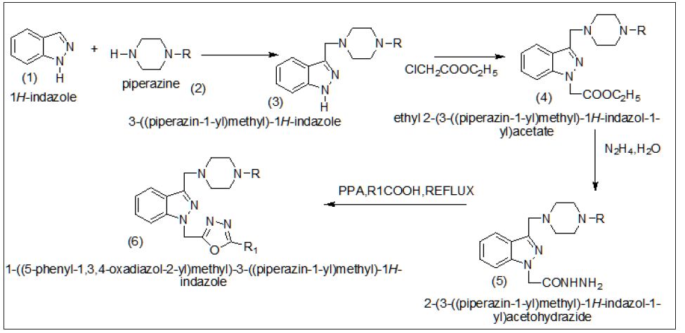
Figure 1:

Table 1

Experimental Section

Chemistry

Chemicals and reagents used in the current study were of analytical grade. The reactions were monitored by thin layer chromatography (TLC) on Merck pre-coated silica GF254 plates. Melting points were determined using a Mettler Toledo FP62 capillary melting point apparatus (Mettler-Toledo, Greifensee, Switzerland) and were uncorrected. Infrared spectra were recorded on a Perkin-Elmer Spectrum One series FT-IR apparatus (Version5.0.1) (Perkin Elmer, Norwalk, CT, USA), using potassium bromide pellets; the frequencies were expressed in cm-1. The 1 H- and 13C-NMR spectra were recorded with a Varian Mercury-400 FT-NMR spectrometer(Varian, Palo Alto, CA, USA), using tetramethylsilane as the internal reference, with chloroform- CDCl 3 as solvent, the chemical shifts were reported in parts per million (ppm) and coupling constants (J) were given in hertz (Hz). Elemental analyses were performed on a LECO 932 CHNS instrument (Leco-932, St. Joseph, MI,USA) and analyses for C, H, and N were within } 0.4% of the theoretical values.

General procedure for the synthesis of compounds (3)

1H-indazole (1) (2 mmol, 235 mg) was dissolved in 20 ml of ethanol-water (1:1) solution, and formaldehyde 37% (3mmol) and substituted piperazine (2) (2 mmol) were added. The mixture was stirred at room temperature and the reaction was controlled by TLC in benzene: methanol (9:1) and toluene: ethyl acetate: diethylamine (75:25:1).At the end of the reaction, the precipitate was filtrated, dried, and recrystallized using an appropriate solvent. Yield: 45%: mp 179.7 ◦C. IR (KBr) cm-1: ν 3130 (N-H), 3095-2756 (C-H). 1H-NMR (CDCl3): δ 8.10 (bs,1H, 1H-indazole N-H), 7.77 (d, 1H, indole H4 , J = 7.6), 7.36 (d, 1H, 1H-indazole H7 , J = 8), ,6.92- 6.82 (m, 3H, 1H-indazole H2, H5, H6), 3.79 (s, 2H, C-CH2 -N), 3.20 (t, 4H, piperazine H3, H5 , J = 4.8), 2.68(t, 4H, piperazine H2, H6 , J = 4.8). Anal Calc.: C, 77.35; H, 7.35; N, 14.42%, found: C, 78.16; H, 6.94; N, 14.25%

Ethyl2-(3-piperazin-1-yl)methyl)-1H-indazole) acetate(4)

An equimolar mixture of 3-(piperzin-1-yl) methyl)-1H-indazole (3) and chloro ethyl acetate were dissolved in dimethyl formamide solvent and to this reaction mixture anhydrous K2CO3 was added and the reaction mixture was stirred at room temperature (350C) for 8 hours and the progress of the reaction was monitored by TLC using cyclohexane and ethyl acetate solvent mixture (7:3) as eluent the reaction mixture was kept overnight. After completion of the reaction the solvent was evaporated on rota-evaporater. The gummy solid was separated and it was recrystallized from -2-propanolpetrolium ether (800c) solvent mixture. The crystalline solid was found to be -2-(3-formyl-1H- 1H-indazole) acetate. With a yield of 75% and mp 143-1450C.The indole-3-carbaldehyde used in the present studies was purchased from Aldrich Company and was used without any for their purification. Yield 75%, m.p.:143-1450C.

Yield: 55%: mp 185.7 ◦C. IR (KBr) cm-1: ν 3150 (N-H), 3095- 2782 (C-H). 1H-NMR (CDCl3): δ 7.60 (d, 1H, 1H-indazole H4 , J = 7.6), 7.20 (d, 1H, 1H-indazole H7, J = 8), ,6.95-6.85 (m, 3H, 1H-indazole H2, H5, H6), 3.85 (s, 2H, C-CH2 -N), 3.25 (t, 4H, piperazine H3, H5 , J = 4.8), 2.70(t, 4H, piperazine H2, H6 , J = 4.8), 1.29 (t,3H, J=13.2Hz, CH3 of ethyl group), 4.13 (q, 2H, J=13.2Hz, CH2 of ethyl group),. Anal. Calc. for: C, 78.32; H, 7.26; N, 14.42%, found: C, 78.18; H, 6.70; N, 14.15%,

2-(3-((piperazin-1-yl)methyl)-1H-indazole-1-yl) acetohydrazide(5):

A solution of 4 (0.01mol) and hydrazine hydrate (0.015) in ethanol (20ml) was refluxed for 5 hours. The reaction mixtures was cooled and poured in to ice-cold water with stirring. The separated solid was filtered, washed with water and recrystallized from ethanol.

Yield: 50%: mp 180.7 ◦C. IR (KBr) cm-1: ν 3160 (N-H), 3070- 2780 (C-H). 1H-NMR (CDCl3): δ, 7.65 (d, 1H, 1H-indazole H4 , J = 7.6), 7.35 (d, 1H, 1H-indazole H7 , J = 8), ,6.80-6.85 (m, 3H, 1H-indazole H2, H5, H6), 3.80 (s, 2H, C-CH2 -N), 3.25 (t, 4H, piperazine H3, H5 , J = 4.8), 2.70(t, 4H, piperazine H2, H6 , J = 4.8), ,4.28(s,2H,-NH2), ). 4.36 (s, 2H N-CH2-C =O), 4.98 (s,1 H,-N-NH), Anal. Calc. for: C, 78.32; H, 7.26; N, 14.42%, found: C, 78.18; H, 6.94; N, 14.25%.

1-(( phenyl (1,3,4-oxadiazol-2-yl)methyl)-3-(piperazine- 1-yl)methyl)- 1H-indazole 6(a)

A mixture of 2-(3-((piperazin-1-yl) methyl)-1H-indol-1- yl) acetohydrazide (5) (0.01 mol) and substituted carboxylic acid (0.01 mol) was heated at 100-120 oC in presence of excess polyphosphoric acid (PPA) for 4-5 h. After cooling, the mixture was poured into crushed ice, and neutralized with 5% aq.NaHCO3 solution. The precipitated solid was filtered and purified using column chromatography (petroleum ether: ethyl acetate, 9:1).

Yield: 60%: mp 190.7 ◦C. IR (KBr) cf-1: ν 3150 (N-H), 3050- 2750 (C-H). 1H-NMR (CDCl3): δ, 7.65 (d, 1H, 1H-indazole H4 , J= 7.6), 7.35 (d, 1H, indole H7 , J = 8), ,6.80-6.85 (m, 3H, indole H2, H5, H6), 7.35-7.45(m,5H,phenyl group),3.80 (s, 2H, C-CH2 -N), 3.25 (t, 4H, piperazine H3, H5 , J = 4.8), 2.70(t, 4H, piperazine H2, H6 , J = 4.8),Anal. Calc. for: C, 78.32; H, 7.26; N, 14.42%, found: C, 78.18; H, 6.94; N, 14.25%.

1-(( tollyl (1,3,4-oxadiazol-2-yl)methyl))-3-(piperazine- 1-yl)methyl)- 1H-indazole 6(b)

Yield: 58%: mp 195.0 ◦C. IR (KBr) cm-1: ν 3100 (N-H), 3020- 2720 (C-H). 1H-NMR (CDCl3): δ, 7.60 (d, 1H, 1H-indazole H4 , J = 7.6), 7.30 (d, 1H, indole H7 , J = 8), 7.40-7.55(m,4H,phenyl group) ,6.60-6.65 (m, 3H, 1H-indazole H2, H5, H6), 3.60 (s, 2H, C-CH2 -N), 3.75 (t, 4H, piperazine H3, H5 , J = 4.8), 2.50(t, 4H, piperazine H2, H6 , J = 4.8), 2.43(s,3H,-CH3 ),2.40(s,3H,phenyl attached CH3 group) , Anal. Calc. for C, 70.32; H, 7.15; N, 14.20%, found: C, 70.18; H, 6.94; N, 14.10%

1-((chlorophenyl(1,3,4-oxadiazol-2-yl)methyl))-3- (piperazine-1-yl)methyl)-1H-indazole (d)

Yield: 53%: mp 160.0 ◦C. IR (KBr) cm-1: ν 3020 (N-H), 3090- 2710 (C-H). 1H-NMR (CDCl3): δ, 7.10 (d, 1H, 1H-indazole H4 , J = 7.6), 7.20 (d, 1H, 1H-indazole H7 , J = 8), 7.15-7.40(m,4H,phenyl group),6.20-6.15 (m, 3H, 1H-indazole H2, H5, H6), 3.20(s, 2H, C-CH2 -N), 3.10 (t, 4H, piperazine H3, H5 , J = 4.8), 2.20(t, 4H, piperazine H2, H6 , J = 4.8),, Anal. Calc. for: C, 65.32; H, 6.15; N, 12.20%, found: C, 65.18; H, 6.24; N, 12.10%.

1-((Bromophenyl(1,3,4-oxadiazol-2-yl)methyl))-3- (piperazine-1-yl)methyl)-1H-indazole 6(e)

Yield: 51%: mp 165.0 ◦C. IR (KBr) cm-1: ν 3000 (N-H), 3010- 2710 (C-H). 1H-NMR (CDCl3): δ, 7.15 (d, 1H, 1H-indazole H4 , J = 7.6), 7.40 (d, 1H, 1H-indazole H7 , J = 8), 7.05-7.25(m,4H,phenyl group), ,6.10-6.15 (m, 3H, 1H-indazole H2, H5, H6), 3.10(s, 2H, C-CH2 -N), 3.15(t, 4H, piperazine H3, H5 , J = 4.8), 2.20(t, 4H, piperazine H2, H6 , J = 4.8), Anal. Calc. for: C, 65.15; H, 6.15; N, 12.20%, found: C, 65.10; H, 6.04; N, 12.05%.

1-((nitrophenyl(1,3,4-oxadiazol-2-yl)methyl))-3- (piperazine-1-yl)methyl)-1H- indazole 6(f)

Yield: 49%: mp 175.0 ◦C. IR (KBr) cm-1: ν 3020 (N-H), 3020- 2750 (C-H). 1H-NMR (CDCl3): δ, 7.20 (d, 1H, 1H-indazole H4 , J = 7.6), 7.00 (d, 1H, 1H-indazole H7 , J = 8), 7.30-7.40(m,4H,phenyl group),6.20-6.25 (m, 3H, 1H-indazole H2, H5, H6), 3.15(s, 2H, C-CH2 -N), 3.20(t, 4H, piperazine H3, H5 , J = 4.8), 2.25(t, 4H, piperazine H2, H6 , J = 4.8), Anal. Calc. for C, 64.15; H, 4.15; N, 8.20%, found: C, 64.10; H, 4.04; N, 8.05%.

Anti-Bacterial Activity

The anti bacterial activity of synthesized compounds was studied by the disc diffusion method against the following pathogenic organisms. The gram-positive bacteria screened were staphylococcus aureus NCCS 2079. The gram negative bacteria screened were Escherichia coli NCCS 2065 and pseudomonas aeruginosa NCCS 2200. The synthesized compounds were used at the concentration of250 μglml and 500μglml using DMSO as a solvent the Cefaclor 10μglml disc was used as a standard. (Himedia, Laboratories Ltd, Mumbai).The test results presented in the (Table 1), suggest that 4b,4d,4e exhibit high activity against the teased bacteria, the rest of the compounds were found to be moderate active against the tested microorganisms.

Antifungal activity

The antifungal activity of synthesized compounds was studied by disc diffusion method against the organisms of Penicillium and Trichophton. Compounds were treated at the concentrations of 500μglm and 1000μglml using DMSO as solvent. The standard used was Clotrimazole 50 μglml against both organisms. The test results were presented in the (Tables 2 & 3).

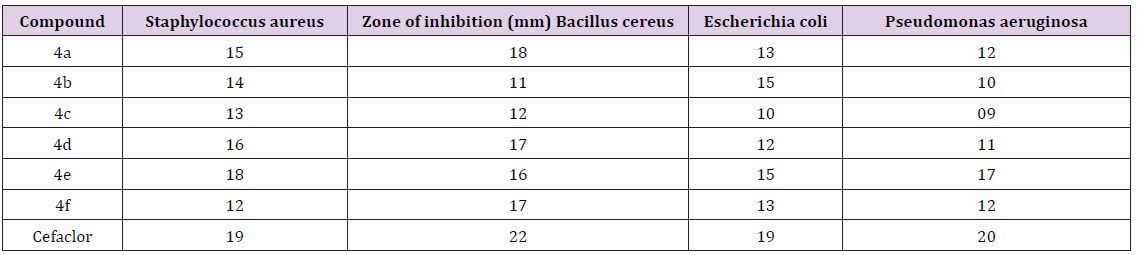
Table 2: Antibacterial activity by disc diffusion method of indazole linked 1, 3,4 oxadiazole 4(af).

Table 3: Antifungalactivity.
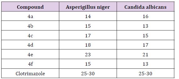
Acknowledgement

a) It’s my pleasure to express my thanks to Department of Chemistry for giving an opportunity to do research.

b) I express my sincere thanks to P.RAVISANKARA REDDY (Sr. Excutive in Biological E Ltd company, shameerpet, Hyderabad), who is giving valuable guidance during my research.

For more Articles: Biomedical Journal Impact Factor: https://biomedres.us

Open access Journals on Surgery

Difficult Laparoscopic Cholecystectomy

Mini Review

Laparoscopiccolecystectomy( L C ) hasdramaticallyreplacedconventional open cholecystectomy [1]. LC was introduced by Mouret in 1987. It is one of the most common procedures being performed by the general surgeons all over the world. With more and more endeavors being made in the field of laparoscopy, more and more complicatedcases, which were relatively contraindicated a few years ago, are now being performed laparoscopically. Advantages of LC are a better cosmesis, shorter hospital stay, shorter recovery time and decreased morbidity [2]. Now a days LC has become the gold standard not only for routine gall bladder removal due to asymptomatic or symptomatic gall stones but also for the treatment of acute gall bladder inflammation [3]. One of the most important aspects of safe LC is a meticulous dissection of the structures in the triangle of Calot. Cystic ductan dcysticartery must be recognised and carefully dissected to prevent intraoperative injury of major bile ductsandrigh the paticartery or excessive intraoperative bleeding. Rate of conversion from laparoscopic to open technique can be minimized by adhering to the basic principles of surgery and keeping in mind the critical anatomy while doing the safe and meticulous dissection [4]. In certain circumstances difficult LC can be expected. Difficult LC can be anticipated based on the pre-operative and intra-operative factors such as old age, male sex, historyof multiple attack so frecent origin or repeated admissions, diabetes mellitus, previous upper abdominal surgery, liver cirrhosis and patients presenting with acute cholecystitis, pancreatitis or cholangitis [1-4].

Difficult LC is considered in cases of dense adhesions at the triangle of Calot, contracted and fibrotic gall bladder, previous upper abdominal surgery, acutely inflammed and gangrenous gall bladder, empyema of the gall bladder, Mirizzi ̍s syndrome, previous cholecystostomy and cholecystogastric or cholecystoduodenal fistula [4,5]. Every case should be considered as difficult until completed successfully. Level of difficulty may vary with the skill and experience of the surgeon but conditions, mentioned above, are really difficult, irrespective of the experience and skill of the surgeon. Bile duct injury is the most catastrophic event that can happen to the patient during LC, leaving the patient with high morbidity and high treatment cost. Measures taken to prevent bile duct injury are careful dissection of the triangle of Calot, keeping in mind the anatomy of the hapato biliary system and possible an atomic variations and step-by-step progression until removal of the gall bladder [6]. In cases of unclear anatomy and failure to progress in laparoscopic dissection, conversion to the open procedure should be done and it should not be takenas a failure or a complication on the part of the surgeon [6,7].

Conclusion

Difficult LC can be expected in certain circumstances. Some scoring and validation systems have been recommended to predict difficult cases. Step-by-step meticulous dissection of the triangle of Calot should be performed to avoid bile ductinjury. Conversion to open procedure should be done in cases of unclear anatomy or failure to progress in laparoscopic dissection.

For more Articles: Biomedical Journal Impact Factor: https://biomedres.us

Open Access Journals on Obstetrics Gynecology

Vulva cancer and HIV

Introduction

Vulvar carcinoma is more and more common, especially in young patients because of viral infections. The objective of the study was to report images of a rapid onset of epidermoid carcinoma on HIV 1, unknown and untreated.

Observation

It was Mrs. SB, aged 42, married, primigeste, with a history of menarche at 14 years, twin birth and concept of low genital infection, received at the oncological consultation of 24/05/2017, for vulvar swelling evolving for 3 months. A examination, it had a vulva tumor budding 6 x 6 cm, interesting the 2 lips, the entire left lip and 2/3 of the right with infiltration of the bladder trine, inguinal ganglia indurated and a flow of odorless pus at the pressure of the tumor (Figure 1) at that date, colposcopy was normal. The diagnosis of suspected neoplasia of the vulva with destruction of the lips (large and small) was posed with biopsy samples and a request for paraclinical investigations exploring the field, after counseling on neoplasia and on the possible positivity of testing for sexually transmitted diseases including HIV.

As of 12/07/2017, it was reviewed with worsening and infiltration of the entire vulva to Mont the venus [Figure 2] and histological confirmation results (of appearance corresponding to invasive squamous cell carcinoma of the vulva), serology positive for HIV 1 and TPHA / RPR (syphilis), CD4 count at 265 / mm3, CRP positive at 80 mg / dl and moderate anemia at 9g/dl. A palliative treatment was offered to him with his orientation, after counseling, in the house of his treatment day with Anti Retro Virus (ARV).

Comments

The three-month delay in the diagnosis of our case and that of Amourak [1], would be linked to the patient’s ignorance of the sexually transmitted infections with ulceration, premary signs such as pruritus, swelling (our case), evolution and severity of the cancerous disease and its HIV status. Vulvar cancer appeared in a young woman under 50 years old instead of 70 years old. The frequency of HIV-related tumors increases 20 years younger than in the general population [2]. This increase in the number of young people is linked to the observation of human papillomavirus (HPV) infections [3] and HIV [4]. Syphilitic lesions would be the gateway to HIV infection including a drop in CD4 count and acute outbreak of historical vulvar cancer between 2 visits at 7 week intervals without treatment. Delay in diagnostic confirmation can be linked to poverty and the lack of integration of HIV testing and management into cancer consultation. The vulvar smear on primary lesions of the vulva for an early diagnosis of precancerous lesions and their correct treatment in medical, gynecological or dermatological environment, could improve the prognosis of the patients of this association.

Conclusion

The association cancer of the vulva and HIV is rare. She had a rapid course in a woman in 7 weeks. The sensitization of the population, education for self-examination, the treatment of precancerous lesions and the risk of HIV infection would allow earlier diagnosis.

Medical Images

About a rapid evolutionary thrust of vulvar cancer and historical HIV at the Gynecological Obstetrics Department of Donka National Hospital, Conakry, Guinea.

Journals on Surgery

Dilemma in Diagnosis-Huge Preauricular Sinus Mimicking Parotid Tumor

Abstract

Objective: This paper reports a case of huge preauricular sinus mimicking parotid tumour to highlight the difficulty in obtaining a correct diagnosis.

Background and Case Report: This case describes a 51-year-old gentleman who presented with a huge, soft and painless swelling over left parotid region which was progressively increasing in size over two years. The swelling appeared to push the left ear lobule upward and on palpation, the swelling did not go beyond the zygomatic arch. Fine needle aspiration was unsatisfactory. Ultrasonography scan showed left parotid tumour. The diagnosis of preauricular sinus was made only upon excision of the mass under general anaesthesia. Post operatively, the recovery was good and there was no complication.

Conclusion: Though it is extremely rare, preauricular sinus should be considered as a differential diagnosis of painless soft parotid swelling.

Keywords: Preauricular sinus; Parotid

Introduction

Preauricular sinus is a benign congenital malformation of the preauricular soft tissue, first described by Van Heusinger in 1864 [1]. Preauricular sinuses are formed from incomplete or defective fusion of the six auditory hillocks during embryological development of auricle at sixth week of gestation. We would like to report a case of a huge preauricular sinus as a rare differential diagnosis of a parotid swelling.

Case Report

A 51-year-old gentleman presented to us with a painless swelling over the left parotid region for duration of two years. The swelling gradually increased in size and there were no episodes of inflammation or infection of the swelling. He had no significant past medical or surgical history. Examination revealed a 7x5cm swelling over left parotid region, elevating the ear lobe and with multiple pits all over the swelling and cheek region (Figure 1). On palpation, the swelling was doughy in consistency, non-tender. His facial nerve function was grossly normal. There were no palpable neck nodes. Fine needle aspiration for cytology was performed of the swelling which was unsatisfactory. Ultrasonography of the left parotid swelling showed a well-defined, homogenous, hypoechoic mass at the left parotid gland measuring 7.3×5.0x2.0cm, with no internal vascularity seen. A differential diagnosis of pleomorphic adenoma was made. With the above clinical and imaging correlation, patient was planned for left superficial parotidectomy. Left modified Bailey incision was made. A preauricular cyst with sinus tract was revealed. (Figures 2 & 3) The surrounding skin was adhered to the swelling which was able to be dissected of by meticulous dissection. The preauricular cyst with sinus tract was completely excised with preservation of left parotid gland. Patient was discharged day one post operatively with no complication. Patient was well in three months of follow-up with no signs of recurrence.

Figure 1: The excised preauricular sinus mass measuring about 7x5cm.

Discussion

Preauricular sinus has an incidence of 0.1 to 0.9% in Western populations and a higher incidence of 4% and 10% among Blacks and Orientals [2]. This condition can occur either sporadically or via inheritance. Unilateral preauricular sinus is more common than bilateral. Over 50 % of preauricular sinus cases are unilateral and likely to be sporadic while in 25 to 50% of cases, preauricular sinuses are bilateral and are more likely to be hereditary [3]. Preauricular sinus can usually be noticed as a small pit adjacent to the anterior margin of the ascending limb of the helix, rarely along the posterosuperior margin of the helix, tragus or lobule. Most of the preauricular sinus are asymptomatic and do not require any treatment. Patient may present with persistent discharge from the preauricular sinus or signs of acute infection of sinus tract. In our patient, there was presence of unilateral preauricular sinus pit over the anterior margin of ascending limb of left helix, with multiple pits surrounding the overlying skin of the swelling. However, this did not give rise to any discharge.

Infected preauricular sinus requires administration of antibiotics. Incision and drainage is necessary in the presence of abscess. Surgical excision of the sinus tract is then carried out after resolution of the acute infection to prevent recurrent preauricular sinus infection. Incomplete excision of sinus tract leads to recurrence of preauricular sinus. Recurrence rate ranged from 22 to 42% has been reported in cases where preauricular sinus is excised with traditional method without any aid for tract delineation or visualisation [4]. Robert J et al reported a new inside-out technique for excision of preauricular sinus where no recurrence was noted [5]. Gan EC et al. reported a low recurrence rate of 2.4% by performing excision of preauricular sinus under microscope guidance and under methylene blue and probe guidance [4]. From our literature review, there were no reported cases of preauricular sinus presenting as parotid swelling. In view of a huge and soft swelling over left parotid region, the differentials include sebaceous cyst and benign parotid neoplasm. The possibility of left preauricular sinus was never thought of as clinically and ultrasonographically looked more like a mass arising from parotid gland.

Conclusion

In conclusion, though it is extremely rare, preauricular sinus should be considered as a differential diagnosis of painless soft parotid swelling. Computed tomography scan might assist in differentiating a huge preauricular sinus which mimics a parotid tumour. Complete excision is deemed necessary to prevent recurrence.

Summary

a) Preauricular sinus is formed from incomplete or defective fusion of the six auditory hillocks during embryological development of auricle at sixth week of gestation.

b) This case describes a 51-year-old gentleman who presented with a huge, soft and painless swelling over left parotid region which was progressively increasing in size over two years. Clinical and ultrasonographical findings favour the diagnosis of parotid tumour.

c) The diagnosis of preauricular sinus was made only upon excision of the mass under general anaesthesia. Post operatively, the recovery was good and there was no complication.

d) Though it is extremely rare, preauricular sinus should be considered as a differential diagnosis of painless soft parotid swelling.

For More Articles: Biomedical Journal Impact Factor: https://biomedres.us

Otorhinolaryngology Open Access Journal

Dilemma in Diagnosis-Huge Preauricular Sinus Mimicking Parotid Tumor

Abstract

Objective: This paper reports a case of huge preauricular sinus mimicking parotid tumour to highlight the difficulty in obtaining a correct diagnosis.

Background and Case Report: This case describes a 51-year-old gentleman who presented with a huge, soft and painless swelling over left parotid region which was progressively increasing in size over two years. The swelling appeared to push the left ear lobule upward and on palpation, the swelling did not go beyond the zygomatic arch. Fine needle aspiration was unsatisfactory. Ultrasonography scan showed left parotid tumour. The diagnosis of preauricular sinus was made only upon excision of the mass under general anaesthesia. Post operatively, the recovery was good and there was no complication.

Conclusion: Though it is extremely rare, preauricular sinus should be considered as a differential diagnosis of painless soft parotid swelling.

Keywords: Preauricular sinus; Parotid

Introduction

Preauricular sinus is a benign congenital malformation of the preauricular soft tissue, first described by Van Heusinger in 1864 [1]. Preauricular sinuses are formed from incomplete or defective fusion of the six auditory hillocks during embryological development of auricle at sixth week of gestation. We would like to report a case of a huge preauricular sinus as a rare differential diagnosis of a parotid swelling.

Case Report

A 51-year-old gentleman presented to us with a painless swelling over the left parotid region for duration of two years. The swelling gradually increased in size and there were no episodes of inflammation or infection of the swelling. He had no significant past medical or surgical history. Examination revealed a 7x5cm swelling over left parotid region, elevating the ear lobe and with multiple pits all over the swelling and cheek region (Figure 1). On palpation, the swelling was doughy in consistency, non-tender. His facial nerve function was grossly normal. There were no palpable neck nodes. Fine needle aspiration for cytology was performed of the swelling which was unsatisfactory. Ultrasonography of the left parotid swelling showed a well-defined, homogenous, hypoechoic mass at the left parotid gland measuring 7.3×5.0x2.0cm, with no internal vascularity seen. A differential diagnosis of pleomorphic adenoma was made. With the above clinical and imaging correlation, patient was planned for left superficial parotidectomy. Left modified Bailey incision was made. A preauricular cyst with sinus tract was revealed. (Figures 2 & 3) The surrounding skin was adhered to the swelling which was able to be dissected of by meticulous dissection. The preauricular cyst with sinus tract was completely excised with preservation of left parotid gland. Patient was discharged day one post operatively with no complication. Patient was well in three months of follow-up with no signs of recurrence.

Discussion

Preauricular sinus has an incidence of 0.1 to 0.9% in Western populations and a higher incidence of 4% and 10% among Blacks and Orientals [2]. This condition can occur either sporadically or via inheritance. Unilateral preauricular sinus is more common than bilateral. Over 50 % of preauricular sinus cases are unilateral and likely to be sporadic while in 25 to 50% of cases, preauricular sinuses are bilateral and are more likely to be hereditary [3]. Preauricular sinus can usually be noticed as a small pit adjacent to the anterior margin of the ascending limb of the helix, rarely along the posterosuperior margin of the helix, tragus or lobule. Most of the preauricular sinus are asymptomatic and do not require any treatment. Patient may present with persistent discharge from the preauricular sinus or signs of acute infection of sinus tract. In our patient, there was presence of unilateral preauricular sinus pit over the anterior margin of ascending limb of left helix, with multiple pits surrounding the overlying skin of the swelling. However, this did not give rise to any discharge.

Infected preauricular sinus requires administration of antibiotics. Incision and drainage is necessary in the presence of abscess. Surgical excision of the sinus tract is then carried out after resolution of the acute infection to prevent recurrent preauricular sinus infection. Incomplete excision of sinus tract leads to recurrence of preauricular sinus. Recurrence rate ranged from 22 to 42% has been reported in cases where preauricular sinus is excised with traditional method without any aid for tract delineation or visualisation [4]. Robert J et al reported a new inside-out technique for excision of preauricular sinus where no recurrence was noted [5]. Gan EC et al. reported a low recurrence rate of 2.4% by performing excision of preauricular sinus under microscope guidance and under methylene blue and probe guidance [4]. From our literature review, there were no reported cases of preauricular sinus presenting as parotid swelling. In view of a huge and soft swelling over left parotid region, the differentials include sebaceous cyst and benign parotid neoplasm. The possibility of left preauricular sinus was never thought of as clinically and ultrasonographically looked more like a mass arising from parotid gland.

Conclusion

In conclusion, though it is extremely rare, preauricular sinus should be considered as a differential diagnosis of painless soft parotid swelling. Computed tomography scan might assist in differentiating a huge preauricular sinus which mimics a parotid tumour. Complete excision is deemed necessary to prevent recurrence.

Summary

a) Preauricular sinus is formed from incomplete or defective fusion of the six auditory hillocks during embryological development of auricle at sixth week of gestation.

b) This case describes a 51-year-old gentleman who presented with a huge, soft and painless swelling over left parotid region which was progressively increasing in size over two years. Clinical and ultrasonographical findings favour the diagnosis of parotid tumour.

c) The diagnosis of preauricular sinus was made only upon excision of the mass under general anaesthesia. Post operatively, the recovery was good and there was no complication.

d) Though it is extremely rare, preauricular sinus should be considered as a differential diagnosis of painless soft parotid swelling.

For More Articles: Biomedical Journal Impact Factor: https://biomedres.us

Journals on Medical Sciences

Planned Teaching Programme Regarding Basic Life Support in Terms of Knowledge and Skill of Pre-University College Students in a Selected College of Moodbidri, Dakshina Kannada District

Introduction

Basic life support is commonly taught to the general public and the new generation as these may be the only ones present in the crucial few minutes before emergency personnel are available. Cardiopulmonary resuscitation is a technique of basic life support for the purpose of oxygenating the brain and heart until appropriate definitive medical treatment can restore the normal heart and ventilatory action. Cardiopulmonary resuscitation, more commonly known as CPR, is a basic life support procedure for people whose heart and lungs have ceased to function effectively. Cardiovascular disease remains the most common cause of death in developed countries and is increasing in number in developing countries. In USA, 48% of all deaths (1994) were due to cardiovascular disease. In 1997 the death rate was 35% but 68% of these deaths occurred before reaching the hospital. In India, in 1994, the annual death rate due to cardiovascular disease was 18% [1-5]. In 2002 the death rate increased to 26%. National academy of science and national council in 2002 emphasized to rediscover the value of teaching CPR in colleges. In 1998 American Health Association began a large-scale evaluation of CPR in colleges in the US. Experts strongly recommended the development of CPR programmes in colleges to ensure widespread learning of CPR and other basic life support skills, because 70-80% of cardiac arrest occur at home [5-10].

Objectives of the Study

a) To assess the knowledge and skill of pre-university college students regarding basic life support.

b) To prepare and conduct planned teaching programme on CPR technique for pre-university college students.

c) To evaluate the effectiveness of planned teaching programme on CPR technique among pre-university college students.

d) To find out the significant association of knowledge and skill with selected demographic variables.

Operational Definitions

a. Effectiveness: The extent to which the planned teaching programme has achieved the desired effect in improving the knowledge and skill on basic life support.

b. Planned teaching programme: It is a systematically organized teaching strategy as basic life support.

c. Basic life support: It is a specific level of pre-hospital medical care provided by trained responders, including emergency medical technicians, in the absence of advanced medical care – demonstration of mouth-to-mouth ventilation and external cardiac compression by one rescue method.

d. Knowledge: It refers to the ability of the sample in giving correct responses to the questions asked as measured by the knowledge questionnaire.

e. Skill: It refers to the proficiency or dexterity that is acquired after the demonstration of the CPR technique on manikin, as measured by the observation checklist.

f. Pre-university college students: Students refers to those studying in the second year science course in selected pre-university college at Moodbidri.

Hypotheses

H1: The mean post-test knowledge score of pre-university college students will be significantly higher than the mean pretest knowledge score regarding CPR technique.

H2: There will be significant association between knowledge scores of pre-university college students and selected demographic variables.

Population

The population for the study comprised of 30 students studying in the selected pre-university college.

Samples

The sample of the study comprised of 30 students studying in pre-university college.

Sampling Technique

Simple random technique was used to draw the sample. In this study, the investigator adopted a lottery method to choose 30 students from the sampling frame.

Research Design

The research design adopted for present study was one group pre-test post-test design adopted in the evaluative research approach for collection and analysis of data. The primary objective of the teaching programme was to impart education on basic life support to the randomly selected sample of 30 pre-university college students assessed in terms of mean gain in knowledge test and mean gain in skill test.

Development of the Tool

A self-administered questionnaire was prepared to assess the knowledge on basic life support CPR Adult 1 and observation checklist regarding the skill about CPR technique – basic life support adult rescuer method.

Implications

Curriculum of nursing should prepare the nurses to assess the life-threatening conditions and their management. Adequate classroom teaching and demonstration regarding basic life support will be beneficial. The nursing staff and students can be taught to impart health education to pre-university college students regarding basic life support. Regular education programmes conducted by the nursing personnel, both in the hospital and in the community area will help in the management of life-threatening conditions. The nurse can play an important role in helping the adolescents regarding basic life support. Administration can also take initiatives in imparting health information regarding basic life support through different teaching methods. Individual and group teaching can be arranged for people in the hospitals, schools, colleges and other community settings. There is ample scope for nurses to conduct research in areas like various strategies that can be used in effective management of life-threatening conditions.

Recommendations

a) A similar study can be conducted on a larger sample for wider generalization.

b) A similar study can be conducted with a control group.

c) A comparative study can be carried out on knowledge of basic life support in terms of CPR among university college students.

d) Similar study can be conducted on school children, school teachers, parents, fire force, and traffic policemen.

e) Similar studies could be undertaken using other teaching strategies like video film, film shows or telephonic instruction.

f) A study could be conducted as knowledge regarding basic life support in terms of CPR among nursing students/staff nurse/nursing tutors.

Summary

Heart action and respiratory effort are absolute requirements in transporting oxygen to the tissues. One of the main organs to suffer from oxygen starvation is the brain, which may sustain damage after 4 minutes and irreversible damage after about seven minutes. The heart also rapidly loses the ability to maintain a normal rhythm. Following cardiac arrest, effective CPR enables enough oxygen to reach the brain to delay brain death and allows the heart to remain responsive to defibrillation attempts. CPR takes a lot of effort and may keep care provides from helping others. The PTP was prepared with the aim of improving the knowledge of pre-university college students related to CPR technique.

For More Articles: Biomedical Journal Impact Factor: https://biomedres.us