Dental Open Access Journal

Upper Crossed Syndrome and Dentistry Why the Need for Concern

Abstract

Occupational hazard plays a detrimental role in influencing the quality and quantity of work of dental health care personnel. Basic understanding of the same is essential in terms of improved productivity and progress. Among the work related health concerns, musculoskeletal disorders are perhaps the most common and debilitating. Dentists are prone for improper posture and associated muscle strain. Upper crossed syndrome is an incorrect posture associated musculoskeletal condition with high prevalence. This review article will brief on upper crossed syndrome and its implications in dental workforce, with focus on precautionary and therapeutic measures.

Introduction

Occupational hazard is an umbrella term, encompassing various short and long term risk factors encountered in the work place. Being un heedful of the potential work environment risk factors will lead to vulnerable state. Dental profession presents a unique platform relating to work burdens and ailments. Occupational Safety and Health Administration has outlined the involved factors related to dentistry, its recognition, control and prevention. Major risk factors included, but essentially not limited are biological, chemical and pharmaceutical hazards, musculoskeletal disorders and ergonomic issues, noise and vibration related hazards and psychological issues [1]. Musculoskeletal disorders (MSD) are key contemporary dental occupational hazard and its reported incidence is as high as 81.4% [2]. The work related pain was mostly attributed to shoulder, lower back and neck region [3]. Lower back pain had high prevalence of 64%, closely followed by 60% neck pain, with simultaneous pain in multiple sites tagging behind [4]. Females are found to be at higher risk than that of a male dentist.

The concerned symptoms were reported to occur early in the profession with chronicity noted due to typical late intervention. Reported consequences are socioeconomic burden on the professional with reduced working hours, even early retirement [5]. Long working sesions, sustained static position, awkward posture combined with that of recurrent, repetitive, precise and highly controlled movements are the reasons attributed.5 More frequent and prolonged the aforementioned factors happens to be, more pronounced is the resultant injury, owing to strained muscle, bone and joints. Dental professionals are more prone for prolonged bending; twisted trunk and continuous preordained static posture [6]. A static posture could be one that has been withheld during the whole period of the exertion. In dentistry, static postures with repetitive motion and poor ergonomics leads to a vicious pathway. During a specified treatment time, constrained posture with awkward position of head and trunk is highly observed [7].

Upper crossed Syndrome

Poor posture has been associated with musculoskeletal imbalance and one such commonly reported condition is Upper crossed syndrome (UCS). Improper posture has been associated with muscle tension and limited mobility [8]. Literature reports that the posture frequently adopted in the upper extremity is forward head posture [9]. UCS is the term coined by Janda and used for such misalignment. He has stated that the maintenance of a stooped sitting posture for an extended period of time is the major predisposing factor [10]. It is also known by other terms such as proximal or shoulder girdle crossed syndrome.

The overactive muscles on one side of the neck are found to counteract the underactive muscles of the adjacent side, creating an ‘X’ pattern. This comprises of reciprocal inhibition, wherein muscles on one side are lengthened to compensate for the shortened, contracted muscle on the other side of the joint. With prolonged continued postural imbalance, the intensity of the muscular imbalance increases over time, creating a vicious cycle [10]. The resultant clinical presentation are the complaint of neck and shoulder pain, cervicogenic headache, hunched upper back and rounded shoulders [8]. Owing to the constrained dental clinical work field aka oral cavity, rapid, repetitive, controlled and/ or forceful movements in conjunction with sustained awkward posture, the dental personnel are highly predisposed to UCS [4]. As previously stated, in the literature ample epidemiological studies are published establishing the association of occupational factors and MSD in dentistry. A cross sectional study analysing 220 dental students has ascertained the tendency of increased tightness in the neck extensor and pectoral muscles in dentists [11]. This supports the hypothesis of heightened risk of the dental professionals for developing muscular imbalance and especially, UCS.

However the methodological difficulties are key predicament in generalising the prevalence of such muscular imbalance. Inconsistencies in study design, classification method, outcome variable and interpretation of the results has generated significant controversy in this field. Rapid upper limb assessment (RULA) is a standard method used to assess the ergonomic sitting posture [12]. The number of movements, static muscle force and work are the aspects evaluated in RULA. Muscular length test, muscular strength test, cervical range of motion, forward head posture measurement by craniovertebral analysis, rounded shoulder measure and kyphosis measure are some assessments that will outline the severity of the clinical entity presented [13]. Subsequent to identification of the UCS, proper corrective measures need to be undertaken to correct and limit the muscular imbalance. A recent study assessing 30 dentists with UCS were analysed in a eight week exercise program [14]. A 30 – 60 minutes workout was carried out, with exercises being selected on the basis of individual needs. Warm up, light exercises, special strength and resistance training was the aspects of the training program, with significant positive outcome.

In the upcoming dental profession, preventive program aimed at establishing the habit of improper posture development is critical in curbing this vicious pathological process. Rigorous stress and training on workplace posture maintenance should be stressed upon in dental educative system. Appropriate use of ergonomic instruments will also reduce the unnecessary muscle load. A concept known as ‘Active physical movement’ during treatment procedures has been advocated by many researchers. Changing posture at regular interval, taking micro breaks, using stretching exercises in between the procedure will potentially reduce the ill-effects of static posture. The stretching exercise is advocated especially in the opposite direction of the static and repetitive posture [15]. Regular work out and/or yoga seems to be beneficial from biomechanical view, but studies doesn’t show consistent pattern in this regard [16]. Cultivation of healthier posture habits, early diagnosis and compensatory muscle stretching and relaxation exercises, especially in cervical region could prevent the incidence and severity of UCS.

Conclusion

Neck, a structurally delicate body segment tasks the huge burden of supporting the human head. Improper body posture inherent in dentistry, paves way for increased incidence of neck pain and UCS. Numerous suggestions are put forth for altering the assumption of awkward posture, including improved equipment ergonomics, favourable workspace up gradation, physical therapy and active life style adaptation. Early identification and prevention of UCS is crucial in preventing further degenerative changes and treating the musculoskeletal pain. Understanding how best to develop to awareness regarding prevention of UCS is a singular agenda of this research field still remaining. Modifying the dentists view on monotonous work, improving awareness of the posture embraced, positive ergonomic improvements perhaps be the cost effective and has long standing benefits.

For More Articles: Biomedical Journal Impact Factor: https://biomedres.us

Journals on Dentistry

Application of Nanobiomaterials in Endodontics

Abstract

Nowadays, the application of Nanobiomaterials in dentistry is increasing. The combination of biomaterials with nanotechnology leads to the term of “Nano biomaterial”. Nanotechnology aims to control over material properties at nanoscale and over the precision and sensitivity of various tools and devices in different technologies. In the endodontic science, Nanobiomaterials are utilized in different aspects such as: instruments and materials modifications, improvement of root canal disinfection and obturation, root repair materials, local anesthesia and repair and regeneration of pulpal tissue. The aim of the present study was to reviews the research findings and future possible applications of Nanobiomaterials and nanotechnology in endodontics.

Keywords: Endodontic; Nanomaterial; Nanotechnology

Introduction

Dental pulp is a unique low compliance tissue with specific characters different from similar loose connective tissues. Its encasement within the relatively rigid and unyielding dentinal walls, its particular rich neurovascular supply, a powerful immune response and scarcity of collateral circulation may lead to rapid degeneration and necrosis [1]. Dental caries, traumatic injuries and iatrogenic procedures could affect the health of dental pulp and trigger immune response within the pulpo-dentin complex [2,3]. Bacterial byproducts and products from the dissolution of the organic and inorganic constituents of dentin, mechanical and thermal injuries during cavity preparation, toxicity of restorative materials and more importantly micro leakage at the interface of dentinal walls and restorative materials could alter the existent balance within the pulp and cause irreversible pulpitis and pulp necrosis [4,5].

Clinical endodontics is mainly directed towards curing or preventing apical periodontitis. The microbial infection of the pulp via their toxins and noxious metabolic byproducts, in addition to the presence of disintegrated pulpal tissue are the primary causes of apical periodontitis [6]. Nonsurgical root canal treatment has a high degree of predictability with favorable outcome rates of up to 95% for the treatment of teeth with irreversible pulpitis [7] and up to 85% for necrotic teeth [8]. Endodontic treatment is based on the main two integrated phases: cleansing and shaping [9]. Cleansing and shaping procedures are directed towards the mechanically debridement, disinfecting the root canal system with irrigants and medicaments, and finally optimized canal geometrics for adequate obturation and seal [10,11]. The mechanical debridement is aimed to prepare all the root canal surfaces in a fully incorporated form into the original canal shape. Moreover, preparation errors such as perforations, zips, transport and etc. should be absent and as much as radicular dentin should be left to avoid vertical root fracture [12,13].

Endodontic instruments, both hand- held and engine-driven, are available for root canal preparation. Since the early 1990s, with the advent of nickel-titanium, various instrument designs and modalities have been produced in this regard [14]. Surface quality is an important factor in the function and durability of NiTi instruments and superficial defects such as metal flash, roll over and cracks may lead to the instrument fracture [15] (Figure1). Electro polishing the surface and coating it with titanium nitride have been recommended for promotion of the surface quality [16,17]. Currently, nanomaterials, with a smaller size, are being suggested for surface modification and reduction the incidence of failure in the rotary nickel-titanium files [18].

Figure 1: A separated nickel-titanium instrument in the mesiobuccal canal of mandibular first molar.

Nanoscaffolds for pulp regeneration, bioceramics for retrofilling, and repair materials are other applications of nanotechnology in the endodontic treatment (Figure 2). Nanorobots and nanoterminators are also new technologies for local anesthesia with fewer side effects and complications [24]. Nanotechnology has revolutionized all aspects of science and endodontic is no exception. Nano sized particles with significantly superior properties compared to the similar materials at larges scales of measurement have improved the quality of treatment. Understanding of dental tissue at the nanoscale, enabling the precise design of materials and instruments with ultrafine architecture and improving the present techniques in clinical dentistry have significantly promoted the quality of treatment.

Figure 2: Deep pulpotomy for apexogenesis. a. Extensive carious exposure in an immature mandibular first molar with a history of spontaneous pain. b. After complete caries removal and hemostasis, the radicular pulp is overlaid with mineral trioxide aggregate (MTA).

Two concerns remain in this regard:

a) Nanotechnology should make its way from laboratories to clinical practice.

b) The significant potential for misuse and abuse of this technology on a scale and scope should not be overlooked.

For More Articles: Biomedical Journal Impact Factor: https://biomedres.us

Journals on Environmental Science

Evaluating Antimicrobial activity of Aloe vera Plant Extract in Human Life

Abstract

Aloe vera is a perennial draught resistant plant containing more amount of gel in fluctuating amount to different season. Aloe plant is containing a variety of mineral elements and calcium, iron, and zinc. Aloe gel also contains polysaccharides, proteins, lipids, sugars, minerals, phenolic compounds, glucomannan and acemannan. Its composition accelerates wound healing, activating macrophages, stimulating immune system as well antibacterial and antiviral effects. Acemannan is effect as indirect anti-microbial activity through its ability and stimulate phagocytic leukocytes. Such types, it has more antimicrobial value like medicines. The aim of this study are evaluating antimicrobial activity of aloe vera gel such as antibacterial, antifungal, and antiviral activity to which solve human problems in life.

Keywords: Aloe Plant; Gel, Antibacterial Activity; Antifungal Activity; Antiviral Activity

Introduction

Aloe vera (A. barbadensis Miller L.) is most biologically active among 400 species [1-4]. The genus Aloe belonging to family Alliaceae is a succulent plant of 80-100cm in height which matures in 4-6 years and survives for nearly 50 years under favorable conditions. The plant is native to southern and eastern Africa along the upper Nile in the Sudan, and it was subsequently introduced into northern Africa and naturalized in the Mediterranean region and other countries across the globe. Aloe is commercially cultivated in Aruba, Bonaire, Haiti, India, South Africa, the United States of America, and Venezuela [1] while the finest quality of Aloe is grown in desert of Southern California. According to World Health Organization (WHO), aloe is the best source for obtaining a variety of drugs [2], because this plant can survive in both hot and cold temperatures. Plant extracts represent a continuous effort to find new compound against pathogens. Approximately 20% of the plants found in the world have been submitted to biological test, and a substantial number of new antibiotics introduced on the market are obtained from natural or semi synthetic resources [3]. Aleo gel is bactericidal against (bacteriostatic) for common wound infecting bacteria (in vitro). The aloin A and B (barbaloin) are 1,8-dihydroxyanthracene glycosides [4], after oral administration these are not absorbed in the upper intestine, and hydrolyzed in the colon by intestinal bacteria. It has following antimicrobial activities which generally observed after oral administration in 6-24 hours.

Anti-microbial Properties: The anti-microbial activity of aloe juice was investigated by agar disc diffusion against bacteria, fungi and yeast [5]. Aloe juice showed anti-bacterial activity against the Gram -ve bacteria (Pseudomonas aeruginosa, Klebsiella pneumonniae, E.coli and Salmonella typhimurium) and Candida albicans (in vitro) A. hydrophilia and E. coli and not against any fungi or yeast tested. Similar results have been obtained for anti-microbial activity of the aloe juice against Gram +ve bacteria by Alemdar and Agaoglu [6] (Mycobacterium smegmatis, Staphylococcus aureus, Enterococcus faecalis, Micrococcus luteus and Bacillus sphericus). Heggers et al. [7] tested Aloe vera gel against ten bacterial strains (Staphylococcus aureus, Streptococcus pyogenes, Streptococcus agalactiae, Escherichia coli, Serratia marcescens, Klebsiella sp., Enterobacter sp., Citrobacter sp., Bacillus subtilis and Candida albicans), at 90% concentration aloe gel that was effective against all the organisms but at the 70% concentration only against S. pyogenes. [8] tested preserved aloe gel extract and an unpreserved aloe extract against Pseudomonas aeruginosa, Enterobacter aerogenes, Staphylococcus aureus and Klebsiella pneumoniae. It was found that preserved Aloe gel extract was more effective in controlling bacterial growth. Aloe vera gel was shown to inhibit the growth of gram positive bacteria, Shigella flexneri and Streptococcus pyogenes [9].

Antibacterial Activity: Aloe vera gel is a bactericidal agent causing against Pseudomonas aeruginosa and acemannan and prevent human lung epithelial cells from adhering in a monolayer culture [10]. Using a rat model, suggested that the antibacterial effect of the Aloe vera gel (in vivo) could enhance the wound healing process by eliminating the bacteria that contributed to inflammation [7]. The aloe extract was potent against three strains of Mycobacterium (M. fortuitum, M. smegmatis and M. kansasi) and a strong antimycobacterial activity against M. tuberculosis as well as antibacterial activity against P. aeruginosa, E. coli, S. aureus and S. typhi. Thus, Aloe secundiflora could be a rich source of antimicrobial agents [11]. Aloe vera was shown to inhibit microbes like Staphylococcus aureus [12], Candida albicans [13] Pseudomonas aeruginosa [14], and Klebsiella pneumoniae. It has indirect antimicrobial property through self ability to stimulate phagocytic leukocytes [15].

Antiviral Activity: Aloe gels are effective antiviral agent in several ingredients, such as acemannan reduced herpes simplex infection in two cultured target cell lines. Lectins fractions of aloe gel directly inhibited the cytomegalovirus proliferation in cell culture, perhaps by interfering with protein synthesis [16]. A purified sample of aloe emodin was effective against infectivity of herpes simplex virus Type I and Type II and it was capable of inactivating all of the viruses, including varicella zoster virus, influenza virus, and pseudo rabies virus [17]. In some cases anthroquinones are treated herpes simplex virus under electron micrograph examination. Such types result is indicated that anthraquinone extract from aloe plant variety directly enveloped viruses. These fluctuations are indirectly effected due to stimulation of the immune system. The anthraquinone aloin also inactivates various enveloped viruses such as herpes simplex, varicella zoster and influenza [18].

Antifungal Activity: Aloe gel is evaluated on the mycelium development of Rhizoctonia solani, Fusarium oxysporum and Colletotrichum coccodes that showed an inhibitory effect of the pulp of aloe vera on F. oxysporum at 104 μl L-1. The liquid fraction is reduced rate of colony growth at a concentration of 105 μl L-1 in R. solani, F. oxysporum and C. coccodes [19]. A fresh leaf extract of A. barbadensis and A. arborescens have anti-fungal potential against Aspergillus niger, Cladosporium herbarum and Fusarium moniliforme and inhibit the growth of fungi [20]. Aloe juices are anti-inflammatory, anti-arthritic activity, antibacterial and hypoglycaemic effects [12] for bacteria inhibit growth of Streptococcus and Shigella species In Vitro. Agarry et al., [12] showed that aloe gel inhibited the growth of Trichophyton mentagrophytes (20.0 mm), while the leaf possesses inhibitory effects in Pseudomonas aeruginosa and Candida albicans. Another aloe constituents are includes saponins. These are soapy substances of gel that capable of cleaning and perform strongly antimicrobial against for bacteria, viruses, fungi and yeasts [21].

Conclusion

Aloe plant have important role in antimicrobial activity in everyday life. Aloe gel is mostly use in humanity for cosmetic, burn and medicinal application. Aloe plant has major role in the promotion of recombinant-DNA based product, targeting compounds of value to be isolated and produced in stable and realistic quantities. Such type aloe is a “wonder plant” because it use in multiple problems like antiseptic, anti-inflammatory agent and help in relieving like diabetes, and being a cosmetic field. The aloe plant is need to a greater research emphasis for better utilization of this plant in humankind welfare, it remains for us to introduce to ourselves and thank the nature for its never-ending gift. Furthermore, study all principles of aloe vera needs to be evaluated in future for scientific using, so that its other therapeutic uses can be widely explored. Isolation and maintenance procedures of aloe products are require special care and these have been established after painstaking efforts.

For More Articles: Biomedical Journal Impact Factor: https://biomedres.us

Journals on Physiology

Soil pH, Ca and Mg Stability and pH Association with Temperature and Groundwater Silicon

Abstract

Objective: It is generally known that pH, Ca and Mg have changed remarkably during 1961-90, but their inter-areal variation seems not have been fully discussed nor explained. Parameters of cropland have been earlier associated with CHD mortality. Silicon (Si) and temperature (Temp) have been earlier assessed with human CHD mortality. In this study we have assessed soil values of 21 Rural Centers (RC) from the 1960’s, 1970’s and 1980’s, [(60’s) (70’s), (80’s)] with respective mean annual temperatures from 1981-2010 and mean groundwater (gw) Si content.

Results: Combined regression by pH(60’s) and pH(70’s) explained pH(80) by 96.8 % (p < 0.001). Ca. (60’s) and Ca.(70’s) explained respectively Ca.(80’s) by 99.3 % (p < 0.001) and Mg.(60’s) and Mg.(70’s) explained respectively Mg.(80’s) by 92.3 % (p < 0.001). Combined regression by Temp and Si explained pH (80’s) by 86.5 % (p < 0.001). Respectively combined regression by Temp and pH (80’s) explained gw Si by 77.5 % (p < 0.001).

Conclusion: Groundwater silicon is associated with the soil matrix factor (sources of groundwater silicon/silicon colloids?), which seems with temperature to explain inter-areal pH variation and obviously is associated with relative local stability of cropland Ca and Mg (and soil fertility). Maybe now is the time to increase Si trials in agriculture.

Abbreviations: RC : Rural Centers; FMI: Finnish Meteorological Institute; ETS: Effective Temperature Sum

Introduction

It is generally known that pH, Ca and Mg have changed remarkably during 1961-90 [1,2,3] but their inter-areal variation seems not have been fully discussed nor explained. Parameters of cropland have been earlier associated with CHD mortality [4]. In this survey based on old statistics has been discovered great stability in inter-RC soil parameters and connection of this stability with temperature and gw Si.

Material and Methods

Soil data is from Eurofins Viljavuuspalvelu as in our earlier publications [1-4]. The soil values [1-3] given as 5 year periods (1961-65, 1966-70,..) were combined to decade periods [(60’s), (70’s), (80’s)]. Values of “(9).Kymenlaakso” and “(10).Etelä- Karjala” were combined to”(9;10).Kymi” by weighting the soil values by their cropland areas in 1988 (4,5). Respectively values of “(17).Keski-Pohjanmaan” and “(18).Oulun” were combined to “(17;18).K-Pohjanmaan, Oulun” and used as such in statistics. Available provincial Si.gw.m data from Geologic Survey of Finland [5] have been changed to approximate RC values by its provincial values as such if RC was totally inside of the province, in other cases by weighting the different Si.gw.w values of different provinces by their cropland areas. Area weights selected for this study as earlier from 1988 [4,5]. The values of RC temperatures were determined by benefiting the map of RCs in Official Statistics of Finland [6] and the map of Finnish Meteorological Institute (FMI) [7], by selecting their central commune and then visually estimating its place between the temperature lines.

Results

Table 1 shows that the range of periodical changes varied between 5.4 (pH) and 15.8 % (Ca).

Table 1: Changes of the mean RC soil values between the 1960’s and 1980’s.

Combined regression by pH (60’s) and pH (70’s) explained pH (80’s) by 96.8 % (p < 0.001).

Combined regression by Ca. (60’s) and Ca. (70’s) explained Ca. (80’s) by 99.3 % (p < 0.001)

Combined regression by Mg. (60’s) and Mg. (70’s) explained Mg. (80’s) by 92.3 % (p < 0.001)

Combined regression by Temp and Si explained pH.(80’s) by 86.5 % (p < 0.001). (Fig.1)

Computation gave equation: pH [Temp; Si (mg/L)] = 6.02 + 0.16*Temp – 0.11*Si (mg/L).

Combined regression by Temp and pH (80’s) explained Si by 77.5 % (p < 0.001):

Computation gave equation: Si (mg/L) = 38.5 + 1.14*Temp – 6.12* pH.

Discussion

Values (60’s) and (70’s) explained inter-RC variations (80’s) in pH, Ca and Mg by 92-99 %. This stability in proportional inter- RC soil values seems not remarkably to have been affected by the equal nation-wide soil liming recommendations. Soil data have been collected by a private enterprise “Viljavuuspalvelu Oy” (since 2014 “Eurofins Viljavuuspalvelu Oy”) depending on the activity of the farmers. Additionally the number of Mg samples was lower than the number of “basic” samples until 1985, e.g. in 1966-70 the number of Ca samples was ca 400,000, but by Mg “only” ca 33,000” [8]. Obviously less biased sample series could show even higher associations, e.g. combined regression by provincial Si.gw and their capital Temp (4) can explain soil pH variation more than 90 %. This proportional inter-RC stability in the soil factors could explain the stability in proportional CHD mortality between provinces [9]. Association of groundwater with local food could (at least have been) promoted by long-root plants like alfalfa and red clover, which could benefit gw better than short-root plants. Evotranspiration (E0) [10] on Finnish croplands varies with the effective temperature sum (ETS) [11]

Figure 1: Combined regression by pH.(60’s) and pH.(70’s) explained pH.(80’s) by 96.8 % (p < 0.001).

Figure 2: Combined regression by Ca.(60’s) and Ca.(70’s) explained Ca.(80’s) by 99.3 % (p < 0.001).

Figure 3: Combined regression by Mg.(60’s) and Mg.(70’s) explained Mg.(80’s) by 92.3 % (p < 0.001).

Figure 4: Combined regression by Temp and Si explained pH.(80’s) by 86.5 % (p < 0.001). Computation gave equation: pH [Temp;Si (mg/L)] = 6.02 + 0.16*Temp – 0.11*Si (mg/L).

Figure 5: Combined regression by Temp and pH.(80’s) explained Si by 77.5 % (p < 0.001): Computation gave equation: Si (mg/L) = 38.5 + 1.14*Temp – 6.12* pH.

E0 = -100 + 0.388 *ETS (mm year-1), i.e. ca 2.5 – 4 million l/ha.

If one million liters of this came from gw, plants could get annually on the average 15 kg Ca, 3.8 kg Mg/ha and 6.5 kg Si/ha [6]. These values respond ca 1/4 of Ca and 1/3 of Mg total supply by fertilizers at the first half of the 1950’s [12] and possibly several folds the Si given in fertilizers, because in the 1970’s Si/Mg ratio in Finnish food was 0.008 [13]. Losses of Ca and Mg have been replaced, maybe now is the time to correct the Si losses [14-16].

Conclusion

Groundwater silicon is associated with the soil matrix factor (sources of groundwater silicon/silicon colloids?), which seems with temperature to explain inter-areal pH variation and obviously is associated with relative local stability of cropland Ca and Mg (and soil fertility). Maybe now is the time to increase Si trials in agriculture.

For More Articles: Biomedical Journal Impact Factor: https://biomedres.us

Journals on Dental Medicine

All-Ceramic versus Metal-Ceramic Tooth Supported Single Crowns with a Minimum Follow-Up Time of 3 Years; Survival and Complications: A Systematic Literature Review

Abstract

Purpose: To assess the 3-year survival rates and the incidences of biological and technical complications of all-ceramic and metal-ceramic tooth-supported single crowns.

Material and Methods: An online research of literature on Pub med was conducted independently by 3 reviewers to identify the clinical studies performed from 2000 up to 2017 and completed by a manual research. Keywords, inclusion and exclusion criteria were well-defined.

Results: The research revealed 501 titles and led to a final analysis of 31 full text articles. Only 6 studies met the inclusion criteria and reported on 215 metal-ceramic and 501 all-ceramic TSSCs. The meta-analysis of the included studies indicated an estimated survival rate of metal-ceramic TSSCs of 90.14% (95% CI: 89.72%-91.35%) after 3 years. This was a little bit higher than the estimated 3-year survival rate of Lithium-disilicate (89.54%; 95% CI: 87.18%-91.89%). However the 3-year survival rate, of Zirconia TSSCs was statistically much higher (92.01%; 95% CI: 89.58-94.43%). When the outcomes in anterior and posterior regions were compared, Zirconia and Lithium-disilicate TSSCs exhibited survival rates of 91.64% and 91.12% respectively, in the posterior region, where metal-ceramic TSSCs exhibited only 90.14% as a 3-year survival rate. As for the anterior region, we could only compare the TSSCs made out of Lithium-disilicate and Zirconia as the metalceramic ones were all laid on the posterior regions. Zirconia performed the highest 3-year survival rate in the anterior region (93.54%; 95% CI: 91.08-96%) whereas Lithium-disilicate showed only 87.5%. This was due to the low level of the mechanical properties of such material, which clinically indicated by major chipping and tooth fracture.

Conclusion: Survival rates of metal-ceramic crowns were nearly similar to those reported for most types of all-ceramic TSSCs in the posterior region. Weaker Lithium-disilicate-based ceramics should be limited to application in the anterior region, even if they expressed a higher survival rate in the anterior regions. Zirconia-based TSSCs should not be considered as a primary option due to their high incidence of technical problems.

Keywords: Tooth-supported single crowns; Fixed dental prosthesis; All-ceramic; Metal-ceramic; Survival; Technical complications; Biological complications

Introduction

Nowadays; all ceramic prostheses are considered as an established alternative compared to metal-ceramics. They offered more favorable esthetics by miming so naturally the optical properties of teeth [1].

Another more recent factor influencing the choice of ceramic materials is the pronounced cost for high precious metals as gold. The main defect of the firstly introduced ceramics as the feldspathic ones was the mechanical stability that limited the indication for all ceramic restorations to anterior regions and to single units fixed dental-prostheses. In the last years, many new dental ceramic materials were developed in order to increase the mechanical stability of the all-ceramic restorations and still maintaining the esthetic benefit. Among those materials; leucite/lithium-disilicate glass ceramics and oxide ceramics such as alumina and Zirconia that appeared to be very promising for different indications [2,3]. Reconstructions made of these more recently developed ceramics were placed at posterior sites and even included multiple-unit fixed dental-prostheses. Our systematic review focused on toothsupported single crowns, as the published studies reporting on this type of FDPs, were so limited and our objective was to obtain an estimate of the long term success, survival and complication rates of all-ceramic single crowns versus the metal-ceramic ones over an observation period of at least 3 years and to compare the biological and technical complication rates of all-ceramic TSSCs with the ones made out of metal-ceramic.

Material and Methods

An electronic literature search was carried out from the data banks MEDLINE, using the database Pub Med. The indexing language based on keywords has been used according to the formula ‘’PICO’’

The keywords were used after being audited if they were MeSH ones.

The formula PICO: Population, Intervention, Comparison and Outcomes, the ‘’PICO’’ for this systematic review was defined as follows:

Population: Anterior and/or posterior tooth-supported single crowns.

Intervention: All-ceramic tooth-supported single crowns.

Comparison: Metal-ceramic tooth-supported single crowns.

Outcomes: Clinical survival rates, and technical and/or biological complication rates.

The search was limited to human studies in dental journals written in English language. Articles published from 2000 up to 2017 and including the inclusion criteria.

The following detailed search terms were used and the search strategy was follows:

P and I: crowns[MeSH] OR crown[MeSH] OR dental crowns[MeSH] OR crowns, dental[MeSH] OR Denture, Partial, Fixed[Mesh])) OR (crown*[all fields] OR fixed partial denture*[all fields] OR FPD[all fields] OR FPDs[all fields] OR fixed-dental prosthesis[all fields] OR fixed dental prostheses[allfields] OR FDP[all fields] OR FDPs[all fields] OR bridge*[all fields].

C: Ceramic [MeSH] OR ceramics [MeSH] OR metal-ceramic restorations [MeSH])) OR (ceramic*[All Fields] ORall-ceramic [all fields] OR Dental Porcelain[All Fields] OR metal-ceramic[All Fields].

O: Survival [Mesh] OR survival rate [Mesh] OR survival analysis[Mesh] OR dental restoration failure[Mesh] OR prosthesis failure[Mesh] OR treatment failure[Mesh].The combination in the builder was set as ‘’P & I AND C AND O’’.

Titles and abstracts of the searches were independently screened by two reviewers for possible inclusion in the review. Furthermore, the full text of all studies of possible relevance was then obtained and spilt into literature on single crowns. The literature on single crowns was independently assessed by three of the reviewers. Any disagreement regarding the assortment articles was resolved by discussion.

Inclusion criteria

a) The additional inclusion criteria for study selection were:

b) Studies with a minimum mean follow-up period of 3 years.

c) Prospective studies.

d) Randomized Controlled Trial.

e) Cohorts.

f) Studies between 2000 and 2017.

g) Studies reported details on the characteristics of the reconstructions, on materials and methods, on the biological complications and their appearance and on the results.

h) Studies had to include and follow-up at least 10 patients.

i) Patients of studies had been examined clinically at regular intervals.

j) Studies on tooth-supported single crowns, fixed units with metal and ceramic frameworks were considered.

k) Data on the survival and failure of TSSCs and descriptions of the biological and technical complications had to be reported.

Exclusion criteria

The following study types were excluded:

a) In vitro or animal studies.

b) Studies with a mean follow-up time less than 3 years.

c) Clinical or case reports.

d) Narrative review.

e) Retrospectives studies.

f) Fixed implant prosthesis.

g) Partial removable dentures.

h) Plural fixed dental prosthesis (PFDPs).

Data extraction

Data on the following parameters were extracted: Author(s), Title, Journal, Year of publication, Study design (cohort, metaanalysis, randomized controlled clinical trials, prospective case series, prospective study, and prospective clinical study), Population (Planned number of patients, Sex, Age), number of patients at the end of the study, Drop-out rate, Mean age, Operators (practitioners), Material framework, Brand name of framework material, Veneering material, Brand name of veneering material, Type of manufacturing procedure, Number of single crowns, Location of single crowns (anterior, posterior, maxilla, mandible), Reported mean followup time, Published single crowns survival rate, Number of single crowns lost (total, anterior, posterior), Reported biological complications (caries, periodontal and endodontic diseases, root fracture, tooth fracture, marginal discrepancy), Reported technical complications (framework fracture, minor chipping, major chipping, loss of retention), Reported number of TSSCs free of complications, Specificity of the study, Aesthetic properties, Biological properties.

Statistical analysis:

Survival was defined as the tooth-supported single crowns remaining in situ with or without modification during the observation period. Failures included every type of complication that lead to the removal or the replacement of the restoration or the loss of the tooth. Complication may led or not to a failure. Failure and complication rates were calculated by dividing the number of events (failure or complication) in the numerator by the total toothsupported single crown exposure time in the denominator.

The numerator could usually be extracted directly from the publication. The total exposure time was calculated by talking the sum of:

a) Exposure time of tooth-supported single crowns that could be followed for the whole observation time.

b) Exposure time up to a failure of the tooth-supported single crown that were lost due to failure during the observation time.

c) Exposure time up to the end of observation time for toothsupported single crowns that did not complete the observation period due to reasons such as death, change of address, refusal to participate, non-response, chronic illnesses, missed appointments and work commitments.

For each study, event rates for the tooth-supported single crowns were calculated by dividing the total number of events by the total tooth-supported single crown exposure time in years. The total exposure was determined by multiplying the total number of tooth-supported single crowns by the mean follow-up time, no more precise information. For further analysis, the total number of events was considered to be Poisson, disturbed for a given sum of tooth-supported single crown exposure years and Poisson regression with a logarithmic link-function with a total exposure time per study as an offset were used [4].Robust standard errors were calculated to obtain 95% confidence intervals of the summary estimates for the event rates [4,5].

To assess heterogeneity of the study specific event rates, the Spearman goodness-of-fit statistics and associated p-value were calculated. If the goodness-of-fit p-value was below 0.05 three year survival; proportions had been calculated via the relationship between event rate and survival function S, S(T)=exp(-T* event rate), by assuming constant event rates [5]. The 95% confidence intervals for the survival proportions were calculated by using the 95% confidence limits of the event rates. Multivariable Poisson regression was used to formally compare construction subtypes and to assess other study characteristics. All analyses were performed using Stata®, version 13.1.

Results

The research on MEDLINE using the Boolean-equation had identified 501 articles. During the preselecting step, 437 articles were excluded based on titles and the year of publication. After reading, other 33 articles were excluded based on the exclusion criteria adopted in the study. Among the 31 selected articles, only 3 corresponded to studies on tooth-supported single crowns. 3 more articles were added based on manual research. (Figure 1) Among the 6 selected articles, 4 corresponded to Prospective clinical studies [6-9], one to a Randomized controlled trial [10] and another one to a Prospective case series [11], evaluating 3 to 15 years follow-up of the single restorations with all ceramic and metal ceramic materials. The articles included in this systematic review are listed in Table 1 by author, study design and patient characteristics.

Figure 1: Research strategy and included studies on TSSCs.

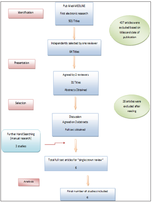
Table 1: Study and patient characteristics of the reviewed studies of tooth-supported single crowns.

The 6 clinical studies included one 15-years follow-up [6], one 10-years follow-up [8], one 7-years follow-up [7], one 5-years follow-up [9] and two 3-years follow-up [10,11]. The studies included patients between the age of 32 and 60 with a follow-up rage of 6 months. The proportions of patients, who could not be followed for complete studies periods or at least 3 years, were calculated for all studies as a Drop-out rate and ranged from 3.33 % to 36.23 %. (Table 2) According to material and manufacturing procedures, two studies compared all-ceramic crowns made out of zirconia (one study on CAD/CAM System; cercon smart ceramics˖) [10] the remaining on Procera and Lava systems [9], to metal ceramic crowns [9,10]. Furthermore, one study was reporting on metal-ceramic tooth-supported single crowns [8], one on crowns made out of Lithium with IPS e-max press [6] and two on crowns made out of Zirconia with the CAD/CAM technology [7,11] (Table 2).

Table 2: Study and tooth-supported single crowns characteristics.

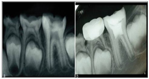
For metal-ceramic TSSCs only noble metal or high noble metal alloys were used for the framework; based on the conventional lost wax technique [8-10] and the veneering was based on the manual layering technique with a standard firing process (Figure 2), using ; feldspathic ceramic [8], low fused porcelain [10] and non-specific porcelain [9] (Table 2).For all-ceramic TSSCs the layering material used during the various studies was as follows: fluorapatite veneering ceramic [6], feldspathic porcelain [7,11] and layering ceramic [9,10]. The veneering technique was based on manual layering [6-11] (Figure 2), the slow cooling protocol [7] and various CAD/CAM manufacturing procedures such as Procera system and 3M ESPE [9] (Table 2). In all included studies, preparation guidelines according to the manufacturer’s recommendation were considered. Different measurement methods were used to exanimate the TSSCs at the recall appointments; in order to identify the technical and biological complications. In two studies [7, 8] remarkable complications of TSSCs were discovered and the criteria used were not specified. For biological and technical complications the California Dental Association criteria (CDA) was used in one study, in the company of periodontal parameters [14] such as plaque index, gingival index [6]. The technical complications were evaluated according to Heintz/Rousson 2010 chipping criteria in two studies [6, 10]. The United States Public Health Service (USPHS) criteria were used to evaluate technical issues in one study [9]. Both technical and biological complications were defined based on a specific clinical evaluation according to Hickel et al. in one study [11].The estimated survival rates of 716 TSSCs ranged from 80% to 93.54%. Which were predominantly posterior single reconstructions? (Table 3).

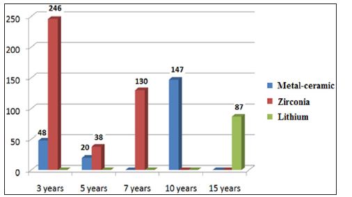
Figure 2: Tooth-supported single- crowns mean follow-up time according to material.

Crown survival

Overall, in the 3 studies [8-10] reporting on MC TSSCs with a mean follow-up of 7 years ± 6 months. An estimated annual failure rate of 0.051 was reported, translated into an estimated 3-year survival of metal-ceramic crowns of 90.14%. In comparison, all ceramic crowns had an annual failure rate ranging between 0.015 and 0.15, translating into overall estimated 3-year survival rates ranging between 80% and 93.54% (Table 3). This was based on 5 studies [6-11] on all-ceramic crowns included in this analysis (Table 3). The survival rates of all-ceramic crowns differed for the various types of ceramics. One study [6] reported on Lithium glass ceramic and rendered an estimated 3-year survival rate of 89.54%. This survival rate was a little bit lower than the one reported for the gold standard, metal-ceramic crowns (Table 3).Tooth-supported single crowns made out of zirconia had a significantly higher estimated 3-year survival rate compared to metal-ceramic crowns. The zirconia-based crowns reached an estimated 3-year survival rate of 92.01% (Table 3). Table 3: Annual failure and survival rates of single crowns (estimated evaluation after 3 years). Anterior vs. posterior regions TSSCs were distributed as 103 crown in anterior regions and 603 crown in posterior regions. The distribution of TSSCs according to material was as follows: none of MC TSSCs; 38 crowns were made of lithium [6] and 65 of zirconia [7-11].For the posterior regions we have, 215 metal-ceramic single crowns [8-10] and 388 all-ceramic crowns [6-11], in which 49 crown were made out from Lithium [6] and 339 from zirconia-oxide and zirconium [7-11]. When outcomes of anterior and posterior tooth-supported single crowns were compared no statistically significant differences of the survival rates were found for metal-ceramic crowns, as they were all made on posterior regions for the 3 studies analyzed (Table 4). For the entire 215 MC TSSCs posed in the posterior region, we had an estimated failure rate of 0.015. For the crowns made out of Lithium, there was a significant difference of survival rates between anterior region, which exhibited an estimated 3-years survival rate of 87.5% and the posterior region with an estimated 3-year survival rate of 91.12%. 38 crowns made out of Lithium and posed in anterior region, showed a higher estimated annual failure rate of 0.079 compared to the 49 crowns posed in posterior region, which showed a failure rate of 0.04 (Table 4). Table 4: Annual failure rates and survival 3 year estimates of crowns placed anterior and posterior. Crowns made out of Zirconia, showed significantly lower survival rates in the posterior region than the anterior (93.54 % vs. 91.64 %). 65 crowns were posed in the anterior region with a failure rate of 0.015, which was lower compared with the 339 crowns posed in the posterior region and showed a failure rate of 0.035 (Table 4). For all-ceramic crowns there is no significant difference of survival rates between anterior and posterior regions (91.31% vs. 91.57%).For the total number of TSSCs, there was no statistically difference between the anterior and the posterior regions, which exhibited 3-year survival rates of 91.31% vs. 91.06% respectively (Table 4). Over all, in the anterior region, the crowns made out of Lithium showed lower 3-years estimated survival rate compared to SCs made out of Zirconia (87.5% vs. 93.54%). Whereas in the posterior region the several types of SCs could be compared and showed a statistically difference between the MC SCs and those made out of Lithium and Zirconia, with 90.14% vs. 91.13% and 91.64% as a 3-years estimated survival rates, respectively. Finally to summarize, we could compared all-ceramic and the metal-ceramic SCs only in the posterior region, as none of the metalceramic crowns were placed on the anterior. A 3-year estimated survival rate was calculated as an over-all result for both MC SCs and CC SCs and showed a statistically difference between both of them (90.14% VS 91.57%) (Table 4). Technical and biological complications Table 5 and 6 display an overview of the incidences, the estimated annual complication rates and the cumulative 3-years complication rates of technical and biological complications for metal-ceramic and the two types of all-ceramic TSSCs, as well as the statistical differences between the crown types. Technical complications: Non optimal margin, marginal discrepancy, minor and major chipping, crown fracture, post fracture, loss of retention, anatomical form and ‘’the coping fracture, the veneering fracture the occlusal wear, the marginal adaptation and the anatomical form’’ according to the USPHS criteria (Table 3) and described as Bravo (B).The solutions adopted for the different technical complications were: a) A clinical check-up for the non-marginal adaptation was adopted as a solution for a one case reported as an all-ceramic SC made out of Lithium during a mean follow-up time of 11.4 ± 3.8 years in one study [6], no clinical or technical intervention was need. b) The minor chipping was the most common complication reported and the polishing of ceramic was the solution to adopt for 2 metal-ceramic single crowns reported in one study [10,11] single crown made out of Lithium [6] and 4 single crowns made out of Zirconium [7,10]. c) The loss of retention were reported for 9 all-ceramic crowns in two studies [6,7]; 4 crowns had been lusted adhesively and 2 had been cemented conventionally [6] for the 3 crowns reported in the second study [7] the technique of cementation was not mentioned. d) The unsatisfying anatomical form and contact point was reported on 152 all-ceramic single crown made out of Zirconium and zirconia-oxide in on study [11], no intervention was need, only a clinical follow-up was planned based on crown check-up and hygiene motivation. e) The Bravo criterion reported on the minor chipping, the occlusal wear, the slight probe catch and the slightly contoured according to the USPHS criteria (Table 3); was considered as technical complication affected one metal-ceramic single crown and 7 all-ceramic single crowns made out of zirconium with the Lava and Procera systems (Figure 3) in one study [9]. The polishing and the clinical control were the solutions to adopt. Figure 3: Survival rates by 3 years; Anterior versus posterior regions according to materials The major chipping, the crown fracture, the post fracture and the marginal discrepancy were reported as failure and the crown replacement was need. Charlie and Delta criterion according to the USPHS criteria were reported as failure and the loss of crown was a fact [9]. I. 7 crowns were lost by major chipping ; 4 crowns made out of Lithium [6], one crown made out of zirconium [10] and 2 metal-ceramic SCs [8]. II. The crown fracture was reported in two studies [6,8] and affected one all-ceramic crown made out of Lithium [6] and 2 metal-ceramic SCs [8]. III. The crown replacement by marginal discrepancy was reported on one crown made out of Lithium [6] as well as the post fracture. IV. For Charlie and Delta (Table 3) according to USPHP criteria; 9 crowns were lost in which 2 metal-ceramic crowns and 7 all-ceramic crowns made out of Zirconium [9]. According to Table 5; the ceramic chipping was a common problem, and overall occurred similarly at the metal-ceramics and the all-ceramic crowns. Furthermore, for metal ceramic crowns, minor chipping was the most frequent technical complication with a cumulative 3-years event rate of 0.88% (95% CI: 0-2.69). For allceramic a tendency to major chipping of the veneering ceramic was more observed for Lithium than the zirconia oxide (2.18% VS 0.23%), opposite to minor chipping with 0.68% cumulative 3-year event rate for Zirconium crowns vs. 0% for the Lithium ones. Crown fracture rarely occurred for metal-ceramic crowns (cumulative 3-year complication rate 0.44%; 95% CI: 0-1.34%). Overall, this problem occurred significantly more often for ceramic crowns. The Lithium glass ceramic exhibited the highest 3-year crown fracture rate of 1.09% (95% CI: 0.85-2.75%) when the Zirconia-based single crowns showed only 0.68% as an event rate. (95% CI: 0-1.6%). With the exception of zirconia-based crowns, loss of retention was not a predominant technical problem. Only crowns made out of zirconium exhibited loss of retention during a 3 years follow up time (estimated 3-year complication rate 0.23%; CI: 0-1.08%). Only zirconia TSSCs exhibit an unsatisfying anatomic form as a technical complication with a 3-years event rate of 34.87% (95% CI: 19.03- 50.73%) (Table 5). Table 5: Overview of technical complications for different types of SCs. All-ceramic vs. metal-ceramic technical complications According to (Table 7); ceramic chipping affected much more the metal-ceramic crowns than the all-ceramic crowns with a cumulative 3-years complication rate of 0.88% vs. 0.56% for minor chipping and 0.44% vs. 0.37% for major chipping. For crown fracture the all-ceramic crowns exhibit a superior cumulative 3-years complication rate compared to metal-ceramic crowns (0.75% vs. 0.44%). The loss of retention, the marginal discrepancy and the clinically controlled not-satisfying form and contact point affected only the all-ceramic crowns with a cumulative 3-years complication rate of 0.19% (95%; CI: 0.00-0.89%), 0.18% (95%; CI: 0.00-0.5%) and 28.82% (95%; CI: 15.72-41.92%) respectively. For the USPHS (Table 3), the criterions Bravo, Charlie and Delta were considered as technical complications that leaded to clinical control, intervention and loss of the crown respectively and they occurred significantly more often for all-ceramic crowns than metal-ceramic ones (2.08% vs. 0.88%). During the 3-years follow-up time, metal-ceramic TSSCs a superior rate of SCs free of technical complication in comparison with the all-ceramic ones. (92.36% vs. 62.05%) Biological complications Loss of abutment tooth vitality, abutment tooth fracture, endodontic and periodontal infections, recurrent caries lesions, lesion of the adjacent mucosa and extraction due to infection (caries, periodontal or endodontic infections), were reported as biological complications for TSSCs. The solutions adopted for different biological complications were: (Table 10) Root treatment for crown laying in 14 posterior metal-ceramic TSSCs [10] and 9 posterior all-ceramic TSSCs made out of zirconium.5 TSSCs were canal treated for endodontic infection; 2 anterior TSSCs made out of Lithium [6], one metal-ceramic TSSC and 2 all-ceramic TSSCs made out of zirconium [10]. A periodontal treatment was adopted for the several periodontal infections and diseases on 3 posterior Lithium TSSCs [6] and 48 posterior Zirconia TSSCs. [11]Only one case was reported for recurrent caries treatment on one TSSCs made out of zirconium. [7]The lesion of the adjacent mucosa was clinically controlled on 40 posterior Zirconia TSSCs. [11] the tooth fracture and the extraction due to infection (recurrent caries, periodontal and endodontic diseases) were reported as failures, during the different studies (Table 7). Two anterior abutment teeth were lost by fracture with crowns made out of lithium. [6] 28 TSSCs were extracted due to infections (periodontal and endodontic diseases and recurrent caries). 13 lithium TSSCs [6], 4 Zirconia TSSCs [7,10] and 11 metal-ceramic TSSCs. [8,10] According to Table 6; the periodontal diseases were common biological complication and the most frequent for Zirconia TSSCs (3-year complication rate 11.24%; 95% CI: 7.71- 14.76%). This problem less frequently occurred for lithium and metal-ceramic TSSCs with 3-year complication rate of 1.09% and 0.85% respectively. In addition, the marginal discrepancy and the lesion of the adjacent mucosa were also predominantly found for Zirconia TSSCs (3-year complication rate 9.17%; 95% CI: 6.73- 11.62%). This complication occurred significantly less frequently for Lithium with 3-year complication rate of 1.09% (CI: 0.85- 2.75%) and not existing for metal-ceramic TSSCs. Furthermore, the endodontic diseases were a common biological problem for the different types of TSSCs, with a cumulative 3-years complication rates for Metal-ceramic, Lithium and Zirconia of 2.2%, 1.09% and 0.46% respectively. The loss of vitality by root-canal-treatment for prosthodontic reasons affected only the Zirconia and the metalceramic TSSCs and showed a cumulative 3-years complication rates of 2.06% and 6.18% respectively. Finally, tooth fracture affected only the Lithium TSSCs with a cumulative 3-years complication rate of 1.09%; CI: 0.85-2.75%, when the caries affected the TSSCs made out of metal-ceramic and Zirconia with cumulative 3-years complication rates of 0.88% and 0.92% respectively. Table 6: Overview of biological complications for different types of SCs. All-ceramic VS metal-ceramic biological complications According to Table 7; endodontic and periodontal diseases, caries and loss of tooth vitality for prosthodontic reasons were the most common biological complications for all-ceramic and metalceramic TSSCs. Periodontal diseases occurred significantly high frequently for all-ceramics with a cumulative 3-years complication rate of 9.48% (95%; CI: 6.37-12.2%), when metal-ceramic TSSCs showed only a cumulative 3-years complication rate of 0.85% (95%; CI: 0-1.09%).For marginal discrepancy and the affection of adjacent mucosa, only all-ceramic crowns showed the unfolding of this event with a cumulative 3-year complication rate of 7.77% (95%; CI: 5.7-10.08%).Endodontic diseases affected both metalceramic and all-ceramic TSSCs, with a 3-year complication rate of 0.85% VS 9.48%, respectively. The loss of abutment tooth vitality for prosthodontic reasons, was also a common problem, but significantly occurred on metal-ceramic TSSCs compared to the allceramic ones (6.18% VS 1.7%).Recurrent caries were a rare event, but affected both metal-ceramic and all-ceramic TSSCs with a 3-year cumulative complication rate of 0.88% VS 0.76%, respectively. The tooth fracture happened only on all-ceramic TSSCs with a 3-year cumulative complication rate of 0.18%.Metal-ceramic TSSCs showed a 3-years free-biological event rate of 84.89%, when allceramic TSSCs showed only 74.73%. Table 7: Overview of technical and biological complications for metal-ceramic versus all-ceramic TSSCs. Discussion This systematic review focused on the results of prospective clinical studies and case series [9,10] that would compare head-tohead the different core materials of TSSCs along with a randomized controlled trial. Retrospective and in vitro studies, narrative review, case and clinical reports and studies with a mean follow-up period less than 3 years were excluded to summarize the available information about survival and complication rates of TSSCs after a period of at least 3 years. Even with follow-up time of at least 3 years, some clinicians may argue that such a period is still too short to obtain reliable information on survival and complication rates [12,13]. After an observation period of 3 years, the lowest annual failure rates were seen for Zirconia TSSCs (0.031 per 100 SCs years). Multivariable random-effect Poisson’s regression showed that Lithium TSSCs had significantly higher annual failure rates of about 0.057 per 100 SCs years. Furthermore, the analyzing study reporting on lithium TSSCs [6] with a mean follow-up time of 11.4 +/- 3.8 years had expressed an annual failure rate about 0.195 per 100 SCs years, translating into 10-year survival rate of 89.7%. This highest statistically significant failure rate of TSSCs may have his origin from the limited indication of the Lithium disilicate at the posterior regions. This study evoked the potential updating indication of such core material at the posterior regions. But it is still always limited especially for molars because of the high fracture rates. For Gianluca M [7] Reporting on zirconia TSSCs with a 7-year mean follow-up time, a failure rate of 0.054 per 100 SCs years had been reported. When Mutinelli S et al. [9] had reported an annual failure rate of 0.105 for CC TSSCs made by the Lava system, while the Procera ones had a failure rate of 0.201 for a 5-years mean follow-up time. This difference may have cause from the several types of manufacturing procedures. One study reported on zirconia TSSCs with a failure rate of 0.015 by 3 years, this statistical expressed difference amounts to both the observation period and the laying sites (posterior and anterior regions). The choice of zirconia as a core material for SCs, both in posterior and anterior sites, has been increasing over time with clinical results that seem quite comparable to the gold standard MC TSS restorations, although clinical trials are very few, to date specially on TSSCs. Zirconia SCs showed a success rate of 93% after a 2 years observation period, with a favorable soft tissue response, in a limited sample size of 15 Cercon crowns (DENTSPLY Degudent, Hanau, Germany). Another investigation with a longer observational period (3 years), performed on 204 Procera zirconia SCs delivered in a private practice, showed a survival rate of 93%; in this study, 16% of complication were recorded (6% loss of retention, 2.5% extraction of abutment teeth, 5% persistent pain, 2% porcelain chipping) [14]. For the MC gold standard, a clinical observation of 10 years mean follow-up time [8] had expressed a failure rate of 0.054 per 100 crowns which remain the same during the first 3 years of this study. For another one with a 5-years mean follow-up time; TSSCs had a failure rate of 0.1 per 100 crown which had been expressed lower in the first 3-years of observation (0.05 per 100 crown) [9]. This result was caused by the occurrence of biological complications. MC TSSCs had expressed a highest annual failure rate compared to the zirconia ones (0.051 vs. 0.031 per 100 crown), this is due to the occurrence of biological complications such as recurrent caries, periodontal and endodontic diseases and specially the lost of the abutment tooth vitality. Even if we cannot ignore that Zirconia SCs had expressed much more biological complications specially periodontal diseases in population of 414 TSSCs analyzed during this study, this can only be the fact of the precision of fit for a such material which depends on various factors, like; differences in manufacturing systems, individual characteristics of the SCs (regions), core-porcelain ratio, framework architecture, effect of veneering and influence of aging in same reported cases. As to softmachined 3Y-TZP restorations, the precise numerical compensation required by such a system of the enlargement ratio of the model is a paramount factor, strictly dependent also on the composition and homogeneity of pre-sintered zirconia blanks that should be consistent and precise [1]. Overall the lithium TSSCs had expressed a closer failure rate to the MC ones. But it is still higher compared to the gold standard SCs (0.057 vs. 0.051). This may due to the occurrence of technical complications on Lithium TSSCs in anterior regions. Technically, ceramic chipping, crown fracture and unsatisfactory anatomic form were the main complication of the all-ceramics. This problem was most specifically found when weaker ceramic materials were used. [6] The same observation was made when the outcomes of the SCs in anterior and posterior regions were compared. Metalceramic SCs performed a higher failure rate comparing to lithium SCs when it comes to the posterior regions (0.051 VS 0.04) whereas the Zirconia ones had the lowest annual failure rate (0.035 per 100 crown).For the anterior regions we can only compare the CC TSSCs, as the MC ones were all laid in the posterior regions. (Table 7) Biologically, all-ceramic TSSCs seemed to perform better than the gold standard, MC SCs. Significantly more loss of abutment tooth vitality and endodontic diseases was reported for MC TSSCs. While periodontal affections and tooth fracture, were more expressed on CC TSSCs especially to the Zirconia ones. This is caused by the unsatisfactory anatomic form of the last mentioned type of SCs. (Figure 3). In the present review, 3 studies [8-10] on posterior MC TSSCs were available as well as a few numbers of studies evaluating allceramic TSSCs [6-11]. The results of the present review, hence, may be considered more robust with more impact of the daily practice and on pending further studies. In present review, it was shown that all-ceramic crowns made out of Lithium can be recommended as an alternative treatment option to the MCs for TSSCs in anterior and posterior regions [6]. But still less stable in the posterior sites, so they are remaining until now, for anterior indication. Hansel K [8] had evoked another factor that may affect the choice of the core material which is Bruxism. MC TSSCs showed good longevity on vital posterior teeth especially in the case of Bruxism. This may due to the amortization properties of the gold standard SCs. The review also indicated that zirconia based SCs performed less well in clinics, despite the enhanced mechanical stability of this oxide ceramic. Failure due to extensive fracture of the veneering ceramic, loss of retention and non-satisfying anatomic form were found as technical problems for this type of ceramic crowns. The more recent clinical studies showed that despite all developments and efforts for the improvement of the veneering procedures of zirconia and lithium frameworks, the problem of chipping ceramic has not been eliminated yet [12-14]. The trend toward an increasingly extended use of all-ceramic SCs is an undeniable reality in FDPs. After the development era, dental ceramics introduced in the last 20 years exhibit different, favorable and promising esthetic and mechanical properties. At the moment, there is no one ceramic material that equally excels in all these characteristics. The choice of one specific typology of ceramic, rather than the latest fashion, should be based on a careful evaluation of the very advantages and disadvantages of the material related to the specific dental application [14]. Consequently, allceramic TSSCs should not be considered as the primary treatment option for now, and patients need to be thoroughly informed about current limitations. Another factor influencing the choice of the material for TSSCs in daily clinical practice is the biological outcome of the reconstructions.

For More Articles: Biomedical Journal Impact Factor: https://biomedres.us

Surgery Journal

Factors Influencing the Status of the Surgical Margin in the Resection of Oral Squamous Cell Carcinoma

Abstract

Aim: Surgical margin status in the resection of oral squamous cell carcinoma (OSCC) is a significant prognostic indicator of recurrence and long term outcome. We sought to investigate the factors (patient, tumour and surgical) at time of surgery that influenced the ability to achieve adequate surgical margins.

Method: We retrospectively reviewed patients who had undergone primary resection of OSCC. Over a 4-year period (2012-2015) 100 patients were surgically treated. Histological derived margins were classified as clear (≥5mm), close (<5mm) or involved (tumour present at resection margin).

Results: Overall, 49%, 45% and 6% had clear, close and involved margins respectively. Of the 100 patients, 28 had stage I, 21 stages II, 7 stages III and 44 stage IV diseases. No relationship was evident between margin status and sex, age (<65), surgical access or individual surgeon. Maximum tumour diameter and depth of invasion were significant factors relating to poorer margins (p=0.015 and 0.021). Tumour site appeared to have no impact upon margin status. The histological feature of bone invasion had a significant impact upon poorer margins (p=0.015), as did a positive node status (p=0.0054). We were unable to correlate lymphovascular or perineural invasion with margin status.

Discussion: We highlight tumour factors which appear to influence the margin status of resected OSCC, notably tumour size and depth, nodal spread and bone invasion. These all correlate to advanced stage disease being more difficult to treat. Our findings further stress the importance of being able to identify and delineate tumour mass intra-operatively to facilitate a clear resection margin.

Keywords: Head and Neck cancer; Oral squamous cell carcinoma; Surgical margin; Tumour Resection

Introduction

Recently published UK data stated a 5-year disease-free survival of 74% and a local recurrence rate of 10% for patients with surgically treated oral squamous cell carcinoma (OSCC) [1]. Whilst several factors account for poor outcomes within the treatment of OSCC, surgical margin clearances a recognised negative prognostic indicator of local recurrence with mixed evidence of its impact upon long-term survival [2-8]. Current UK guidelines set the goal of 10mm as the gold standard for macroscopic margin clearance [9]. Allowing for specimen shrinkage this relates to a 5mm pathological margin. In the oral cavity, arguably more so than other sites, this may be difficult or indeed impossible taking into account the need to preserve nearby vital structures.

Furthermore, the ability to compare data on surgical margin significance is marred by the lack of clarity of what defines an ‘involved’, ‘close’ and ‘clear’ margin. The involved margin can either be considered as<1mm [10] or the presence of frank tumour at the resection edge [11]. The widely accepted definition of a close margin is that of <5mm clearance [9]. The importance of the definition of the close and involved surgical margin is paramount when determining the need for adjuvant radiotherapy. With such emphasis placed on the determination of this characteristic and the potential for post-operative morbidity, several authors have argued for 4mm [12] or even 3mm [8] to be considered a close margin, to better guide adjuvant treatment. Whilst studies have identified factors that influence the adequacy of the surgical margin, including tumour site [2,13], advanced T-stage [2,13,14], tumour size [2,14], depth of invasion [14] and histological features [2], few have drawn a direct comparison of these variables to the adequacy of the resection margin. With this in mind we sought to investigate what factors (patient, tumour and surgical) influence the surgical margin when resetting OSCC and the potential impact this has upon surgical management.

Method

Our inclusion criteria were defined as all head and neck cancer patients who had undergone surgical resection for primary oral SCC. From the period 2012-2015, 100 patients met our inclusion criteria and were included within the study. All patients had been seen through a designated head and neck oncology clinic and investigated as appropriate with biopsy and imaging, and MDT discussion. Four Consultant surgeons operated during the 4-year period. The following datasets were collected: demographics (age, sex), clinical (stage, site, surgeon, surgical access) and pathological (margin status, histological features). Histological assessment was performed according to a standardized protocol producing a consistent OSCC dataset, with specimens examined by a Consultant Pathologist.

In line with previously published reports of a similar nature [2] and to allow comparison, we used the following definition of surgical margins: Clear-No evidence of tumour at 5mm or greater from the margin, Close-Tumour within 5mm of margin, Involved- Evidence of frank tumour at margin. Univariate analysis was used to compare the aforementioned variables to the three margin groups. Chi-square test was used to analyse contingency tables and the Mann-Whitney U Test was used to compare nominal variables (margin status) to continuous variables (tumour depth). A p-value of <0.05 was considered statistically significant.

Results

Of the 100 patients, 28 had stage I, 21 stages II, 7 stages III and 44 stage IV diseases. Forty-nine patients had clear margins, 45 had close margins and the remaining 6 patients had involved margins. In all involved cases the deep margin was the site of involvement. With regard close margins, 40% (18) were close on the deep margin, 42.2% (19) on the lateral/mucosal margin and 17.8% (8) close on both margins. The study cohort consisted of 54 male and 46 female patients. For the purpose of categorising age, a cut off of 65 was used, with 61 patient’s ≥ 65 years of age. No significant correlation was found between margin status and gender or age.

The most commonly used surgical approach was per oral (86%), with lip split (2%), Weber-Ferguson (4%) and Transcervical (8%) approaches also utilised. No association was found between surgical access and margin status. Furthermore, no significant difference was found between operating surgeon and margin status. Seventy-three patients underwent micro vascular reconstruction and 74 patients received modified radical neck dissection. There was no significant difference in surgical margin status between the micro vascular and non- micro vascular cohorts. For the purposes of data continuity, when no neck dissection was performed in the clinically node negative neck then nodal metastasis and extracapsular spread was recorded as not present (N0). Thirty-five percent of patients received post-operative radiotherapy.

With regard tumour site, the majority were tongue cancers (45%), followed by cancers of the alveolus (21%). No association was found between site and margins status. Whilst tongue cancers exhibited the majority of involved margins, this was likely skewed due to higher patient numbers in this group. Tumours of the lower alveolus appeared to have proportionately more close margins and all palatal tumours were excised with close margins. Tumours of a greater maximum diameter exhibited poorer margins (p=0.015), with the majority lying in the T2 category of 21-40mm and almost all of those >40mm in size demonstrating close or involved margins. Average depth of invasion for clear vs. close/involved margins was 7.5mm and 11.7mm respectively. To evaluate tumour depth, as no clear validated categories are available, the Mann Whitney U test was used to compare clear vs. close/involved margins to depth of invasion on a continuous scale. A greater depth of invasion was significantly associated with close/involved margins (p=0.021). When evaluating nodal status, there was a significant relationship between patients with nodal metastases and extra capsular spread, and inadequate margins (p=0.014).

Histopathological data revealed a significant relationship between bone invasion and inadequate margins (p=0.015). There was no significant association between margin status and the other histological variables of lymphovascular invasion, perineural invasion, severe dysplasia, and non-cohesive growth front and tumour differentiation. Using descriptive analysis, tumours with perineural and/or lymphovascular invasion had a greater proportion of close margins compared to other histological variables. The majority of tumours appeared to have a moderately differentiated non-cohesive invasive tumour front (75% and 86%), which may explain why the low numbers in other categories negated any clear trend in these variables towards margin status.

Discussion

If the adequacy of the resection margin for OSCC is an indicator of outcome and need for adjuvant therapy, then the aim of this study was to identify what factors, present at the time of surgery, both impact and are within our control to influence the quality of our resection. Our involved margin status of 6% is in line with previously reported figures (4.5-22%), but forming a direct comparison is difficult. Whilst those studies that use the definition of an involved margin as tumour at the resection have an average of approximately 5-10%, those that state <1mm as involved exhibit skewed data with figures as high as 24% [6]. Perhaps for this reason, recent research has focused on the definition of the close surgical margin, as this is seen as the ‘grey’ area of deciding whether adjuvant treatment is required [5,8].

It was our hypothesis that we would discover that larger tumours and those more posterior in the mouth would have a higher frequency of close or involved margins, due in part to difficulty in surgical access and resection. However, from our data, tumour site does not appear to influence adequacy of resection margin. In a series of 110 patients, Lawaetz et al. reported that floor of mouth cancers had significantly poorer margins when compared to tongue cancers [13] and this finding was mirrored by Nason et al. who found that in 277 patients, tongue cancers had significantly clearer margins than other sites [8]. The ability to manoeuvre the tongue within the mouth when compared to other ‘fixed’ anatomical sites potentially explains these previous findings. Like Sutton et al., we were unable to find this correlation between site and margin status [2], perhaps due to a disproportionately large number of tongue tumours compared to other sites.

Evaluating surgical factors, the surgeon and complexity of the procedure had no impact upon the margin status. The majority of our resections were performed via oral access, which leaves us unable to fully analyse the impact of more complex surgical approaches. Within our cohort, tumours of increasing diameter and depth of invasion had significantly poorer margins (p = 0.015 and 0.021), supporting the findings of Sutton et al. and Girardi et al. We assume these relationships relate to difficulty in surgical resection, whereby either the surgeon identified the tumour margin and chose to perform a close resection to preserve nearby vital structures, or was unable to accurately assess the tumour margin in the first instance and performed a normal wide excision, as may be the case with a deep irregular infiltrating tumour. As in other studies [2], the presence of nodal metastases was significantly correlated to poorer margins (p=0.0054), with 5 of the 6 patients with involved margins demonstrating nodal metastases.

However, the presence of extra capsular spread did not appear to have a correlation to margin status. The above findings draw the conclusion that tumours with increased aggression and metastatic potential demonstrated poorer surgical margins. These findings are further highlighted when comparing overall stage of disease to margin status. Those with early stage I and II disease had a significant trend towards clearer margins when compared to late stage III and IV disease (p=0.02). Whilst previous research has reported a strong link between inadequate margins and the features of the invasive tumour front and perineural and vascular invasion [2], like Lawaetz et al. and Girardi et al. we were unable to replicate these findings [13,14]. Of all the histological characteristics we analysed, the presence of bone invasion was the only variable significantly associated with inadequate margin status.

In resections of large tumours infiltrating bone, the desire is undoubtedly to preserve as much hard tissue as possible to broaden reconstructive options, which may explain this finding. Sutton et al. noted that given the correlation between pathological findings and margin status, then resection clearance “should be regarded as a product of aggressive tumour behaviour in addition to, or even rather than, inadequate surgical resection”. Their suggestion was the potential use of larger initial biopsies, to better identify those pathological characteristics and prepare the surgeon for a wider excision. This notion is obviously balanced against the surgical morbidity in resection and reconstruction and the desire to preserve vital structures. One option is the use of intra-operative frozen sections, but its clinical application is contentious for reasons of cost and reliability [15].

If our findings are applied to clinical practice, the conclusion is that the only parameters we are able reliably to use to predict margin status are those of the already adoptedpre-operative staging assessment of tumour size, depth of invasion and nodal metastasis. McMahon et al. discussed the importance of using an ‘anatomical approach’ in the resection of OSCC [16]. When comparing surgical margin adequacy between 2 cohorts a decade apart, they demonstrated that the use of modern imaging and in particular post-acquisition processing to better define tumours reduced the involved margin rate from 37% to 5%. A similar approach was taken by Ota et al., who used a combination of US, CT and MRI to evaluate buccal SCC cancers and define them preoperatively into 3 categories based upon depth of invasion in relation to the buccinators muscle [17]. They reported increased local control and long term survival when compared to previous surgical methods. As the deep aspect was responsible in all of our involved margins, and over half of the close margins, such a technique may be applicable to other sites in the mouth. Whilst the above enhanced pre-operative assessment can improve outcomes, the ability of the operating surgeon to better visualise and delineate the tumour mass intra-operatively is the gold standard.

To these effect agents such as Toluidine blue [18] and Lugol’s Iodine [19] have been suggested, with apparent positive outcomes. The use of Lugol’s iodine is currently the topic of a UK based multicentre RCT [20]. Perhaps the most exciting advancement is the use of 3D navigation to improve tumour resection rates. Using this technology the surgeon is able to define the tumour on preoperative imaging and set a tumour “distance wall” correlating to a safe resection margin in relation to nearby vital structures, this is then used intra-operatively to guide the exact resection [21]. This dataset represents the beginning of an evolving head and neck database within our unit. As such we accept that the most significant limitation of our study is the absence of any recurrence or long term survival data, and relatively low patient numbers.

Conclusion

If we accept that surgical margin status is a significant negative prognostic indicator of outcome in the treatment of OSCC, then efforts should continue to better identify factors relating to and methods to improve the standard of resected margins. Our findings add weight to the evidence that the characteristics of tumour invasion to deep tissues and bone together with nodal metastases correlate to the adequacy of surgical margins, regardless of tumour site or surgical procedure complexity. It remains to be seen how we can better improve surgical margins by delineating tumour mass intra-operatively.

For More Articles: Biomedical Journal Impact Factor: https://biomedres.us

Agricultural Journal

Economics and Importance of Beekeeping

Opinion

Apiculture or Beekeeping is the art and science of collecting, processing honeybee colonies of desired species having them in specified and standard boxes, installing at appropriate sites, managing optimum number of colonies scientifically round the year and harnessing both direct and indirect benefits of the activities. As such a degree or high qualification is not essential in order to work in this profession. Apiarists can be developed and trained to handle the enterprise. There is vast potential and scope from diversification in Apiculture i.e. besides honey its offers scope for production and marketing of other bee products like bee pollen, bee propels, bee way bee venom and Royal selling. Honeybees can also be managed as and when required for pollination of field and Horticultural crops and for hybrid seed production in vegetables and other bee pollination crops technologies for the production of different products i.e. Royal jelly, bee pollen, bee porpoise, bee venom, Queen bees, package bees etc. now available in India Thakur 2008.

In Haryana state, commercial beekeepers are keeping Apis mellifera L.. bee whose queen is highly prolific and lay about 1500-2000 eggs per day during honey flow season. Therefore the colonies always remain in good strength. At present Haryana state is one of the leading state in India in honey production in the year 2004-05 there were only 28,000 colonies from which about 275mt of honey (years 2015-16) about 3,05,000 bee colonies from which about 4100mt honey in produced annually. Haryana has vast resources of bee flora, there is a great scope for further expansion of beekeeping in the state. In Haryana, where land holding is less than 0.75ha beekeeping can provide better food, balanced nutrition and employment to small and marginal farmers. It can also provide the unemployed and underemployed persons with full employment and extra income. A honey bee is member of the genus Apis, primarily distinguished by the production and storage of honey and the construction of perennial, colonial nests from wax Table 1.

Table 1: Economics of Beekeeping.

Currently only 7 species of honeybee are recognized the best known honey bee species Apis mellifera L. (western honey bee) which has domesticated for honey production and crop pollination. Honey bees present only a small fraction of roughly 20,000 known species of bees. But only members of the genial Apis are true honey bees. Two species of honey bee Apis mellifera L. and Apis cerana indica F. (Indian Honey bee) are often maintained fed and transported by beekeepers. Modern bee hives also enable beekeepers to transport bees, moving from field to field as the crop needs pollinating and allowing the beekeeper to charge for the pollination services. They provide, revising the historical role of the self employed beekeeper and favoring large scale commercial operations Table 2.

Table 2: Economics of Honey production and returns.

Bee Keeping Industry

Beekeeping is an ideal activity which provides supplementary income to a large number of rural, hilly and tribal production and also for horticulturists, agriculturists, hobbyist etc. because of the rich flora available in abundance in the country. Any beginner who wants to start beekeeping should known some of the aspects of beehives, tools, locations where honey bees can be kept with its attempt to known honey bee and its start with one should known and learn more about honey bee and its requirement in order to make honey bee work for beekeeper. Most valuable return of the industry is the honey, wax and pollination service rendered by bees which increase yield of many of the agricultural and horticultural crops.

*1Total of 60,000+42,000+20,000+10,000=1,32,000

*2 Total of 1,32,000+17,500=1,49,500

Return expenses = Honey production-Net profit

272000-149500=1, 22,500 Rs/-

Bee’s increase (25×1800) = 45,000 Rs/-

Net profit annual =1, 67,500 Rs. Which is excellent profit?

For More Articles: Biomedical Journal Impact Factor: https://biomedres.us

Orthopedics Open Access Journal

Kienbock Disease, a Tertiary Care Experience from the Developing World

Abstract

Introduction: Kienbock’s disease is a form of osteonecrosis of lunate, first described in 1910 by Robert Kienbock. It occurs most commonly in men aged 20 to 40 years of age. Its exact etiology is still under study but most hand surgeons believe to be a multi-factorial origin and some time establishing the diagnosis is a challenge especially in unaccustomed hands. Management of Kienbock’s disease is focused on alleviating pain and halts the worsening disease process [1]. Our study aims to determine the functional and radiological outcome after surgery for Kienbock’s disease.

Objective: To determine the functional and radiological outcome after surgery for Kienbock’s disease.

Methods: The retrospective review of patients managed operatively for Kienbocks disease at Aga Khan University Hospital Karachi. Kienbock disease was classified according to Lichtman and Ross Classification. Patients underwent various procedures including surgical decompression and vascular bone grafting, etc. Radiological variables and outcomes were assessed.

Results: Of the 7 patients, 3 (42.9%) were men and 4 (57.1%) women. The right side was involved in 5(71.4%) patients, and 2(28.6%) had a left sided Kienbock disease. Duration of symptoms ranges from 8 months to 84 months with mean 31.12 +/- 26.63. Post operative x-rays 6 months follow up which showed Ståhl index minimum 0.29 to maximum 0.45 with mean 0.36 (sd .055), Nattrass index ranges from 0.76 to 1.74 with mean 1.4 (sd 0.31) and Radioscaphoid angle varies from 46.3 to 60.6 with mean 51.7 (sd 5.4).

Conclusion: We concluded that revascularization procedures are effective treatment in stage II and IIIa. Limitation was limited number of patients which encourage multi-centre trial to prove the efficacy of treatment.

Introduction

Kienbock’s disease is a form of osteonecrosis characteristically affecting the lunate, first described in 1910 by Robert Kienbock who identified the changes in the proximal portion of the lunate and affecting the radiolunate articulation [2]. It is characterized by lunate sclerosis, cystic changes, fragmentation and articular surface collapse on plain radiograph [3,4]. It occurs most commonly in men aged 20 to 40 years of age. Its exact etiology is still under study but most hand surgeons believe to be a multi-factorial origin and some time establishing the diagnosis is a challenge especially in less experienced hands [5]. Litchman et al. [6] provided four progressive radiological stages of the disease which can be used access the progression of disease. Management of Kienbock’s disease is focused on alleviating pain and halt the worsening disease process [7]. Various standard modalities are used to treat this disease, including nonsurgical management, vascularised bone graft (VBG), joint levelling procedures, intercarpal arthrodesis, proximal row carpectomy and total wrist arthrodesis [8,9]. Gupta et al in 2014 presented their experience in 12 patients and found improvement in the functional outcome after treatment [3]. Our study aims to determine the functional and radiological outcome after surgery for Kienbock’s disease.

Patients and Methods

The retrospective review of patients managed operatively for Kienbocks disease from January 2005 to December 2015 at Aga Khan University Hospital Karachi. All adult patients with radiological evidence of Kienbocks disease were included. Kienbock disease was classified according to Lichtman and Ross Classification of Lunate osteonecrosis. Patients underwent various procedures including surgical decompression and vascular bone grafting, carpel fusion and iliac bone grafting, radial shortening, and external fixator. Radiological variables and outcomes were assessed by viewing appropriate pre-operative, post-operative and 6 month follow-up radiographs. Stahl index, Nattrass index and Radioscaphoid angle were calculated. Data was analyzed using SPSS 20.

Results

Of the 7 patients, 3 (42.9%) were men and 4 (57.1%) women. The right side was involved in 5 (71.4%) patients, and 2 (28.6%) had a left sided Kienbock disease. Duration of symptoms ranges from 8 months to 84 months with mean 31.12±26.63. According to Lichtman and Ross Classification of Lunate osteonecrosis, stage II had 3, stage IIIA had 1, stage IIIB had 2 and stage IV had 1 patient. Three patients with stage II were underwent decompression of lunate bone and vascular bone grafting, in stage IIIB one patient had radial shortening and vascular bone grafting and 2nd patient underwent lunate decompression and bone grafting with unilateral external fixator application, stage IV patient had wrist reconstruction with carpel fusion with iliac bone grafting. Postoperatively patients were immobilized with splint for 6 weeks with exception of patient with external fixator (Tables 1 & 2).

Table 1: Characteristics of patients in the present study (n = 7).

Table 2: Radiological outcomes of patients in the present study (n = 7).

Post operative 6 months follow up x-rays which showed Ståhl index minimum 0.29 to maximum 0.45 with mean 0.36 (sd .055), Nattrass index ranges from 0.76 to 1.74 with mean 1.4 (sd 0.31) and Radioscaphoid angle varies from 46.3 to 60.6 with mean 51.7 (sd 5.4). One of our patient developed persistent wrist pain and later on diagnosed as Osteoid osteoma. Two and half years later we did curettage with bone grafting. Patient treated with no recurrence.

Discussion

Kienbock disease though very rare but presents with agonizing wrist pain affecting the life of working individual. Situation would be more worrisome if they end up in primary care physicians. The radiographic findings don not correlate to the severity of symptoms [1]. A recent meta analysis showed subjective pain relief in all patients whereas object improvement in grip strength and range of movement in all patients treated surgically either joint levelling surgeries or VBG [1]. Our study also showed improvement with joint levelling procedures for negative ulnar varience. Impairment of vascularity is also thought be part of disease process and vascularised bone grafting, which could be expected to restore the biological environment. In our study 5 patients who received vascularised bone grafting showed improvement [10]. One of our patient developed persistent wrist pains one year post operatively and later on diagnosed to be Osteoid osteoma. He was managed by curettage with bone grafting. Due to very limited disease volume evaluation of multicentre data may help enhance better understanding of the disease. Though the present study is small case series we found decompression and vascularised bone grafting has provided promising results.

Conclusion

We concluded that revascularization procedures are effective treatment in stage II and IIIa. Limitation was limited number of patients which encourage multi-centre trial to prove the efficacy of treatment.

For More Articles: Biomedical Journal Impact Factor: https://biomedres.us

Journals on Surgery

The Impact of the Multi-Disciplinary Team Approach In Managing Palliative Head and Neck Cancer Patients: A Review

Abstract

End of life care in head and neck cancer (HNC) is a complex process, addressing symptomatic, functional and psychosocial needs. In order to provide a high level of care the role of each member of the palliative care multi-disciplinary team (MDT) is vital. This review article discusses end of life care in HNC and the palliative care pathway. We highlight the contribution of each palliative care MDT member and the role of the head and neck surgeon in end of life care.

Introduction

The World Health Organisation define palliative care as: “an approach that is aimed at improving the quality of life of patients and their families facing the problem associated with life-threatening illness, through the prevention and relief of suffering by means of early identification and impeccable assessment and treatment of pain and other problems, physical, psychosocial and spiritual”[1]. For the head and neck cancer patient who presents with advanced, incurable or recurrent disease, the role of palliative therapy is vital to ensure both quality of life and quality of dying, with as far as possible a symptomless end of life. Current estimates are that 60% of patients with head and neck cancer present with stage I or II disease, and of those 40% presenting with advanced disease, 10% will have metastatic “incurable’ disease at presentation [2]. Furthermore, as five-year survival data for head and neck cancer approaches two thirds [3], it should be at the forefront of the head and neck surgeons mind that one third of patients will require some form of palliative care. This essay will discuss the impact of the specialist palliative multi-disciplinary team (MDT) upon the head and neck cancer patient, briefly describing non-oncological palliative care treatment modalities and their application by various members of the palliative MDT.

Why the Need for Specialists in Palliative Care medicine and the MDT?

It is paramount for the modern surgeon to appreciate the needs of the palliative head and neck cancer patient; hence having a thorough understanding of the reasoning behind specific palliative care interventions. Palliative interventions are directed by patient presenting signs and symptoms (Table 1). However, palliative care of head and neck cancer is further complicated by site specific squeal that arise in the location of the tumour. Such symptoms are dysphagia, oral problems, and airway obstruction with feeding and communication difficulties. Tumour fumigation, fistula formation and hemorrhage are serious end-stage consequences [4,5]. As can be seen, these are complex and distressing outcomes that require specialist input. In 1987, palliative medicine was recognized as a medical specialty [6]. Palliative medicine provides care relating to symptom management, management of complex psychosocial and spiritual issues, terminal care, and decision making in uncertain progressive situations [6]. In the UK, palliative care services are offered in hospital, community (including hospice care), and outpatient clinic/day therapy settings.

Table 1: Palliative care problems experienced by head and neck cancer patients.

This palliative care service is delivered as part of a specialist MDT, co-operating with numerous other specialties, including head and neck surgery. According to the NHS England Cancer Quality Improvement Network System and the Royal College of Physicians, the palliative care MDT comprises of ‘core/specialist’ and ‘extended’ members [6,7]. Core/specialist members include the palliative medicine team, palliative clinical nurse specialists (CNSs), specialist physiotherapist, specialist occupational therapists and specialist social workers. Several extended members, together with the primary surgeon and oncologist make up the MDT (Table 2). The need for such a diverse MDT becomes apparent when one considers the medical, ethical and psychosocial challenges that arise in end of life care [8]. As Schuman [8] state, palliative care requires “proactive consideration of quality of life, functionality, symptom control and other patient-centered objectives” [8]. At times these factors can be in conflict and the ‘team approach’ is essential to balance needs of care.

Table 2: Members of the specialist palliative care MDT.

The palliative Care Pathway

The palliative needs of a patient should be addressed at initial treatment planning and regularly throughout on-going phases of treatment [2]. If not already involved, the onus is on the surgeon to seek help from a CNS and/or Consultant in palliative medicine [9]. Roland and Bradley listed factors that influence the palliative care decision-making process for head and neck cancer patients. They separated these into patient, tumour and clinician related (Table 3) [2]. With these factors in mind, the planning and provision of palliative care should begin as soon as incurable head and neck cancer is diagnosed, and continue until death; utilizing a MDT approach. The palliative care pathway begins with surgical or nonsurgical oncological treatment to prolong life, followed by terminal non-oncological symptom control and progression onto the end of life pathway (Figure 1). The National Institute for Health and Care Excellence (NICE) describes 6 steps for end of life care to ensure all needs of the patient and family are met with co-ordination of relevant services (Figure 2). In the UK, the Liverpool Care Pathway currently guides end of life care.

Table 3: Factors that influence the palliative care decision making process for head and neck cancer patients.

Figure 1: The palliative care pathway.

Figure 2: NICE 6 steps for end of life care.

Where and when to Provide Palliative Care

The location where terminal patients receive treatment should enable a pain free, peaceful and dignified death, with relatives presents [10]. Ideally, the patient should be allowed to decide where they wish to die. The location may also govern the quality of care patients receive and the provisions in place to ensure this care is delivered; such as access to palliative care specialists in hospices, or Macmillan CNSs at home. A recent study by Kamisetty et al. [11] found that for UK oral cancer patients treated with palliative intent, 34% died in hospital, 22% in a hospice, 22% in their own home, and 22% in a nursing, residential or old people’s home. In a similar study, assessing UK head and neck cancer patients, Ethunandan et al. [10] reported 63% died in hospital, 19% in a hospice and 16% at home. Despite long held social opinion that dyeing at home is preferable, this group argued that the shift to hospital deaths is due in part to a societal change in pastoral and family support. They also noted that the reliance, especially in complex head and neck cancer patients, on technology may be driving palliative care towards the hospital setting. Whilst a hospice setting arguably provides the best access to technology and the palliative MDT, this group reported 53% of patients requiring emergency admission in the final month of life that led to terminal events.

The establishment of specialist centers and specialist palliative care networks is a requirement of the NHS National Cancer Peer Review Programme [12]. Such ‘Expert Centers’ can provide improved psychosocial support to patients and families and better contact between head and neck surgeons with patients and families [13]. Kwon et al. [33] described the characteristics of patients attending a ‘supportive care centre’ (this name was chosen because the term ‘palliative’ was seen as a barrier to referral from physicians) [14]. They grouped patients into ‘early referrals’ (expected survival > 2years) or ‘late referrals’. A significantly greater proportion of early referral patients had head and neck cancer, compared to the late referral patient group (67% vs 6%). These patients were younger, less likely to be married, more likely to suffer from alcoholism and attended services more regularly. This data has ramifications for head and neck surgeons, and the early attendance and referral of such patients to palliative services. The outcome of the above findings is that while head and neck surgeons should respect patient’s wishes regarding location of palliative treatment, they should be increasingly prepared to facilitate and partake in the palliative pathway in hospital as these numbers increase. Furthermore, as Kwon et al [33]. Demonstrate, head and neck cancer patients are more likely to seek palliative care earlier, and thus may present to the head and neck outpatient clinic with palliative needs.

The Impact of Palliative Clinical Nurse Specialists

In the UK, we are fortunate to have access to head and neck oncology and Macmillan palliative CNSs. These nurses are based either in hospital or the community and play a significant role in specialist palliative services in the UK [16]. In a qualitative descriptive study, Howell. Investigated the activities and patient interactions of community palliative care CNSs [17]. They described how palliative CNSs act as ‘liaison points in a complex health service’ and were involved in the assessment, care planning, intervention and evaluation of terminal patients. In such a difficult time for both patients, careers and relatives, CNSs require the ability to make real-time decisions, co-ordinate care in complex situations, and communicate between several teams. Furthermore, CNSs provide the emotional care and support for cancer patients, often overlooked and not delivered elsewhere [18]. CNSs appear to have key roles both in the hospital setting and the hospice/community. CNSs have been reported as acting as triage leaders for the hospitalbased palliative care team; improving the triage process, team efficiency and timely access to care for patients and families [19]. In a similar leadership role, Brockis examined the function of a palliative CNS in the emergency department and acute medical unit [20]. They found that for patients with palliative and end of life needs, 15% were assessed early by the CNS and avoided admission with discharge back home or directly to a hospice. In addition they were able to reduce hospital stay and provide early provision of specialist palliative care for those patients admitted. In the hospice environment, in addition to routine care, CNSs have a key managerial role to facilitate palliative care meetings and adhere to the palliative care ‘gold standards framework’ [21]. While the above roles of CNSs seem generic to all cancer patients, these interactions will overlap with head and neck cancer patients. Furthermore, as previously mentioned, the location of head and neck cancer creates specific palliative symptoms related to airway and eating/speaking difficulties. Macmillan and palliative CNSs receive education, often at a postgraduate level, specific to their field and are therefore experts in managing terminal head and neck cancer patients [22]. It can be seen from the above, that CNSs are truly the lynch pin in the delivery of specialist palliative care. They will be the first port of call for patients in outpatient head and neck cancer services [23], and will assess and co-ordinate both hospital and community based palliative care for head and neck cancer patients.

Allied Healthcare Professionals in the Palliative MDT

As described, the palliative care MDT comprises of extended members. In this section we discuss the role of several of these teams. Specialist dietitians are able to provide nutritional counseling and provide oral nutritional supplements to palliative head and neck cancer patients. As Ardillo states of head and neck cancer patients: “the unique set of side effects of the disease process and treatment cause the patient to develop nutritional challenges” [24]. In a recent review, Hayward and Shea listed factors relating to nutritional issues in head and neck cancer patients (Table 4) [25]. It can be seen that the impact of these issues upon the nutritional needs of head and neck cancer patients can be complex, requiring calculation of protein, calorific and fluid requirements, and may require supplemental feeding [22]. The SALT team plays a key role in the assessment and recommendation of treatment for head and neck cancer related dysphagia. The SALT team can also provide screening before treatment (such as radiotherapy) to provide information on the impact of this therapy upon swallowing and communication [26]. In the palliative patient, this may involve the prescription of food thickeners, or referral for a gastrostomy tube. It is the responsibility of the head and neck surgical team to be able to recognize swallowing difficulties and refer appropriately to SALT. For the terminal head and neck cancer patient, the outcome may be conservative management, nonetheless the SALT team can provide practical advice and support for such patients. For this purpose, Zuydam et al. [27] have described the use of the University of Washington Quality of Life swallowing domain and the MD Anderson Dysphagia Inventory, as useful tools to grade when a referral to SALT is required and to grade the impact of swallowing difficulties upon the patients QOL [27]. Physical exercise and physical therapy have been shown to have beneficial effects for palliative cancer patients: improving quality of life, physical and psychosocial functioning [28]. Treatments that are offered by palliative physiotherapists include: physical exercise (standing, walking, etc), relaxation therapy (massage) and breathing treatment. In a randomized clinical trial of terminally ill cancer patients, a combination of massage and physical exercise was shown to significantly reduce pain and improve mood [29]. Further evidence exists for the overall psychosocial benefit of physical therapy to promote coping with symptom burden [26].

Table 4: Nutritional issues affecting head and neck cancer patients.

Palliative Drug Therapy

One of the mainstays of care delivered by the specialist palliative team takes the form of various drug therapies. For this reason palliative drug therapy is discussed below. Palliative drug therapy commonly includes a variety of analgesics, combined with other symptom controlling medication such as anti-emetics, cough suppressants and corticosteroids [30]. One of the most common symptoms experienced by terminal patients with head and neck cancer is pain [4,5,30]. WHO guidance is clear regarding the analgesic ‘pain ladder’ and the escalation from non-opiod drugs, through to weak and then strong opioids. Whilst this assumption of cancer pain needing appropriate analgesics in the palliative phase is straightforward, evidence exists that both environment and healthcare worker education impacts upon the amount and thus adequacy of palliative pain control. Lin et al. [5] demonstrated that morphine doses significantly increased when head and neck cancer patients were admitted to a hospice. As the level of education of staff increased, so did morphine doses; noting a continued misunderstanding and fear of strong analgesics among health professionals. Furthermore, they stated that the education level of patients impacted upon correct opioid dosage, with those of a low education level fearful of ‘the myth of addiction’. Interestingly, in their patient cohort, tongue cancer required higher doses of morphine than laryngeal, oropharyngeal and floor of mouth cancer. The key finding from this study was that survival time significantly increased with change in morphine dosage. To evaluate the impact of drug therapy upon palliative treatment in head and neck cancer patients, Bisht et al. [30] assessed quality of life (QOL) at baseline and then 1 and 2 months after the initiation of treatment [30]. Most frequently prescribed drugs were analgesics, but patients also received cough suppressants, anti-emetics, multivitamins, anti-ulcer agents, corticosteroid and antibiotics. Patients received a mean 8.7 different drugs. They demonstrated a statistically significant improvement in QOL and reduction in pain score, with the use of correct palliative drug therapy in this cohort. Especially important in head and neck cancer patients, is the method of drug administration, taking into account the symptoms of dysphagia and other oral difficulties [10]. Devices such as syringe drivers provide continuous infusions, often of multiple drug cocktails, and can facilitate an easier transition from hospital care to the home environment. Clearly, as surgeons and members of the palliative MDT, we should be educated and able to competently prescribe palliative drugs and recognize patients with increased needs. In this aspect of care the palliative medicine team and pain team can be very valuable.

Psychosocial Support

Treatment for head and neck cancer, both the surgical and palliative/terminal phase have a profound impact upon the psychosocial well being of the patient. It is established that head and neck cancer impacts upon several facets of quality of life [31,32] and the psychosocial challenges faced by head and neck cancer patients are many and complex (Table 5) [33]. For the terminal patient, manifestations of psychological distress, such as depression and anxiety, can have an adverse outcome on the quality of dying. In a study of 481 terminal cancer patients and 381 carers, Chang et al. reported that the level of ‘burden of care’ was the factor that most predicted satisfaction about overall care in both patient and carer groups [34]. For these reasons, the psychosocial support provided by the specialist palliative care team is invaluable for the terminal head and neck cancer patient. Palliative medicine doctors and especially CNSs are experienced in dealing with these patients and their emotional needs [18]. When required, psychologists and psychiatrists can be added to the extended MDT to deliver psychological therapy and/or medication.

Table 5: Psychosocial issues experienced by patients with head and neck cancer (issues pertinent to terminal patients highlighted in bold).

Involvement of Family and Carers

Despite not being part of the formal MDT, the involvement of family and carers in the care of the terminal head and neck cancer patient should not be overlooked. Specifically the care provided, but also the impact of the cancer upon the family and carers. Family members often assume the role of a carer, feeling enormous responsibility and emotional burden. The demands of care lead to significant practical life changes, and induce financial and psychological effects upon the family [35]. Interestingly, it is often the carers that feel a greater burden of care than the patients [34]. Verdonckde Leeuw demonstrated that 20% of partners of head and neck cancer patients had clinically significant levels of emotional distress; this was related to the presence of feeding tubes, feeling worried and incapable of taking action, and due to disruption in daily living [36]. Furthermore, the patients themselves perceive this burden of care. In a recent survey of 386 head and neck cancer patients, Precious et al. [36] identified nearly half of patients having family members as carers, with one third feeling that their care was a considerable burden and ‘very hard’ for their careers [37]. The palliative care team should be aware of the limitations of care that families can provide and the impact of this care giving upon their lives. For the surgical team, the awareness of such a caregiver burden is imperative. As the clinician seeing the palliative patient regularly, in the outpatient or inpatient setting, we are best placed to offer support. Providing clear information related to the cancer and the specific care requirements of the terminal patient can be useful for carers to understand the care provided and the overall process of dying. Furthermore, self-help groups can be recommended or carers directed for further support, often from the palliative care CNS.

How Can the Head and Neck Surgeon Contribute to the Palliative Pathway?

The above sections have described the members of the palliative MDT and the care they provide, with the aim of highlighting areas where the surgeon can interact and assist this process. It can be seen that the head and neck surgeon is most likely to be involved in the palliative and terminal phases of care. However, other specific roles are worth mentioning. Firstly, as surgeons with a knowledge of oral pathology we are best placed to deliver guidance when managing the oral problems experienced by palliative patients [2]. As listed in Table 1, these include dry mouth, mucositis, candidacies and oral ulcers. The surgeon can also influence the patient perception of psychosocial support. Ledeboer et al. [13] reported a positive relationship between a single surgeon attending to patient care and higher levels of psychosocial support [37]. They also reported that communication between the surgeon, MDT and patient/family was poor and an MDT logbook to improve continuity of care resulted in a reduction of psychological problems. Finally, surgeons are arguably best placed to evaluate the impact of palliative care interventions upon QOL, from the beginning of treatment to the terminal phase. Thus, we have a key role in ongoing palliative care research.

Conclusion

In summary, palliative care should be thought of as an on-going continuum during head and neck cancer treatment once active therapy has ceased, not just end-of-life care. The head and neck surgeon should be aware of the problems facing terminal head and neck cancer patients, with knowledge of how to treat and when to refer such patients. Where possible, involvement of the specialist palliative team should be sought early in the palliative pathway. Furthermore, an understanding of the members of this team and the extended members of the MDT is crucial in meeting the diverse and complex needs of palliative and terminal head and neck cancer patients. Finally, the needs of family and carers should always be borne in mind, with the confidence to involve clinical nurse specialists or other support groups/resources. Upon writing this essay I am reminded of the favorite saying of my former Consultant in Clinical Oncology: “palliative and end of life care is the most important care you will you will ever deliver to a patient, unlike other areas of medicine you only get one chance to get it right!”. Head and neck surgeons and all members of the palliative MDT would do well to remind ourselves of this.

For More Articles: Biomedical Journal Impact Factor: https://biomedres.us

Chemical Science Open Access Journal

Bio sorption Studies of Chromium Ions with Modified Chicken Feathers

Abstract

Modified chicken feather has been used as biosorbent for removing chromium ions from aqueous solution. The adsorption capacity tests were performed on an equilibrium batch basis. The parameters such as contact time, biosorbent dose, pH etc were optimized and were found to be 2 hrs, 100 mg & pH 6.0. Equilibrium isotherms were analyzed for the adsorption process and Freundlich adsorption isotherm model was found to fit the data well. The kinetics of adsorption followed pseudo-second order model.

Keywords: Modified Chicken Feather; Bio sorption; Chromium ions; Freundlich isotherm; Pseudo second order

Introduction

Heavy metals such as Hg, Pb, As, Cr, etc are harmful to soil, water bodies and aquatic life. They are leached out mainly from industrial wastes, mines and research labs [1]. They produce acute toxicity in plants, animals and micro organisms. Due to bio accumulation and bio magnification through food chain they cause chronic effects even at lower concentrations. Heavy metals are generally removed from environment by precipitation, reverse osmosis, coagulation, flocculation etc [2-7]. These processes have many drawbacks, which include selective or partial removal of metal ions and high operational cost. Bio sorption can be used for effective removal of the heavy metals from environment. The major advantages of biosorption are low cost, high efficiency, regeneration, metal recovery etc. Bio sorption is largely influenced by pH, the concentration of biomass and the interaction time.

A very big problem of the agriculture industry is managing the enormous amount of waste generated by poultry processing enterprises. The scientific usage of feathers as a renewable material offers both economic and environmental benefits. The adsorbing ability of chicken feathers (CF) as potential biosorbent for the removal of hazardous substances from effluents is due to their high surface area and several reactive functional groups [8-11]. CF consists of keratinous proteins with many functional groups such as -COOH, -NH2, S-S- which can be treated with suitable chemical reagents to get chemically modified CF biosorbent.

Experimental

The reagents used were ethanol, methanol and HCl and were of analytical grade. UV-Visible spectrophotometer (shimadzu-1800), Mechanical shaker (Rotek, Model number REC27255A2), pH meter, XRD (Rigaku Miniflex X-ray Diffractometer with Cu Kα radiation) etc were used.

Preparation of Biosorbent: The chicken feathers (CF) collected from poultry farms were cleaned, washed in water and ethanol and cut into pieces of size 5mm. CF was modified using an equimolar mixture of methanol and HCl for two hours. 10 g CF was mixed with 6% (v/v) CH3OH and 2% (v/v) HCl in a 250 ml double necked flask and placed on a hot plate at 80 0C with constant stirring for 3 hours. The reaction mixture was filtered, washed with distilled water and kept for drying [12]. Modified CF biosorbent was characterized by XRD & FTIR [13].

Preparation of Adsorbate: The stock solution of chromium metal ion was prepared in the range (1-10) × 10−5 M.

Batch Adsorption Experiments: The adsorption studies were carried out in batches in different conditions of pH, contact time, amount of adsorbent, temperature etc to check the propensity of adsorption process. In each 100 ml conical flasks, 25 ml of chromium solution was taken along with 100 mg of adsorbent and shaken for 2 hours in an orbital shaker which was then kept for 24 hours for saturation. Thereafter supernatant liquid was filtered through Whatmann Filter Paper No.42 and the amount of chromium ion adsorbed was determined spectrophotometrically at λmax 540 nm. The amount of chromium ion adsorbed per unit biosorbent (mg metal/g of biosorbent) was calculated using Equations (1) & (2)

Equation 1:

Where Co & Ce represent initial and final equilibrium concentrations (mg/L), V is the volume of Adsorbate taken, W is the weight of the biosorbent and qe is the amount of dye adsorbed at equilibrium.

Results and Discussion

Characterization of the Biosorbent

Chemical modification of chicken feather was carried out with methanol as shown in Figure 1. Modified CF was characterized using XRD and FTIR. IR showed peaks in the range 1600-1700 cm-1 is due to -NH and -C=O stretching vibrations of the amide group. In the case of modified CF, this peak becomes sharp at 1653 cm-1 due to the formation of random coils at the expense of a α-helix and β-pleated sheets .The appearance of intense peak at 1740 cm-1 is due to the -C=O stretching vibration of the aliphatic ester of the modified CF [13]. XRD patterns of CF and modified CF are shown in Figure 2. The peak at 9.9 indicates α- helix configuration and a peak at 19 is due to stranded secondary structure. The modified CF peaks show decreased intensity. The slight shift of 2ϴ values confirms the decrease of the β-sheet content and partial cleavage of α- helix network [13] (Figures 1 & 2).

Figure 1: Chemical modification of chicken feather was carried out with methanol.

Figure 2: XRD patterns of CF and modified CF.

Adsorption Studies

Figure 3 shows the variation of adsorption efficiency of CF with pH for chromium ions it was found that maximum adsorption occurs at pH 6.0. The effect of variation of contact time on the adsorption of metal solution was also studied and optimum time was found to be 2 hours as shown in Figure 4. From Figure 5 we can see that adsorption of the metal by modified CF also depends on the amount of sorbent used and the optimum amount was found to be 100 mg.

Figure 3: Variation of adsorption efficiency of CF with pH for chromium ions.

Figure 4: Effect of contact time on adsorption of matal.

Figure 5: Optimum time was found to be 2 hours.

Adsorption kinetics

Figure 6 shows the effect of contact time on adsorption of chromium onto modified CF at 100 mg/L. Ho’s pseudo-second order model (Eq. 3) was used.

Figure 6: Effect of contact time on adsorption of chromium onto modified CF at 100 mg/L.

Equation 2:

Kinetic studies show that the data fits well in the pseudosecond order plot and is shown in Figure 7.

Figure 7: Kinetic studies show that the data fits well in the pseudo-second order plot.

Equilibrium Studies

The Freundlich adsorption isotherm was applied for the adsorption of metal on modified CF.

The Freundlich equation is represented as:

Equation 3:

Figure 7 is the plot of equilibrium isotherm for the sorption of chromium ions on chemically modified CF. The data fits well in the Freundlich isotherm. It also shows the dependence of temperature on the adsorption of chromium ions on the sorbent which confirms the endothermic nature of biosorption.

Conclusion

This study showed that modified chicken feather could be used as a potential biosorbent for the removal of Cr (VI) ions from aqueous solution. The biosorption process was affected by contact time, temperature, pH and biosorbent dosage. The thermodynamic studies indicated the endothermic nature of the biosorption process. The Freundlich isotherm model was found to be the most suitable in describing the equilibrium of the biosorption process. The kinetics of adsorption followed pseudo-second order model.

For More Articles: Biomedical Journal Impact Factor: https://biomedres.us