Chemical Science Open Access Journal

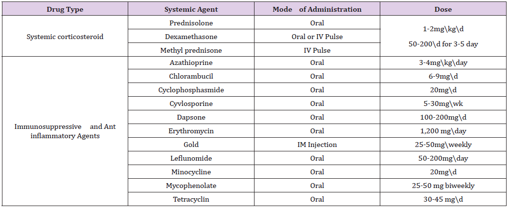
Bio sorption Studies of Chromium Ions with Modified Chicken Feathers

Abstract

Modified chicken feather has been used as biosorbent for removing chromium ions from aqueous solution. The adsorption capacity tests were performed on an equilibrium batch basis. The parameters such as contact time, biosorbent dose, pH etc were optimized and were found to be 2 hrs, 100 mg & pH 6.0. Equilibrium isotherms were analyzed for the adsorption process and Freundlich adsorption isotherm model was found to fit the data well. The kinetics of adsorption followed pseudo-second order model.

Keywords: Modified Chicken Feather; Bio sorption; Chromium ions; Freundlich isotherm; Pseudo second order

Introduction

Heavy metals such as Hg, Pb, As, Cr, etc are harmful to soil, water bodies and aquatic life. They are leached out mainly from industrial wastes, mines and research labs [1]. They produce acute toxicity in plants, animals and micro organisms. Due to bio accumulation and bio magnification through food chain they cause chronic effects even at lower concentrations. Heavy metals are generally removed from environment by precipitation, reverse osmosis, coagulation, flocculation etc [2-7]. These processes have many drawbacks, which include selective or partial removal of metal ions and high operational cost. Bio sorption can be used for effective removal of the heavy metals from environment. The major advantages of biosorption are low cost, high efficiency, regeneration, metal recovery etc. Bio sorption is largely influenced by pH, the concentration of biomass and the interaction time.

A very big problem of the agriculture industry is managing the enormous amount of waste generated by poultry processing enterprises. The scientific usage of feathers as a renewable material offers both economic and environmental benefits. The adsorbing ability of chicken feathers (CF) as potential biosorbent for the removal of hazardous substances from effluents is due to their high surface area and several reactive functional groups [8-11]. CF consists of keratinous proteins with many functional groups such as -COOH, -NH2, S-S- which can be treated with suitable chemical reagents to get chemically modified CF biosorbent.

Experimental

The reagents used were ethanol, methanol and HCl and were of analytical grade. UV-Visible spectrophotometer (shimadzu-1800), Mechanical shaker (Rotek, Model number REC27255A2), pH meter, XRD (Rigaku Miniflex X-ray Diffractometer with Cu Kα radiation) etc were used.

Preparation of Biosorbent: The chicken feathers (CF) collected from poultry farms were cleaned, washed in water and ethanol and cut into pieces of size 5mm. CF was modified using an equimolar mixture of methanol and HCl for two hours. 10 g CF was mixed with 6% (v/v) CH3OH and 2% (v/v) HCl in a 250 ml double necked flask and placed on a hot plate at 80 0C with constant stirring for 3 hours. The reaction mixture was filtered, washed with distilled water and kept for drying [12]. Modified CF biosorbent was characterized by XRD & FTIR [13].

Preparation of Adsorbate: The stock solution of chromium metal ion was prepared in the range (1-10) × 10−5 M.

Batch Adsorption Experiments: The adsorption studies were carried out in batches in different conditions of pH, contact time, amount of adsorbent, temperature etc to check the propensity of adsorption process. In each 100 ml conical flasks, 25 ml of chromium solution was taken along with 100 mg of adsorbent and shaken for 2 hours in an orbital shaker which was then kept for 24 hours for saturation. Thereafter supernatant liquid was filtered through Whatmann Filter Paper No.42 and the amount of chromium ion adsorbed was determined spectrophotometrically at λmax 540 nm. The amount of chromium ion adsorbed per unit biosorbent (mg metal/g of biosorbent) was calculated using Equations (1) & (2)

Equation 1:

Where Co & Ce represent initial and final equilibrium concentrations (mg/L), V is the volume of Adsorbate taken, W is the weight of the biosorbent and qe is the amount of dye adsorbed at equilibrium.

Results and Discussion

Characterization of the Biosorbent

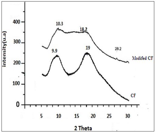
Chemical modification of chicken feather was carried out with methanol as shown in Figure 1. Modified CF was characterized using XRD and FTIR. IR showed peaks in the range 1600-1700 cm-1 is due to -NH and -C=O stretching vibrations of the amide group. In the case of modified CF, this peak becomes sharp at 1653 cm-1 due to the formation of random coils at the expense of a α-helix and β-pleated sheets .The appearance of intense peak at 1740 cm-1 is due to the -C=O stretching vibration of the aliphatic ester of the modified CF [13]. XRD patterns of CF and modified CF are shown in Figure 2. The peak at 9.9 indicates α- helix configuration and a peak at 19 is due to stranded secondary structure. The modified CF peaks show decreased intensity. The slight shift of 2ϴ values confirms the decrease of the β-sheet content and partial cleavage of α- helix network [13] (Figures 1 & 2).

Figure 1: Chemical modification of chicken feather was carried out with methanol.

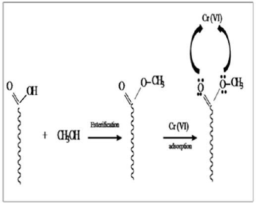
Figure 2: XRD patterns of CF and modified CF.

Adsorption Studies

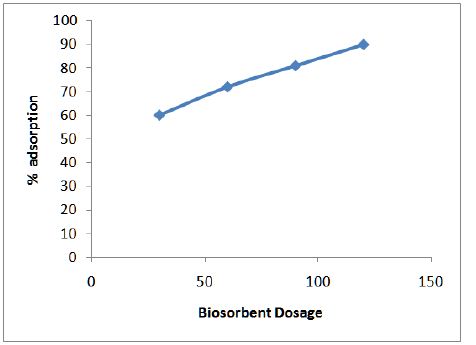
Figure 3 shows the variation of adsorption efficiency of CF with pH for chromium ions it was found that maximum adsorption occurs at pH 6.0. The effect of variation of contact time on the adsorption of metal solution was also studied and optimum time was found to be 2 hours as shown in Figure 4. From Figure 5 we can see that adsorption of the metal by modified CF also depends on the amount of sorbent used and the optimum amount was found to be 100 mg.

Figure 3: Variation of adsorption efficiency of CF with pH for chromium ions.

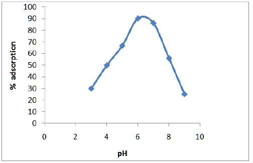
Figure 4: Effect of contact time on adsorption of matal.

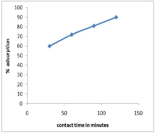
Figure 5: Optimum time was found to be 2 hours.
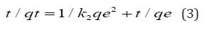
Adsorption kinetics

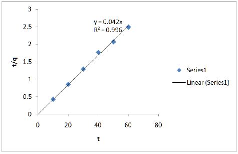
Figure 6 shows the effect of contact time on adsorption of chromium onto modified CF at 100 mg/L. Ho’s pseudo-second order model (Eq. 3) was used.

Figure 6: Effect of contact time on adsorption of chromium onto modified CF at 100 mg/L.

Equation 2:

Kinetic studies show that the data fits well in the pseudosecond order plot and is shown in Figure 7.

Figure 7: Kinetic studies show that the data fits well in the pseudo-second order plot.
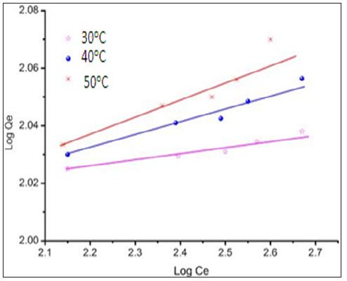
Equilibrium Studies

The Freundlich adsorption isotherm was applied for the adsorption of metal on modified CF.

The Freundlich equation is represented as:

Equation 3:

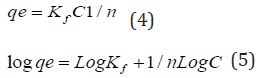
Figure 7 is the plot of equilibrium isotherm for the sorption of chromium ions on chemically modified CF. The data fits well in the Freundlich isotherm. It also shows the dependence of temperature on the adsorption of chromium ions on the sorbent which confirms the endothermic nature of biosorption.

Conclusion

This study showed that modified chicken feather could be used as a potential biosorbent for the removal of Cr (VI) ions from aqueous solution. The biosorption process was affected by contact time, temperature, pH and biosorbent dosage. The thermodynamic studies indicated the endothermic nature of the biosorption process. The Freundlich isotherm model was found to be the most suitable in describing the equilibrium of the biosorption process. The kinetics of adsorption followed pseudo-second order model.

For More Articles: Biomedical Journal Impact Factor: https://biomedres.us

Journals on Philosophy

Affording Visual Causal Epistemologies in Epidemiology

Mini Review

One of the challenges of 21st Century sciences is how to deal with and manage huge amounts of raw data [1] Using several computational tools, scientists are able to capture, process and, finally, to understand that data. The visual aspects of this understanding process are of the utmost importance due to the specific cognitive mechanisms that make possible human thinking [2]. Epidemiology is a very complex research field devoted to the study of health and the causes of illness [3]. The difficulty of establishing sound statistical relationships between sets of events and some causal outcomes [4] has been the main source of debates within the field [5-7] Although epidemiologists and physicians have tried to avoid philosophical debates [8] about causality, it has been impossible to not be aware of the intrinsic and insurmountable problem of working with so complex amounts of data. From the simple one-hit paradigm of early epidemiology [9,10] to current multi-causal webs of determinants [11], new challenges have emerged. A possible solution for the management of such sets of data has been to invest into visual causal methods: directed acyclic graphs (henceforth, DAG). These methods have allowed a visual quantitative approach to epidemiology [12-14] that fits perfectly with the current research trends in cognitive sciences which defend the power of extended and enhanced ways of using informational tools, which afford new and sound ways of processing information.

In this brief mini report I suggest several interesting corollaries of the implementation of such techniques:

a. The necessity of the enhancement of visual argumentative methods in scientific practices. A few examples, such as Venn diagrams or Feynman’s diagrams [15,16] have still not reached a full implementation of Western scientific cultures dominated by classic written methods.

b. The power that these visual methods give to not only human analysis but to the more fruitful interactions between human thinking and machine thinking in data-mining processes [17].

c. The epistemic reliability and soundness of visual reasoning, beyond the classic critics to these methods.

d. The necessity of more analysis on the culturally embedded values into visual thinking (related on traditions on spatial uses during writing processes, to prevent some possible mistakes as well as to define better this new thinking space [18-20].

Taking into account these ideas, we’ll be able to define a new reasoning scenario for epidemiological researchers that could even be exported to other domains, not only from classic sciences but also to social sciences or even humanities. This new visual quantitative methodologies may make possible a new set of scenarios for the advancement of knowledge thanks to the implementations of tools which impulse the cognitive creativity, closer to naturalized and easily affordable ways to deal with information.

For More Articles: Biomedical Journal Impact Factor: https://biomedres.us

Food Sceience Journal

Factors Contributing to Low Productivity and Food Insecurity in Bungoma County, Kenya

Abstract

Food insecurity within households is a risk to people’s livelihoods. If not addressed in good time it could results into a disaster that will require foreign intervention for that affected community. Households in Bungoma county of Kenya were noted to be vulnerable to food insecurity due to low productivity and this state threatened peoples livelihoods. The objective of this study was to examine the physical, economic, environmental and social factors that led to low food production in Bungoma County, Kenya. A cross-sectional survey design was used in the study and a cluster (multi-stage random) sample size of 384 households was selected. Tools used for data collection were questionnaires, interview guides, focus group discussions and observation checklists. Data was analyzed using descriptive and inferential statistics. The study found that land size was small; the road network was poor and disorganized market systems. The cost of farm inputs was high as well as high poverty levels.

Climatic variability affected crops and animal production. Social support, traditional beliefs and culture which discriminated against women were key risk factors that contributed to low farm production, making households vulnerable to food insecurity. Based on the findings, the study concluded that low farm productions were attributed to physical (Poor road networks and small land size), economic (poverty and high cost of farm inputs), environmental (climate variability and deforestation) and Social (cultural belief and negative attitude) factors. The study recommended that costs of farm inputs should be subsidized, improve road network system and sensitize people on positive cultural practices and attitude change to allow both gender participation on issues of food security.

Key words: Farm productivity; Household food insecurity; Bungoma County

Introduction

Background

Food is a basic necessity of life. It is a basic means of sustenance and key for healthy and productive life. If Kenya is to continue to cut down on health costs and compete in a global economy, it should ensure adequate food security and nutrition within households. Food insecurity within households is a risk to people’s livelihoods. If not addressed in good time it could result into a disaster that will require foreign intervention for that affected community. The economic development of any nation is dependent on the productive capacity of human resources which is however a function of how well fed they are. Poor farmers have little or no access to credit, particularly short-term seasonal credit for farming Audsley et al. [1]. Under such circumstances, households lack economic capacity and therefore are at a risk of being vulnerable to food insecurity. Crucial information on the type of interventions that can be most effective in increasing productivity, reducing hunger, targeting the most needy, informing preparedness and developing contingencies is lacking in most communities in Kenya Lautze et al. [2].

Problem Statement

Available literature indicates that Bungoma County is food insecure and also records a poverty index of 52.9% compared to the National index of 46%, while the food poverty stands at 43% KNBS [3]. There is documentary evidence that Bungoma County has many stakeholders dealing with food security issues being led by the County Government GOK [4]. This would give an impression high production and food sufficiency at household level but it is not the case. Food situation reports dating way back to 2011, show insufficient food stocks among households in Kenya GOK [5]. Records of studies done in Bungoma county revealed household food insecurity NALEP [6], Muyesu [7], KARI [8] and Ndienya et al. [9]. Many families in Bungoma County take one meal a day, in contrast to the recommended three meals per day UNICEF [10]. Due to this controversy, the study was set up with the objective to examine factors that led to low productivity within households, making them vulnerable to food insecurity despite the County’s interventions.

Objective: The objective of this study was to examine physical, economic, environmental and social factors that led to low productivity and made households vulnerable to food insecurity in Bungoma County, Kenya

Contribution to the Field: The study will give recommendations to guide policy makers on issues of food security. This paper contributes to the knowledge bank important for scholars. It is arguable that findings of this study with a focus on Bungoma County will inform similar studies in other counties in the entire country.

Significance of Work: The outcome of the study will guide decision-makers at all levels in formulating food policies. Reliable and timely information on the incidence and causes of low productivity, food insecurity and malnutrition will be documented. Recommendations from the study is expected to assist households understand the crucial factors of production and risks of food insecurity and be able to appropriately plan their farming schedules.

Research Methods and Design

Materials: The study targeted household heads whose food security depended on farming. Community groups (women groups, men groups, youth groups and self-help groups) were targeted for focus group discussions. Opinion leaders, Non-Governmental Organizations, Community Based Organizations/Non-State actors, Faith Based Organizations and Government officials were selected as key informants.

Setting: This study was done in four sub-counties of Bungoma County; they included Bumula, Bungoma West, Mt. Elgon and Bungoma North. The County is located on the Southern slopes of Mt. Elgon, and lies between latitude 00 281 and latitude 10 301 North of the equator, and longitude 340 201 East and 350 151 East of the Greenwich Meridian.

Procedure: The research work adopted a cross-sectional survey research design and the variables examined were physical, environmental, social and economic factors. The population for the study was household heads, key informants and formal organized groups. A cluster (multi-stage random) sample size of 384 households- calculated using a formula from the book of Mugenda [11] was selected from household’s population of 1,553,655 KNBS [12]. This study utilized both primary data collected from the field and secondary data from archival sources. Data was collected using semi-structured questionnaires administered to the selected household heads. Four (4) Focus Group Discussions were held and each group was composed of eight to twelve (8-12) members of mixed gender. Twenty (20) key informants purposely chosen from opinion leaders, Government departments, Faith based organizations, Non-governmental organizations were interviewed. More information was obtained from observation checklists [13,14].

Analyses: The quantitative data were organized, coded and edited by a process called data cleaning Punch [15-17]. The statistical package for social sciences (SPSS) was used to analyze data. Two analyses were made. Descriptive analyses was done by use of means, modes, standard deviations, variance, percentages, and frequencies) while inferential analyses was by use of chi-square test and Spearman rank order correlation analysis.

Results and Discussion

Physical Factors and Vulnerabilities to Food Insecurity

Various physical factors were identified as contributors to low productivity. These included small land size for farming, non use of fertilizer and certified seeds. The soil was infertile and this led to low yields, poor infrastructure, and disorganized marketing system [18]. Chi-square tests revealed a significant relationship between physical factors and production levels in the county (p-value = 0.035; < 0.05). It was also established that markets were few and far apart from farmers. The distribution of farm produce outlet included; farm gate level, neighbors, local or open markets and others. International markets fetches better prizes but unfortunately, all households interviewed had no idea of existence of export market. Very little produce was sold to supermarkets, meaning low incomes that could not enable farmers to purchase certified seeds or other food items not produced on the farm [19,20]. The seasonal roads as well as lack of means of transport made farmers to sell their produce at low prices on the farm. Besides this, farmers did not have government permits and certificates of operation to enable them penetrate the supermarkets in the country.

Economic Factors

In order to earn a living and be food secure households engaged in the following activities: Dairy production, maize farming, horticulture, banana farming and petty trade. Most of the households depended on farming with some shifting from subsistence to business farming to raise income. Similar views were found by Makhanu et al. [21] working in the same region; this shift in attitude to do farming as a business reflects current trends of blending specialization and diversification to reap optimal benefits by smallholder farmers. This was also observed by similar studies as captured by government policy initiatives in Agriculture GOK [22]. The economic factors that contributed to low productivity and food insecurity were listed as high levels of poverty and high cost of farm inputs. Due to high cost of farm inputs like fertilizers and certified seeds, majority of the farmers planted uncertified maize seeds (number name) and without fertilizer. As a result of planting uncertified seeds, the cereal yields were so low that it hardly sustained a household for three months after harvest. Horticulture farming was affected due to non-use of chemicals to control pests and disease [23-25].

Environmental Factors

Environmental factors contributing to food insecurity were found to be natural calamities like drought, floods, hailstones and inadequate / unreliable rainfall. Crops on farms were at the risk of natural calamities like hail stones [26-28]. Too much rainfall led to floods which damaged both properties and livelihoods. Human activities like cutting of trees led to deforestation and this resulted into soil erosion. Erosion made soil unproductive as the soil nutrients are washed downstream, hence food insecurity for such households. Other factors established were pests anddisease outbreaks which were a risk to both crops and animals [29]. This finding is comparable to a study done by Ahmed et al. [30], which revealed that increasing vulnerable environmental conditions such as diminished biodiversity, soil degradation or growing water scarcity can easily threaten food security for people dependent on the products of the land, forests, pastures, and marine environments for their livelihoods. These findings also support Kenya Government recommendations for adapting to climate change like; conservation farming, right land use practices that reduce emissions of greenhouse gases GOK [31].

Social Factors

A key social factor contributing to vulnerability was the gender of the household head. The study established that 80% households were headed by men while 20% were women. All decisions in the household were made by men. In many cases men were found to be the cause of food disasters in their own homes. Women had no say in decision making concerning food issues where men were heads [32-34]. Men made final decisions in relation to land allocation for different crops, when to market farm produce and the use of cash from sale of farm produce. The study further revealed that women were in the same category with children, so they could not be allowed to make final decisions in the households. One Man, during focus group discussion quoted the Holy Bible (Genesis 2:18) [35] where he said ‘women were made to assist men), therefore they should always be subordinates to us’. This notion made households vulnerable to food insecurity as productive ideas from women may not be adopted. The findings were similar to the study done by Lautze et al. [2] who found out that positive traditional value, customs and ideological beliefs contributed to social vulnerability of any given household.

Focus group discussions recorded that culture prohibited working on the farm during bereavement and this contributed to low productivity incase funeral occurred during planting season. Farming activities may be stopped for periods exceeding three weeks. This can be crucial as even a small period of time lost affects agricultural production Africa Progress Report [36], Delgado [37]. Laziness, idleness among the youth and theft of farm produce while in the farm and store were mentioned as contributing factors to food insecurity. Key informants quoted lack of knowledge on production and storage as factors making households vulnerable to food insecurity. This was also revealed by household interview results, where 61% of the household heads only attained primary level of education, meaning they were limited in knowledge and the level of understanding of new farming technologies [38,39].

Conclusion and Recommendations

Farm production by Households in Bungoma County were found to be low and hence making them vulnerable to food insecurity because of the following factors; physical (Poor road networks and markets), economic (poverty and high cost of farm inputs), environmental (climate variability and deforestation) and Social (cultural belief and negative attitude). The study recommended that the County Government of Bungoma should subsidize costs of farm inputs and make it accessible to farmers, the road network system should be improved to ease transportation to access markets for farm produce, people should be sensitize on positive culture practices and attitude change to allow both gender participation on issues of food security.

For More Articles: Biomedical Journal Impact Factor: https://biomedres.us

Dentistry Journal

Comparative Evaluation of the Bleaching Efficacy of Colgate Visible White, Close-Up Diamond Attraction Compared With Pola Office Plus

Abstract

Bleaching is very common and costly procedure for whitening of teeth. As the aesthetic concern of the patient’s increases these days, the introduction of wide range of less costly whitening products in the market also increases which promises whitening in few applications.

Aim: The objective of this in vivo study is to check the bleaching efficacy of Colgate Visible White, Close-up Diamond Attraction compared with Pola Office Plus.

Materials & Methods: 15 patients were selected and randomly divided into three groups according to whitening agents used. Each group contains five patients i.e. n=5.The application time is as per recommended by the product instructions. For the evaluation of shade selection – color of both canines in upper jaw was evaluated visually before and after bleaching. Tooth shade was evaluated using VITA classical shade guide.

Results: After statistical analysis a significant difference was seen in shade change between pre and post treatment in each group. However, no significant difference was found between all the three groups.

Conclusion: Tooth whitening using minimally invasive techniques is enjoyable for the clinician and allows the patients to smile with confidence.

Key words: Bleaching; Colgate visible white; Close up diamond attraction; Pola office plus

Introduction

Patients increasingly seek to have an attractive smile, as it is considered to be synonymous with health. This growing demand for an enhanced esthetic appearance has led to great development of bleaching products. The color of teeth is influenced by a combination of their intrinsic color and the presence of any extrinsic stains that may form on the tooth surface. Tooth discoloration can be treated professionally with in office or home bleaching. Pola Office+ is the advanced version of the well known in-office tooth-whitening system, Pola Office, which has been used successfully for several years. Pola office +contain desensitizing agents, such as potassium nitrate, to reduce postoperative sensitivities in vital teeth. Also, the whitening tooth pastes have gained in popularity for treating extrinsic stains. The effectiveness of toothpastes at reducing or removing extrinsic dental stain has improved with the introduction of more whitening toothpastes onto the market [1]. A key feature of whitening toothpastes is that they include proteolytic enzymes that remove extrinsic stains from teeth. It is claimed that some whitening toothpastes also remove pellicle (external membrane) from a tooth surface [2].

Apart from containing whitening agents, whitening toothpastes also commonly contain bicarbonates and fluoride to promote caries protection. In Turkey, it has been suggested that further clinical studies are required to help clinicians decide exactly which whitening toothpaste(s) to recommend to patients [3]. A cursory appraisal of the ingredients suggests that most whitening toothpastes are formulated to control extrinsic dental stain rather than to change the natural colour through a bleaching action [4]. The market continues to expand as more tooth whitening dentifrices become available and clearly it is important to evaluate these products. Many clinical techniques such as the Lobene Stain Index (1968) [5], the Shaw and Murray Stain Index (1977) [6], and the comparison of tooth colour with a Vita shade guide have been used to investigate the reduction of extrinsic staining of teeth. Many clinical studies into the whitening effect of dentifrices have been reported, with study periods differing from two weeks to six months [7].

Aim of the Study

The objective of this in vivo study is to check the bleaching efficacy of Colgate Visible White, Close-up Diamond Attraction compared with Pola Office Plus.

Materials and Methods

An initial sample of 15 patients aged from 18 to 30 years was selected from patients who came to our department for tooth bleaching. A randomized, single-blind (examiner-blinded) trial was conducted. The examiner and all staff were blind to the treatment assigned throughout the study. Study inclusion criteria (evaluated at a first examination) were as follows: age ≥18 years, absence of gingival recession or restorations in upper-front teeth, good oral hygiene and gingival health, no previous tooth bleaching procedure, tooth shade of A2 or above on the Vita ClassiÊl Shade Guide scale (Vident, Brea, Calif, USA.) ordered by value, and, in the case of females, not being pregnant or in breast-feeding period. Subjects were excluded if they used tobacco products, had aesthetic restorations, which could become discolored, or were already taking chromogenic oral products, such as chlor hexidine, or medications that could stain the dentition. Before the bleaching treatments, baseline numerical shade values were obtained for central incisors and canines of both hemiarches in accordance with the Vita Classical Shade Guide (Figure 1 & 2). Shade tabs were arranged in the sequence recommended by the manufacturer, assigning each tab with a number from 1 to 16 (B1, A1, B2, D2, A2, C1, C2, D4, A3, D3, B3, A3.5, B4, C3, A4, C4). The shade was recorded by two independent examiners not otherwise involved in the study (K coefficient = 0.85, standard error = 0.09), who were blinded to the materials used. Group 1(n-5) patients were assigned Colgate visible white tooth paste. Group 2 (n-5) were assigned Close up diamond attraction toothpaste. Subjects were instructed to brush their teeth twice a day, in the morning after breakfast and in the evening before sleeping. Subjects were not given any instructions about drinking and eating, and were allowed to follow their previous patterns for these activities. In Group 3 (n-5) patients bleaching was performed by Pola office +. The dual-barrel syringe system of Pola Office+ always delivers a consistent mixture of freshly activated gel that can be easily applied with a fine applicator tip, even in hard-toreach areas .Excess gel was removed with a cotton pellet. A thin layer of gel was applied on the vestibular surface of the tooth. The gel was left on the tooth for eight minutes and then removed using a surgical aspirator tip. The application steps were repeated up to three times, so that the material is on the tooth for a maximum of four times for 8minutes (32minutes in total) in one session.

Results

Fifteen patients completed the study protocol, with a mean age of 23.8 years (range, 18-30 years). No adverse effects on soft tissue were reported by the patients or observed by the examiners. After statistical analysis, a significant difference was seen in shade change between pre and post treatment in each group. (Table 1 & 2) However, no significant differenc was found between all the three groups (Figure 1-4).

Figure 1: Values Were Obtained.

Table 1: N Par Tests.

Table 2: Wilcoxon Signed Ranks Test.

Discussion

In this study, whitening scores were evaluated to assess the validity of whitening toothpaste advertisement sclaiming that whitening will be observed after four weeks’ use of whitening toothpaste. The whitening effects of the toothpastes were compared during this short period. Visual assessment with shade guides, computer analysis of digital images, colorimetry, and spectrophotometry can all be successfully used to measure the color change of teeth in longitudinal tooth bleaching studies [8]. Digital images offer an objective shade difference value, but the light and positioning of the subject must be standardized and a robust mathematical transformation algorithm must be used [9]. We used the Vitapan Classical shade guide ordered by value, considered a valid and reliable method for color assessment [10], and applied in previous investigations [1,11-13]. Other more sophisticated measurement systems are available [10,17,18] but do not offer a more reliable accuracy, since different color measurements can be obtained for the same tooth according to the positioning of the probe tip of the device [14].The Vita shade visual assessment has previously been used successfully in many clinical studies [12-15]. It was therefore used in this study.

Previous studies have reported that whitening dentifrices produce a greater reduction and/or inhibition in extrinsic staining of natural teeth than standard commercial toothpaste formulations [16-18]. The active ingredients of toothpastes either include enzymes that dissolve pellicle proteins and inhibit staining, or contain chelating agents that have stain-dissolving properties. Alumina, dicalcium phosphate dihydrate, and silica are some of the abrasives that are included in whitening toothpastes [19]. In the present study, two whitening toothpastes were evaluated. Colgate visible white was used which consists of silica and polyphosphates that removes and prevent stains on teeth. The high cleaning silica in this toothpaste is similar to the abrasive materials used for cleaning and polishing teeth to remove surface stains. The polyphosphates are widely used to prevent surface staining due to plaque formation. Other toothpaste used was close up diamond attraction based on blue light technology.

It consists of sorbitol, hydrated silica, trisodium phosphate. Hydrated silica has been shown to have great cleaning and stainremovalability [20]. It has been suggested that another factor for whitening effect of toothpaste is pH Price. Reported that whitening toothpastes had a mean pH of 6.83 (range 4.22 to 8.35) [21]. The abrasive in toothpastes should ideally provide stain removal without causing wear of the tooth [22]. Lima. Reported that the enamel wear caused by these dentifrices still needs to be assessed before they are recommended for routine use [23-30]. In future studies, this criterion may be added for the evaluation of whitening toothpastes. In the present study Pola Office + was used which showed significant difference in pre and post treatment results. With SDI’s new in-office system, Pola Office+, patients can have whiter and brighter teeth in less than one hour. The easy-to-handle, dual barrel and auto-mixing syringe system is economical and optimizes the workflow of the practice. In addition, the desensitizing agents and neutral pH make Pola Office + the ideal tooth-whitening material for vital and non-vital teeth. The active ingredient of Pola Office+ is 37.5 % hydrogen peroxide and the gel rapidly releases peroxide ions upon contact with the tooth, enabling a shorter contact time compared with its competitors

Results

Tooth-whitening toothpastes are effective for removal of superficial stains and for providing a whitening effect. Tooth whitening using minimally invasive techniques is enjoyable for the clinician and allows the patients to smile with confidence.

For More Articles: Biomedical Journal Impact Factor: https://biomedres.us

Journals on Dentistry

Anti-Microbial Peptides and Their Speculative Role in Periodontitis

Abstract

Antimicrobial Peptides (AMPs) are present in the oral cavity in the form of defensins and human cathelicidin LL-37 (from neutrophil granules) and histatins along with pdefensins 1 and 2 (from salivary glands and gingival epithelial cells). The oral micro flora organisms that play an important role in the pathogenesis of periodontal disease are opportunistic pathogens, that are highly proteolytic and this activity is known to contribute to nutrient acquisition, tissue destruction and de-regulation of inflammatory responses. Furthermore, the production of proteases enables oral bacteria to evade killing by antimicrobial peptides, thus contributing to the virulence of such opportunistic pathogens, which could have implications for the use of antimicrobial peptides as therapeutic agents to treat periodontal disease. Hence, this review summarizes the suggestive role of AMPs in periodontal disease.

Key words: Immunity; Anti-Microbial Peptides; Periodontitis

Abbreviations: AMPs: Antimicrobial Peptides, LAP: Lingual Antimicrobial Peptide, HNP: Human Neutrophil Peptide; hBD: Human Beta- Defensins

Introduction

Humans often require a multilayered defense mechanism to function smoothly and combat micro-organisms and pathogens. The fundamental guarding complex for almost all human organisms comprises of innate immunity [1]. It is this defense mechanism that helps discern a wide variety of agents, known as pathogens, and distinguish them from the organism’s own healthy tissue [1]. The oral cavity is a manifold interaction where sundry organisms, both commensal and destructive types, intercommunicate and escalate in the environment [2]. The noteworthy quality of the oral cavity lies in the presence of specialized interaction between tooth (hard tissue) presenting itself from the underlying gingival epithelium (soft tissue).

Ideally in the oral cavity, the tooth structure often encompasses a layer of pathogenic biofilm termed “Dental plaque,” that compromises the surrounding epithelium via its incessant exposure to microorganisms [3]. This is when the comeback of the epithelium to these insults determines the overall condition of the gingival sulcus. It is evident that the oral epithelium works in several ways for the conservation of the underlying tissues. As a physical barricade, it can counter unbroken microbial oppositions from dental plaque by the production of chemokines, cytokines, and antimicrobial peptides (AMPs), which enhance inflammation and immune response in oral epithelial tissues [4]. These epithelial antimicrobial peptides are considered to be paramount for the innate immunity of the host [5].

AMPs are also prime contributors that enable the stabilization between health and disease in this complex ecosystem [6]. Exhibiting a wide spectrum of antimicrobial activity against grampositive and gram-negative bacteria, yeasts and certain viruses, they possess the ability to prevent various oral periodontal diseases including bacteria, fungal and viral infections [7]. The reaction involving multiple AMPs to a single pathogen of infection prevents the consequences of antimicrobial resistance. Several families of antimicrobial peptides have been studied in the oral cavity which includes α-defensins, β-defensins, calprotectin, adrenomedullin, histatins, and cathelicidin [8]. The aim of this review paper is to view the crucial role of these molecules against periodontal diseases and its function in host immunity. The article also sheds light on the mechanism of action and the types of identified AMPs.

Types of Anti-Microbial Peptides involved in Periodontitis

The AMPs react to the periodontopathogenic bacteria in a synergistic manner, whereby they secrete chemical innate immunity signal molecules like interleukin, chemokine and cytokines that attract neutrophils at the site and caution the host response [7]. Conducive with the amount of microbial exposure, they also produce natural AMPs and proteins. By acting as an integral part of the hosts natural innate environment, the oral epithelium, polymorph nuclear leukocytes (neutrophils) and saliva, all concurrently and solitarily bestow to this response [9,10]. These responses involve several salivary antimicrobial peptides, the β-defensins manifested in the epithelium, the α-defensins expressed in neutrophils, and the cathelicidin, LL-37, in both epithelium and neutrophils [11-13].

Defensins

The first AMPs identified in the oral epithelium were defensin, Lingual Antimicrobial Peptide (LAP) [14]. The defensin families are prominent AMPs that reside in the host environment, which include the alpha-defensins (human neutrophil peptide; HNP) and human beta-defensins (hBD) [15]. Defensins are stored in secretory granules, with expanding levels observed during periodontitis (Figure 1). Their mechanism of action involves destroying phagocytosed bacteria upon fusion with the secretory granules from the phagocytic vacuoles they reside in [16]. Being a part of the host’s natural environment, expressed in gingiva, tongue, salivary glands and mucosa, they are present in conditions like oral inflammation, carcinomas, etc. Defensins have also shown to inhibit LPS-stimulated inflammatory responses in host cells, which is a primary causal mechanism in the pathogenesis of periodontal disease [16]. Defensins are allocated into subfamilies of α- and β-defensins. Human beta-defensins (hBD) are elementally revealed in epithelial cells, while alpha-defensin are predominantly expressed in neutrophils [17]. Segregated in terms of their cysteine motifs, dually both of them share a homogenous secondary structure, and are opulent in cationic residues [18,19].

Figure 1: Mode of activation of hNPs in periodontal tissues.

In the α-defensin subfamily, four of the six α-defensins, human neutrophil peptide -1, -2, -3, and -4, are fabricated and gathered in neutrophil granules while the other two α-defensins, HD-5 and -6, are synthesized and stored in the granules of paneth cells, specialized epithelial cells at the crypts of Lieberkuhn of the small intestine [17,20,21]. In the β-defensin subfamily only hBD -1, -2 and -3 are substantially expressed in the oral cavity [16,22]. hBD-3 along with the respective three mentioned above are markedly expressed in epithelial cells that encrust some tissues and organs, fundamentally skin [23]. In non-inflamed gingival tissues and gingival crevicular fluid, hBD-1 and hBD-2 have been expressed, with maximum levels at gingival margin [22]. Elevated levels of HNP1-3 have been noted in cases of periodontal infections, indicating a strong correlation See Table 1 for summary.

Table 1: Summary of the role of defensins.

Cathelicidin (LL-37)

Used markedly as one of the predominant identifying tools in inflammatory periodontal disorders, this class of AMPs incorporates a mature peptide and cathelin domain. It is a multifunctional peptide, comprising of 37 amino acids [24]. Active against gram positive and gram negative bacterias, it directly binds to the LPS of bacterial cells, and is native to the oral cavity in several sites, including the buccal and tongue mucosa, gingival crevicular fluid [24]. By activating antigen-presenting cells, it presents as a hemoattractant for immune cells, including monocytes, T cells, etc. There is a specific correlation amongst multiplication in LL-37 levels and periodontal inflammation [25-28]. It is also known to cause an elevation in mucosal thickness in the gingiva. Furthermore, studies have also demonstrated its tissue specific effects on cancer cells [27,28]. Their mode of action involves intracellular killing of the phagosomes after phagocytosis of the bacteria, where the AMP in the neutrophil is severed into a fully developed peptide (Table 2).

Table 2: Summary of the role of Cathelicidin.

Histatins

These AMPs are exclusively resided in the salivary glands, i.e. parotid and submandibular salivary duct cells. Histamine 1, 3 and 5 are found to be predominant of the total histatin proteins (85%) in the saliva [29-32]. They have a major role in fungicidal activity, having a noteworthy role in oral candidiasis restraint, especially histatin-5, which is the most significantly active against candida species as well as bactericidal activities against Porphyromonas gingivalis and Streptococcus mutans, which play a key role in the etiopathogenesis of periodontal disease and dental caries ,respectively [30,32]. Significant linkage occurs between xerostomia and oral candidiasis. It is also noteworthy that the antifungal action is shared between alpha-defensin and histatin, which require ATP transport found in active mitochondria.

Secretory Leuko Protease Inhibitor (SLPI)

Another AMP identified from the parotid salivary gland is SLPI. Manifested in neutrophils, epithelial cells and salivary glands, this protein suggests anti-inflammatory action at the site of infection [33]. Supplementary to having fungicidal action, it has shown to inhibit contamination of human monocytes and is also conveyed in oral tumor tissues as well as inhibition of the HIV virus [34,35]. SLPI is associated with wound repair and its expression is elevated following wound healing [35,36]. The elevated quantity of SLPI following periodontal treatment is an evidence of inflammation resolution activities [36].

Summary

The capacity of these inborn antibiotics is only at its inception of its recognition, as amplified natural announcement or as novel relieving agents [8,37]. This class of proteins is a captivating target for periodontal diseases [37]. In times of periodontal disease, AMPs amalgamate with other inflammatory proteins and maintain inflammatory molecules and pathways. It can be concluded that salivary AMPs have a prospective capacity to be recognized as initial markers of periodontal infections [38]. Thus, a dual approach is mandatory to comprehend the task of hosts’ immunity in response to, against periodontitis [37]. This involves more comprehensive statistics about HNP and hBD, LL-37, and other oral antimicrobial peptides and proteins, along with their mode of action and clinical significance [38-50]. These peptides are peculiar in keeping the level of bacteria in control, having distinctive as well as dual roles in maintaining oral health. Not only can they act as diagnostic or prognostic biomarkers, but also the enhancement of new peptide agents can be a signaling method for future investigation and testing (Table 3).

Table 3: Significance of AMPs.

Moreover, Antimicrobial peptides are potentially important as novel therapeutic agents against periodontal disease diagnosis and potential treatment and probably show most immediate promise for development as topical adjuvant agents in conjunction with conventional periodontal therapy in the treatment of periodontal disease. This makes them promising for oral diseases, as topical application of antimicrobial agents is easy and appropriate, and their use would not contribute to resistance to antibiotics normally used in treatment of more life-threatening bacterial infections. All these findings will have direct implications for new understanding of oral innate immune responses and the development of potential new and innovative therapeutic interventions for periodontal disease.

For More Articles: Biomedical Journal Impact Factor: https://biomedres.us

Journals on Orthopaedics

Periosteal Osteosarcoma of Radius Mimicking Synovial Sarcoma

Abstract

Periosteal osteosarcoma is a rare surface osteosarcoma. It has a benign course with good prognosis. We report a case of periosteal osteosarcoma in an old lady arising around the elbow and mimicking like synovial sarcoma with calcification in the periarticular tissue. The mass biopsied proved osteosarcoma with osteoid matrix. Patient underwent definitive treatment and followed up for five years with no evidence of recurrence.

Key words: Periosteal osteosarcoma; Calcification; Synovial sarcoma

Abbreviations: ESR: Erythrocyte Sedimentation Rate; CRP: C-Reactive Protein; MRI: Magnetic Resonance Imaging; PET: Positron emission tomography; IHC: Immuno Histo Chemistry

Introduction

Periosteal osteosarcoma belongs to the group of surface osteosarcoma. It is a rare tumour representing 1-2% of surface osteosarcoma. The tumour can mimic various benign pathological conditions when it arises around the joint and cause diagnostic dilemma. We report such a case.

Case Report

63 year old female presented with right elbow swelling which was insidious onset and progressive of 6 months duration. Dull aching pain followed swelling. Pain present at night and not relieved with analgesics. No significant loss of weight. Systemic examination appeared normal. A solitary globular swelling of dimension 5x5x4cm present over the right elbow extending from proximal to mid forearm. Skin over the swelling was stretched with irregular surface. Swelling was firm to hard in consistency. Plane of swelling was deep to the muscle. Elbow range of motion was 50-80 degree with thumb drop and metacarpal drop. Blood investigations showed normal ESR and CRP ruling out the possibility of infection. Radiological evaluation revealed a large ill-defined lobulated lytic lesion with osteoid matrix noted within the soft tissue on volar and radial aspect of the proximal forearm. Presence of cortical destruction in the neck (metaphysis) and proximal shaft (diaphysis) of radius due to infiltration of tumour (Figure 1). MRI scan showed a large soft tissue component with mineralization and calcification. Periosteal breach with infiltration of tumour into the radial metaphysis, adjacent muscles and posterior interosseous nerve (Figure 2). Differential diagnosis of synovial sarcoma was considered. Incisional biopsy was inconclusive between low grade osteosarcoma (surface variety) and spindle cell synovial sarcoma (Figure 3). Immuno-histochemistry markers proved negative for synovial sarcoma. PET scan showed no evidence of metastasis or skip lesions. Patient counseled and opted for amputation over limb salvage. Wide local excision with above elbow amputation was performed. Tumour margins were negative and hence postoperative chemotherapy wasn’t started. Now post op 5 years with no evidence of recurrence or metastasis (Figure 4).

Figure 1: Radiograph of right elbow anterior-posterior(A) and lateral(B) views showing a large ill-defined roughly lobulated, lytic lesion with osteiod matrix noted within the soft tissue of volar and radial aspect of proximal right forearm. Infiltration of tumour is seen into the neck (metaphysis) of radius.

Figure 2: MRI(1.5 Tesla) of right elbow Axial T2 weighted (A) and Coronal T2 weighted (B) section showing T2 hyper-intensity in the metaphysis and in the soft tissue with cortical discontinuity. Focal T2 hypo-intensity in the soft tissue suggesting calcification.

Figure 3: Microscopy slide of low power 10X (A) and High power 40X (B) showing pattern less sheets of tumour cells with ill-defined cytoplasm with moderate aniso-nucleosis. Delicate seams of osteiod with calcification are seen among the tumour cells.

Figure 4: Anterior-posterior radiograph of the right humerus with amputation stump at follow up (5 years) showing no recurrence.

Discussion

Periosteal osteosarcoma is defined as a tumour with its epicenter outside medulla. Stippled calcification present in the tumour with or without medullary involvement [1]. Tumour commonly involves in the 3rd and 4th decade. Incidence is more common in tibia, femur and humerus. In our case, tumour is seen involving the proximal radius which is an uncommon site. Radiologically synovial sarcoma was considered as a differential because of its para-articular involvement and presence of diffuse calcification. Microscopy proved negative for synovial sarcoma because of the absence of monophasic or biphasic pattern. IHC markers were negative for cytokeratin and epithelial membrane antigen. Central medullary osteosarcoma was also excluded due to absence of a central destructive lesion with periosteal reaction. Characteristic sunburst and codman triangle with varying osteoid matrix in the soft tissue was absent [2]. Periosteal osteosarcoma has a good prognosis with high ten year survival. Primary concern of recurrence is because of positive margins and inadequate tumour resection. Lung metastasis is a problem when there is invasion of the vascular system. Presence of a meta-chronous lesion increases the frequency of metastasis. Treatment options are amputation, loco-regional control and limb salvage. Regular follow up is needed because periosteal osteosarcoma is prone for recurrence. Metachronous lesions are to be watched for, if genetic mutations are suspected [3]. Treatment for recurrence is chemotherapy and palliative radiotherapy.

Conclusion

Periosteal osteosarcoma follows a slow course and its occurrence in uncommon sites can mimic various pathological lesions. Swellings with para-articular involvement and diffuse calcification should be investigated with biopsy and IHC markers to institute prompt management. Adequate surgical clearance with chemotherapy is needed to prevent recurrence.

For More Articles: Biomedical Journal Impact Factor: https://biomedres.us

Journals on Radiology

A Comprehensive Review on Pemphigus Vulgaris

Abstract

Pemphigus vulgaris is a life threatening chronic autoimmune disease characterized by the formation of intraepithelial blisters on the skin and mucous membranes. Pemphigus vulgaris initially manifests in the form of intraoral lesions which spread to other mucous membranes and the skin. Pemphigus vulgaris (PV), the most common and important variant, is an autoimmune blistering disease characterized by circulating pathogenic IgG (immunoglobin) antibodies against desmoglein 3 (Dsg3), about half the patients also having Dsg1 autoantibody. Oral lesions are initially vesiculobullous but readily rupture, new bullae developing as the older ones rupture and ulcerate. Biopsy of perilesional tissue, with histological and immune staining examinations, is essential to the diagnosis. The diagnosis is based on clinical findings and laboratory analyses, and it is usually treated by the combined administration of corticosteroids and immune suppressants. Detection of the oral lesions can result in an earlier diagnosis. The current review focuses on the etiopathogenesis, diagnosis and current treatment of pemphigus vulgaris.

Key words: Pemphigus; Oral mucosa; Autoimmune bullous disease

Introduction

Pemphigus vulgaris (PV) is a chronic autoimmune mucocutaneous diseases characterized by the formation of intraepithelial blisters. It is a rare disease (0.1-0.5 cases/100,000 inhabitants/yr), with onset usually in the fifth or sixth decade of life [1]. Various research have considered pemphigus as idiopathic in nature but strong evidences suggest it to be a disorder which is autoimmune in nature and is due to the presence of production of antibodies PV results from an autoimmune process in which auto antibodies are produced against the desmoglein protein which his adhesive protein and is responsible for holding the cells of epithelium together [2]. Dsg 3 is mainly present in the oral cavity while the desmoglin 1 is present over the skin region. The serum antibodies responsible for PV are always IgG type and these antibodies act against the desmoglein adhesion protein and lead to formation of bulla [1] (Table 1). The diagnosis of PV is based on three independent set of criteria: clinical features, histology, and immunological tests [3]. Research advances have expanded the therapeutic arsenal against PV, which now includes treatments with: pulse therapy (intravenous infusion of very high doses of immunosuppressant for a short time period); high doses of intravenous immunoglobulin; plasmapheresis; immunospecific immunoadsorption; extracorporeal photopheresis with exposure of serum to psoralens and UVA; antagonists of tumor necrosis factor a (TNFa); cholinergic antagonists; and anti- CD20 monoclonal antibodies (e.g., rituximab) [4].

However, no treatment has demonstrated superiority over the others Beutner [5]. The separation of cells, called acantholysis, takes place in the lower layers of the stratum spinosum, whereas according to Jordan et al it was noted that IgG auto antibodies are present in the sera which acts against the desmoglein protein which is responsible for cell to cell adhesion. Mahoney M et al. [6] said that the underlying mechanism responsible for causing the intraepithelial lesion of PV is the binding of IgG auto antibodies to desmoglein 3, a transmembrane glycoprotein adhesion molecule present on desmosomes. Some investigators believe that binding of the PV antibody activates proteases, whereas more recent evidence supports the theory that the PV antibodies directly block the adhesion function of the desmogleins. The separation of cells, called acantholysis, takes place in the lower layers of the stratum spinosum. Electron microscopic observations show the earliest epithelial changes as a loss of intercellular cement substance; this is followed by a widening of intercellular spaces, destruction of desmosomes, and finally cellular degeneration with ulceration. In a non immunologic reaction, a drug directly stimulates monocytes or lymphocytes to release cytotoxic chemical mediators. No immune response is involved in this situation, and these reactions are not antibody dependent.

Investigation and Diagnosis

Davenport S et al. [7] mentioned that diagnosis of pemphigus depends on biopsy confirmation of intraepithelial vesicle formation, acantholysis and the presence of Tzanck cells. Ettlin DA [8] discussed that demonstration of immunoglobulins especially IgG and complement in the intercellular space by direct immunofluorescence (DIF) is a very reliable test for pemphigus vulgaris. Scully [9] said that Indirect Immunofluorescence studies enable a search for circulating auto antibodies in the patient’s serum and are usually performed after direct immunofluorescence studies reveal antibody deposits in the mucosa or skin. Bhol K et al. [10] reported that ELISA is a quantitative method for measuring antibody levels and a useful test for the diagnosis of pemphigus. This method is more sensitive and specific than IIF. It is also superior to sophisticated immunoblotting techniques. Davenport S et al. [7] entioned that diagnosis of pemphigus depends on biopsy confirmation of intraepithelial vesicle formation, acantholysis and the presence of Tzanck cells. Ettlin DA [8] discussed that demonstration of immunoglobulins especially IgG and complement in the intercellular space by direct immunofluorescence (DIF) is a very reliable test for pemphigus vulgaris. Scully [9] said that Indirect Immunofluorescence studies enable a search for circulating auto antibodies in the patient’s serum and are usually performed after direct immunofluorescence studies reveal antibody deposits in the mucosa or skin. Bhol K et al. [10] reported that ELISA (enzyme linked immunosorbant assay) is a quantitative method for measuring antibody levels and a useful test for the diagnosis of pemphigus. This method is more sensitive and specific than indirect immunoflouroscence. It is also superior to sophisticated immunoblotting techniques.

Management of Pemphigus Vulgaris

Silverman S L et al. [11] proposed that treatment protocol for the initial/induction phase for PV typically involves the use of moderate to high dose(60 to 80 mg/day in a single morning dose) corticosteroids and an adjunctive immunosuppressive drug, azathioprine, 100 to 150 mg/day, for steroid-sparing effect, thereby reducing steroid-induced side effects. Moderate to high doses of corticosteroids administered daily, as a single dose, for 2 to 3 weeks are generally safe and well tolerated. Potential side effects of longterm steroid therapy have been extensively reviewed Tan-Lim R and Bystryn [12] stated that Plasmapheresis is the process by which plasma is removed from blood using a cell separator as antibodies are contained within plasma, plasmapheresis results in the removal of the pathogenic PV auto antibodies. Multiple case series have evaluated the efficacy of plasmapheresis in treating PV.20-23 Of the 28 patients evaluated in these studies, 18 (64%) experienced complete remission, 6 (33%) experienced partial remission and 4 (22%) had no clinical improvement. Adverse effects encountered included systemic infections, acute hepatitis, thrombocytopenia, anemia, hypocalcemia, nausea, dizziness, urticaria, fever, and hypotension. In a multicentric study (n=40) were randomized to receive prednisolone alone or prednisolone plus large-volume plasma exchange. While plasmapheresis failed to demonstrate a therapeutic benefit in this study, it has been suggested that an additional immunosuppressive (i.e, cyclophosphamide) or immunomodulatory (i.e., IVIg) therapy may be required to prevent the rebound production of pathogenic auto antibodies associated with disease flares. High dose intravenous immunoglobulin (IVIg) has been reported to be of benefit as a steroid-sparing agent in the management of patients with PV. In a review of eight reports high dose IVIg was effective in 17 of 18 patients .Cyclophosphamide is an alkylating cytotoxic agent that is a highly effective steroid sparing agent. In a review of five studies with a total of 51 patients, complete remission was achieved in 32% of patients. The rate of remission for each of the five studies that were reviewed was very wide (0%-80%), reflecting the small number of patients in each study. Cyclophosphamide has been used as pulse therapy (1 g intravenously every four weeks) [12].

Rook AH et al. [13] mentioned that Extracorporeal photo chemotherapy [ECP] is a new form of immunotherapy which involves the extracorporeal photo inactivation of peripheral blood cells by 8-methoxypsoralen in the presence of ultraviolet A irradiation, followed by re administration of the cells. There are only 2 small case series and 2 case reports in the literature that document the use of ECP for refractory PV. Of the 9 PV patients treated with ECP in these studies, all experienced significant clinical improvement, while no adverse effects from ECP were noted. Shimanovich I et al. [14] said that Immunoadsorption resulted in a dramatic clinical response and a rapid decline in desmoglein-specific IgG auto antibodies. Recently, a small case series demonstrated that IA, administered in combination with rituximab, may result in long-term remission. In all studies, IA was safe and well tolerated. Harman KE et al. [15] concluded that systemic corticosteroids are still the most frequently used drugs and best-established therapy in pemphigus. The guidelines for managementof PV by the British Association of Dermatologists recommend patients with mild disease to receive an initial prednisolone dosage of 40 to 60 mg/d and in more severe cases, 60 to 100 mg/d. If no response is achieved within 1 week, the dosage is increased by 50% to 100% until disease control. Yeh SW et al. [16] reported that adjuvant drugs are usually administered in combination with systemic corticosteroids so as to reduce corticosteroid related side effects and increase efficacy. Conventional adjuvants include various immunosuppressive agents such as azathioprine, mycophenolate mofetil, methotrexate, cyclophosphamide, chlorambucil and cyclosporine, as well as antiinflammatory agents such as gold, dapsone, colchicine and a variety of tetracycline antibiotics (Table 1).

Table 1: Drug Regimes in Management of Pemphigus Vulgaris.

Bystryn JC et al. [17] discussed that Intravenous Immunoglobulin (IVIg) is a fractionated and purified blood product derived from the plasma of between 1,000 and 15,000 healthy donors per batch. It contains a high concentration of IgG and has a broad range of antibodies directed against pathogens, foreign antigens, and selfantigens. Although its exact mechanism of action remains unclear, IV IgG is associated with a rapid and selective decline in the serum levels of pathogenic PV auto antibodies. Jacobi A et al. [18] stated that TNF-á antagonists, tumor necrosis factor may be beneficial for the treatment of PV as experimental studies have demonstrated that TNF-á plays a role in the acantholytic process. Two case reports document the successful use of Infliximab for refractory PV. Anhalt G et al. [19] concluded that selective therapy using Intravenous Desmoglein 3 peptides was developed to suppress the production of anti-desmoglein 3 antibodies through inactivation and/or deletion of disease-associated CD4+ T lymphocytes. Joly P et al. [20] mentioned that Rituximab is a chimeric murine/human IgG1 anti-CD20 monoclonal antibody that targets pre-B and mature B lymphocytes, resulting in complement and antibody-dependent cytotoxicity and apoptosis. Multiple case reports suggest that rituximab is an effective treatment option for PV. The largest clinical study evaluating the use of rituximab in PV has been a case series of 14 patients with refractory PV in which 12 (86%) experienced a complete remission at 3 months after a single cycle of rituximab.

Schmidt E et al. [21] concluded that Rituximab is a chimeric murine/human IgG1 anti-CD20 monoclonal antibody that targets pre-B and mature B lymphocytes, resulting in complement and antibody-dependent cytotoxicity and apoptosis. Rituximab reduces circulating B cells, thereby preventing their maturation into antibody-producing plasma cells. Of the 18 patients with refractory PV reviewed, 3 (17%) experienced complete remission, 4 (22%) experienced clinical remission with further therapy required and 11 (61%) experienced partial remission.

Conclusion

The diagnosis of PV at an early stage when it is limited to oral cavity requires careful assessment and correlation of the clinical appearance, histological features and immunofluroscence findings. Their exist various treatment modality for treating Pemphigus which may range from topical and systemic steroid to current immunomodulators. The incidence of remissions in pemphigus is unclear, because these are usually reported at a single point in the evolution of the disease. Thus, it is uncertain whether treatment simply suppresses the manifestations of the disease and consequently must be continuously administered, or induces complete and long-lasting remissions that permit therapy to be discontinued. The prognosis of untreated oral lesions is a progression that involves other mucosa, including the skin. When treated, the prognosis depends on the age of the patient, the initial severity, the extent of lesions, the interval between symptom onset and start of treatment, and the drug dose required to control the disease, among other factors.

For More Articles: Biomedical Journal Impact Factor: https://biomedres.us

Journals on Medical Sciences

Nodular Fascitis of Zygoma- A Case Report

Abstract

Nodular Fasciitis is a benign reactive lesion of fibroblast that presents as a solitary, well circumscribed, rapidly growing soft tissue mass. It is most commonly located in the upper extremities, chest and trunk. Nodular fasciitis is rarely found in the Head & Neck region. Although benign it can be associated with a malignant process because of its rapid growth. Hence accurate diagnosis is important to avoid unnecessary and often mutilating surgery. We present a case of nodular fasciitis over zygoma in a 10 yr old child and its management.

Introduction

Nodular Fasciitis was first described by Konwaler, who named it pseudo sarcomatous fasciitis because of its resemblance to sarcoma. Nodular Fasciitis is also known as psedosarcomatous fibromatosis, proliferative fasciitis and infiltrative fasciitis [1-4]. It is an idiopathic, reactive, self limited proliferation of fibroblast and myofibroblast most commonly found in subcutaneous tissue. It commonly presents as a solitary, well circumscribed, rapidly growing soft tissue mass that may be painful of tender. Lesions most commonly occurs on upper extremities (43%), followed by trunk (25%), lower extremities (43%) and head & neck (10%). Although head & neck region is rare in adults, it is the most common site in pediatric population. Has an approximately equal sex distribution. Nodular Fasciitis is a benign lesion that can often be confused with myofibromatosis or a sarcoma due to its rapid growth, rich cellularity and mitotic activity. Hence it is important to distinguish the lesion from a more aggressive condition [5-10].

Case Presentation

A 10 year old child was referred to our college from a child hospital with a provisional diagnosis of osteoma of zygomatic bone. History reveled that there was presence of a small painless progressively enlarging swelling over right zygoma region for past 2months [11-13]. On examination, a single solitary nodule of size 1.5 x 1.5 cm, spherical in shape was seen over the area of right zygoma (Figure 1). The lesion was firm; surface was smooth and fixed to the underlying zygomatic bone. On applying pressure firmly in an upward and downward direction, slight mobility can be appreciated. Conventional x-ray was taken and it revealed a soft tissue density mass [14-17]. FNAC was performed and it was suggestive of an osteoma. Following the FNAC, an ultrasonography was done which revealed a well-defined homogenous hypoechoic mass with slight internal echo lateral to right zygomatic arch and features were suggestive of a benign soft tissue lesion (Figure 2). There was evidence of tissue intervening the lesion and the outer surface of zygomatic arch [17-21]. On doppler ultrasonography, no evidence of internal vascularity was found. Following the findings in ultrasonography, another FNAC was repeated for the second time and report was suggestive of a spindle cell neoplasm probably neural or fibrohistocytic lesion. As the patient was only 10 years old and a soft tissue lesion was suspected, an MRI was planned. Urea and creatinine test was done and it was within the normal range. An MRI was taken and it reveled 15x10x10mm well-defined T1 isointense and T2 hyper intense lesion in subcutaneous plane over the right zygomatic arch (Figure 3). The lesion is not suppressed on fat-saturation sequence. The lesion is not eroding the underlying bone.

Figure 1: Spherical in shape was seen over the area of right zygoma.

Figure 2: Homogenous Hypoechoic Mass with Slight Internal Echo Lateral to Right Zygomatic arch and Features were Suggestive of a Benign Soft Tissue Lesion.

Figure 3: T1 Isointense and T2 hyperintense lesion in subcutaneous plane over the right zygomatic arch.

Surgical procedure

The patient was taken up for local surgical excision. Through a transfacial approach an incision was made along the direction of the relaxed skin tension lines. Layer wise dissection done to reach the plane of the lesion. The mass was easily dissected out from the surrounding tissues and was submitted for histopathology.

Gross examination of the specimen showed a well circumscribed 1×1.5cm yellowish white mass. Microscopically, bland spindle cells arranged in short fascicles and set in a myxoid to fibrous stroma with extravasations of red cells. Mitosis were evident but no gross atypical were noticed (Figure 4). Hence it was interrupted as nodular fasciitis. Post operatively recovery was uneventful. Three months follow up revealed; there was no evidence of facial nerve paresis and no residual facial contour deformity.

Figure 4: Mitosis was evident but no gross atypia were noticed.

Discussion

Nodular Fasciitis can occur anywhere in the body. Most common location in adults being upper extremities (39-54%), especially in the volvar aspect of the elbow. They can also be found on the trunk (15-20%), lower extremities (16-18%) and head & neck region (7-20%). In children however head and neck region is the most common site. Approximately 10% of all lesions are found in children. Nodular Fasciitis most commonly presents as a rapidly growing firm nodule of one month duration or less. Mostly as solitary, round to oval subcutaneous nodules with involvement of deep fascia and muscle in small percentage of patients. The cause is unknown but has often been linked to local injury or trauma. Radiographic evaluation with MRI renders an accurate assessment of the degree of soft tissue extension. Proper diagnosis requires histological confirmation. Nodular Fasciitis typically shows well circumscribed, encapsulated nodules composed of spindle cells. Treatment is most commonly by local surgical excision. However in some cases recurrence has been associated with incomplete excision of the lesion. When lesions recur alternate diagnosis of malignant process should be considered.

Summary

Nodular Fasciitis is an uncommon benign, idiopathic, proliferation of fibrous tissues. It is important to distinguish it from a malignant process because it can be successfully treated with complete local excision.

The importance of the nature of fascitid relates to the potential for overtreatment on the basis of microscopic features and its rapid growth. It should be emphasized that nodular fascitis is most common in the head & neck region of children and should be considered in the differential diagnosis of all fibrous lesions of that region.

For More Articles: Biomedical Journal Impact Factor: https://biomedres.us

Journals on Chemical Science

Rare Earth Elements Resources and Different Recovery Techniques from Egyptian Ores

Introduction

In fact, bastnaesite, monazite and xenotime are the most important rare earth-bearing minerals known as sources for extraction of rare earths by hydrometallurgical approaches to produce intermediate products in the form of rare earth-chlorides, fluorides, halides, and oxides. In the following paragraphs I will summarize some of the rare earth elements extraction using variety of their extraction techniques from some treated Egyptian ores. Leachability of lanthanides from the radioactive pegmatite pockets found in Gabal Abu Furad area, southwest Safaga city, limited by latitudes 26°37′ and 26°43′ and longitudes 33°36′ and 33°43′E. Central Eastern Desert, Egypt. It was investigated using different acids at different concentrations, liquid-to-solid ratios, contact times and temperatures in order to determine the optimum recovery conditions. The best leaching efficiency was obtained with 40g/l HCl solution; 8hrs contact time, 90°C temperature and 1:5 (g/ml) solid: liquid ratio. Afterwards, lanthanide stock solutions were prepared by applying the optimum leaching conditions. Lanthanides were then precipitated by either 10% oxalic acid or 20% sodium hydroxide solutions. Electron scanning microscope (ESM) analysis was utilized to determine the qualitative percentage of the existed lanthanides. It was found that thorium percentage was around 10.05%. High percentages of light lanthanides were detected, e.g., La (11.16%), cerium (39.97%), Pr (6.21%), Nd (13.99%), Sm (2.00%), and Gd (1.06%) [1].

The study area of El-Garra El-Hamra is one of the igneous masses located in the southern part of the Western Desert of Egypt. This work has been carried out to investigate the process of gadolinium separation from REEs cake obtained from El-Garra El-Hamra ore concentrate. It is considered as a new approach in the Nuclear Materials Authority of Egypt. Firstly, the optimum leaching conditions achieved are 200 gm/L H2SO4, 1/2 solid/liquid ratio, 4 hours agitation time, 125 mesh size and at 90OC temperature. Then by solvent extraction method using 1mol/L D2EHPA middle REEs were extracted (Figures 1 & 2). The organic extractant was stripped by using 2mol/L HCl and Gd pregnant solution was obtained. Then, the relevant optimum factors were 0.3 mol/L D2EHPA diluted by kerosene, 10 minutes contact time, and 4/1 organic/aqueous ratio giving 87% Gd extraction. Then, the scrubbing of the loaded extractant was by 1mol/L HCL followed by Gd stripping using 5M HCl, contact time of 30 min. and 1/3 organic/aqueous ratio. The strip solution was subjected to evaporation and little amount was dried to obtain GdCl3 powder having a purity of about 81% associated with YCl3 4% [2].

Figure 1: Geologic map of Gabal Abu Furad, Central Eastern Desert, Egypt (modified after Mahmoud, 1995 – Dardier and El Wakeel 1998).

Figure 2: Map of Egypt showing location of the study area.

The study area of El-Garra El-Hamra is one of the igneous masses located in the southern part of the Western Desert of Egypt. In this study, it is emphasized upon the Y extraction because as far as the author is aware, it is the first time to prepare Y concentrate from the local ores. Thus, the prepared sample is enriched in U, Th, REE and Y and hence it is suitable for such study. In the Y extraction process the REE cake would first be dissolved in 8 N HNO3 and then contacted by the organic solvent TBP (100% v/v for 15min. while using an O/A ratio of 1:1). By applying the counter current solvent extraction system, it is expected that 2 stages are sufficient to achieve almost complete Y extraction. Scrubbing step would be performed upon the loaded solvent to remove most of the undesired elements (Figure 3 & 4). This is done by using distilled water for 2cycles. Y loaded TBP solution would then be subjected to the stripping step by 6 N H2SO4 acids when using A/O ratio of 1:2 for 60min. Also, it is expected that 5 stripping stages are sufficient to strip out most of the Y content while the depleted TBP will be recycled to the regeneration tank. Y could be precipitated by adjusting the stripped solution at pH 7 by using ammonia solution and then by adding 12 % oxalic acid drop by drop during stirring until almost complete precipitation of Y.

Figure 3: The Geographical map of EI-Hamra area, South western Desert Egypt.

Figure 4: Location map of Qattar and prospect area Gabal Qattar, Northern Eastern desert, Egypt.

The obtained Y oxalate was then claimed at 550OC for 2hours to obtain yttrium oxide Y2O3 [3]. Phosphogypsum (PG), a residue of the phosphate fertilizer industry that has relatively harmful concentrations of rare earth elements (as lanthanides) is accumulated in large stockpiles and occupies vast areas of land. The present work was oriented to decrease the concentration of lanthanides by leaching of PG using the synergism of methyl iso-butyl ketone (MIBK) and Tri-octyl amine (TOA) in kerosene modified by 1-octanol. The factors which affect the leaching process such as agitation time, concentration of the solvent, liquid/solid ratio, and temperature were optimized. Based on the experimental results, about 92.1% of lanthanides were successfully transferred from the PG into the solvent phase. Nitric acid has been used to strip the lanthanides from the organic solvent. The precipitation of 93.4% of lanthanides was carried out using oxalic acid under the desirable conditions. A decontaminated product that can be safely used in many industrial applications was obtained [4].

Gebel Gattar area is considered one of the most promising uranium mineralization occurrence located in the northern Eastern Desert of Egypt, located at the intersection of latitude 27O 7/ 30// N and longitude 33O 17/ 5// E covering about 2 km2. Hydro thermal alterations of Gabal Gattar granitic mass at GII occurrence exhibited an increase in Ni, Cu, Zn, Pb, Mo, Nb, Rb beside HREEs. A technological sample from GII occurrence was subjected to extraction of REEs through both leaching and precipitation processes. The optimum leaching conditions achieved were as follows: 40 g/l HNO3 acid, 12 hours contact time, 1/2 S/L ore/acid ratio, 60 mesh grain size and room temperature (25OC) giving REEs leaching efficiency of about 97%. REEs were directly precipitated using sodium hydroxide (20%) where REEs hydroxide was obtained at pH 8.5 [5]. The above text through a light on some applications of extracting rare earth elements performed in my dear country Egypt, I hope it may be benefit for the interested readers.

For More Articles: Biomedical Journal Impact Factor: https://biomedres.us

Journals on Chemistry

Silica Chloride Catalyzed Efficient Synthesis of 2,3-Dihydroquinazoline-4(1h)-Ones Derivatives In Water

Abstract

Describe method consist of eco-friendly procedure for the preparation of 2,3-dihydroquinazoline-4(1H)-ones from equimolar 2-aminobenzamide and substituted aromatic aldehydes in presence of 20mol% of silica chloride. Green impact of reaction significantly enhanced due to use of water as solvent and recyclable silica-chloride as catalyst. Good to excellent yield of products, simple working strategy and easy purification are the advantage of present methodology

Keywords: Quinazoline; Silica-chloride catalyst; Water mediated; Green methodology

Introduction

Search of expeditious and cost-effective methodologies to replace tedious, low productive traditional methodologies gains its own importance. Now a day’s green methodologies has attract significant attention and environmentally benign, recyclable, chap solid catalysts get ultimate reputation. Such methodology offers to obtained complex pharmaceutically important molecules or intermediate by possibly viable ways. Such methodologies shine with imminent light when water incorporates as solvent, due to its non-toxic, green, cheap nature and biochemical consequence [1,2].

Quinazoline has been occupied distinct position in nitrogen containing heterocycles due to its spectacular wide spectrum of pharmaceutical properties. Various reports of quinazoline underline its widely biopharmaceutical activity like, anticancer [3- 5], antibacterial [6-8], antiinflammation [9,10], antituberculosis [11], antihypertension [12] and antidiabeties [13]. Such wide spectrum of quinazoline strongly demands possible derivatisation to test out for further pharmaceutical possibilities. Various methods have been proposed to obtained quinazoline analogues using catalysts like ammonium bromide[14], Zirconyl chloride [15] Heteropoly acids [16], Gallium (III) triflate [17], Titanium oxide nano-particles [18], Starch solution [19], cyanuric chloride [20] and Cyclodextrin sulphonic acid [21].Most of these methodologies are suffers from long reaction time, high temperature, use of expensive catalyst and tedious work procedures. ‘On water’ reports [22] of quinazoline synthesis by using expensive catalyst increase cost of reaction.

Readily accessible solid support catalyst like Silica chloride for the synthesis one pot quinazoline has not attempted. Such solid supported catalyst simplifies the reaction procedure and do not pass on unpleasant toxic residue to environment. Silica, perhaps most easily available substance and extensively used as support in reactions [23]. Much organic reactions and used silica chloride as efficient catalyst [24-28]. In continuation of our previous research work [29] to develop fast, naturally benign, productive methodology for small and fused heterocyclic compounds, we intended to developed facile, efficient, cost-effective and easy workup method for the synthesis of quinazoline derivatives. Here, introduce facile methodology as shown in scheme 1 for syntheses of quinazoline derivatives (Figure 1).

Figure 1: 1: Syntheses of Quinazoline Derivatives.

Experimental

The reagents and solvents were purchased from Alfa aesar and Aldrich Chemical companies and used without further purification. All compounds obtained were describe for open head capillary tube for their melting point and are uncorrected. The samples were analyzed by FT-IR spectroscopy (JASCO FT/IR-460 plus spectrometer). 1NMR and 13CNMR spectra of compounds were recorded on a Bruker DRX-400 Avance instrument in DMSO-d6.

Preparation of Silica-chloride

To a well-stirred silica gel (20 g) in DCM (50 mL) was added to this slow drop wise SOCl2 (20 g) introduced at room temperature. After stirring for 1-2 hour, the solvent was removed under reduced pressure to dryness. The silica chloride thus obtained was used in the describe experiments as catalyst [28].

General Procedure

In a RBF containing 20 ml of water was added 2-aminobezamide (0.01mol 1.36gm), substituted aldehyde (0.01mol) and portion wise SiO2-Cl (2 gm 20 mol%) with stirring. The reaction mixture was stir at room temperature. Progress of reaction was monitor by thin layer chromatography (TLC) using Ethyl acetate-Hexane. After completion of reaction, reaction mixture was filter off and filtrate neutralized by saturated solution of sodium bicarbonate, brine and extracted with ethyl acetate. Organic layer dried on anhydrous sodium sulphate and evaporated in reduced pressure to afford pure product after recrystallization from ethanol. Representative compounds were scan for spectral data and found satisfactory agreement with reported.

Silica Chloride as Reusable Catalyst

Catalyst obtained after filter off from first reaction cycle, and dried at 110°C for 2 hours and used for second cycles. Process was repeated until trace product formation takes place during the course of reaction (Table 2).

Spectral data of representative compounds

A. 2-phenyl-2,3-dihydroquinazolin-4(1H)-one; (1) m.p.= 219°C, 1HNMR (400 MHz, DMSO-d6): δ= 8.27 (s, 1H), 7.61 (d, 1H), 7.50 (d, 2H), 7.31-7.41 (m, 3H), 7.22 (t, 1H), 7.06 (s, 1H), 6.72 (d, 1H), 6.69 (t, 1H,), 5.75 (s, 1H) ppm; IR (KBr): 3310, 3014, 1671, 1630, 1523 cm-1.

B. 2-(4-methoxyphenyl)-2,3-dihydroquinazolin-4(1H)-one; (2) m.p. 184°C, 1HNMR (400 MHz, DMSO-d6,) δ: 8.28 (s, 1H), 7.70 (d, 1H), 7.51 (d, 2H), 7.30-7.35 (m, 1H), 7.10 (s, 1H), 7.04 (d, 2H), 6.83 (d, J=7.6 Hz, 1H), 6.76 (d, 1H), 5.79 (s, 1H), 3.83 (s, 3H); IR (KBr): 3292, 3161, 2830, 1655, 1614, 1543, 1480, 1393, cm-1

C. 2-(p-tolyl)-2,3-dihydroquinazolin-4(1H)-one; (4) m.p.= 224°C. 1HNMR (400 MHz, DMSO-d6): δ= 8.17 (s, 1H), 7.60 (d, 1H), 7.41 (d, 2H), 7.26−7.17 (m, 3H), 7.02 (s, 1H), 6.75 (d, 1H), 6.71 (t, 1H), 5.70 (s, 1H), 2.31 (s, 3H) ppm. IR (KBr): 3312, 3065, 1656, 1611, 1542, 1487 cm-1.

D. 2-(4-Bromophenyl)-2,3-dihydroquinazolin-4(1H)- one; (5) m.p. 1950C; 1HNMR (DMSO-d6, 400 MHz) δ: 8.28 (s, 1H), 7.58-7.65 (m, 3H), 7.43 (d, 2H), 7.22 (d, 1H), 7.14 (s, 1H), 6.71 (d, 1H), 6.65 (d, 1H), 5.77 (s, 1H); IR (KBr): 3300, 3174, 3029, 1672, 1656, 1600, 1482, 1430.38, 1290, 1133 cm-1.

Results and Discussions

Series of reactions were performed to optimized reaction condition including amount of catalyst with respect to yield of product. Room temperature and ‘on’ water was kept as fix reaction parameters. 2-aminobenzamide and p-methoxy benzaldehyde were taken for model reaction and various reaction condition were applied. At began, model reaction was done without catalyst to ensure necessity of catalyst and was found little productive (Table 1). When silica without surface modification introduces as catalyst (Table 1) reaction was fruitful, but yield of reaction was not satisfactory. Good to excellent results obtained after introduction of silica chloride as catalyst. Amount of silica chloride were vary in search of optimum amount of catalyst from 5 mol % to 25 mol %. It was observed that (Table 1) 20 mol% of catalyst offers optimum product.

Silica chloride catalyst reusability study was performed with model reaction (Table 2). First two cycles offers excellent yield of product and productivity decreases after third cycles. It was observed that when reused at fifth time gave 18% of product, as this reading matches with no-catalyst (Table 1) assumed complete neutralization of efficiency of catalyst and no further reusability were checked. By keeping 20mol% of silica chloride as optimum amount of catalyst further derivatisation were performed. Study of effect of various substituents on the yield of reaction was performed (Table 3). It was observed that electron releasing groups present on aromatic aldehydes offers more conversion of product. p-OCH3, p-Br, p-CH3 were found highly productive, whereas, p-NO2 benzaldehyde was the less productive substituent. The representative products structures were substantial from their spectral data and found satisfactory agreements with reports in literature [20-30] (Figure 2).

Figure 2: Hypothetical pathway of progress of reaction.

Table 1: Optimization of amount of Silica-chloride using model reaction strategy.

Isolated yields Reaction condition: 2-aminobenzamide (0.01 mol), p-methoxy benzaldehyde (0.01 mol) stirred in water (20 ml) at rt. for 60 minutes.

Table 2: Optimization of amount of Silica-chloride using model reaction strategy.

aIsolated yields,

Table 3: Showing Quinazoline derivatisation with respect to yield of reaction, time and physical constant of obtained products.

aIsolated yields; Reaction condition: 2-aminobenzamide (0.01 mol), p-methoxy benzaldehyde (0.01 mol), silica-chloride (20mol%), stirred in water (20 ml) at rt.

Conclusion

In conclusion, an efficient and simple method for the synthesis of quinazoline analogues has been described using readily available and cost effective silica chloride as a heterogeneous catalyst. The green reaction profile and mild reaction conditions are main advantage of this method. Reaction takes place at room temperature by simply stirring method, with operational simplicity offers excellent yields.

For More Articles: Biomedical Journal Impact Factor: https://biomedres.us