Journals on Pharmacy

Patterns of International Coauthor Collaboration in Bioinformatics

Abstract

Objective: To investigate journal features by collecting data from Medline and to visualize the journal characteristics of Bioinformatics.

Method: Selecting 11,411 abstracts, author names, countries and MESH (medical subject heading) terms by a keyword “Bioinformatics”[Journal] on October 31, 2017 from the Medline, we applied social network analysis (SNA) and Google Maps to report following features:

a. Nation distribution and coauthor collaboration,

b. Journal features represented by paper MESH terms.

We found that

a. The most number of papers are from nations of U.S. (4175, 36.58%) and Germany (1010, 8.85%)

b. The most linked MESH terms are algorithms and software.

Keywords: MESH terms; Authorship Collaboration; Google Maps; Social Network Analysis; Medline

Introduction

An interdisciplinary field of science developing schemes/ methods and software tools for understanding and utilizing biological data for health care is popular in recent years [1]. By searching keyword Bioinformatics from Medline library on October 31, 2017, we found 228,865 published papers in which 3,928 with bioinformatics in title. Bioinformatics combines computer science knowledge, statistics and engineering to analyze and interpret the biological data using mathematical and statistical techniques has become an important part of many areas of biology in a short span of time. However, the pattern of international coauthor collaboration as well as the main MESH (medical subject heading) term [2,3] is still unclear.

An apocryphal story often told to illustrate the concept of cooccurrence is about beer and diapers sales. It usually goes along with both beer and diapers sales which were strongly correlated [4-6] in a market place. As such, all possible pairs of our observable phenomena can be combined and analyzed using computer techniques. However, we have not seen any computer algorithms that help us select the most possible pairs co-occurred with each other till now.

Social network analysis (SNA) [7-9] has applied to authorship collaboration in recent years. It is because co-authorship among researchers that forms a type of social network, called co-author network [10]. We are thus interested in using SNA and Google Maps to display the most pair relations for a journal in international author collaboration and MESH terms.

Aims of the Study

Our aims are to investigate journal features by collecting data from Medline and to visualize the journal characteristics of Bioinformatics in following representations:

a) Nation distribution and coauthor collaborations,

b) Journal features represented by paper MESH terms.

Methods

Data Sources

We programed Microsoft Excel VBA (visual basic for applications) modules for extracting abstracts and their corresponding coauthor names as well as MESH terms on October 31, 2017 from the US National Library of Medicine National Institutes of Health (Medline) by a keyword “Bioinformatics”[Journal]. Only those abstracts published by Bioinformatics and labelled with Journal Article were included. Others like those labelled with Published Erratum, Editorial or without author name(s) were excluded from this study. A total of 11,411 abstracts were retrieved from Medline since 1999.

Data Arrangement to Fit SNA Requirement

We analyzed 11,411 papers with complete data including authors’ countries, names, and MESH terms. Prior to visualized representations of research findings using SNA, we organized data in compliance with the SNA format and guidelines using Pajek software [11]. Microsoft Excel VBA was used to arrange data fitting the SNA requirement.

Graphical Representations to Report

We combined SNA and Google Maps to present the distribution of nations and their corresponding collaborations by separating isolated and clustered nodes (e.g., nations). The bigger bubble means the more number of authors (including their coauthors) in papers. The wider line indicates the stronger relations between two nodes. Community clusters are filled with different colors in bubbles. Similarly, keywords of MESH terms represent the research domain for Bioinformatics, the stronger relations between two MESH terms can be highlighted through the SNA, like the concept of co-occurrence about beer and diapers sales. The presentation for the bubble and line is interpreted in results.

Statistical Tools and Data Analyses

Google Maps [12] and SNA Pajek software [11] were used to display visualized representations for Bioinformatics. Author-made Excel VBA modules were applied to organize data. Gini coefficient [13] is used to measure the strength of a role in a network: the higher is the Gini, the stronger is the role in the network.

Result

Authors’ Nations and their Relations

A total of 11,411 papers with complete authors’ nations based on journal article since 1999 are collected. The most number of papers are from nations of U.S. (4175, 36.58%) and Germany (1010, 8.85%). The distribution of coauthor nations is present in Figure 1. The closest relation is linked by U.S. and Taiwan, see the widest line in (Figure 2). All coauthors connected to Taiwan can be shown in Figure 3. After we click the bubble and the diagram. Interested readers are recommended to practice it by clink the link in reference [14].

Figure 1: International author collaborations in bioinformatics.

Figure 2: International author collaborations in bioinformatics with links.

Figure 3: International author collaborations in bioinformatics focused on a specific nation/region.

Keywords to Present the Journal Research Domain

The most linked Keywords denoted by MESH terms are algorithms, software, *algorithms sequence analysis, dna/*methods, information storage and netrieval/*methods, and sequence analysis/instrument/Methods, see (Figure 4). The closest relation is between algorithms and software with a highest frequency of 848. Two terms of algorithms and sequence alignment/*methods (760) follow [15].

Figure 4: Main keywords using Mesh terms to describe the Journal of Bioinformatics dispersed in clusters.

Discussion

In this study, we found that

a. The most number of papers are from nations of U.S. (4175,36.58%) and Germany (1010,8.85%);

b. The most linked MESH terms are algorithms and software.

Using Google Maps to show the relations of author collaboration and MESH term to represent the features of a Journal that is never seen in previous published papers.

Many previous researches [7-9] have investigated coauthor collaboration using SNA. However, the results have not been incorporated with Google Maps to clearly show the international author pattern. An apocryphal story often told to discover the cooccurrence about beer and diapers sales [4-6]. However, we have not seen any that demonstrates a concrete way to show how to conduct this exploration and to present informative messages to readership. Furthermore, what are the most popular terms that present in journals of Bioinformatics have been investigated in [Figure 4].

Incorporating Google Earth, Google Maps and/or network visualization with Pajek software, one can overlay the network of relations among addresses in scientific publications on the geographic map. We demonstrated and provided illustrations with hyperlinks [14,15] for interested authors to practice in their own ways. There are several limitations that should be concerned in future. First, the interpretation and generalization of the conclusions of this study should be carried out with caution because the data were merely extracted from a single journal. It is worth noting that any attempt to generalize the findings of this study should be made in the similar journal domain with similar topic and scope contexts.

Second, although the data were extracted from Medline and carefully dealt with every linkage as correct as possible, the original downloaded text file including some errors in symbols which are hard to deal with and might lead to some bias in the resulting nation distribution. Third, the social network analysis is not subject to the Pajeck software we used in this study. Others such as Ucinet [16] and Gephi [17] are suggested for readers to use in future.

For More Articles: Biomedical Journal Impact Factor: https://biomedres.us

Journals on Orthopedics

Reduction of Distal Radius Fractures under Haematoma Block

Abstract

Introduction: Most common fracture in elderly patients is distal radius fracture. The most common method of management is closed reduction and immobilization. The aim of the study is to evaluate the analgesic effects of hematoma block for closed reduction of distal radius fracture.

Materials and Methods: A prospective study was carried out among 50 patients of age >60 years of either sex who had fracture distal radius between 2015-2016. The patients having multiple fractures, pathological fractures were excluded from the study. After taking informed written consent, reduction of fracture was done following after infiltration with 2% lignocaine into fracture hematoma site. Pain score was compared by VAS before, during and after manipulation. Time taken from presentation at emergency department to reduction and discharge from hospital was also recorded.

Results: 50 patients of mean age 65.1 years, male: female 22:28 with fracture distal radius were studied. Mean time from admission to fracture reduction was 2.05 hours. Discharge time from hospital after reduction of fracture 2.0 hours .Average VAS during reduction was 0.94. 10 minutes after reduction VAS was 0.20.

Conclusion: For closed reduction of distal radius fracture, hematoma block with lignocaine is safe and effective alternative to other form of anaesthesia.

Keywords: Eldely Patient; Distal Radius Fracture; Haematoma Block; Close Reduction

Abbreviations: PR: Pulse Rate; RR: Respiratory Rate; BP: Blood Pressure; SO: Oxygen Saturation

Introduction

Distal radius fracture is most common fracture of musculoskeletal system. It accounts for about 16% of all fractures treated at emergency department [1-4]. Fracture distal radius is most common in elderly and more in females than males [5- 7]. It usually occurs due to minor fall than severe trauma [8,9]. Various methods are used to decrease the pain during closed reduction of fracture radius like brachial plexus block, intravenous regional anaesthesia, general anaesthesia, conscious sedation and haematoma block. Each of these methods has certain merits and demerits [7]. The aim of the study was to evaluate the result of hematoma block for closed reduction of distal radius fracture.

Materials and Methods

This was a prospective study done in 50 patients of age >60 years of either sex at PCMS&RC, Bhopal, M.p in department of orthopaedics and Anesthesia, between 2015-2016.cases were posted for closed reduction of distal radius fracture at orthopaedic OT. After taking and informed written consent, the patients were taken for manipulation under hematoma block. The following parameters were recorded for study:

a. Demographic data

b. Pain score by VAS before, during and 10 minutes after reduction (VAS on scale of 0-10, zero no pain and ten being intense pain).

c. Time from presentation to hospital to reduction of fracture and time taken for discharge from hospital following reduction.

Patients having multiple fractures and pathological fractures were excluded from the study.

Preanaesthetic evaluation was done in all the patients of study. Inside OT, suitable intravenous line was secured and multipart monitor connected for continuous monitoring of Pulse Rate (PR), Respiratory Rate (RR), Blood Pressure (BP) and Oxygen saturation (SpO2). Following proper sterilization of the affected part, hematoma was confirmed by aspiration of 1-2mL of old hematoma blood. 10mL of 2% lignocaine was injected into the hematoma at the dorsal aspect of wrist and also into the adjacent periosteum in an aseptic manner. Massaging was not done after lignocaine injection. Reduction of fracture was allowed 10-15 minutes after injection of the drug. Immobilization was done following reduction by plaster of paris cast. Patients were not given any other analgesic before the procedure. Pain score by VAS was recorded before, during and 10 minutes after reduction of fracture. Total time in presentation at emergency department to reduction and discharge from hospital after reduction of fracture was recorded.

Result

Demographic data like age and sex were comparable in both the groups in Table 1. The above table shows demographic characteristics of the study population. The mean age in years was 65.01 years. The ratio of male: female was 22:28. Table 2 shows time taken from admission to reduction and also reduction to discharge from hospital. Average time taken for admission to reduction was in our study was 2.05 hrs, and average time taken from reduction to discharge from hospital is 2.0 hrs. Table 3 shows the VAS Score 10 minutes before reduction, during reduction and 10 minutes after reduction. The average VAS score 10 minutes before reduction was 7.68, during reduction was 0.94 and 10 minutes after reduction was 0.2 recorded. The graphical representations VAS score with tme given in Figure 1.

Table 1: Demographic data like age and sex were comparable in both the groups.

Table 2: Shows time taken from admission to reduction and also reduction to discharge from hospital.

Table 3: Shows the VAS Score 10 minutes before reduction, during reduction and 10 minutes after reduction.

Figure 1: The graphical representation of VAS scores with time.

Discussion

There are different methods to relieve pain during reduction of fracture of distal radius as described by different authors each having its own merits and demerits. Haematoma block has been studied as a procedure for fracture reduction by various authors. Kendal et al. [5] studied hematoma block in 1995. The increasing cost and time taken for general anaesthesia in comparison to hematoma block for reduction of distal radius made hematoma block more popular. Singh et al. [4] did a comparative study between haematoma block and conventional sedation in 1992 and found that pain score in hematoma block was significantly low in comparison to sedation group [10]. Compared results of brachial plexus block and haematoma block for reduction of fracture radius in 2008. He found no difference in analgesic effect between the study groups. Funk [7] in a study compared the VAS during reduction of fracture radius between IV general anaesthesia and hematoma block and found that VAS in general anaesthesia group was zero and hematoma block was 3.7.

Demographic data in our study is comparable to other studies showing mostly elderly and females affected by this fracture [5]. In our study, VAS during reduction was 0.94. This is different from the result obtained by Funk. This might be due to waiting for 10 minutes after giving lignocaine into the hematoma for the block to be effective – the procedure followed in our study.

Post reduction pain score in our study is comparable to study by Funk. We obtained a score of 0.20. This could be explained by the fact drug administered was lignocaine- a local anesthetic, which also contributed to post procedure analgesia. In our study, we found a significant reduction in mean time from admission to reduction of fracture and discharge from hospital in compare to other modality of anesthesia; this is similar to the study of Funk. Usually, other modality of anaesthesia requires an operation theatre, anaesthesia machine, a source of oxygen, fasting protocol of at least 6 hours and continuous monitoring, which is time consuming and costlier. Haematoma block for reduction of distal fracture radius can be done as Non-Operating Room Anaesthesia – NORA, at emergency department itself. Moreover, it can be done in a set up where a skilled anesthetist is absent or proper OT set up is not available.

Limitations

During hematoma block by infiltration, the close fracture maybe converted to an open fracture giving way for the entry of microorganisms thereby causing infection [11]. However, in our study, we did not witness any such complication, which could be because of strict aseptic protocol being followed.

Conclusion

Thus, we conclude that hematoma block for reduction of distal fracture radius is a safe, simple and effective alternative to other type of anesthesia. It is also safe procedure in compare to other modality of anesthesia but should be given in the presence of qualified anesthetic.

For More Articles: Biomedical Journal Impact Factor: https://biomedres.us

Chemistry Journal

Marine Drugs Research in Saudi Arabia

Opinion

Recently drug discovery and development program based on the sustainable use of marine biodiversity have attracted much attention, because many scientists believe that its integration with recent advances in biotechnology not only promises economic benefits but also promotes the protection and conservation of marine biodiversity. Some biotechnological innovations have enabled to generate ecologically and environmentally sound approaches, which contribute greatly to the sustainable use of marine biodiversity. This emerging multidiscipline is especially interesting to be developed in Saudi Arabia as a wide country with highly diverse marine resources. Therefore here we will deal with developing a concept about how drug discovery based on marine natural products can be implemented to promote the sustainable use, protection and conservation of marine biodiversity as well as to secure economic benefits. Marine Organisms such as sponges and corals are broadly known as rich sources of novel and useful bioactive marine natural products as marine cancer drugs. However, it is not only elaborate pharmaceutically useful compounds but also produce a lot of toxic substances.

One of the most important societal contributions of applied chemists has been the isolation and identification of toxins responsible for seafood poisoning. Outbreaks of seafood poisoning are usually sporadic and unpredictable because toxic fish or shellfish do not produce the toxins themselves, but concentrate them from organisms that they eat. Most marine toxins are produced by microorganisms such as dinoflagellates or marine bacteria and may pass through several levels of the food chain. The identification of marine toxins has been one of the most challenging areas of marine natural products chemistry. The major occupation of marine natural products chemists for the past two decades has been the search for potential pharmaceuticals. It is difficult to single out a particular bioactive molecule that is destined to find a place in medicine. However, many compounds have shown promise. Marine organisms produce some of the most cytotoxic compounds ever discovered, but the yields of these compounds are invariably so small that natural sources are unlikely to provide enough material for drug development studies. These organisms are frequently colonized by bacteria. Some of these bacteria can be pathogenic or serve as beneficial symbionts. Therefore, these organisms need to regulate the bacteria they encounter and resist microbial pathogens.

Many of the natural products isolated from marine invertebrates share structural homology with compounds of microbial origin, leading to the hypothesis that the marine compounds are actually produced by microorganisms living in association with the invertebrates. Marine invertebrates are indeed largely sessile, filter-feeding organisms that contain a complex assemblage of symbiotic microorganism. Thus, the real producer of biologically active compounds from marine invertebrates is always uncertain. Detailed investigation of bioactive metabolite symbioses is a field that is still in its infancy. Whilst marine compounds are considered in current pharmacopoeia, it is anticipated that the aquatic environment will become an invaluable source of novel bioactive compounds in the nearest future. It was known that many of these natural products act as regulators of specific biological functions. Some of them have pharmacological activity due to their specific interactions with receptors and enzymes.

The development of marine compounds as therapeutic agents is still in its early stage due to the lack of an analogous ethno-medical history as compared with terrestrial habitats, together with the relative technical difficulties in collecting marine organisms. The systematic investigation of marine environments is reflected in the large number of novel compounds reported in the literature over the past decade. Some of these agents have entered preclinical and clinical trials, and it may be expected that this number will increase in the future. It was known that the isolation of new anticancer agents derived from marine sources has been based on the collection of marine macroorganisms. The progress in scuba-diving techniques and deep-water collection instruments has been pivotal in the collection programs implemented by academic and pharmaceutical groups. Recently, a lot of research programs are emerging to exploit marine microorganisms and the results are promising. These studies have demonstrated the capability of marine bacteria to produce compounds not available from terrestrial sources. They also have led to an increase in knowledge of the many bioactive compounds produced by these microorganisms. Nowadays, the compounds are systematically tested for relevant biomedical properties including antiproliferative effects. The major screening system is carried out by the National Cancer Institute of the USA. This system looks for selective activity in a panel of more than 60 human tumor cell lines. Alternative strategies employ a more mechanistic-based approach, with systems designed to screen for substances with inhibitory properties towards specific enzymatic reactions. This type of assay offers specificity and can focus on a number of discrete drug targets. The potentially confounding effects of toxic components are also avoided, permitting the screening of crude extracts from marine organisms. Thus, the search for new and useful bioactive natural products from marine soft corals and sponges has been one of the main research subjects and that research will be performed within several projects funded by King Abdulaziz City for Science and Technology in Riyadh, Saudi Arabia, and by many Saudi Arabia universities.

For More Articles: Biomedical Journal Impact Factor: https://biomedres.us

Orthopaedics Open Access Journal

Triflange Acetabular Cup for Severe Acetabular Bone Defect

Introduction

Acetabular bone defects in revision hip arthroplasty are a challenging problem. Its treatment relies on preoperative and intraoperative assessment of the size of the defect, presumed quality of the bone, integrity of the acetabular columns and presence of pelvic discontinuity. Several surgical techniques advocated for treating pelvic discontinuity are ilio-ischial cages, plate fixation of structural allograft, triflange cups, Steinmann pin fixation, acetabular revision with additional pelvic screws, acetabular reinforcement rings, oblong cups and cup-cage reconstruction [1- 5]. Severe cases of acetabular bone loss often require an implant specifically made to match the patient’s individual anatomy. Triflange acetabular component is a patient-matched implant designed in partnership with the surgeon, using the patient’s own computed tomogram (CT) scan data. We present a case with severe acetabular bone loss treated with a custom triflange acetabular cup.

Case Report

A 59-years old lady presented with gradual worsening pain in her right groin and thigh for a year. More recently, she has noticed progressive shortening of her right leg. The pain with the limb length discrepancy was affecting her function including activities of daily living. She had undergone a primary uncomplicated right total hip arthroplasty in 1986 at age 39 years which was subsequently revised in 1993 for aseptic loosening. She had a past history of Hepatitis C viral infection which she supposedly contracted from a blood transfusion. Other medical illnesses of note were hypothyroidism, osteoporosis and chronic back pain. On examination, she had an obvious limb length discrepancy of 10 mm, a positive Trendelenberg’s test, and painful hip range of movements. She was a lean built lady with a BMI of 18. Her preoperative Oxford hip score was 11/48. The plain radiographs (Figure 1) showed loosening of the acetabular component with extensive osteolysis of the periacetabular bone and ballooning of the medial wall. A CT scan was also obtained to establish the extent of bone loss (Figure 2). The 3D reformat images highlighted the significant bone loss further and were utilized to design the patient specific triflange acetabular cup. The custom made triflange acetabular cup was used as it allows for screws to be inserted in to the ilium, ischium and pubic bones to allow fixation of the cup in to the pelvis (Figure 3). A liner was then cemented in the triflange acetabular cup which articulates with the head of the femoral component. Patient has been recently reviewed in the clinic, twelve-months postoperatively, the hip was pain-free and stable (Figure 4).

Figure 1: Preoperative radiographs- The right hip acetabular component has loosened and penetrated through the wall of the acetabulum. a. AP view of the pelvis b. Lateral view of the hip joint.

Figure 2: Preoperative CT scan- This is a computed tomography scan of the same patient, demonstrating extensive bone loss, medial acetabular wall fracture with migration of the implant. a. Axial reformat, b. Coronal reformat.

Figure 3: Clinical photographs showing the plastic model for trial and the final custom triflange acetabular cup.

Figure 4: Postoperative radiograph-Six-month postoperative radiographs showing the triflange custom acetabular implant in situ. a. AP view b. Lateral view.

Discussion

Patients with large acetabular defects and pelvic discontinuity pose a difficult problem for surgeons. The goal of treatment in these cases is fixation with a stable acetabular construct, permitting healing of the discontinuity, and ultimately biological fixation of the acetabular component through osseointegration. If the healing potential of the bone stock is considered to be sufficient, but the defect too large to allow sufficient anchoring with a standard hemispherical uncemented cup, alternative methods of fixation may be required. Extensive bone grafting, porous metal augments, cage or cup cage or triflange acetabular cup may be necessary to restore the pelvic bone stock and augment the reduced acetabular rim. Large defects associated with reduced bone quality should be treated with distraction. Distraction is achieved by a highly porous metal component with metal augments or with the use of custom triflange acetabular components [6]. A cup-cage construct might help to augment construct stability. A polyethylene cup is then cemented into the reconstruction cage [7-8]. The custom designed triflange acetabular components are titanium plasma sprayed, porous or hydroxyapatite coated acetabular components with flanges to aid fixation on to ilium, ischium and pubic bones.

The construct provides the advantage of initial rigid stability until further stability is achieved through the biological fixation of the implant. The use of this implant however requires recognition of the bone loss or pelvic discontinuity in advance to facilitate design and production of the implant and surgeon’s co-operation with the manufacturers to check the images, planning and plastic model before the final product is produced using reverse engineering techniques. A prototype of the implant is created using reconstruction of the patient’s CT scan data into a 3D bone model (Figure 3). The pelvic model informs the surgeon of the areas of bone to be removed to implant the prosthesis and the trajectory of the screws. In addition, the custom design allows a relatively uncomplicated surgical technique without the need to shape, fit or fix allograft or to bend and fix cages, or augments [9]. Published studies report a high complication rates, particularly instability with an incidence of up to 30% with the use of these implants [10]. The costs of these custom implants however, were found to be comparable to the trabecular metal cup-cage construct [11]. In summary, the triflangeacetabular cup provides a viable solution for difficult acetabular reconstructions involving severe bone loss or pelvic discontinuity by achieving stable and rigid initial implant fixation on host bone, while it achieves osseointegration.

For More Articles: Biomedical Journal Impact Factor: https://biomedres.us

Journals on Chemistry

A New Look at the Origin of the Immune System and New Immune Theory of Aging: Lymphocyte Regulation of Cellular Growth of Somatic Tissues: (History and Modern Concepts)

Abstract

Specialized cell populations should exist to carry out intercellular regulation of the growth of various types somatic tissues (“cellular hyper cycle”). Based on the data of immunology and cell biology, it is suggested that such a system should be represented by specialized T-lymphoid cells. The function of the regulation of growth of various types of somatic tissues may be more primary in phylogenies in the formation of the immune system (new theory of the origin of the immune system). The age-related immune decrease in the function of such regulatory cells may be the main mechanism of aging of the organism’s self-renewing somatic tissues and may determine an age-related decrease in the growth potential of tissues of an aged organism (new immune theory of aging).

Keywords: Cell Growth Regulation; Immunity; Theory of Immunity; Aging; Theory of Aging

Introduction

The problem of regulating cell division and cell growth is one of the most fundamental problems of both theory and practice of contemporary biology and medicine. Modern theories of aging suggest also a close relationship of aging and the processes of growth and development, also consider the age-related decrease in cellular self-renewal of tissues due to their decreased tissues growth potential [1-4]. The most important level of regulation of cell growth is the interaction between growing cell populations, in order the unity of the whole organism. To describe this process, we have proposed a theory of self-organizing “cellular hyper cycle” based on the general theory of hyper cycle [5]. “Hyper cycle” in general is defined as “a concept of intrinsic self-organization that determines the integration and coordinated evolution of a system of functionally related self-replicating units” [6]. The hyper cycle theory is based on the evident inevitability of the generation during evolution of higher order regulatory relations between selfreplicating units and systems of lower order (e.g., between cells or cell populations), which form a new unified super system of a higher level. In terms of immunology and theories of “regulatory nets,” this can be presented as positive selection specific clones, e.g., lymphocytes, during lymphocytes-organ interactions.

Here we briefly set out our main ideas about that:

i. Part of immune cells directly affect cellular growth of somatic cells of different types,

ii. This function is primary for the immune system and defines the primary development of regulatory cells of the immune system (new theory of the origin of the immune system),

iii. Age-related deficiency of these lymphocytes leads to a reduction of cellular growth of somatic tissues and determines tissue degeneration during aging (new immune theory of aging).

T-Lymphocytes as Regulators of Somatic Cell Growth

The importance of lymphocytes for morphostasis and regeneration processes has long been known, even before the formation of modern immunology [7,8], as a stimulating effect of immuno-tropic agents. Both theoretical concepts and experimental data clearly indicate that the main role in regulation of somatic cell growth belongs to T-lymphocytes-regulators (helpers and suppressors), namely, to their nearest precursors (presumably, nonspecific cells-regulators of somatic cell growth – CRP) and to T-cells, which participate in the “syngenic mixed lymphocyte culture” (sMLC), and react to own cells of organism, unlike immune cells, which react to “alien” (for the specific CRP). Funds on regeneration processes [9]. The greatest attention was paid to studies concerned with the lymphocyte transfer of “regeneration information”: lymphocytes taken from animals with regenerating tissue of any type and transferred to intact syngenic animals were found to induce mytoses and cell growth of the appropriate type of tissue [10-12].

The transfer of the hyper plastic reaction by lymphocytes is possible for any tissue and any process, e.g., isoproterenol-induced hypertrophy of the salivary gland of rodents, functional heart hyperplasia etc. [13-15]. It was shown also that signs of bone tissue growth-osteoporosis are corrected by transfer of lymphocytes from healthy animals; moreover, parathyroid hormone, which is specific for osteoblast, produces the effect through T-lymphocytes, which have receptors to this hormone unlike the osteoblast [16]. The regulation of fibroblast proliferation by T-lymphocytes, including the release of fibroblast-specific lymphokines, is well known. General growth inhibition, e.g., dwarfism in mice, can be prevented by the transfer of lymphocytes from healthy animals; and T-lymphocytes have receptors to the growth hormone and their number increases in the period of the animals’ growth. There is a known paradox of “nude” mice, which contradicts the theory of tumor supervision (tumor supervision was thought to be the main evolutionary factor in lymphoid system development): the incidence of spontaneous and induced tumors in thymus-free mice is decreased rather than increased; moreover, the transfer of lymphocytes from normal mice restored the usual incidence of tumors in them; the decreased regeneration and tissue renewal (the dystrophy syndrome) in these animals cannot be explained only by decreased immunity and bacterial infection [17,18]. The “graft-versus-host” reaction leading to the inactivation of the host’s own lymphocytes are accompanied by typical “nonimmune” symptoms a decrease in the induced regeneration, tissue degeneration, etc.

Facts are known of cases where lymphocytes stimulate and inhibit tumor growth, and these effects are not reduced to the typical “killer” or other purely immune phenomena [19,20]. Thus, lymphocytes are involved in the objectives a number of processes of regulation of cellular growth of different types of somatic cells and may not only be responsible for the immune processes of defense against infection or rejection of “alien” and the tumor tissue.

New theory of the Origin of the Immune system

The system of T-cell-regulators is much more complex than the systems of T- and B-effectors of immunity, and moreover, T-cells of the immune system recognize a foreign antigen in а complex with antigens of the major histocompatibility complex. The science of immunology presents thorough studies and detailed descriptions of the so-called “Syngenic Mixed Lymphocyte Culture” (sMLC), when T-cells, mainly of the T-helper and T-suppressor type, show a vigorous proliferation in response to the organism’s own cells, including nonimmune ones; these are “surplus” cells in classical immunology and they can be removed by antisera without changes in the ordinary immune reactions [13,21,22].

Therefore, we have suggested that the function о regulation of cell growth of somatic cells is phylogenetically more ancient and more important, then immunity itself. T- And B- effectors of immunity are phylogenetically younger [5,23-26]. In this case, the immune system is part of more general and complex system – of cell Growth Regulation System (CGRS):

i. A special system regulating somatic tissue growth (CGRS) must exist on the intercellular level, which originated, developed, and became complicated very early during evolution;

ii. The CGRS which include certain populations of T-lymphocytes (for contemporary mammals) are units of this system;

iii. Other types of cells can also form units of this system, and this permits us from following its development during evolution (first and foremost, these are macrophages and large granular lymphocytes);

iv. This system mediates many regulatory influences of hierarchically higher systems (e.g., hormonal influences);

v. During evolution, it necessary to regulate cell growth in the multicellular organism and not supervise tumors that caused a generation of the complicated T-lymphoid system, which is considered in immunology to be a regulatory system only for immunocytes:

vi. CGRS can be isolated and studied, their activity can be influenced, and they can be used to obtain specific and nonspecific regulatory factors, which affect various physiological and pathological processes;

vii. The CGRS is a special system separate from the immune system of the organism and responsible for more general and important tasks than the immune system;

viii. The immune system can be considered to be a specialized part of these system.

This function (somatic cell growth regulation) is primary for the immune system and defines the primary development of regulatory cells of the immune system in phylogenesis (new theory of the origin of the immune system in phylogenesis).

A new immune-Regulatory theory of Aging

After the appearance of the immune theory of formulated aging by F. Bernet etc., functions of the immune system during aging were comprehensively studied and their definite relation to the aging process was shown: age-related progressive atrophy of the thymus and lymphoid tissue as a whole, a decrease in expected lifetime with a decrease in the number of circulating T-lymphocytes, the similarity of senile changes and the consequences of early thymectomy and other immuno-deficiencies, immune disorders, and damaged expression of histocompatibility antigens in progeria, etc. Age-related changes are found in all functions of the immune system, especially those of the immune T-system: atrophy of the thymus, spleen and lymph nodes; a decreased number of peripheral T-cells and increased number of immature lymphocytes due to delay in their differentiation; a decreased number of T-cell precursors; a pronounced decrease in the production of thymus hormones associated with the activation of T-suppressor mechanisms and nonspecific T-helpers and T-suppressors to lead to the release of autoimmune processes; a decreased variety of lymphocyte antigen decreased production of interleukin-2 by T-helpers pronounced decrease in lymphocyte activation by organism’s own cells, i.e., a decrease in syngeneic lymphocyte culture; decreased anti-tumor resistance of the organism etc.

However, attempts to theoretical link two processes: agerelated increased lymphoid dystrophy all decreased self-renewal of other tissues, which is the main mechanism of aging in selfrenewing tissues have failed. This age-related decrease in selfrenewal of varies tissues is known to be accompanied by the development of a generalized G1/S block of proliferation: the cells prepared for proliferation is increased, but they are not stimulated for proliferation-there is a lack of growth-stimulating external factors. However, the reason for this block is well known in immunology for lymphocyte-effectors of senile animals; produced by an imbalance in the functions of T-lymphocyte-regulators. This is manifested by a reduction of the total number of T-regulators and an increased fraction of T-inhibitors. These data, along with the known sharply decreased sMLC in senility, correlate with above-mentioned concepts of the CGRS existence in organism. We have proposed a new lymphoid theory aging: a decrease in cell renewal (physiological regeneration) during aging is preceded by degeneration of the thymus and T-lymphocytes of CGRS; were proposed the immune theory of aging [23-26]. It is also known also that symptoms of ageing can be transferred to syngeneic transfer of lymphocytes from old mice [27]. Effects on thymus mediated by through the pituitary gland and the hypothalamus [28]. Age-related changes of the thymus could be reversed by transplantation of the hypothalamus from young mice to old animals, accompanied by the symptoms of their rejuvenation [29]. This is consistent with the known immune theory of aging develops and clarifies the specific mechanism of the influence of lymphocytes on tissue ageing [30- 31].

With regard to significance of the decreased cell growth potential of somatic tissues in the aging process of the multi-cellular organisms, a new immune theory of aging has been developed, which emphasizes the important role of age-related deficiency of the lymphocyte-dependent regulation of somatic tissue growth during aging: age-related deficiency of lymphocyte-dependent regulation of somatic tissue growth is a crucial mechanism of aging in multi-cellular organisms, that determines the decrease of selfrenewal and growth of tissues in old age and therefore age-related tissues degeneration [25,26]:

a) A decrease in the self-renewal of cells is a leading mechanism somatic tissue aging.

b) The decrease of cell growth of somatic tissues during aging is determinated by changes in the system of lymphoid regulation of somatic cell proliferation.

c) The essence of this changes in the CGRS in senility is an increase in the fraction of CGRS inhibitors and an absolute decrease in total number of CGRS.

d) The disrupted ratio between the stimulating and inhibiting types of CGRS results in a decreased rate of somatic cell passage from G1 phase and generation of the G1/S block in tissues of aged animals.

e) The declining proliferative activity somatic tissue cells increases the “aged” cell fraction and “senile” changes are caused by the manifestation normal properties of such cells with a prolonged life time, which are in a permanent state of differentiation.

f) Since there are continuous processes of cell renovation going on in tissues, processes of adaptation, hypertrophy etc. take place along with aging processes.

g) Change in the CGRS result from the continued influence of regulators, which limit the growth of the organism after growth termination, with the involvement of the hypothalamic – pituitary system and thymus.

Thus, the function of T-lymphocytes is suggested decrease sharply during aging due to changes in the organism’s regulatory systems. We have discovered a variety of experimental evidence and have shown that it is possible to reactivate and rapidly regenerate the growth potential of somatic cells under the influence of the immunity and to reverse the symptoms of aging in the experiment [32,33]. Thus, the proposed new immune-age theory is not only of theoretical interest but also makes it possible to use the whole potential of immuno-pharmacology to counteract one of the crucial mechanisms of aging: the age-related decrease in cell self-renewal in the multi-cellular organisms, including mammals and humans.

For More Articles: Biomedical Journal Impact Factor: https://biomedres.us

Journals on Medicine

College Students Attitude toward Elderly Persons after Aging Simulation Experience

Abstract

Objective: To evaluate the impact of the Aging simulation experience on students’ attitudes towards the elderly.

Method: One hundred fifty six college students (mean age: 22.74) were recruited and were assigned either experiment group (EG) or control group (CG). Students in EG were dressed in an aging simulation suits(SAKAMOTO) and wore Aging simulation glasses (GERT, Germany), which transformed students into a frail senior citizen, suffering from weaken muscle, impaired vision, hearing loss, kyphosis and joint stiffness. With aging simulation suits, students performed daily activities for 2 hours in three sessions in 5 areas which include a bedroom, living room, bathroom, kitchen and dining room. Then they went outside to experience real life such as taking public transportations, going for grocery shopping, watching movies, etc. for another two sessions. Student’s attitudes toward older adults were measured with the Ageing Semantic Differential before and after the intervention.

Results: There was a statistically significant improved in attitude in EG towards the elderly following four weeks of the aging simulation program. Attitude toward seniors in EG was significantly positively changed (t=2.47, p<.05) while that of CG was negatively changed. All three subscales–“Instrument-ineffective”, “Autonomous-dependent” and “Personal acceptability-unacceptability turned significantly positive after the experience.

Key words: Aging simulation; Attitude; Students; ASD

Introduction

The aging speed of South Korea is apparently faster than that of developed countries; The percentage of individuals over 65 years is predicted 38% in 2050(National Statistical Office, 2017). As this rapid aging phenomenon, one of the critical challenges is to prepare our future workforce to care for seniors in this enlargement and unique group. Attitudes have long been associated with behavior. The theory of planned behavior Ajzen [1] suggests a causal relationship between attitudes, intention and behavior. According to the theory, attitude towards particular population, such as older adults, will affect intentions, which eventually behavior toward them. Positive attitude toward older adults leads a favorable behavior and actions. A more personalized learning may allow students to develop their own beliefs, values, and attitudes based on their own experience and reflections.

There has been considerable research examining student attitudes to aging, the majority focusing on nursing and medical students Jeong, Lucchetti, Lucchetti, Douglass [2-4]. Researchers have reported significant improvement in participants’ empathy and attitude towards in the health care system after integrating an aging simulation game. However, only limited number of studies applies this on students from other service professions [5-9]. Therefore, the purpose of the present study was to examine whether the Aging simulation experience would cause more positive attitudes toward the aged in the general college students.

Materials and Methods

Subjects: One hundred fifty-six students were assigned either the experimental group (EG) or the control group (CG). EG participated in aging simulation program for six times, while CG attended the basic introduction of social work.

Aging simulation program: The aging simulation program was conducted for four times a month at the KN University Aging simulation center. Students wore aging simulation suits and glasses and performed daily activities for 3 hours in three sessions at the five areas of the center including a bedroom, living room, bathroom, kitchen and dining room. Then they performed the two-hour particular activity at the community level such as using a public transportation either bus or subway, going to grocery shopping, visiting the public library, and going to movie theater, etc. Aging simulation suit (Sakamoto Co., Japan) (Figure 1) used weights and straps to resists movement to mimic the effect of muscle loss and limit dexterity of the students to make everyday tasks difficult. Aging simulation glasses (GERT, Germany) simulate six different eye disease including macular degeneration, unilateral retinal detachment, cataract, diabetic retinopathy, glaucoma and retinitis pimentos (Figure 1). Students had the aging simulation experience guided by a principal investigator and six trained staff members. The contents and the order of the aging simulation program were presented as in (Table 1).

Table 1: Aging simulation program content and procedure.

Figure 1: Aging experience.

Measures: The Ageing Semantic Differential (ASD) were administered to measure if aging simulation experience change students attitudes toward older adult. The ASD was developed by Sanders, Montgomery, Pittman and Balk well (1984) and had permission for utilizing for a research by the author. The ASD consists of 20-item that assess the attitudes or perceptual predisposition of students towards older adults. Each item consisted of bipolar adjective reflecting extremes of words that are used to described elderly persons with a response range of 1-7, for example wise (1) to foolish (7). A score between one and three represents a more positive attitude toward older adults (they are wiser than foolish), a score of four represents a neutral response (neither wise nor foolish), and a score five to seven represents a negative attitude towards older adults (they are more foolish than wise). The lower ASD score indicates a more positive view of older adults. The ASD can be broken down into the subgroup “Instrument-Ineffective,” “Autonomous Dependent,” and “Personal Acceptability-Unacceptability.” The validity and reliability of the original ASD was Cronbach’s alpha 0.96, and the Cronbach’s alpha in the present study was 0.931 indicating a high degree of internal consistency for the overall scale.

Statistical analysis: Data analysis was conducted using SPSS version 21.0 (IBM, Chicago, IL, USA). Students provide a unique ID code that enables their pre- and post-scores to be matched for analysis. Students who completed both pre- and post- scores to be matched for analysis were included. Descriptive statistics were used to summarize the quantitative data. Homogeneity between the groups was analyzed using a t-test, and Fisher’s exact test. Paired t-test and t-test was adopted for testing changes of attitudes toward the elderly.

Results

Demographics: In total 156 students completed pre- and post- ASD survey and emphasize survey (response rate 93%) allowing them to be matched. Sixty-seven percent of students were female and 33% male with mean age of 22.74. Twenty-eight percent of students were the freshman, 33% sophomore, 25.6% junior and last of them are senior. The majority (62.8%) of the respondents had ever lived with elderly persons, and only 8.3% of the respondents live with parents and grandparents in the present. 62.8% reported they had felt and experienced that the society turned to the aging community.

Ageing Semantic Differences: The mean total score of 20 items on the ASD survey pre-test in EG was 78.17, which decreased significantly post-test to 74.81(t= 2.467, p=.016), indicating on average students’ attitudes become increasingly positive toward older adults (Table 2). In contrast, a total score of ASD in CG was increased (Table 2). Three subscales on the ASD changed significantly from pre- to post-test. “Instrument-ineffective” scores in EG significantly decreased from 44.62 to 42.86 (p<.048) in contrast to significantly increase in CG. “Autonomous-dependent” scores in both EG and CG significantly decreased (p<.001), and “Personal acceptability-unacceptability” scores in EG decreased from 14.60 to 13.45 (p=0.007) in contrast to unchanged in CG.

Table 2: ASD scores pre- and post-aging simulation experience.

*p<.05, **p<.001.

Discussion

The quasi-experimental study investigated the effect of aging simulation experience on college students’ attitude towards the older adults. In results, aging simulation experience positively influences college student’s attitude and gave a meaningful learning opportunity to understand the physiological changes with aging. These opportunities provide the students with a greater understanding of each professional’s role and their role in the aged community. With the super-aging society’s approaches, there is need to develop the Aging simulation experience as the 101 curricula in the university.

For More Articles: Biomedical Journal Impact Factor: https://biomedres.us

Journals on Periodontology

Grayish Metal to White Ceramic; Iso Standards Are the Only Passports for Ceramic Dental Implants

Introduction

The long-term success of titanium osseointegrated implants in periodontally healthy patients has been documented in various studies [1]. However, additional data are still needed to confirm the long-term predictability of dental implants in general. Titanium and titanium alloys are commonly used as dental implant materials. The process of integration of titanium with bone has been firstly termed by Brånemark [2] as “osseointegration”. Currently, most of the commercially available implant systems are made of pure titanium or titanium alloy. However, even though titanium alloys were exceptionally corrosion-resistant because of the stability of the TiO2 oxide layer, they are not passive to corrosive attack [3]. Moreover, one of the most famous problems regarding titanium is hypersensitivity [4,5]. Due to the possible negative effects of titanium, the clinical application of implants made from different novel ceramic biomaterials has become more active. Such ceramic materials include single- and poly-crystal alumina [6], bioactive glasses [7], hydroxyapatite [8], and zirconia [9]. To date, there are several commercially available zirconia implant systems on the market [10]. Some provide both one- and two-piece designs and the others provide only one-piece designs. In order to bring dental implants into markets, they should firstly pass several mechanical tests like fatigue and dynamical loading tests.

These tests are mainly related to the ability of implant to withstand loading strength as a simulation to what is comparable to the oral cavity. Loading tests for dental implants can be denoted according to predefined standards or norms (i.e. ISO, DIN, or EN). For instance, DIN 50100 describes a load-controlled fatigue testing design at constant load amplitudes on metallic specimens and components. The endurance limit can be displayed, for example, in a fatigue strength diagrams [11]. However, this standard is not usually applicable for testing dental implants. ISO 13356:2015 specifies the requirements and corresponding test methods for a biocompatible and bio-stable ceramic bone-substitute material based on yttriastabilized tetragonal for use as a material for surgical implants. This norm imposes that a maximum of 25 wt% of monoclinic phase is present in test specimens after an accelerated aging test (134°C in a humid atmosphere with an air pressure of 0.2 MPa) [12]. ISO DIN 14801:2016 [previously known as ISO 14801:2007] specifies a method of dynamic testing of single post endosseous dental implants of the trans mucosal type in combination with their premanufactured prosthetic components [13,14], and is used in 162 member countries around the world. It is most useful for comparing endosseous dental implants of different designs or sizes [15].

This international standard is not a test of the fundamental fatigue properties of the materials from which the endosseous implants and prosthetic components are made, and, moreover, is not applicable to dental implants with endosseous lengths shorter than 8 mm nor to magnetic attachments. While ISO 14801:2016 simulates the functional loading of an endosseous dental implant under “worst case” conditions, it is not applicable for predicting the in vivo performance of an endosseous dental implant or dental prosthesis, particularly if multiple endosseous dental implants are used for a dental prosthesis. In our opinion, although ISO standards are equipped to encounter all possible loading situations that could take place in the mouth, they still lack more real conditions that should be taken into consideration. To simulate intraoral aging to the extent possible and, in particular, address the degradation susceptibility of metastable zirconia ceramics, an experimental setup by Spies et al. [15] tried to add some modifications that differed from ISO 14801. The mentioned norm does not include horizontal loading components or degradation accelerating environmental factors. By placing the samples of the mentioned study in a warm fluid of 60°C during the dynamic loading procedure, the applied testing protocol was designed to account for the specific nature of zirconia ceramics and its behavior in aqueous environments.

Furthermore, ISO 14801 dictates the simulation of a 3mm bone recession. In another important trial to enhance the testing conditions of ISO 14801, Castolo et al. [16] tried to use finite element analysis to assess the influence of design parameters on the mechanical performance of an implant in regard to testingconditions of ISO 14801 standard. In their study, an endosseous dental implant was loaded under ISO standard 14801 testing conditions by numerical simulation, with 4 parameters evaluated under the following conditions: conditions of the contact surface area between the implant and the loading tool, length of the fixation screw, implant embedding depth, and material used for implant stiffness. Finite element analysis was used to compare the force that needed to reach the implant’s yield and fracture strength. It was shown that finite element analysis made it possible to evaluate 4 performance parameters of a dental implant under ISO standard 14801 conditions. Under these conditions, the contact surface area was found to be the major parameter influencing implant performance.

Conclusion

Zirconium implants have an obvious esthetic advantage over titanium implants being “pure white”, making them indistinguishable from natural teeth. Fracture, corrosion, fatigue, the possible abrasion actions that take place within the connected parts of implant, and other relevant terms are all important mechanical factors that should be taken into consideration before introducing ceramic dental implants in the market. Such mechanical features should be tested through previously defined standards or norms. To date, two separate international ISO standards are available for testing dental implants; namely ISO 13356 and ISO 14801. However, there is still a recent debate regarding these currently applicable ISO standards due to the fact that they are not addressing the in vivo aging behavior of zirconia dental implants to verify their real pre-clinical safety.

For More Articles: Biomedical Journal Impact Factor: https://biomedres.us

Journals on Surgery

Rare Diagnosis of Benign Schwannomas after Right Thyroid Lobectomy in A 12-Year-Old Female

Abstract

Schwannomas are benign neoplasms arising from neural sheath cells. Schwannomas most commonly arise in the head and neck region, most commonly developing from the vestibulocochlear nerve. On rare occasions, primary schwannomas have been described originating from the thyroid. Traditional diagnostic modalities are often ineffective in diagnosing schwannomas of the thyroid preoperatively, leading to an incidental finding after surgical intervention has occurred. The case we present is of a 12-year-old female who underwent a right thyroid lobectomy after two fine needle aspirations that raised concern for follicular malignancy, Hurthle cell type. Final pathology revealed a benign, S-100 positive, primary schwannoma of the thyroid.

Keywords: Thyroid Schwannoma

Introduction

Schwannomas are benign, peripheral nerve neoplasms originating from neural sheath cells (Schwann cells) [1]. These well-circumscribed, encapsulated masses develop from nerve roots throughout the body, with 25% to 45% originating in the head and neck [2]. In a majority of cases, benign Schwannomas arise from the vestibulocochlear nerve (CN VIII), followed by trigeminal nerve (CN V) and facial nerve (CN VII) [2]. On rare occasions primary Schwannomas have been reported in the thyroid gland, with the first case reported in the literature in 1964 [3]. Since then, a vast majority of cases reported of schwannomas involving the thyroids are in adult patients. In 2004, one case was published involving a twelve-year-old female [4].In 2010, a primary schwannoma of the thyroid was reported in a fourteen-year-old male [5]. The case we present is an additional schwannoma of the thyroid in a twelveyear- old female.

Case Report

A twelve-year-old female patient first presented to the University of Kansas Pediatric Department with increased rightsided neck swelling after experiencing recent upper respiratory tract infection. The neck was non-tender. There was no reported dysphagia, dysphonia or hoarseness and no hyperthyroid or hypothyroid symptoms were noted. Past medical history was only significant for mild asthma with occasional use of an albuterol inhaler. No surgical history was reported and no family history of thyroid disorders or thyroid cancer. The patient did not smoke or consume alcohol. Physical exam revealed right neck swelling that was non-tender to palpation. Thyroid function tests were within normal limits. An ultrasound was ordered and revealed a right thyroid hypoechoic mass in the middle portion of the thyroid at and along the isthmus measuring 2.3 cm by 1.4 cm by 1.8 cm (Figure 1). The margins were slightly lobulated and irregular with some vascularity seen at the inferior medial edge. Mild mass effect was noted on the adjacent trachea with no evidence of tracheal invasion. The left thyroid lobe was unremarkable. Small, normal appearing lymph nodes were noted within the neck. At this point, the differential diagnosis included a complex hemorrhagic cyst versus a hypovascular solid thyroid neoplasm.

Figure 1: A right thyroid hypoechoic mass in the middle portion of the thyroid at and along the isthmus measuring 2.3 cm by 1.4 cm by 1.8 cm.

The patient was referred to University of Kansas Pediatric Surgery for further evaluation. The patient continued to be asymptomatic with physical exam noting fullness of the right thyroid that was non-tender to palpation. At this time it was recommended that a biopsy by fine needle aspiration be obtained and a repeat ultrasound be completed in two months. A repeat thyroid ultrasound was obtained two months after the initial study and revealed a stable right thyroid mass in the medial right lobe and isthmus measuring 2.1 x 1.5 x 1.9 (Figure 2). The differential continued to be hemorrhagic cyst versus benign or malignant neoplasm. A biopsy by fine needle aspiration was again encouraged. Three months following initial presentation a biopsy was obtained by fine needle aspiration. Cytology was suspicious for a follicular neoplasm, Hürthle cell type and the patient was referred to Surgical Oncology at the University of Kansas. During the evaluation by surgical oncology, the patient continued to report no dysphagia, dysphonia or hoarseness and no hyperthyroid or hypothyroid symptoms. Physical exam remained unchanged with a palpable, non-tender right-sided neck mass noted with no lymphadenopathy noted. Repeated thyroid function tests were again within normal limits. Because the patient presented at such a young age, the case was presented at a multidisciplinary endocrine tumor conference at the University of Kansas. After detailed discussion, it was recommended that a second biopsy by fine needle aspiration be obtained with Afirma molecular genetic testing.

The repeat fine needle aspiration was again suspicious for a follicular neoplasm, Hürthle cell type. The repeat fine needle aspiration using Afirma thyroid FNA analysis was suspicious for malignancy, with the probability of malignancy from 10-30% to 40%. However, the test is not validated on patients under twentyone years old. The gene expression classifier also identified a genetic signature similar to malignant melanoma. As a result of the second fine needle aspiration, combined with the molecular genetic testing, surgical lobectomy with the possibility of bilateral neck exploration and total thyroid ectomy was recommended. The patient underwent right thyroid lobectomy with intraoperative monitoring of the recurrent laryngeal nerve. Surgical findings revealed an enlarged, firm, right inferior thyroid lobe with effacement of the anterolateral tracheal surface, requiring a shave dissection of the nodule off the tracheal rings (Figure 3). No lymphadenopathy was noted within the neck. Final pathology revealed a benign schwannoma in the right thyroid lobe (Figure 4). The mass was less than 4 centimeters, lacked mitotic activity, necrosis or nuclear pleomorphism, all of which are factors that support a benign cellular process. The tumor was positive for S-100 and negative for TTF-1, which supported diagnosis of benign schwannoma.

The case was again discussed in multidisciplinary endocrine tumor conference to determine adequate follow-up. Because pathology showed positive margins at the anterior tracheal margin, risk of recurrence remained. It was decided that the patient would be followed with a thyroid ultrasound six months postop to monitor for recurrence. Subsequent serial ultrasound evaluations over the following three years have demonstrated no evidence of recurrence and the patient continues to do well.

Radiology Images

(Figures 1-4)

Figure 2: A stable right thyroid mass in the medial right lobe and isthmus measuring 2.1 x 1.5 x 1.9.

Figure 3: Gross surgical specimen of the nodule off the tracheal rings.

Figure 4: Gross surgical specimen of a benign schwannoma in the right thyroid lobe.

Discussion

Primary schwannomas of the thyroid gland are exceptionally rare. To our knowledge, less than twenty cases have been reported in the literature, with only two other cases reported involving a child. One case involving a fourteen-year old male [5] and our case being the second case reported of a primary schwannoma in a twelveyear- old female. Fine needle aspiration is the foundation of initial management of a thyroid nodule. The technique has reduced the number of thyroid ectomies and increased the number of malignant diagnoses after thyroidectomy [6]. In the case presented; fine needle aspiration was performed on two occasions. Each fine needle aspiration was suspicious for follicular neoplasm, Hürthle cell type. The results of the fine needle aspiration combined with the ultrasound images raised concern for malignancy, which lead to surgical intervention. It has been suggested that multiple diagnostic modalities combined with fine needle aspiration should be considered. Ultrasound, CT and MRI can be used to help differentiate between solid and cystic masses, but cannot be used to obtain a definitive diagnosis [7]. PET-CT has also been described as having a possible role in pre-operative diagnosis of thyroid schwannomas. It has been shown that both benign schwannomas and malignant peripheral nerve sheath tumors show FDG uptake [8]. This may be useful when distinguishing between schwannoma and other benign masses of the thyroid where FDG uptake would not occur [7], but would have been of little use in our case, due largely to the fact that malignancy was our greatest concern. Recurrence of schwannomas is uncommon and due largely to the inability to fully resect the tumor. Although rare, malignant transformation from benign schwannoma in sites outside the thyroid has been reported in the literature. For this reason we have decided to follow up with ultrasound every six months.

Conclusion

In conclusion, schwannomas originating from the thyroid gland are rare and difficult to diagnose pre-operatively. They frequently present as a painless neck mass and are difficult to distinguish in appearance from most thyroid nodules on ultrasound. Definitive diagnosis of a thyroid schwannoma prior to thyroid lobectomy is an uncommon occurrence and is often found incidentally by pathology after surgical intervention.

For More Articles: Biomedical Journal Impact Factor: https://biomedres.us

Journals on Otorhinolaryngology

Improved Early Stage (T1/2) Oral Tongue Cancer Medial Pathology Margins Using Horizontal Mattress Suture Technique

Abstract

Objectives: Description of a novel surgical technique for partial glossectomy in oral tongue cancer patients with comparison of pathologic margins to conventional technique.

Patients and Methods: Patients with oral tongue cancer underwent partial glossectomy using a new horizontal mattress surgical technique developed by the senior author and were compared retrospectively to an age, sex and stage matched cohort. Anterior, posterior, and medial pathology margins were compared utilizing the student’s t-test for normally distributed variables, and Wilcoxon-Mann-Whitney test for variables which were not normally distributed.

Results: 10 patients underwent partial glossectomy with the new technique. The mean medial pathology margin was significantly greater in the new technique group (1.40 cm vs 0.88 cm, p=0.04). There were no significant differences in anterior margin or posterior margin, age, tumor size or depth of invasion.

Conclusions: The new surgical technique using horizontal mattress sutures for dissection guidance and specimen orientation yielded improved medial pathology margins compared to conventional technique.

Keywords: Partial Glossectomy; Oral Tongue Cancer; Horizontal Mattress; Pathologic Margins; Novel Surgical Technique

Introduction

The oral tongue is the most common site for oral cavity cancers. Squamous cell carcinoma (SCCa) is responsible for over 90% of cases with an estimated incidence of 3.0/100,000 in the United States [1,2]. Males with a history of tobacco and alcohol use are at greatest risk and commonly present with early stage lesions. Treatment involves wide local excision, neck dissection if indicated and possibly post-operative radiation therapy depending on pathological features. The 5 year survival is 75-89% for early stage disease [2].

Unfortunately, locoregional recurrence is the most common cause of treatment failure. Inadequate surgical resection margins are often attributed to local recurrences while tumor depth, occult metastasis, and insufficient treatment of the neck contribute to regional recurrences. Locoregional recurrences of 23% have been reported in T1/T2 oral tongue cancer patients with margins <5mm despite adjuvant therapy [3]. In contrast, local recurrence has been shown in only 11% with tumor free margins, showing that establishing adequate negative margins is imperative to preventing recurrence [2,4]. Operative success in terms of negative margins is largely determined by the surgeon’s experience and frozen section analysis. Achieving clear pathologic margins is often challenging as distortion of the specimen during mobilization may compromise an adequate margin around the specimen. Although various techniques are utilized to remove tongue cancers, information is limited in regards to obtaining consistently acceptable surgical margins. We describe a novel technique to improve accuracy and consistency of tumor resection using horizontal mattress sutures.

Patients and Methods

Patients were prospectively selected between February 2011 and July 2013. All patients of the senior author (NV) with a diagnosis of T1 and T2 squamous cell carcinoma of the oral tongue were eligible for the study provided mattress suturescould be adequately applied around the lesion’s resection margin. Patients with significant tumor extension to the base of tongue and floor of mouth were excluded as the sutures become difficult to insert. Approval for the study was obtained from the University of Oklahoma institutional review board. The operations were performed at the University of Oklahoma Health Sciences Center. Patients who underwent partial glossectomy with the new technique were compared with a cohort of patients who had undergone partial glossectomy with conventional technique i.e. no mattress sutures.

Patients were matched based on age, sex, and stage to a retrospective comparison cohort selected from prior tongue cancer patients of the department. Observations were classified into two groups: new suture technique and conventional technique. Comparisons of margins at the anterior, posterior, and medial aspects of the pathologic specimens were made between the groups. Mean values were compared with the student’s t-test for normally distributed variables, and median values were compared with the Wilcoxon-Mann-Whitney test for variables which were not normally distributed. P<0.05 was considered a significant result.

Description of Procedure

Depending on the extent of resection, a tracheostomy may be required to secure the airway before or after the resection as deemed necessary by the surgeon. The tumor is assessed both visually and by palpation after reviewing preoperative imaging studies. The tongue is tethered using 4-0 silk sutures around its anterior and lateral borders to maintain orientation and provide traction. A 1.5 cm margin5 is then marked around the lesion. A semi-circle is drawn from the posterolateral margin medially around the cancer and connected to the anterior margin on the dorsal surface. A similar line is drawn on the ventral surface and connected to dorsal markings at the lateral surfaces.

To maintain the plane of dissection and preserve margins, 2-0 silk horizontal mattress sutures are placed along the tongue markings in a sequential fashion usually using at CT1 curved needle. Each suture is placed through the dorsal and adjacent ventral surfaces, passed back through the tongue to the dorsal surface and tied. The concave aspect of the needle faces the cancer lesion to maximize suture distance away from the cancer. A suture scaffold is created to maintain normal anatomic position as the specimen is mobilized (Figure 1).

Anterior traction is applied using the tethering sutures for exposure. Resection is performed using electrocautery to dissect along the medial aspect of the mattress sutures. A surgical plane is maintained without undermining of surrounding normal tissue of either the specimen or the residual tongue (Figure 2). The mattress suture prevents tissue stretching and distortion during manipulation and maintains both specimen orientation and integrity for pathological analysis [5]. In most instances of early tongue cancer, the surgeon does not need to manually manipulate the tongue. The tumor is removed having uniform clean edges with the mattress sutures intact on the specimen. Following removal of the specimen, tissue is oriented for frozen section and transported to the pathologist with the mattress sutures intact. Tissue splaying and shrinkage of margin distance that occurs with retraction and manipulation are reduced by these sutures which also maintain anatomic integrity of the excised specimen.

Results

Between February 2010 and July 2013, a total of 10 patients underwent partial glossectomy utilizing the new suture technique. The mean age was 60.8 years old (range: 24-88). The male to female ratio was equal at 1:1. The tumors were all T1 or T2 and ranged in size from 1.2 to 3.3 cm. Sixty percent of the tumors were staged as pT1. The median depth of invasion was 0.65 cm and ranged from 0.4 to 1.6 cm. Upon comparing the two groups, there were no significant differences with regard to the mean tumor size (1.91 cmvs. 1.71 cm, p=0.67) and depth of invasion (0.65 cm vs. 0.80 cm, p=0.73). The mean age was 60.8 years for the new technique group and 61.5 years for the conventional group (p=0.93).

The mean medial pathology margin however was significantly greater in the new suture technique group compared to the conventional group, 1.40 cm vs 0.88 cm respectively (p=0.04). No significant differences were found between the two groups for either the mean anterior margin (1.10 cm vs. 1.25 cm, p=0.79) or the mean posterior margin (1.47 cm vs.1.13 cm, p=0.15). Average tumor free margins were increased by 0.65 cm in the new suture technique group, although this difference was not found to be significant (p=.14).

Discussion

Adequate tumor resection is important for local disease control of oral tongue squamous cell carcinoma. Wide local excision of the tumor remains the primary treatment modality for this subsite as 5-year disease specific survival has been shown to be significantly lower when clear margins are not obtained (43% vs 59% for patients with pathologically clear margins) [6]. It has previously been shown that 84% of errors occurring during intra-operative consultation for margin adequacy were the result of incorrect sampling [7,8]. Also, repeat frozen sections impose potentially avoidable pathology expenses and operative fees to the patient. Our technique for partial glossectomy of early oral tongue cancers describes a simple approach to maintain consistency of pathologic margins and help improve accuracy. Tissue splaying and margin shrinkage are reduced as the horizontal mattress sutures preserve normal anatomic integrity and orientation.

This technique is quick and easy, especially for more anteriorly located oral tongue cancers. Larger lesions extending towards the tongue base can make placement of the sutures difficult however the senior author has used this method in T3 lesions including those that approach the tongue base. The technique is also easy to implement for surgeons in training as is the case in our practice. The scaffolding provides concrete landmarks to assist excision and prevent unnecessary manipulation and interruptions in the procedure to evaluate the surgical plane and margin. We have found that consistent margin distance is more frequently obtained using the horizontal mattress technique. We recognize that we have presented a small number of early stage cases; however, this is a reflection of our referred patient population which tends to be mainly those with advanced stage disease (T3/4) for which this technique has limited application i.e. composite resections with glossectomy for example. This study is also limited by the retrospective nature of the comparison group although they were age, sex, and stage matched. A prospective trial could be undertaken to confirm these results.

For More Articles: Biomedical Journal Impact Factor: https://biomedres.us

Journals on Surgery

AO/OTA 31-A2 Fractures: Long or Short Gamma Nail?

Abstract

Introduction: The Gamma 3 nail is widely used for AO31A1-3 fractures. The aim of this study was to compare outcomes in patients with 31A2 fractures treated with two lengths of Gamma3 nail.

Material: A prospective study of 104 non-pathological 31A2 fractures treated January 2012-January 2014. 14 patients were eliminated due to inadequate follow up. Mean follow-up was 18 months (range 12-36). Average age was 81 years (range 50-99), 78% of the patients were female. We studied two groups: Long Gamma3 Nail (LGN) and Short Gamma3 Nail (SGN). Preoperative variables included: age, medical pathologies, gait, anesthetic risk, associated fractures, hemoglobin and hematocrit values. Postoperatively, we evaluated: functional and radiographic results, quality of life, hemoglobin concentration, hematocrit, transfusion, gait and pain. Intraoperative and postoperative complications were recorded: malunion, nonunion, infection rates, cut-outs and periprosthetic fractures.

Results: We obtained a correct reduction in 73% of cases. We found two intraoperative complications (greater trochanteric fractures) in two LGN cases. 38% of the patients with LGN presented nail tip impaction upon the distal anterior femoral cortex, associated with anterior knee pain. Blood loss was statistically different between groups but neither clinical outcomes nor quality of life presented any differences.

Conclusion: Our results with these two sizes of the Gamma3 Nail in 31A2 fractures showed no overall differences in clinical outcomes and complication rates. Despite this, the LGN presented a statistically significant higher decrease of the postoperative hematocrit and more transfused blood concentrates. We therefore recommend the use of locked SGN to treat the 31A2 fractures.

Keywords: Unstable proximal femur fracture; Unstable pertrochanteric femur fracture; Intramedullary nail; Gamma nail

Introduction

The treatment of proximal femoral fractures is a constant subject of interest due to their high incidence. The present study considers a particular type of fracture characterized by instability, also known as unstable intertrochanteric fracture. This fracture presents a special pattern with a fracture line extending from lateral-proximal to medial-distal affecting the lesser trochanter. The instability of these fractures is exerted by the iliopsoas muscle that inserts in the fractured lesser trochanter and therefore medializes the comminuted postero-medial cortex of the fracture; this reduces the area of contact between the two ends of the fracture, delaying bone callus formation and increasing the risk of implant failure [1]. This fracture´s controversy seems to center on the type of treatment used: extramedullary devices versus intramedullary nail [2-5]. The different biomechanical forces involved because the extramedullary devices to produce distraction of the fracture, which leads to a high percentage of implant failures. Few studies have examined the true behavior of intramedullary nails in these fractures, and the existing publications involve a great diversity of nails, thereby complicating interpretation of the results [6-11]. Many publications showed good results of former gamma nails to treat proximal fractures [12-17]. Recently, with the evolution of the gamma nail many studies published and compared the outcomes of the previous generations with the actual Gamma3 [18-24].

In order to study our experience at the Hospital Clinico San Carlos in Madrid, a retrospective study of the treatment of the intertrochanteric fractures with gamma nail was done [12]. In 2000 a total of 348 intertrochanteric femoral fractures, excluding pathological fractures were reviewed. Among the various data collected, the phenomenon of cut out screw was seen in 21 cases, which accounted for 8% of the series, being therefore the most frequent cause of reoperation. The study of these patients revealed the existence of a combination of factors that could be involved in the failure of the fixation [16]. Some years after, with the new nail generation, we decided to analyze if the results of the long dynamic gamma3 nail (LGN) were comparable to short static gamma nails (SGN) for the treatment of 31A2 fractures in our department.

Materials and Methods

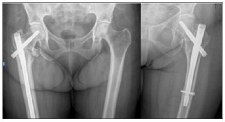
We present a prospective study of 104 non-pathological unstable trochanteric femoral fractures treated with the third generation of Gamma nail ® between January 2012 and January 2014. The patients were aleatorized and divided in 2 groups according to the implant: SGN or LGN (Figure 1). Mean follow-up was 18 months (range 1-3 years). 14 patients were eliminated from the study due to inadequate monitoring (one immediate postoperative death and 13 patients with incomplete record but intact implant in their last visit). The recorded preoperative variables included: patient age, associated diseases, type of gait, anesthetic risk, the presence of associated fractures, hemoglobin and hematocrit concentration and fracture etiology. The preanesthesic risk was collected using the ASA (American Society of Anesthesiologists) classification. The type of gait was classified according to the need of walking aids (cane or walker), the absence of walking ability or the capacity of the patient to walk autonomously without help of any kind. The fractures were classified according to the AO classification (Orthopedic Trauma Association) [6].

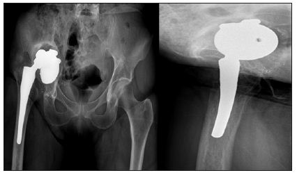
Figure 1: Plain radiographs of unstable pertrochanteric fractures treated with long Gamma 3 nail and short locked Gamma 3 nail.

Postoperatively, we evaluated the following parameters: hemoglobin and hematocrit concentration in the immediate postoperative period, the need for transfusion, the type of gait at discharge from the outpatient clinic and the presence of pain that was scored as follows: no pain, occasional pain, or disabling pain. Functional results and quality of life was queried. Radiographic evaluation was also made including the assessment of fracture reduction and evaluation of cephalic screw location. Intraoperative and postoperative complications were studied; such as malunion, nonunion, infection rates, cut-outs and periprosthetic fractures. Statistical analysis was performed using SPSS 15.0 (SPSS Inc, Chicago Illinois). Student t test was used to compare study outcomes with parametric means. Chi-square test and Fisher´s exact test were used to compare non-parametric means. To level of statistical significance was set as a two-sided P value of 0.05.

The internal resistance of the MFC was determined in a closedcircuit arrangement shown in Figures 1& 2. Closed circuit voltages were measured in different external resistance of 3,000Ω, 2,000Ω, 1,000Ω, 800Ω, 600Ω, 400Ω or 200Ω. Using the equation 1, the corresponding electric currents were calculated. A polarization curve, that was a plot of electric current with voltage, was generated, and the internal resistance Rint was determined by the slope of the regression [11]. Electric current with the correction of internal resistance was calculated as in equation 2.

Results

Preoperative Period

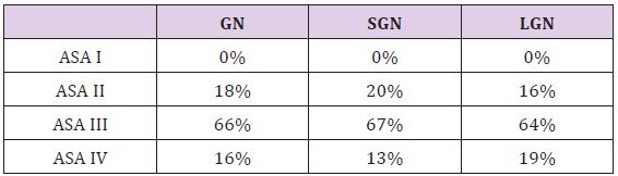
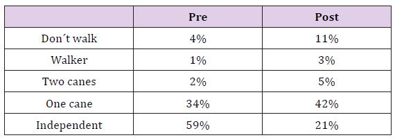
The mean age of the patients was 81 years (range 50-99) and 78% of the patients were female. The associated medical conditions are summarized in Table 1, and are seen to be very prevalent: 88% had at least one pathology and 62% had more than two. By means of the ASA classification: 44% of the patients corresponded to ASA II, 40% to ASA III and 16% were ASA III. The fractures were classified according to the AO classification: 31A2.1: 28%, 31A2.2: 34% and the most commonly found 31A2.3: 38%. We found associated upper limb fractures in 7.1% of cases (distal radius fractures and proximal humerus fractures). Other associated injuries were seen in 5% of the cases, the most frequent was traumatic brain injury which was usually mild. The quality of life prior to fracture was: 25% needed personal care assistance, 33% were independent at home, 39% were independent outside their home and only 3% were able to do physical activities. The type of walking was: 3% unable to walk, 25% used two sticks, 38% used one stick and 34% walked autonomously. The percentages shown refer to the total of patients, which suggests a very high rate of pathological associations in our patients. The two studied groups were homogeneous and the small differences seen in the distribution were not statistically significant (Table 2).

Table 1: Associated medical conditions.

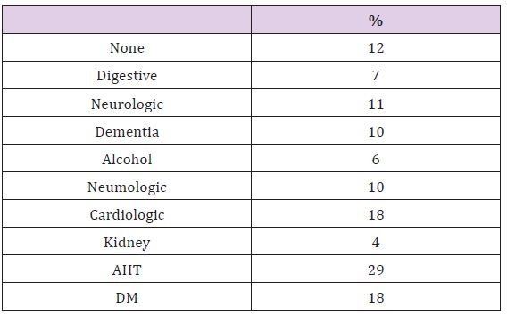
Table 2: Preoperative Study/Implant used.

Surgery

The patients were operated upon following preanesthetic evaluation, an average of 3 days from admission. Spinal anesthesia was performed in 74% of cases, epidural anesthesia in 14% and general anesthesia in 12%. The most commonly used nail was the 130º cervical-cephalic angled nail (52%) and the most common length of the cephalic screw was 90 mm (33%). There were no significant differences between the distribution of angle and the length of the head screw used between both groups. The diaphyseal locking screw of the long gamma nails was dynamic while the short gamma nails had a static distal locking screw. The average time of surgery was 48 minutes (range 30-68). The only intraoperative complication found was a greater trochanteric fracture in two cases treated with long nails.

Postoperative Hospital Period

The local complications in the early postoperative period were limited to a single case of a significant hematoma. The most frequent general complications registered during the immediate postoperative period were urinary tract infection (no differences between groups). Mean hemoglobin in the immediate postoperative period was 9 g/dl (SGN9.5g/dl, LGN8.5g/dl), whereas the hematocritwas 27% (29.2%SGN, 26%LGN). The blood loss recorded was significantly lower in SGN (p=0.05). It was necessary to transfuse an average of 1.4 blood units / patient (SGN0.8, LGN1.65), this difference isn’t statistically but clinical significant.

Full limb loading was allowed after 24 hours. The hospital stay was on average 6 days including the preoperative period (5 SGN, 7 LGN). The destinations were elderly care homes, rehabilitation centers or secondary hospitals in 65% of cases and the patient´s home in 35% of cases. No significant differences were found in hospital stay or destinations.

Outpatient clinic

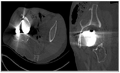
The mean time to fracture consolidation was 3.1 months without differences between groups. To be considered as consolidation an evident bone callus formation had to be present of both the anteroposterior and axial projection. Fracture reduction was evaluated in the postoperative x-ray. In 73% a correct reduction of the fracture was obtained (73% SGN, 74% LGN), 18.5% of the cases didn´t have medial cortex contact (21% SGN, 13% LGN), in 7% the lateral reduction was unsatisfactory (SGN 6%, LGN 10%) while 1,5% presented a complete lack of reduction (SGN 0%, LGN 3%), no significant differences were found. The placement of the cephalic screw was studied in both the anteroposterior radiographs and in the axial, without significant differences between the locations in both groups. In all cases treated with SGN the cephalic screw was placed in the two inferior thirds of the head and neck as in 97% of the LGN. Concerning the axial x-ray, both SGN and LGN head screws were placed in the posterior part of the head. The mean screwsubchondral bone distance was 9 mm (range 4-20 mm). 38% of the patients with LGN presented nail tip impaction upon the distal anterior cortical of the femur, this radiographic impaction was associated with pain in the anterior zone of the knee (Figure 2).

Figure 2:Plain radiographs showing a long Gamma Nail tip impaction upon the distal anterior cortical of the femur.

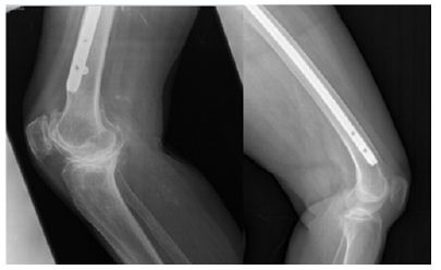
Since this situation is not to be found in the SGN group, it was a significant difference between the groups. There were not any statistically significant differences between groups concerning the late complications studied, even though they were more frequent in the LGN group. We found one case of delayed consolidation that required surgery revision with replacement of the LGN. We also had one LGN with cephalic screw lateral protrusion two months after surgery. Loading of the affected limb was avoided until complete consolidation was obtained and the material was finally extracted. Finally we found one SGN case of cephalic screw cut-off that required extraction and hip arthoplasty (Figure 3). The patients were discharged from the outpatient clinic after an average followup of 24.3 weeks (SGN 24, LGN 25, no differences found). Before the discharge the functional results were evaluated. The type of gait at discharge from the clinic is reported in Table 3. Sixty-eight percent of the patients were free of pain, however, 15% of the LGN reported discomfort in the anterior zone of the knee, 2% of these patients referred a disabling pain. In all cases this coincided with impaction of the nail on the distal anterior cortical layer of the femur but only 3 cases underwent surgery with removal of the implant. There were no statistically significant differences between groups about the lower limb pain. It was described as disabling by only 2% of all the patients.

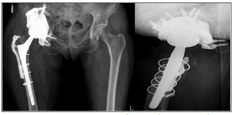
Figure 3: Plain radiographs showing a case with cephalic screw cut-off that ended in arthroplasty.

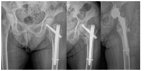
Table 3: Gait pre- and post-operatively.

Discussiont

The results obtained with the two sizes of the Gamma3 Nail in unstable pertro chanteric femoral fractures did not show any important differences in neither clinical outcomes nor complication rates. Theses unstable fractures, also called 31A2 fractures account for 37- 42% of all intertrochanteric fractures of the femoral region [12,16]. The treatment of these fractures is subject to controversy, particularly concerning the use of intramedullary versus extramedullary devices. However, there is one point on which the different studies seem to agree: extramedullary sliding devices (e.g., SHS) are not adequate for fractures of this kind [2- 5]. As a result of their distinct biomechanics, these devices distract rather than compress the fracture fragments thus leading to a high percentage of failures. The great diversity of implants used within the same study contributes to perplex the situation. In series such as those published by Parker, Chinzei, Barton, Michael or Cheng, as many as four different types of nails have been used [6-11]. In our study we have only used the Gamma3 nail (Stryker Howmedica), a fact that allows us to adequately define its behavior. In unstable fractures, intramedullary nailing theoretically affords increased stability since the leverage is less and the proximal portion of the nail is supported against the proximal fragment. In our series, the percentage of implant failure (0,7% cutting phenomenon, 0,7% than with extramedullary fixation and slightly lower than other published intramedullary series [8,9].

Many studies have used intramedullary nails for the treatment of unstable trochanteric fractures [6-11]. On one hand the problem of these studies is that unstable fractures are not only A.2 fractures but also A1.3 and inverse fracture patterns (A3.1, A3.3) and on the other hand the series where only the A.2 fracture was contemplated, the patient series were of limited in size [14]. The advantages of the endomedullary nail are lesser blood loss and a lower percentage of failures, the latter being essential in order to avoid reinterventions that pose an important risks for the elderly patient [12,15,17]. On the other hand, the consolidation rate obtained is high (with only one case of pseudoarthrosis in our series), and with the use of short nails one of the most common complications in endomedullary nailing procedures is diminished: diaphyseal refracture fundamentally associated with the use of long gamma-nails (not seen in our series) [13]. Failure of closed fracture reduction is relatively infrequent, since perfect anatomical reduction is not necessary for implantation, the consolidation can be achieved provided that there is sufficient contact between the bone fragments. However, we agree that a positive medial cortical support allows limited sliding of the neck fragment and achieve secondary stability [8].

This leads to clinical-radiological discordance in many cases. The need to access the fracture focus is only considered in those cases where all the non-aggressive reduction options have been exhausted, and the open reduction might be related to pseudoarthrosis and intraoperative fractures. We agree with Pervez [14] and López-Vega [19] concerning the distal nail locking to get additional stabilization to the nail. In 13% of the patients the nail impacted upon the distal anterior cortical layer of the femur. This situation is more than a simple radiological finding, since in contradiction to others authors, impaction caused clinical manifestations in our patients (pain in the anterior knee region). This circumstance could be related to the characteristics of the femur in our elderly population, which are different to those found in the central European population [15]. The decision to adopt the Gamma3 Nail as our implant in this type of fractures was based on the findings of last year´s studies between the previous gamma-nail and the Gamma3, in which the Gamma3 afforded the best results [18-24]. We agree with most authors that the best way to prevent complications is to ensure a careful surgical technique, with special attention to medullary cavity drilling and the manual insertion of the nail [13]

In our study we recorded a single cutting phenomenon, in contrast to the 6% rate reported in another series reviewed in our hospital, involving 348 trochanteric fractures treated with the short gamma-nail, and where 37% were A.2 type fractures. This could, in part, be related to an improved surgical technique as well as the increased stability afforded by the long gamma3-nail, due to its lesser leverage and increased length [16]. There are no publications to be found that studies the difference between two different types of the same implant. Although our study are not enough to rise statistically significant differences between the groups some are clinical significant. The data obtained allow us to affirm that the clinical and radiological results with SGN and the LGN in the treatment of the AO/OTA 31A2 proximal femur fractures are similar. We found that there are statistically significant differences of transfused blood concentrates and higher decrease in the postoperative hematocrit, the LGN caused more blood loss and could there for potentially cause major medical complications. We found differences in favor of the use of SGN opposite the LGN, such as the presence of pain in the distal third of the femur as some late complications.

For More Articles: Biomedical Journal Impact Factor: https://biomedres.us