Open Access Journals on Medical Imaging

The Impact of Sequencing Genomes on the Understanding of the Origin of Life on Earth

Has all life always been on planet Earth as it is today? Why don’t we see in our Zoo animals like Dinosaurs, Woolly Mammoths, Mastodons or Saber Tooth Tigers? The answer is that life has not always been on planet Earth as it is today. All ancient animals are dead and gone and become extinct. The nucleus of all living creatures carries DNA (Deoxy Ribonucleic Acid) which holds the information not only to store the information but also to pass it on to the next generation. DNA is made of four chemicals called nucleotides bases and they are Adenine (A), Thiamine (T), Guanine (G) and Cytosine (C). They come in pairs. The pairing of A/T bases and G/C bases imply a double helical structure. The one chain of A/T bases running opposite of the other chain of G/C bases. The double helical structure of the nucleotide base pairs solves the mystery of life. It shows how the information to create life is stored, copied and passed on to the future generations.
The essence of life is information and the information to convert non-living chemicals to living creatures is written on the double helical structure DNA. The living cell has no Soul, no Holy Spirit, no Vital Force and no Devine Intervention. Now, we know that Life is a series of coordinated chemical reactions of nucleotide bases. Different life forms are the result of the slow process of Natural Selection. Once we discovered that the secret of life resides on DNA, we could manipulate life that is we could cut paste and copy the DNA to create a new life form in the test-tube that never existed before. This new life form will carry instructions not only to clean up our environmental pollution, but also to provide the most nutritious food for the burgeoning population of the world, to provide new fuel to run the engine of modern society, and to provide new medicine to treat every disease known to mankind.
If you are a religious person and believe that creation of life is a miracle. You understand the origin of life from a different point of view that is from the point of view of religious faith based on belief system that says that there is a Creator who created life on Earth. What it tells me is that to know the way to Creator is to study Creation of life? Most religions believe that Heaven declared the Glory of God, it shows his handy work, there is a Creator who has done it. If that is true than I must study creation trying to read the mind of God. There is a tremendous creation of drive build into the Cosmos. I respect your belief. For scientists, evolution of life is the result of four and a half billion years of biological evolution. We see life evolves everywhere on Earth. For example, during springtime, you see the plants grow and flowers bloom; we see children are born and grow up. Life begins with a single cell and grows up to become a full human being. It is not a violation of natural laws. It is a fact of the fundamental Natural Law. I don’t see the conflict between Science and Religions. I respect people of Faith. Some people have deep faith, that is their spiritual way of knowing the Origin and the Creation of life on Earth.
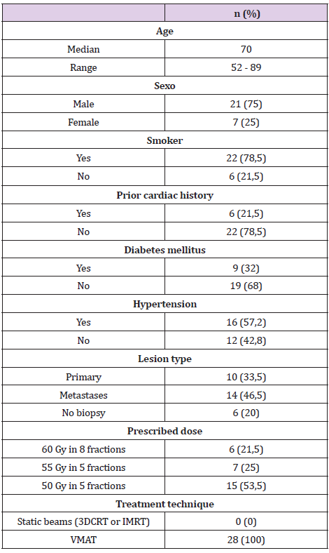
I am a scientist. I look at the origin of life from a different point of view, the rational and scientific way. The Objective truth is verified by the experimental evidence. For example, Water boils at one hundred degrees Centigrade and freeze at Zero degree Centigrade. You could conduct this experiment either in New York or New Delhi, the result should be reproducible, and verifiable. This is how I see the world. Your way of looking at life is different from mine. Your point of view is different, but it is not wrong. It is different. It was Charles Darwin who provided the most rational answer. Charles Darwin was one of the greatest biologists ever lived. In 1859, in his book, the Origin of Species, he stated that Life evolves, and Nature selects. What he meant was that the designs and complexity of living creatures on Earth was due to slow evolutionary processes from the simplest to the most complex species is not by the act of any Divine Intervention, but by the slow process of Natural Selection responding to the surrounding environment.
Species which evolve traits over billions of years to respond to the changing environment survive and the rest of the species that resist evolution die. Their fossils remained trapped in the layers of rocks as the proof of their existence. Soon after the formation of our Solar System about four and a half billion years ago, the hot surface of our Earth cooled. The ancient fossil records show that within a half a billion years, the first life form appeared called the Pre-Cambrian era. During the Pre-Cambrian era which lasted for about 25 million years, there were hundreds of new species evolved from Pre-Cambrian era to the Cambrian Explosion. Most of the pre-Cambrian life forms were unicellular soft tissues creatures which decomposed over the years and their fossils impressions on the rock could be preserved. Only creatures evolved hard shells near the beginning of the Cambrian Explosion were fossilized in the earliest sedimentary rocks. From the pre-Cambrian era, the only creatures that left their fossils behind are the Trilobites, the multicellular crab like creatures which crawled at the bottom of the ancients’ riverbeds.
Darwin critiques argue that the earliest life should be unicellular creatures not multicellular Trilobites. They forgot that unicellular soft tissue creatures don’t fossilized and there were millions of soft tissue creatures during the Pre-Cambrian Era. As we approach near the Cambrian Explosion during the 25 million years, the multicellular hard-shell creatures appeared. The only hard-shell creatures from the Pre-Cambrian era like Trilobites left their fossils behind. Darwin’s critiques will be proved wrong. We have recently learned a technique to extract DNA from the fossils. Using the new technique, a group of German scientists extracted DNA from our ancient ancestors Neanderthal and completely sequenced (decoded) their Genome. Neanderthal died over 30,000 years ago. We could use the same technique to extract the DNA of creature of Pre-Cambrian Era. Any fossil or their impression left on the pre-Cebrian rocks could be extracted and sequence to prove the evolution of life from the simplest to the complex forms.
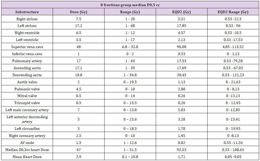
The toolkit developed during the sequencing of human genome helped us sequence the fossils of all ancient creatures for comparison. Now, we can sequence the simplest genome of a microbes, from mouse to monkey to men and compare to see how the simplest to complex organism evolved. How the four nucleotides, the building blocks of life, originated on Earth by the interaction of Carbon, Nitrogen, Oxygen to form nucleotide and how they organized themselves to become alive. Life is a series of coordinated chemical reactions of basic building blocks called the nucleotide bases. If you sequence the genome from the simplest to the most complex life form and compare their genomes, you see how the same four nucleotide aggregate differently over ions in response to the surrounding environment (Figure 1).

biomedres-openaccess-journal-bjstr

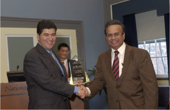
Figure 1: Dr. Khan is the Discoverer of AZQ (US Patent 4,146,622 & 4,233,215), a Novel Experimental Drug Specifically Designed to shut off a Gene that causes Brain Cancer for which he receives a 17-year Royalty for his invention (License Number L-0I9- 0I/0). To this date, more than 300 research papers have been published on AZQ. The award ceremony was broadcast live worldwide by the Voice of America (VOA). Dr. Khan is the first Indian to receive one of America’s highest awards in Medicine.

Cambrian Explosion

Darwin had the greatest foresight. By comparing the fossils, he brought from Galapagos, he saw the evidence of evolution. Planetology is the study of layers of rocks to trace the evidence and ecology of plants and animals from the distant past to the present day. Most fossils are found in the sedimentary rocks and clay deposited on the layers of rocks. One layer deposited on the top of other. Trapped in these layers are millions of years old fossil at various stages of evolution. As the rivers dried up, the sedimentary rocks become hard. The sedimentary rocks unfold like pages of a gigantic book. The earliest fossil of simple structures is found in the lowest or the oldest layers. As he examined younger and younger rocks, he found complexity of structures. No human bones were ever found in any of these ancient rocks. During the pre-Cambrian era, about 450 million years ago when the climate changed, the Cambrian explosions occurred when the frozen Earth began to warm.
The single cell living creature instead of growing by asexual reproduction began to grow by sexual reproduction. The interaction of two separate chromosomes resulted in variations in gene pool which led to divergence of life forms and evolution from the simplest to the more complex life forms called the Cambrian Explosion of life. The progeny of the recombinant genes produced complexity. Only those recombinant daughter cells which carry genes that produced functional proteins survived and the rest died. The proof of the Cambrian Explosion is trapped in the fossil record which lasted for about 25 million years. Extracting fossils from the ancient, eroded rocks is a real challenge. The erosion of sedimentary rocks over the years is due to rain falls, windstorms, running waters, and transportations of the rocks. Once DNA extraction is purified from the fossils, its genome could be sequenced, and its date could be estimated by Radioactive Dating method (Figure 2).

biomedres-openaccess-journal-bjstr

Figure 2: His Excellency, Dr. A.P.J. Abdul Kalam.

Radioactive Dating

Radioactive dating provides accurate measurements of the age of the ancient rocks and the fossils trapped inside those rocks. Heavy elements with large nucleus are unstable. Over a long period of time, their nucleus falls apart to more stable elements. By becoming stable, they release radiations such as alpha, beta and gamma radiations and they are called radioactive elements such as Radium, Thorium, Rubidium, Uranium etc. All radioactive elements release radiations at a steady measurable rate over millions of years. By measuring the ratio of the radioactive elements and its unstable end-product, it is possible to measure the age of the rock and the fossils trapped inside that rock. For example, radioactive element Uranium (Atomic weight = 238) over millions of years break down slowly through various isotopes to a more stable element Lead (Atomic Weight = 206). Uranium first breaks down to element Radium (Atomic Weight = 226) which is further broken down to Polonium (Atomic Weight = 218) which is further broken down to its another isotope Polonium (Atomic Weight = 210) and to its most stable element Lead (Atomic Wight = 206). Radioactive decay is a slow process. Over a million-year, one Gram of Uranium break down to 1/7000 Gram of element Lead. By measuring the ratio of the amount of Uranium to the amount of Lead in a rock, we can calculate the age of the Uranium rock and its fossils trapped inside it (Figure 3).

biomedres-openaccess-journal-bjstr

Figure 3: Discoverer of anti-cancer AZQ, after receiving 2004, Vaidya Ratna, The Gold Medal, One of India’s Highest Awards in Medicine, At The Rashtrapathi Bhavan (Presidential Palace), in Delhi, India, During a Reception held on April 2, 2004.

The Geologic Clock

Let me scan the origin of life on planet Earth from the very beginning to the present day. The slow evolutionary changes can be explained from three and a half billion years to the present day. If we were to examine the fossil record based on the Geologic Time Scale, we can divide this time period into three great eras. First, The Paleozoic Era which starts from the very beginning of the Pre-Cambrian Era from the 100 million to 400 million years ago. Second, The Mesozoic Era beginning from 230 million years ago to 70 million years ago. Third, The Cenozoic Era beginning from the 63 million years ago to the present day. During the Pre-Cambrian Era, primitive life forms appeared. No fossil was found of the soft body creatures except some the impression of their fossils are found on the ancient rocks. During the end of the Pre-Cambrian era, some hard-shell fossils, made of Calcium Carbonate, like Trilobites are found. During Cambrian Era, about 100 million years ago, primitive animals appeared such as Algae, Orthopods; much later sponges, worms and mollusks appeared.
A treasure of fossils was discovered in British Columbia, Canada, called the Burgess Shale, found in the Canadian Rockies of Canada. These are the fossils left behind by the Middle Cambrian Era. Part of this treasure is on display at the Smithsonian Museum in Washington, D.C., Among those fossils were sea cucumber, worms and Trilobites. During the Ordovician Period, from 425 to 500 million years ago, bony life forms appeared which include Tetracorals, echinoids, asteroids appeared for the first time on the primitive Earth. During the Silurian Period, (from 425 to 405 million years ago), brought the most dramatic changes in the Earth’s atmosphere. Plants appeared for the first time. Up to this time period, Earth’s atmosphere was full of Nitrogen gas released by the cooling the hot Nitrate Rocks. Earth’s atmosphere also contained the Carbon dioxide contributed by the Volcanic eruption. Plants carry Chloroplast Genome a procryptic life forms captured in an early evolutionary process.
Genes in the Chloroplasts genome have the ability to capture Carbon dioxide from the atmosphere and in the presence of sunlight and convert to its food the Carbohydrate and release Oxygen as the by-product. For the following 60 million years, at the beginning of the Devonian period, the cooled part of the planet Earth, was carpeted by the early plant life called the Blue Green Algae. Its main function was to absorb Carbon dioxide from the atmosphere and convert it to the Carbohydrate its food and release Oxygen as a part of photosynthesis. Over millions of years, enormous amount of Oxygen was released in the atmosphere. By the end of the Silurian Period, the composition of the Earth’s atmosphere was changed from pure Nitrogen gas to 80% Nitrogen and 20% Oxygen gas. The gas Oxygen is extremely reactive, it reacted with the Oceans Iron forming the Iron oxide. Billions of tons of Iron Oxide deposited on the Ocean floor.
Oxygen is toxic to the Anerobic life forms. Creatures survive in the presence of Oxygen thrived while Anerobic life forms died. Complex life forms appeared. In the Oxygen atmosphere, fossils of Fish and Amphibians were found along with the fossils of spiders, millipedes, insects, and corals were discovered. The time, period from 345 to 310 million years is designated as the Mississippian period, during which the fossils of more complex life forms appeared which included Corals, Branchipodids, and Foraminifers. From 210 to 280 million years ago appeared the Great Coal bearing layers of rocks known as the Pennsylvania Period. It saw the low land of great swamp forming the Coal forest. This period saw the appearance of Clams, shell-fish, reptiles, and amphibians. From 280 to 230 million years ago, called the Permian Period which observed the swampy part of the surface developing Coal-forest plants such as Conifers, Tongue-fern, Oak, insects, beetles and dragon flies. Because of the Climatic changes, the plants and animals of the Permian Period become extinct.
This marked the end of the Paleozoic Era. The Mesozoic Era began about 165 million years ago. It brought the Age of Reptilian. With this Era came the Birds, Mammals, Insects and Flowering Plants including Elm, Oak, Maples became common. New mountains range slowly appeared. From 230 to 180 million years ago which is called The Triassic Period saw the appearance of Dinosaurs that mighty beast that ruled Planet Earth for about 150 million years. They all died when a meteorite structs Planet Earth about 65 million years ago. They left behind their foot prints as their fossil around the world. During the Jurassic Period, rain-forest spread everywhere the Dinosaurs dominated the land, but the Ocean was dominated by the Plesiosaur, the monstrous carnivorous of the Seas. The Creataceous Period which begins about 135 to 70 million years ago, marked the development of sedimentary rock made of Chalk. The moment of the Tectonic Plates formed the mountain range from Andes to Rockies, from Antarctica Northwestern Asia. Plants thrived during the Creataceous Period. The fossil record showed the appearance of Trees, shrubs, including Magnolia, Oak, Maples, Birch, Holly, and Ivy which provided food for mammals, birds, reptiles, and insects. Dinosaurs spread on all seven continents. Their fossils are found all over the planet. As I said above, they all disappeared around 65 million years ago, when a meteorite structed at the Northern Mexico. The Cenozoic Era, called The Age of Mammals, began about 70 million years ago to the present day. The climatic changes resulted in the cooling of the polar regions and warming climatic temperature everywhere. This climate change stayed on to the present day. The Cenozoic Era, is dominated by the Flowering Plants, and reptiles are replaced by Mammals. Birds continue to expand everywhere. Finally, the Quaternary Period in which we now live began with the melting of the 10,000 feet thick ice sheet over much of the Northern hemisphere in which four glaciers advanced which lasted about 11,000 years.
As the ice sheet melted away, it created suitable atmosphere for the emergence of human being. Humans appearance on Earth is a matter of only a few million years. Have we found human fossil during any of the Geologic periods from the Pre-Cambrian to Cenozoic Era? The answer is no. In 1974, the first human fossil, Lucy, Australopithecus afarensis skeleton was discovered, in a 3.2-million-year-old rock found by anthropologist Donald Johansson in Hader, valley in Ethiopia. Chimps were living in the Great Rift Valley for the past 25 million years. A more advanced form of the Chimp called Austral opthecus appeared in East Africa. He was an advanced forest man called Homo habilis. He was a hunter gatherer of food who built tools. He was a direct ancestor of Man and who lived about 20 million years ago. Next was the apeman, Pithecanthropus, who lived about 500,000 years ago in Java and China. Neanderthal man lived in Europe. He was also a hunter gatherer and lived about 100,000 years ago.
They all died about 30,000 years ago. Cro-Magnon finally evolved modern brain. Cro-Magnons, a term derived from the Cro-Magnon rock shelter found in southwestern France, where the first human fossils were found in 1868. Darwin’s extraordinary prediction was confirmed by sequencing genomes or reading the book of lives of over a thousand species on Earth. Of all the experiments in Biology, the Sequencing of Human Genome was the greatest accomplishment of all times. On April 3, 2003, Dr. Francis Collins, the Director of my Institutes, The National Institutes of Health (NIH) announced that we read the book in which God created life. We completely read the book of life of a human being letter by letter, word by word, sentence by sentence and chapter by chapter all 46 volumes called the Chromosomes carrying the 24,000 chapters called the genes and its text written in four nucleotides containing six billion four hundred million letters. In a few sentences, he described the completion of the Human Genome Project.
The greatest biological experiment ever conceived by human mind. It will answer the most fundamental questions we have asked ourselves since the dawn of human civilization. What does it mean to be human? What is the nature of our memory and our consciousness and our development from a single cell to a complete human being; the biochemical nature of our senses; the processes of our Aging. The scientific basis of our similarities and dissimilarities. Similarities that all living creatures from a tiny blade of grass to mighty Elephant, including man, mouse, monkey, mosquitoes, and microbes all are made of the same chemical building blocks and yet they are so diverse that no two individuals are alike, even identical twins are not identical they grow up to become two separate individuals. Essential components of life are RNA, DNA, Proteins, Carbohydrates, Lipids, and Hormones. We always wonder how these non-living chemicals could get together to create living creatures. When did life evolve? Where was it evolved? And how life evolved? Evolution of Life on Earth is not a miracle.
Life could have been evolved on Earth’s surface such as on the oldest rocks found in Australia or it could have been evolved at the bottom of the Ocean floor where Black Smokers are formed with Lava emerging from under sea volcanoes reacted with surrounding Hydrogen Sulfide gas which provides energy for life forms such as tubeworms and crabs who thrive on the Ocean floor. Life also could have been evolved underground. Soil sample brought by miners from the gold mines in South Africa two miles deep underground contained micro worms. Such life form could be cultivated on a Petri dish containing Agar mixed with nutrients. Early life could have been unicellular life forms. When harvested within 24 hours, the Petri dish could be filled with microbial life. Could life have been brought on Earth by meteorites. Early Earth has no Water. Billions of Comets brought Water on Earth. Would it be possible that some of those Icy mountains contained life giving essential components? Life could also be evolved on the surface of Earth. A million-lightning strike Earth each day.
Could it be possible that at some remote corner of the Earth, Lightning struck at cloud of gases such as Ammonia, Methane and Sulfur on a Phosphate containing rocks making the essential components of life like nucleotides which combined to form RNA Which not only carry information like DNA and perform function like amino acids. The polymerization of Formaldehyde in the atmosphere could produce Carbohydrates another essential component of life. The presence of Acetonitrile, Carbon dioxide, Water in the presence of Ultraviolet light could produce the nucleotides such as Adenine (A), Thiamine (T), Guanine (G) and Cytosine (C) forming a binary code leading to RNA which start replicating itself creating the first living anaerobic creature. RNA molecule can catalyze reaction like enzyme such as protein, but also it could store information like DNA. Were there creature in the RNA world which thrived in the absence of Oxygen. Since no human was present to witness the formation and evolution of first life on Earth, we rely on its presence from the early fossils found in the layers of ancient rocks.
Once a single replicating living cell appears on Earth, complexity develops. In other words, all complex life forms are evolved from simple life forms. Since no humans were present to observe the beginning of life on Earth, we deduce their evolutionary developments from their fossil records. Fossils are the remains of the pre-historic life forms. To become fossilized, the species must have developed hard parts such as bone or shell and must be trapped in mud which slowly become hard rock. Soft tissue creatures do not fossilize; their tissues decomposed. As I said above, the Earth was formed about four and a half billion years ago. The hot Earth cooled by the bombardment of the icy comets. Every drop of Water on Earth was brought by the icy comets. The first life form appeared on Earth about a billion year after the Earth was formed about four and a half billion years ago. Over billions of years of evolutionary process, chemicals reacted together to create Life. One of the most essential components of Life that is Amino Acid was created in the Lab.
In 1953 Stanley Miller, the student of the Nobel Laureate, Harold Urey, at the Chicago University conducted an experiment in the Lab to create life’s essential components the amino acids. He created primitive Earth like conditions in the Lab. He took two flasks connected with a condenser. One flask contained water vapors and the other filled with gases found on the primitive Earth such as Methane, Carbon dioxide and Ammonia. To mimic thunder and lightning, a source of energy, on Earth, he sparked electric current in the flask. The high energy electric spark, split the stable molecules of Nitrogen, Oxygen, and Carbon, producing extremely reactive ions which reacted with one another recombing to produce a more stable new molecule. Within a week, the clear solution in the flask became pink and dark. The analysis of the colored material showed the formation of Amino Acids, the essential building blocks of life which perform all body functions. In similar experiments, Francis Crick and Lesli Orgel, attempted to synthesize Nucleotides the replicating molecules which carry information to make life. Using Formaldehyde, the other essential components of life such as sugars and hormones were synthesized.
Chromosomes are thread-like structures located inside the nucleus of animal and plant cells. Each chromosome is made of double strand of a long chain of four nucleotides wrapped around with protein called the deoxyribonucleic acid (DNA). As I stated above, this is the information molecule which is passed on from parents to offspring, DNA contains specific instructions that make each type of living creature unique. As the living creatures evolve, the complexity increases as the number of chromosomes increases. The evolution of life on planet Earth is extremely slow process. About a billion years after the formation of Earth that is about four and a half billion years ago, life appeared. Bacteria Phage Phi-X 174 is perhaps the smallest organism, and it is made of over 5,000 nucleotide bases. It carries a single Chromosomes so has most bacteria. As evolution proceeded, chromosome number increases, and complexity appeared in both plants and animals to survive in the changed environment. For example, while bacteria have a single chromosome, Jack Jumper Ant has two Chromosomes.
Yellow fever mosquito has six chromosome; Fruit fly has 8 chromosomes; Swamp wallaby has 10 chromosomes; Nematode has 12 chromosomes; the Australian daisy has 12 chromosomes; the spider mice, Aloe Vera and cucumber has 14 chromosomes; Garlic has 16 chromosomes; Itch Mite has 17 chromosomes; Radish, Carrot, Cabbage and passion fruit have 18 chromosomes; Maize and Cannabis have 20 chromosomes; Bean and Virginia Opossum have 22 chromosomes; Snail, Melon, Rice, Sweet Chestnut have 24 chromosomes; Edible frog has 26 chromosomes; Axolotl has 28 chromosomes; Beg Bug has 29 chromosomes; Giraffe, American mink and Pistachio have 30 chromosomes; Yeast, European honey bee, American badger and Alfalfa have 32 chromosomes; Red fox, Sunflower and Porcupine have 34 Chromosomes; Yellow mongoose, Tibetan sand fox, Starfish, Red panda, Meerkat and Earthworm have 36 chromosomes; Tiger, Sea otter, Sable, Raccoon, Pig, Lion and European mink have 38 chromosomes; Mouse, Mango, Hyena, Ferret, Beaver and Peanut have 40 Chromosomes; Wolverine, Wheat, Rat and Oats have 42 Chromosomes; Dolphin, Sable antelope, and Human have 46 chromosomes; Water buffalo, Tobacco, Potato, Orangutan, hare, Gorilla, Deer mouse, and Chimpanzee have 48 chromosomes; Zebrafish, Water Buffalo, Striped skunk, Pineapple have 50 chromosomes; Spectacled Bear, Platypus, and Cotton have 52 chromosomes; Sheep, Hyrax, Racoon dog and Capuchin monkey have 54 Chromosomes; Strawberry, Gaur, and Elephant have 56 chromosomes; Woolly mammoth has 58 chromosomes; Yak, Goat, cow/Bull, American Bison, Bengal fox, have 60 chromosomes; Gypsy moth, Donkey, and Scarlet Macaw have 62 chromosomes; Mule has 63 chromosomes; Guinea pig, Spotted skunk, Horse and Fennec fox have 64 chromosomes; Gray fox, Red deer, Elk and Roadside hawk have 68 chromosomes; White-tailed deer have 70 chromosomes; Black nightshade and Bat-eared fox have 72 chromosomes; Asiatic black bear, and American black bear have 74 chromosomes; maned wolf, have 76 chromosomes; Grey wolf, Golden Jackal Dog, Dingo have 78 Chromones; Turkey, Sugarcane, and Pigeon have 80 chromosomes; Great white shark have 82 chromosomes; Hedgehog genus have 88 chromosomes; Moon Worts, hedgehog Genus and Grape fern have 90 chromosomes; Pitter’s crab-eating rat. Prawn and Aquatic rat have 92 chromosomes; Kamaraj (fern) have 94 chromosomes; Carp has 100 chromosomes; Red vizcacha rat have 102 chromosomes; Walking catfish has 104 chromosomes; American paddlefish have 120 chromosomes; Northern lamprey has 174 chromosomes; Rattlesnake fern has 184 chromosomes; Red king crab has 208 chromosomes; Field horsetail has 216 chromosomes; A. butterfly has 268 chromosomes; black mulberry has 308 chromosomes; Atlas blue has 448 chromosomes; adderstongue has 1260 chromosomes’ here is a Fern called Ophioglossum, which has the highest number of chromosome count of any known living organism, with 1,260 chromosomes.
This fern has roughly 630 pairs of chromosomes or 1,260 chromosomes per cell. The next generation of scientists will have the opportunity to sequence the genomes of all above species and their genes could be added to our GenBank to be used to develop new food, new fuel and new medicine for the burgeoning population of the world. From the above observations, it became clear that humans are not the panicle of achievement of evolution. Other creatures have more chromosomes than us. Our superiority is in achieving consciousness, our language, and our ability to communicate orally and in writing leaving our knowledge for the future generations (Figure 4).

biomedres-openaccess-journal-bjstr

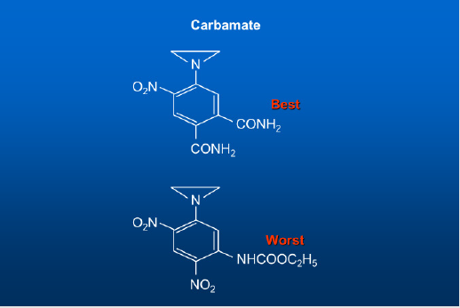
Figure 4: Single Strand DNA Binding Aziridine and Carbamate.

The Impact of Sequencing Human Genome on the understanding of the Origin of Life

As I said above, our entire book of life is written in four genetic letters called nucleotides in a three-letter code called codon, and they are A (adenine), T (thymine), G (guanine) and C (cytosine). These four chemicals are called nucleotide. The essence of life is information which is carried on these four nucleotides. These nucleotides are found in the nucleus of all living cells including humans, plants, and animals. Instruction in a single gene is written in thousands of AT/GC base pairs that are linked together in a straight line and we call them DNA (Deoxyribose Nucleic Acid) – Nobel prize was awarded to Crick, Watson & Morris Wilkins [1] for discovering the double helical nature of the DNA structure which is transcribed into a single stranded of RNA (in mRNA the less water soluble methyl group in Thiamine, T, is converted to more water soluble Uracil, U, by replacing Methyl group with a Hydroxyl group) which leaves the nucleus and moves into Cytoplasm where it is translated in Ribosomes into Amino Acids leading to proteins). When thousands to millions of AT/GC base pairs contain information to make a single protein, we call that portion of AT/GC base pairs a gene (Nobel Prize was awarded to Khorana & Nauenberg for making a functional gene).
A gene is a string of DNA. The starting Codon for a gene is AUG which codes for the amino acid Methionine after several hundred Codons for different amino acids, comes the stop codon. There are three stop Codons, and they are UGG, UGA, UAG. After the stop Codon, no more amino acids are added to the chain, and DNA synthesis stops. If we count all the AT/GC base pairs in a single cell of our body, we will find that there are 3.2 billion pairs of bases present in the nucleus of every cell. The entire AT/GC sequence of 3.2 billion base-pair is called the Human Genome or the book of our life which carries total genetic information to make us. The reading of the total genetic information that make us human is called the Human Genome. In 1990, US Congress authorized three billion dollars to NIH to decipher the entire Human Genome under the title, “The Human Genome Project.” We found that our genome contains six billion four hundred million nucleotides bases half comes from our father and another half comes from our mother.
Less than two percent of our Genome contains genes which code for proteins. The other 98 percent of our genome contains switches, promoters, terminators etc. The 46 Chromosomes present in each cell of our body are the greatest library of the Human Book of Life on planet Earth. The Chromosomes carry genes which are written in nucleotides. Before sequencing (determining the number and the order of the four nucleotides arranged on a Chromosomes), it is essential to know how many genes are present on each Chromosome in our Genome. The Human Genome Project has identified not only the number of nucleotides on each Chromosome, but also the number of genes on each chromosome. A single cell is so small that we cannot even see with our naked eyes. We must use a powerful microscope to enlarge its internal structure. Under an electron microscope, we can enlarge that one cell up to nearly a million times of its original size. Under the electron microscope, a single cell looks as big as our house.
There is a good metaphor with our house. For example, our house has a kitchen, the cell has a nucleus. Imagine for a moment, that our kitchen has 23 volumes of cookbooks which contain 24,000 recipes to make different dishes for our breakfast, lunch and dinner. The nucleus has 23 pairs of chromosomes which contain 24,000 genes which carry instructions to make proteins. Proteins interact to make cells; cells interact to make tissues; tissues interact to make an organ and several organs interact to make a man, a mouse or a monkey. In every cell of our body, we carry sixteen thousand good genes, six thousand mutated (bad) genes responsible for six thousand diseases and two thousand Pseudo-genes that have lost their functions, during evolutionary time. Our genome contains six billion four hundred million nucleotides bases half comes from our father and another half comes from our mother. Less than two percent of our Genome contains genes which code for proteins. The other 98 percent of our genome contains switches, promoters, terminators etc.
The 46 chromosomes present in each cell of our body are the greatest library of the Human Book of Life on planet Earth. The Chromosomes carry genes which are written in nucleotides. Before sequencing (determining the number and the order of the four nucleotides on a chromosomes), it is essential to know how many genes are present on each chromosome in our Genome (Figure 5).

biomedres-openaccess-journal-bjstr

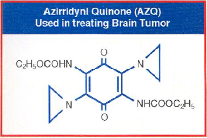
Figure 5: Gold Medal for Dr. Khan.

The Human Genome: The greatest Catalog of Human Genes on planet Earth

Human Genome contains a catalog of traits written on genes in nucleotide sequence. Our Genome also provides a catalog of all 24,000 genes; it also provides the number and location of each gene on the chromosome. The catalog provides 16,000 good genes, 6,000 bad genes and 2,000 pseudogenes (they lost their function). The Human Genome Project has identified the following genes on each chromosome: We found that the chromosome-1 is the largest chromosome carrying 263 million A, T, G and C nucleotide bases and it has only 2,610 genes. The chromosome-2 contains 255 million nucleotides bases and has only 1,748 genes. The chromosome-3 contains 214 million nucleotide bases and carries 1,381 genes. The chromosome-4 contains 203 million nucleotide bases and carries 1,024 genes. The chromosome-5 contains 194 million nucleotide bases and carries 1,190 genes. The chromosome-6 contains 183 million nucleotide bases and carries 1,394 genes. The chromosome-7 contains 171 million nucleotide bases and carries 1,378 genes. The chromosome-8 contains 155 million nucleotide bases and carries 927 genes.
The chromosome-9 contains 145 million nucleotide bases and carries 1,076 genes. The chromosome-10 contains 144 million nucleotide bases and carries 983 genes. The chromosome-11 contains 144 million nucleotide bases and carries 1,692 genes. The chromosome-12 contains 143 million nucleotide bases and carries 1,268 genes. The chromosome-13 contains 114 million nucleotide bases and carries 496 genes. The chromosome-14 contains 109 million nucleotide bases and carries 1,173 genes. The chromosome-15 contains 106 million nucleotide bases and carries 906 genes. The chromosome- 16 contains 98 million nucleotide bases and carries 1,032 genes. The chromosome-17 contains 92 million nucleotide bases and carries 1,394 genes. The chromosome-18 contains 85 million nucleotide bases and carries 400 genes. The chromosome-19 contains 67 million nucleotide bases and carries 1,592 genes. The chromosome-20 contains 72 million nucleotide bases and carries 710 genes.
The chromosome-21 contains 50 million nucleotide bases and carries 337 genes. The chromosome-22 contains 56 million nucleotide bases and carries 701 genes. Finally, the sex chromosome of all females called the chromosome-X contains 164 million nucleotide bases and carries 1,141 genes. The male sperm called chromosome-Y contains 59 million nucleotide bases and carries 255 genes. If you add up all genes in the 23 pairs of chromosomes, they come up to 26,808 genes and yet we keep on mentioning 24,000 genes needed to keep us function normally. A gene codes for a protein, not all 24,000 genes code for proteins. It is estimated that less than 19,000 genes code for protein. Because of the alternative splicing, each gene codes for more than one protein. All the genes in our body make less than 50,000 protein which interact in millions of different ways to give a single cell. Millions of cells interact to give a tissue and hundreds of tissues interact to give an organ and several organs interact to make a human [1-6].
Not all genes act simultaneously to make us function normally. Current studies show that a minimum of 2,000 genes are enough to keep human function normally; the remaining genes are backup support system, and they are used when needed. The remaining genes are called the pseudo genes. For example, millions of years ago, humans and dogs shared some of the same ancestral genes; we both carry the same olfactory genes needed to search for food in dogs. Since humans don’t use these genes to smell for searching food, these genes are broken and lost their functions in humans, but we still carry them. We call them Pseudo genes. Recently, some Japanese scientists have activated the pseudo genes, this work may create ethical problem in future as more and more pseudo genes are activated. Nature has good reasons to shut off those pseudogenes. Our Genome provides the genetic road map of all our genes, past, present and future.
For example, it can tell us how many good or bad genes we inherit from our parents and how many of those gene we are going to pass on to our children. If a family has too many bad genes, and have a family history serious illnesses, they can break off the information flow and stop having children or stop donating mutated eggs and sperms. On April 3, 2003, several groups simultaneously sequenced the entire Human Genome and confirmed that less than two percent of the Genome codes for proteins the rest is the noncoding regions which contains switches to turn the genes on or off, pieces of DNA which act as promoters and enhancers of the genes. Using restriction enzymes (which act as molecular scissors), we can cut, paste, and copy genetic letters in the non-coding region which could serve as markers and which has no effect, but a slight change called mutations in the coding region makes a normal cell abnormal or cancerous (Figure 6).

biomedres-openaccess-journal-bjstr

Figure 6: Dr. A. Hameed Khan, a Scientist at the National Institutes of Health (NIH) USA, an American Scientist of Indian Origin was awarded on April 2, 2004. Vaidya Ratna; The gold Medal, one of India’s Highest Awards in Medicine for his Discovery of AZQ (US Patent 4,146,622) which is now undergoing Clinical Trials for Treating Bran Cancer.

Our Search for Unknown Diseases Has Come to A Closure

There are two most powerful implications of the human Genome Sequencing. One of them is that we have come to closure. What it means is that we have the catalog of all genes in the Human Genome, we can search the entire genome and locate the desired gene. we will not wonder in the wilderness anymore. Everything there is to know about human health and traits are written on these genes in nucleotide sequences. Our Genomes provides the catalog of all genes.

Reference Sequence

We can scan the whole genome (Reference Sequence) for its response to a given situation. When we look at a normal cell and compare with an abnormal cell, we see the differences. Or when we compare their gene expression looking for a specific proteins, from a specific genes and for a specific nucleotide sequence, we can identify a specific mutation responsible for the disease. In the olden days, before sequencing human genome, when a patient visits a physician for some unknown ailment, the Physician would order several tests and would say to his patient, I don’t know what is wrong with you, I will see if any of these tests show if my guess is right and if he is wrong, he will recommend few more tests to see if he could identify the illness. The guesswork and the trial-anderror days are over. Now, after sequencing the human genome, the physician would say to his patient, I don’t know what is wrong with you, but I know where to find it. It is written in your Genome. It would be easy for a Physician to scan the patient entire genome and compare against the Reference Sequence to identify the mutations responsible for causing the disease.
He will refer the patient to a biotechnology Lab. The Lab Technician will take a small blood sample from the patient, separate his WBC, extract DNA, sequence his Genome and compare with the Reference Sequence letter by letter, word by word by word and sentence by sentence and send the result to the Physician who can easily identify the mutations responsible for causing the disease. The result will provide the best diagnostic method to identify a disease. Our Genome is not just a diagnostic road map of our genes, it tells us to clone the good genes and shut off the bad genes. Using the good genes, it also tells us to make its large-scale protein for worldwide use such as Insulin and Human growth hormone. On the other hand, identifying the bad genes and tell us to design novel drugs to shut off bad genes responsible for causing serious diseases.
We have already demonstrated that using the genetic engineering techniques, we can cut, paste, copy, and sequence a good gene for industrial scale preparation as I said above such as Insulin to treat 300 million of diabetic around the world. Genome sequencing of bad genes start a new era of Genomic Medicine which is based on the development of new drugs for treating a disease based on the genetic make-up of the individuals. The next step would be to design drugs to shut off the mutated genes. Gene Therapy will work if the disease is caused by a single gene mutation. Drug Therapy will work if multiple genes are responsible for causing diseases such as Cancers, Cardiovascular diseases, and Alzheimer.

Genomic Medicine

The first step is to cut the human genome with specific enzyme (prepare a Restriction Site Map) at the specific sites using restriction enzymes (molecular scissors such as EcoR1) first accomplished by El Salvador Luria, Max Delbruck, and Hamilton Smith. The fragment of human DNA (a single gene) if not protected will be destroyed by antibody. A naked gene is a piece of DNA (which has a start codon AUG and after a few thousand nucleotide (codons) end at one of the three stop codons UAG, UGA or UGG if not protected by recombinant technology (making a hybrid) that is by recombining with the DNA of Virus, or Plasmids, or Chloroplasts (for plants) which serves as Vectors, will be destroyed by enzymes. One can store the fragments or genes in the Vectors once the human DNA fragment is stabilized in Vectors by recombinant technology; we can not only purify this fragment (genes), but also, we can make millions of copies (clone) of this fragment of DNA by transferring into the host cells such as Bacteria, mammalian cells or Yeast cell which autonomously replicates to produce library of genes.
Each Library contains millions of copies of identical genes that produce same protein. Before the genetic revolution, Insulin is extracted from pancreas of the slaughtered animals which is used to treat old diseases such as diabetes; a tiny fragment of impurity could set anaphylactic shock and kill the patients. Now, highly pure human Insulin produced by Genetic Engineering is used to treat 300 million diabetic patients worldwide without the loss of a single life using the same recombinant technology. Other products of Genomic Medicine such as Growth hormones and hormone proteins to treat Hemophilia by factor VIII protein are being developed as genomic medicines by recombinant technology. The essence of life is information, and the information is located on the four nucleotide bases A-T and G-C. According to Central Dogma of Crick and Watson, the information on DNA is transcribed on RNA which is translated in Ribosome to protein. Attempts are being made to design drugs to attack cancer cells on all three levels that is DNA, RNA and Protein.
Herceptin, a novel class of drug, has been successful in attacking protein. Craig Milo has designed double stranded RNA to shut off gene and prevents its translation into protein. Attack on DNA to shut off a gene was carried out by Ross using highly toxic Nitrogen Mustard. Gene Therapy cannot be applied to multiple genetic defects such as cancers or heart diseases. Drug Therapy could be used to develop novel treatments. Professor WCJ Ross of London University was the first person who designed drugs to attack DNA for Cancer Treatment. He designed drugs to cross-link both strands of DNA that we inherit one strand from each parent. Cross-linking agents such as Nitrogen mustard. The analogs of Nitrogen mustard are extremely toxic and were used as chemical weapon during the First World War. Hundreds of more toxic analogs of Nitrogen Mustard were developed during the Second World War. Solders exposed to Nitrogen Mustard showed a sharp decline of White Blood Cells (WBC) from 5000 cell/CC to 500/CC [7,8].
Children suffering from Childhood Leukemia have a very WBC count over 90,000/CC. Most of the WBCs are premature, defected, and unable to defend the body from microbial infections. Ross rationale was that cancer cells divide faster than the normal cell, by using Nitrogen Mustard he could cross linking DNA and prevent cell division. Once he demonstrated that he could shut off a gene by cross-linking DNA; he could shut off any mutated gene of all 220 tissues present in a human by finding a dye that could specifically color that tissue. He could attach the Nitrogen Mustard group to the dye and attack the cancer genes in any one those tissues. Ross was the first person to use war chemicals successfully to treat cancer. Although such drugs are highly toxic more cancer cell will be destroyed than the normal cells. Over decades, Ross made several hundred derivatives of Nitrogen Mustard as cross-linking agents. Some of the Nitrogen Mustards are useful for treating cancers such as Chlorambucil for treating childhood leukemia (which brought down the WBC level down to 5,000/CC) and Melphalan and Myrophine for treating Pharyngeal Carcinomas.
[9-15]. Because of the high toxicity of Nitrogen Mustard, new drugs could not be developed to treat other types of Oral or Lung Cancers. As I showed above, we sequenced our entire genome, our book of life, letter by letter word by word, sentence by sentence, chapter by chapter all forty-six volumes written in six billion four hundred million genetic letters (nucleotide) of a healthy human being under the Human Genome Project. We can use our healthy Genome as a Reference Sequence for comparison. Using nano capillary method, it took us 13 years to sequence the entire human genome at a cost of $3 billion. Now, we have developed next generation sequencers like Nanopore technology which will sequence the entire genome cheaper and faster. Using biopsy sample, we can take a single cell from the Lung or Oral tumor of smoker, sequence its genome, and compare with the Reference sequence to identify the number and location of all mutations or damage genes caused by smoking. Recently, we also completed the 1000-genome project which will provide thousand copies of the same gene for comparison.
We also learned to convert Analog language of Biology into the Digital language of computer. Now, we can write a program and design a computer to read and compare at the speed of light to some other country. When comparing with the Reference Sequence with the smoker’s gene sequence, it will identify all the mutations with precision and accuracy. Once the mutations responsible for causing Lung or Oral Carcinoma are identified, we can design drugs to shut off those genes. At the London University, I was a graduate student of Professor Ross then his Post-doctoral Fellow and then his Special Assistant. For almost ten years, I worked with Professor Ross making derivatives of Nitrogen Mustard as anticancer agents. While Professor Ross was designing drugs to attack both strands of DNA which are extremely toxic, as a part of my doctoral thesis, I was assigned to design drugs to attack a single strand of DNA.
I was successful in designing a novel class of drugs which attack only one strand of DNA. This class of drugs is called Aziridines [16-18]. I made over 100 Aziridine dinitro-benzamide (CB1954) analogs which attack the DNA of Walker Carcinoma 256 in Rat, a solid aggressive tumor. Using the same rationale, it has taken me about ten years to make (CB1954), a novel drug to shut off a mutated gene responsible for causing Walker Carcinoma 256, a solid aggressive tumor in Rat and about a quarter of a century to make AZQ to shut off Glioblastoma gene in human responsible for causing brain tumor. The following example explains how easy it is to get Lung or Oral cancer by simply smoking a dozen of genetically enhance high Nicotine content Cigarette and how expensive, timeconsuming, and exhaustive it is to find a possible cure. The Drug must be safe and effective. After a year use, if the FDA receives an Adverse Effect Report, the Drug is withdrawn.
All the effort is wasted. Toxicity is measured as the ratio between toxicity of normal cell compared to the abnormal cell. The ratio is called the Therapeutic Index (TI). The TI of most Crosslinking Nitrogen Mustard are ten, the Therapeutic Index of one of the Aziridine (Aziridine dinitro benzamide) CB1954 is (T/I = 70) which showed that CB1954 is seventy time more toxic to cancer cells compared to normal cells. The Walker Tumor not only stopped growing but also it shrank to normal size. I used a simple rationale, the Aziridine attacks a single strand of DNA in acidic medium, particularly the N-7 Guanine. The dye Dinitro-benzamide has great affinity for Walker Tumor. The Aziridine dinitro benzamide (CB1954) stain the tumor. CB1954 acts as a Prodrug that is it remains inactive at neutral or basic pH but activated in acidic solution. As the tumor grows, it uses Glucose as a source of energy. Glucose is broken down to Lactic acid. It is the acid which attacks the Aziridine ring. The ring opens to generate a Carbonium ion which attacks the single strand of most negatively charged N-7 Guanine shutting off the Walker Carcinoma gene.
To continue my work, I was honored with the Institute of Cancer Research post-doctoral fellowship award of the Royal Cancer Hospital of London University. To increase the toxicity of CB1954 to Walker Carcinoma, I made additional 20 analogs. When I attached one more Carbonium generating moiety, Carbamate to the Aziridine Dinitrobenzene, the compound Aziridine Dinitrobenzene Carbamate was so toxic that its Therapeutic Index could not be measured. Because of the safety reason, further work at the London University was stopped. I used the same rationale to continue my work in America when I was offered the Fogarty International Postdoctoral Fellowship Award to continue my work at the National Cancer Institute (NCI) of the National Institutes of Health (NIH) in Bethesda, Maryland, USA. I brought the idea from London University of attacking one strand of DNA using Aziridine, but I do not want to use the same dye Dinitro benzamide.
One day, I heard a lecture at NIH in which the speaker stated that methylated radio labeled Quinone crosses the Blood Brain Barrier. When radiolabeled Quinone is injected intravenously in mice, the entire radioactivity was concentrated in the Brain within 24 hours. I knew that Glioblastoma multiforme, the brain tumor in humans, is a solid aggressive tumor like Walker Carcinoma in Rats. I decided to use Quinone moiety as a carrier for Aziridine rings to attack Glioblastoma. I remember by introducing just one Aziridine and one Carbamate moiety to Dinitro Benzine ring, at the London University I produced such a toxic compound against tumors whose toxicity could not be measured. With the Quinone ring, I could introduce two Aziridine rings and two Carbamate moieties and could create havoc for Glioblastoma. Within three years, I made 45 analogs of Quinone. One of the Quinone carries two aziridines and two carbamate moieties which was so toxic to Glioblastoma.
The tumor stop growing and started shrinking. I named the Diaziridine Dicarbamate Quinone, AZQ. My major concern was how toxic this compound would be to the normal brain cells. Fortunately, brain cells do not divide, only cancer cells divide. AZQ acts as a Prodrug. A Prodrug is compound carrying a chemical by masking group that renders it inactive and nontoxic. Once the prodrug reaches a treatment site in the body, removing the mask frees the active drug to go only where it is needed, which helps avoid systemic side effects. To grow rapidly, cancer cells use Glucose as a source of energy. Glucose is broken down to produce Lactic acid. It is the acid which activates the aziridine and carbamate moieties generating Carbonium ions attacking Glioblastoma which stop growing and start shrinking. My drug AZQ is successful in treating experimental brain tumor because I rationally designed to attacks dividing DNA. Radio labeled studies showed that AZQ bind to the cancer cells DNA and destroy brain tumor and normal brain cells are not affected at all. AZQ is a new generation of drugs.
Not so long ago, these cancers mean death. Now, we have changed it from certain death to certain survival. The immunologists in our laboratories are developing new treatment technique by making radio labeled antigens to attack remaining cancer cells without harming normal cells. We have cured many forms of cancer. We have eliminated childhood leukemia, Hodgkin disease, testicular cancer and now AZQ type compounds which are being developed rationally. While most anti-cancer drugs such as Adriamycin, Mitomycin C, Bleomycin etc., in the market are selected after a random trial of thousands of chemicals by NCI, AZQ is rationally designed for attacking the DNA of cancer cells in the brain without harming the normal cells. We are testing combinations of these drugs to treat a variety of experimental cancers in animals [19-21]. In developing drugs for treatments, we poison bad DNA selectively. All poisons are a class of chemicals that attacks all DNA good and bad alike. Chemicals that cause cancer, at a safe level, can also cure cancer. Science teaches us to selectively attack bad sets of DNAs without harming the good sets of DNAs.
Poisons are injurious to living creatures. There is a small class of chemical, when exposed to humans, disrupt the function of DNAs, and make normal cells abnormal and they are called cancer causing chemicals or carcinogens. I must confess, we still use surgery to cut off a cancerous breast; we still burn cancer cells by radiations; and we still poison cancer cells by chemicals. The largest killer of women is breast cancer. After all the treatment, the remaining cancer cells return as metastatic cells and kill breast cancer patients in three years. A decade from now, these methods could be considered as brutal and savage, but today that is all we have. We hope to develop new treatment for Breast Cancer. Hopes means never ever to give up. As I said above, I rationally design drugs to treat Brain cancer. I am the discoverer of AZQ (US Patent No. 4,146,622 & 4,233,215). I shared a 17-year royalty with two of my colleagues. The discovery of AZQ has been a quarter century long effort starting from the Royal Cancer Hospital, University of London, England and ending in the National Cancer Institute, Washington, America.
Some may think that we are very lucky. The fact is that luck has nothing to do with it. It is a shear hard work. I had already made over one hundred derivatives of Aziridine drugs which tested against experimental animal’s tumors and published with Professor Ross before I came to America and joined NCI (National Cancer Institute). Let me share with you how we sweated for making AZQ. To introduce one successful drug for treating one kind of cancer, over the last 25-year period, I conducted over 500 experiments, out of which 200 drugs were tested in thousands of animals and only 45 drugs were considered valuable enough to be patented by US government and only one drug, AZQ, has recently undergoing extensive several Phase-III clinical trials which showed that patients receiving AZQ live 20 to 24 months longer than the untreated patients. This period gives physicians enough time to develop alternative treatment to eliminate the remaining resistant cancer cells by Immunotherapy. For the discovery of AZQ, I was honored with the “2004 NIH Scientific Achievement Award”, one of America’s highest awards in medicine (Figures 1-6).

Conclusion

The Impact of sequencing human genome on the origin of life is considered. Has all life always been on planet Earth as it is today? The answer is no. The sequencing of hundreds of living species showed that the complexity of life begins with simpler life form over millions of years. We have a common ancestor who came out of a Darwin’s warm little pond over four billion years ago, the proof of our common ancestor came from the sequencing the book of life of many species and comparing their genomes. We discovered that the book of life of all living creature from the tiny blade of grass to the mighty Elephant including Man, Mouse and Monkey is written using the same four genetic letters, that is the nucleotides, Adenine (A) Thiamine (T), Guanine (G) and Cytosine (C) and it is written in double helix. Life is simply a series of coordinated complex chemical reactions inscribed on the strings of the above four nucleotides called the DNA.
The proof of our evolution from the simpler to the more complex forms of life came from the sequencing of their DNA extracted from their fossils trapped in the ancient rocks. If you were to examine, geologic formation of the layers of rocks from Pre-Cambrian era to the present era, you will find no human fossils was discovered until you come to the three million old rocks. Johnson and his team found the first fossil of a bipedal chimp-human in a three and a half million old rocks found near the Haggar Valley in Ethiopia. These were the bones of an 18-year-old woman called Lucy. We have all descended from her. She was the mother of us all. The faster we learn this truth that you and I are brothers and sisters’ children of the same mother, a black woman who was born in Africa three and a half million years ago, the better it is for all of us then and only then men and women of different races, different religions and different nations will respect each other and treat each other like brothers and sisters and time begins now.

For More Articles: Biomedical Journal Impact Factor: https://biomedres.us

Open Access Journals on Medical Informatics

Positive Association Between Short-Term Exposure to High Concentration of Particulate Matter and in Hospital Mortality

Introduction

According to Statistics Poland (GUS) report for the first quarter of 2017, about 13,3 thousand (13%) more deaths were registered in this period than in the corresponding period of 2016 (Table 1) [1]. This was confirmed and elaborated by data published by the National Health Fund (NHF) in 2018 [2]. The analyses conducted showed that in January 2017 the number of deaths in Poland increased by 23.5% compared to January 2016 (number of deaths in this analysis was corrected for age standardized death rate per 100,000 residents, relative to the structure of society in 2010). In the discussed period, lower temperatures were recorded in Poland than in the corresponding months of previous years, as well as extremely high concentrations of PM10 particulate matter in most of the country, reaching an average of 80 μg/m3, were noticed [2]. Research in the United Kingdom, Germany, Spain, the United States and Brazil showed that an average increase in particulate matter of 10μg/m3 increases hospital visits by 1-4% and early all-cause deaths by 0.5–1.5%, which are delayed by 1-2 days in relation to unfavourable aero-sanitary conditions [3,4].
The later effects of such anomalies distributed over subsequent years are quantitatively difficult to estimate. Based on conducted research, it is also believed that in cities with high concentrations of suspended particulates, the risk of death is 15-25% higher than in cities with low particulate pollution [3]. A report by the WHO (2002) states that 800,000 premature deaths of people per year are caused by air pollution [4]. Recently Liu et al. showed that 10μg/m3 increase in mean particulate matter (PM) air concentration causes a 0.44% higher short-term all-cause, a 0.36% higher cardiovascular and 0.47% higher respiratory mortality [5]. Since January 2013, the causes of in-hospital deaths have been analysed in the hospital [6]. The analysed data consists of thousands of patient cases in the form of death records and their causes extracted from the Electronic Health Records (EHR) database. The analyses showed an increase in the number of deaths in January 2017 compared to previous years (Table 1). This study allowed us to examine and compare the association of PM10 air concentration and temperature with allcause, cardiovascular and respiratory mortality in the hospital.

Material and Methods

Bielański Hospital is one of the largest multi-profile hospitals in Warsaw with its own Emergency Department (ED) and about 600 beds, with approximately 100,000 patients hospitalized every year. The analysed data consisted of cases of patient deaths of persons who were admitted to the hospital between January 1, 2013 and June 30, 2018. All medical patient data, including records indicating the causes of death, are stored in the EHR database. Prior to the analysis, all data used for this study were thoroughly anonymized by the Information Technology department of the hospital, so that no personal information was available to the data analysts from the NCBJ. Moreover, all data processing was performed on a dedicated server located inside the hospital. Between January 2013 and June 2018, 6783 death cases were recorded in the EHR of the hospital. For the analysis, we considered only death cases that began as emergencies, i.e. the patient was admitted resulting from a decision made by ED personnel. This requirement was met by 95.7% of cases. Air temperature data was obtained from the archive of meteorological reports of the local, closest to the hospital, weather station [7].
Air pollution data was retrieved from the Polish Chief Inspectorate of Environmental Protection [8]. We used the daily recorded PM10 averages from the measurements performed by the two stations closest to the hospital (with distance to the hospital of 1 and 6.3 km). The reason we concentrated on PM10 levels and not PM2.5 in our analysis is that only PM10 data were available with full coverage for the period under study. For the sake of the analysis, measurements from both stations were averaged for any given day. In cases when only data from a single station were available, this measurement was used instead of the average. There were no analysed days for which data was missing simultaneously from both stations.

Statistical Analysis

An analysis of the anomalies of the variable describing the distribution of deaths over time was conducted on hospital data to determine whether there was an increased number of deaths in the hospital in January 2017 compared to January 2016, as was reported in the nationwide data [1]. To assess the statistical significance of this effect we performed an anomaly detection analysis using the anomalize R library [9]. For this analysis, we used the Seasonal Decomposition of Time Series by Loess (STL) [10] algorithm for decomposing the time series data representing the number of deaths into the trend and seasonality, and the Generalized Extreme Studentized Deviate Test (GESD) [11] for finding anomalies in the remainders. In order to find a possible reason for the increase indeed seen in the data, we used the registered initial cause of death to try to identify which classes of ICD-10 codes contributed significantly to the data of the 2016/2017 winter. Two classes of causes of death were considered: respiratory and cardiovascular, corresponding to Chapters X and IX of the ICD-10 classification. Next, an identical analysis of the anomalies was also performed for the variable describing the concentration level of PM10 particulates in the air.
A cross-check analysis of the anomalies of the number of deaths categorized by the above-mentioned causes was also performed using permutation tests [12]. Next, a generalized linear model with the response variable errors modelled with Poisson distribution was constructed in an attempt to explain the confirmed number of deaths by the ambient temperature, and then the results of this model were compared with another linear model, in which apart from ambient temperature, air pollution levels were also taken into account.

Results

The results of this analysis are presented for all, respiratory and cardiovascular classes defined above (Figures 1A-1C). The analysed data allow to set a threshold of significance level of the anomaly equal to 10-4, which is illustrated by the grey area in the presented figures. The algorithm identifying anomalies found an excess number of deaths caused by respiratory system diseases at the 10-4 significance level in January 2017, while no excess deaths at this level of significance were noted for those relating to cardiovascular causes. To try to find the reason for this excess of deaths caused by respiratory system diseases, we looked at the registered PM10 air pollution data. We used the same algorithm with the same parameters to search for anomalies in these data (Figure 1D). At the 10–4 significance level, an anomaly was found in January 2017, coinciding with the excess number of deaths caused by respiratory system diseases. In order to gain a better understanding of the anomalies detected with the STL/GESD algorithm, additional verification was performed using permutation tests.
Observed number of deaths for each month was compared to data from a similar period observed in the remaining years over a 3-month window. In such approach January 2017 yet again was considered the most anomalous month within the respiratory-related category (with p-value = 0.000064). Moreover, it was observed, that when considering all deaths excluding the respiratory-related ones, January 2017, with p-value=0.154, can by no means be considered anomalous. Therefore, we conclude that the January 2017 excess was caused by respiratory systemrelated deaths. As the next step of the analysis, we decided to check the correlation of the number of deaths with the average daily temperature as this is a well-known effect [3]. We wanted to determine if this effect alone was enough to explain the presented anomaly. In order to do so, a generalized linear model with the response variable errors modelled with Poisson distribution was constructed, linking average daily temperature to the observed number of deaths on a given day.
For each considered case of death, the hospital admission date is used, since time of death approach resulted in a significantly worse data description. The model was constructed using the Maximum Likelihood method and allowed us to calculate the monthly onesided p-values. A low p-value indicates that the observed excess in the number of deaths in a given month is inconsistent with the obtained model, i.e. it should be considered anomalous. The lower the obtained p-value for a given data point is, the more anomalous it should be considered. In such approach, January 2017 is still the most anomalous month, with a p-value of 0.014. Therefore, we conclude that the average daily air temperature alone is not enough to explain the excess of deaths in January 2017. The next step was to consider the model utilizing both temperature and air pollution as input variables. Because there is no known direct mechanism of the impact of air pollution on the human body, the model included the possibility of its interaction within a certain time interval, which remained a free parameter of the model.
In addition, we decided to take into account the possibility that air pollution affects the observed number of deaths only after exceeding a daily threshold of 50 μg/m3, corresponding to the threshold set by EU on daily PM10 mean [13]. Optimal input variables transformations (e.g. window sizes) and models (i.e. the variable sets) were selected using the grid search method and Akaike Information Criterion (AIC) [14]. The best-performing model (the final model) utilized the following variables: 5 days windowed daily air temperature with 12 days of delay 8 days windowed PM10 air pollution average with a threshold of 50 μg/m3 applied after calculating the windowed average The second-best model, utilizing only the windowed temperature-based variable, was rejected, since the Akaike probability that this model does in fact better describe the data than the final model was only 0.077.
Monthly predictions of the average number of deaths obtained with the final model are shown in Figure 2. The observed monthly numbers of deaths are provided for comparison. It can be seen that the predictions of the final model follow the observed values. In the windowed temperature-only based model (i.e. the rejected, second-best model), the p-value obtained for January 2017 (0.02) stops being the most extreme, but is still small (i.e. this month still could be considered anomalous with respect to this model). On the contrary, the p-value obtained with the final model for the same month (0.3) does not allow this month to be considered anomalous any more. Therefore, we conclude that the excess of deaths observed in January 2017 is explained by the observed air temperature and excessive air pollution levels. As the final result, we present the monthly estimated numbers of deaths caused by air pollution (Figure 3). In order to calculate the values shown, the difference was determined between the final model-obtained values presented in Figure 2 and the final model predictions, when applied to the input data with the air pollution-related values zeroed.

The estimated, with the final model, mean number of air pollution-related deaths for the period of January 2017 is 11.1 ± 4.1, which is 8.3% of all deaths observed in that period if the date of hospital admission is taken as the time variable. The total number of air pollution-related deaths in the whole analysed period (January 2013 – June 2018) is estimated to be 70 ± 26, which is 1.1% of all observed deaths in the whole analysed period.

Discussion

The analysis broadens the conclusions presented in the Statistics Poland and NHF reports suggesting a connection between the observed number of deaths with low temperature and high air pollution, confirming at the same time the relationship between high PM10 concentrations in the air and the increased deaths in January 2017, especially those associated with respiratory diseases. It seems that a characteristic feature of the period with the significantly higher number of deaths registered by GUS (January 2017) was the persistence of air pollution levels at a very high level over an exceptionally long (up to ten days) time period without interruptions that would have allowed the body to regenerate, especially in the case of people particularly susceptible to respiratory disorders. It is also important to note that the key aspect of the analysis was to use the emergency hospital admission date as the time variable, not the date of in-hospital death.
One can speculate that the admission date was the same time as the moment the patient was isolated from the adverse impact of the atmosphere: low temperature and air pollution, as well as from the chemical agents accompanying the contamination of the suspended PM. This construct has also ruled out the influence of the hospital environment, which, especially with the elderly and chronically ill, can be an additional risk factor. The study authors and observers of the effects of air pollution emphasize that its short-term effect is observed within hours, and at most days, from the observed contamination [15-17]. The increase in the number of hospitalizations and deaths due to cardiovascular and pulmonary causes was observed both in short and longer time ranges [15-19]. The literature contains comparative analyses of the influence of pollution found inside homes with outdoor air pollution [15,16]. These studies show that the use of air filters in homes reduced the concentration of pollutants by 60-70%, which was accompanied by a significant improvement in vascular endothelial function, after just two days of isolation from the contaminated air [16,20]. This was connected to a change in the influence of inflammatory reactions arising from exposure to air pollution components. It should be emphasized that, until recently, both: the information level about PM10 pollution and alarm level were liberal in Poland when compared to the standards applied in the vast majority of other European countries. Standards for PM10 are set based on the average daily value. In Poland, a level not exceeding 50 μg/ m3 was considered acceptable, whereas the information level was set at 200 μg/m3, and the alarm level at 300 μg/m3, while in most EU countries, the alarm level is set below 150 μg/m3 [21]. In this context, it is interesting to note that on the one hand, the data presented in the analysis best explained the models which assumed that only after exceeding a threshold of 50 μg/m3 for the daily average value of PM10, air pollution affects the number of deaths observed, but on the other hand, this value is several times lower than the alarm value adopted in Poland for the analysed period (300 μg/m3). Recently, the alarm and information levels in Poland were lowered to 150 μg/m3 and 100 μg/m3 respectively [22].
Considering that the best explanations for the anomalies in the increased number of deaths were those models that assumed an already exceeded threshold of 50μg/m3 for the average daily value of PM10 air pollution influences the observed number of deaths, it seems reasonable to take early precautionary measures to isolate patients from air pollutants at levels lower than the current PM10 alarm levels. Has recently been shown to affect mortality also in low PM concentrations, below the current air-quality guidelines, which confirms earlier suggestions that there are no safe thresholds for particulates matters [5,23]. It is also advisable that announcements about the increased risk of severe, often lethal diseases are provided to the public and to health care centres when the combination of risk factors occurs. Among the recommendations repeated in the specialist literature on prevention is the avoidance, particularly by vulnerable people, of the outdoors and to stay indoors with tightly closed egresses during periods of intensified smog.
Experiences from Australia and Japan prove enormous social and economic benefits resulting from a significant and long-lasting reduction in air pollution levels in large cities [4,15]. The presented work draws attention to an important health problem regarding large populations exposed to adverse atmospheric conditions. In light of the presented data, air pollution should be considered a risk factor for premature deaths for various reasons. The literature particularly emphasizes its impact on morbidity and mortality due to cardiovascular problems, which we didn’t confirmed in this study with the small population of hospitalized patients in a short time span.
The Expert Panel on Population and Prevention Science of the American Heart Association 2004 report highlights the risks of exposure to high concentrations of PM in the air, mainly myocardial infarctions, strokes, heart failure and arrhythmias [24-26]. At first, this relationship was defined as mainly relating to the inhalable particles with aerodynamic diameter of 2.5μm or less (PM2.5)- small enough to penetrate deep to alveoli.
Toxic effects of PM2.5 fraction, can be seen very quickly- in a matter of hours or weeks, resulting in cardiovascular and respiratory complications [24,26,27]. Examining the short- term impact of particulate matter on all-cause, cardiovascular and respiratory mortality in 652 cities of the world Liu et al. showed that although the impact of PM2.5 on mortality is slightly greater than PM10, there is a strong correlation between the influence of these two PM fractions (Person correlation coefficient 0.78) [5]. The long-term effects of PM were expressed by a shorter than expected longevity of age groups [28]. At the same time, the influence of the PM10 fraction was determined within very short time periods, counted in days from the occurrence of peak air pollution. It was expressed by an increase in the risk of cardiovascular death by 0.6-1.8% for an increase in the concentration of these particulates by 20μg/m3 [28,29]. Liu et al. analysing almost 60 million deaths in 652 cities in 25 countries around the world showed that enlargement in concentration of PM10 by 10μg/m3 causes increase daily all-cause mortality by 0.44%, cardiovascular by 0.36% and respiratory by 0.47% [5,23].
For PM2.5, this value was 0.6-1.3% for the concentration increase in the air by 10μg/m3 [28] and recently, increase of daily all- cause mortality by 0.68%, cardiovascular by 0.55% and respiratory by 0.74% [5,23]. European studies covered 43 million residents of large cities, showing a 1.5% increase in daily mortality due to cardiovascular problems as a consequence of the increase in PM10 by each subsequent 20μg/m3 [29].

Conclusion

Based on data from the hospital, the analysis is in line with the findings of Statistics Poland on the increased number of deaths in January 2017 and fully confirms the opinions of specialists about the high level of harm caused by poor air quality. It also indicates respiratory system complications as the main cause of the increase in the number of deaths.

For More Articles: Biomedical Journal Impact Factor: https://biomedres.us

Open Access Journals on Medical Research

Anthelmintic Activity in Vitro of Ceratotheca Sesamoides Endl and Striga Hermonthica (Delile) Benth Aqueous Extracts on Haemonchus Contortus Adult Worms

Introduction

Agriculture and livestock employ more than 90% of the population in Burkina Faso [1]. With 26% of export earnings, livestock is the third largest provider of currency after gold and cotton [2]. Livestock provides 38.8% of cash income to households allowing them to access basic social services [3,4]. In 2018, small ruminants breeding represent the second largest activity in the livestock sub-sector in terms of numbers after poultry farming with respectively 15,635,000 number of goats and 10,442,000 number of sheep [5]. Small ruminants is a source of food and nutritional security as well as easily mobilized savings and provides 32% of 30 billion CFA that livestock contribute to the economy of Burkina Faso [1,6]. Despite small ruminant’s socioeconomic importance, their productivity is low due to gastrointestinal parasitosis endemicity. Among these parasites, infestations due to gastrointestinal strong loses are the most dangerous and cause more significant economic losses [1,7]. H. contortus represents the most widespread and the most pathogenic due to its feeding method (hematophagous [8]. Synthetic anthelmintics are commonly used as a means of combating digestive parasitosis caused by gastrointestinal nematodes. These molecules remain unavailable or even inaccessible to rural producers who are the most vulnerable to infestations due to gastrointestinal nematodes [9]. Globally, the excessive use of these antiparasitic has generated resistance to all anthelmintic molecules [10]. The objective of our study is to evaluate the in vitro anthelmintic activity worms of aqueous extracts of two Sahelian herbaceous on H. contortus adult worms.

Material and Methods

Harvest of the Studied Species

Chosen species were collected in the municipality of Dori, capital city of Séno province. These are forage herbs C. sesamoides and S. hermonthica. Both species were harvested between the end of September and mid-October and then dried in the shade at the Regional Direction for Environmental and Agricultural Research (DRREA-Sahel). The plants were identified at the National Herbarium of Burkina Faso (HNBF) respectively under the numbers 8758 and 8759.

Study Environment

The in vitro tests were carried out at the Animal Biology and Health Laboratory of the Center for Environmental, Agricultural and Training Research (CREAF) in Ouagadougou/ Kamboinsin.

Extract’s Preparation

The samples of each dried plant were ground into powder. Aqueous maceration (100g of powder in 900ml of distilled water) was carried out for 24 hours with mechanical stirring at room temperature. The macerations were then filtered three times with hydrophilic cotton and concentrated in the freezer. The concentrated filtrates were subsequently lyophilized.

Biological Tests

Preparations of Stock Solutions

Stock solutions of each extract were prepared by diluting 3g of extract from each plant in 30ml of PBS (1X) to obtain 100mg / ml concentration for each extract. The dilution was homogenized using a sonicator for 3 minutes. From the stock solution two other solutions of lower concentrations, namely 50mg / ml and 25mg / ml of each extract were produced. Levamisole (standard anthelmintic) 2.5mg / ml and PBS (1X) were used as positive and negative controls, respectively.

Harvests of Adult Worms

The adult worms were collected from abomasum of freshly slaughtered sheep at the Kamboinsin slaughterhouse. The abomasum was incised longitudinally then emptied of their contents. The worms were then collected with forceps and placed in an 80 X 15cm petri dish containing PBS (1X).

Mortality Test for Adult Haemonchus Contortus Worms

The adult worm mortality test was carried out according to the modified method of Kaboré. (2009) [9]. The test was carried out using 60X15Cm petri dishes. Three replicates were performed with three replicas for each concentration at each replicate. For each test, 5 adult worms were contacted with 5ml of extract and then incubated for 20 hours. Observations were made after 1h, 2h, 6h, 6h and 20h. After 20 hours, the mortality of the worms was assessed. A worm was pronounced dead if no movement of the head, abdomen or tail was observed after pinching and dipping it in PBS (1X) for 30 minutes. The mortality rate (MR) for each concentration of extract was calculated by the following formula:
MR (%): (Number of adult dead worms / Number of live adult worms placed in the petri dish) X 100.

Statistical Analysis

The data were entered on the Excel Office software16 which was used for the calculation of the means and the Standard deviations. The adults worms mortality rate at different follow-up times was subject of an one factor ANOVA followed by multiple comparison of mean by Tukey contrasts method at 5% significance level. Analyzes was performed using the Rstudio interface 1.4.1717 with Rcmdr version 2.7-1 packages of the R software version 4.1.0.The probit method was used to determine the lethal concentration of each extract which kills 50% of adult worms with SPSS STATISTICA26 software at 5% significance level.

Results

The results show that C. sesamoides aqueous extracts caused high mortality of H. contrortus adult worm compare to the negative control P (< 0.05). Its showed 79.86% of adult worm mortality at high of 100mg/ml while lower doses recorded low mortality rate. The LC50 of 78.74mg/ml was obtained for C. sesamoides aqueous extracts. However, S. hermonthica aqueous extracts recorded low adult worms moratlity rate with an LC50 of 212.13mg/ml (Table 1).

biomedres-openaccess-journal-bjstr

Table 1: Mortality rate of adult H. contortus worms.

Note:
(a, b) difference between column; P: Probabilty

Discussion

C. sesamoïdes whole plant aqueous extract showed in vitro anthelmintic activity on H. contortus adult worms. But S. hermonthica aqueous extract anthelminthic activity on adult worm has been low and less than 25%. Many authors have identified the species H. contrortus as being one of the parasites having a significant impact on the productivity of small ruminants leading to significant economic losses for producers [11,12]. The use of both these two species has no negative impact on the environment as they are annual grasses harvested at their maturity stages and which are naturally consumed by animals. Both species used in this study are widely used in the human and animal pharmacopoeia against several deseases [13,14]. The in vitro tests revealed a H. contortus adult worms sensitivity after 20h contact with the aqueous extract of C. sesamoïdes with a mortality rate of 79.86% while that of S. hermonthica was less vermicidal with a mortality rate of 22.13%. The LC (50) of C. sesamoids Endl was 78.74mg / ml while that of S. hermonthica (Del.) Benth was 212.26mg / ml. Other studies have found results with high doses of aqueous plant extracts to record a high mortality rate of H. contortus adult worms. This is the case with the aqueous extracts of B. aegyptiaca which had 100% of adult worms dead at the dose of 50 mg/ml and that of Artemisia herba alba which recorded a mortality rate of 90% at the dose of 50mg/ml after 6 hours of contact [15]. The difference in mortality rate of this study with ours could be due to the nature of the species and the parts of the plants used. Indeed, our study focused on herbaceous plants so this study focused on woody plants. On the other hand, the anthelmintic tests of aqueous extracts of Cassia obtusifolia showed a mortality rate of adult worms of 11, 1% and 100% respectively with 25mg / ml and 100mg ml after only 2 hours of contact [16]. The similarity of this study with ours is the fact that it concerns an herbaceous species but the difference in effectiveness would be due to the chemical compounds contained in the different plants and also the parts used for the tests. Indeed, our study looked at the whole plant while the other study only looked at the leaves.
The in vitro adult worm’s mortality tests of both whole plants aqueous extracts results, would be due to the presence of certain secondary metabolites such as polyphenols and flavonoids groups. Indeed, several studies have shown that the presence of chemicals groups is the main reason for the anthelmintic activity of many plant species such as Pterocarpus erinanceus, Parkia biglobosa, Morus mesozygia, Albizia adianthifolia, Ficus lutea, Newbouldia laevis and Zanthoxylum [17,18].

Conclusion

Our study results show that C. sesamoïdes aqueous extract anthelmintic activity on H. contortus adult worm was higher and S. hermonthica aqueous extracts. This result would be due to the presence of polyphenols and flavonoids groups in the aqueous extracts of these plant. The use of these two herbaceous species as an anthelmintic in the traditional way would therefore be proven. However, it is essential to complete this study with hydroalcoholic extract in vitro anthelmintic as well as in vivo tests and phytochemical assays in order to determine the main metabolites secondary in origin of this activity.

For More Articles: Biomedical Journal Impact Factor: https://biomedres.us

Open Access journals on Medical Research

Nutritional and Healthy Benefits of Fruits

Introduction

A wide variety of fruits and other plant-based foods provide a wide range of bioactive ingredients like phytochemicals, vitamins, minerals, and fibers Samtiya, et al. [1]. Fruit considered an essential component of the human diet since ancient times, in addition, Date fruits in some regions like desert areas it has been a part of the daily diet, whereas the community depended mainly on eating dates as the main component of their daily meals Vayalil [2]. Furthermore, fruits are used within religious rituals in different regions of the world, Furthermore, fruits are used within religious rituals in different regions of the world. In addition, fruits are used for medical purposes as a part of traditional medicine in different ancient civilizations. For example, Date is one of the oldest fruits that have been used for medicinal purposes due to their unique properties either alone or in combination with other plants Vayalil [2]. Fresh fruits represent valuable resources of numerous vitamins like vitamin C, vitamin A, vitamin B6, vitamin E, .etc. In addition, dry fruits and nuts are considered a good source of important fiber, calories, amino acids, and other important components. Fruits contain important antioxidants that play an important role in altering the metabolic processing and detoxification of different diseases particularly carcinogens and Furthermore, fruits are used within religious rituals in different regions of the world.
It is well documented that, regular consumption of fruits, nuts, and vegetables would alleviate the risk of cardiovascular disease (CVD). Furthermore, there are numerous benefits of fruit consumption in delaying and reducing different health difficulties Hu [4]. Regular fruit consumption as a part of healthy food is considered a proper strategy for controlling various diseases like diabetes and other chronic diseases Lockyer, et al. [5,6]. This work discusses the importance of fruit-eating as the main component of the daily diet, which is considered one of the main vital ways to maintain and prevent different health complications.

Advantages of Regular Consume of Fruit

Fruits are crucial components for a balanced diet. It has beneficial effects on human health and preventing of different diseases. The health effects of fruits attribute to the combination of phytochemicals, fiber, vitamins, and other nutrients, in addition to the sole effects as potent antioxidants and anti-inflammatory agents, thereby reducing risks of the pathogenesis of CVD by counteracting oxidative and inflammation hazards. Therefore, fruits are considered very important to maintain public health Wootton-Beard [7,8].
Fruits provide numerous advantages for the human body that include:
1. Fruit eating represent a potential hope to limit harmful effects on human health.
2. Fruits affect the performance of different systems in the body and different metabolic and physiological processing.
3. Delaying infection with chronic diseases
4. Played a vital role in the management of diabetes mellitus
5. One of the essential components of the staple diet.
6. Provide nutritional ingredients such as vitamins, dietary fiber, carbohydrates, minerals …etc.

Health Benefits of Some Fruits

a. Date Fruits

Date fruits (Phonix dactylifera) are highly nutritious and may have several potential health benefits. Due to phytochemical composition, nutritional values, and potential health benefits date fruit is considered a medicinal food for different diseases inflicting human beings, due to phenolic compounds that are considered the main components that work as antioxidants in date fruits Younas, et al. [9,10].

b. Pomegranate

Pomegranate (Punica granatum L.) is one of the valuable fruits that polyphenol-rich fruit with a high antioxidant capacity compared to other fruit such as cranberry, grapes, or even citrus species Fahmy, et al. [11]. In addition, pomegranate juice has numerous health effects such as antiatherogenic, antioxidant, antihypertensive, and anti-inflammatory effects Basu [8]. Therefore, daily consumption of pomegranate juice increased heart-healthy. Antioxidant components in Pomegranate juice reach three-fold than that in green tea or red wine, due to the higher ability as freeradical scavenging and reducing iron oxidative Stockton [12].

c. Cranberries

Regular consumption of Cranberries (Vaccinium macrocarpon) is very important in the maintenance of health and in preventing CVD and infections in the urinary tract Ruel [13]. Fresh, dried fruits and juice of Cranberries contain various components particularly flavonoids like flavonols myricetin and quercetin, which are considered a significant source of antioxidants and antiinflammatory Skrovankova [14]. Therefore, these substances act as a potent antioxidant, which could prevent and reduce infecting of the urinary tract, through its inhibitory effect on the bacterial activity that causes infections in the urinary tract Nowack [15].

d. Citrus Fruit

Citrus fruits and juice contain various components that include calcium, potassium, phosphorous, vitamin C, Vitamins B, dietary fibers,…etc., which are beneficial for the human body, also, there are various active compounds extracted from citrus used as medicines for heart diseases, and in treatments of hypertension Abobatta [16]. Citrus products have different healthy properties such as anticancer, inflammation, antiviral, antibacterial, and antifungal activity. Furthermore, the enzymes content in citrus juice represented a natural medical component for obesity treatments. In addition to a wide range of various proteins, that reduces fats in the human body Barreca, et al. [17].

Dried Fruits and Nuts

Dried fruits and nuts are an important part of the daily diet for several people in different regions worldwide particularly in arid and semi-arid regions like sub-Saharan since ancient times Shumsky, et al. [18] and in the north of Europe. Dried fruits and Nuts are considered important foods due to their higher nutritional values, and provide healthy bioactive substances for the human body. In addition, it provides a source of energy, higher contents of antioxidants, and minerals. There are different dried fruits like Date fruits (Phonix dactylifera), raisins (Vitis vinifera), goji fruits (Lycium barbarum), and cranberries (Vaccinium macrocarpon and oxycoccus) Jeszka-Skowron, et al. [19]. Currently, there is more attention to the consumption of nuts and dried fruits as a part of the daily diet due to health benefits and their desired flavor. In addition, it provides the human body with requirements for nutrients, bioactive compounds, and antioxidants Chang, et al. [20]. Nuts are characterized by their high content of fats (especially unsaturated fatty acids), polyphenols, and phytosterols, in addition to protein, vegetable fiber, and many useful substances such as folic acid and vitamin E, in addition to their content of sufficient amounts of selenium, magnesium and many other minerals.
Therefore, including nuts such as almonds, walnuts, hazelnuts, cashews, pistachios, peanuts, Brazil nuts, macadamia nuts, and pine nuts in the daily diet provides sufficient amounts of energy for the body. In addition to, numerous beneficial effects such as regulating the body’s immunity, raising body resistance for infection with many diseases.

Advantage of Nuts and Dried Fruits

The bioactive components contents of dried fruits and Nuts play a significant role in preventing cardio-metabolic and noncommunicable diseases through different mechanisms Alasalvar, et al. [21].
There are different health benefits of nuts and dried fruits for the human body that include:
a) Source of energy
b) Antioxidants components
c) Higher vitamins content
d) Source for different minerals like K, Mn, Fe, Mg,.etc

Comparison of Antioxidant Activities and Phenolic Content of Fruits

There are heart-healthy and colorful substances of phytochemicals in fruits particularly carotenoids, and polyphenols, which have been associated with reducing risks of cardiovascular disease. There are various phytochemicals in fruits, whereas the significant groups can be classified as shown in (Figure 1).

biomedres-openaccess-journal-bjstr

Figure 1: Main groups of phytohormones in fruits.

a) Carotenoids

There is a beneficial influence of the consumption of carotenoidrich fruits (Yellow-fleshed fruits) like orange, grapefruit, Apricot, Mango, persimmon, Avocado, Papaya …etc. in the diet on reducing systemic inflammation disease. In addition, eating carotenoid-rich fruits affect positively on inflammation status through increasing plasma concentrations Jahns, et al. [22]. For instance, beta-carotene is an energetic antioxidant and is considered a preventive factor against some cancer diseases and aging Fiedor [23]. Furthermore, carotenoids play important role in protecting optical density, maintenance a clear vision, and keeping the eye healthy. Clinical studies Mitra, et al. [24-26] are well establishing different benefits of carotenoids particularly lutein and zeaxanthin compounds to maintain and protect eyes as follow:
1. Protect the macula from blue light hazards.
2. Enhancing visual acuity.
3. Reducing the harmful effects of free radicals.
4. Minimizing age-related degeneration (AMD) of eye health.
5. Reducing cataracts disease.

b) Phenolics

Fruit is an important source of dietary phenolics. Cranberry, wild blueberry and blackberry fruits contain the highest percentage of total phenolic, while apple fruits contain 33% of phenolic compounds, followed by pomegranate, blueberries, plums, strawberries, and red grapes Manach, et al. [27]. Polyphenols contain numerous compounds like flavonoids, resveratrol, ellagitannins, isothiocyanates, and organosulfur.

c) Flavonoids

The beneficial impact of fruits in reducing cardiovascular diseases could be to their higher content of flavonoids, which play important role in improving cardiovascular health Viuda‐Martos [28,29].

Conclusion

Fruits represent a vital source of phytochemicals, dietary fiber, protein, carbohydrates, vitamins, carotenoids, flavonoids, and other components. Therefore, daily consumption of fruits, nuts, dried fruits, and fruit juice provide important components for body health. There are different kinds of fruits such as Date fruits, oranges, grapefruits, pomegranate, Cranberries, nuts, and dried fruits that must be an essential part of the staple diet. There are important roles of daily eating fruits like management of diabetes mellitus, delaying infection with chronic diseases, altering different metabolic processes, improving the performance of various systems and physiological processing, decreasing hazard effects on the human body, in addition, it provides nutritional ingredients such as vitamins, dietary fiber, carbohydrates, minerals …etc. Fruits have beneficial effects as potent antioxidants and anti-inflammatory agents that alleviate risks of the pathogenesis of the cardiovascular disease, reducing infection of non-communicable diseases.

For More Articles: Biomedical Journal Impact Factor: https://biomedres.us

Open Access Journals on Medical Research

Zinc and COVID-19

After iron, Zinc is the second most abundant trace metal in the human body. Zinc has a main role in various cellular functions such as maintenance of immune health. This property of zinc is of great significance in the COVID-19 pandemic. Studies show that Zinc exhibits antiviral activity through inhibition of SARS-CoV RNA polymerase. It also regulates inflammatory response and modulates antiviral and antibacterial immunity. [1,2]
Studies show that Zinc produces significant effect on viral infections by variation in viral fusion, replication and translation of viral protein. Trial show that Zinc supplementation in COVID 19 patients decreased airway neutrophil infiltration and TNF-α release by impeding NF-kB-dependent transcription of inflammatory genes. [1,2] According to recent research Zinc can be used as adjunct therapy for viral life-threatening infections such as COVID 19. Zinc repletion produces anti-viral effects, moderates the cytokine storm, helps in early recovery, as it causes renewed nucleic acid and protein synthesis. [3] Zinc is an essential nutrient that has antiviral properties. Further research is essential to understand its therapeutic potential against life-threatening viral infections.

For More Articles: Biomedical Journal Impact Factor: https://biomedres.us

Open Access Journals on Medical Research

The Deadly COVID-19 Being in Pandemic Near Two Years may be Man-Made in Inhumanity

Introduction

The deadly COVID-19 has been being in pandemic in Wuhan in December, 2019 and then around the world. Globally, as of 5:08pm CEST, 12 July 2021, there have been 186,638,285 confirmed cases of COVID-19, including 4,035,037 deaths, reported to WHO. As of 11 July 2021, a total of 3,114,766,865 vaccine doses have been administered [1]. And the control of the outbreak and pandemic has been in bad results up to now. But China is just well controlled. Though the Diabetes, Cancers, Cardiovascular diseases, Hepatitis, Chronic obstructive pulmonary disease, and hypertension, et al. have been being at large with heavy burden and have been being suffered from by all Chinese. Which has been being called the facts that China has been being Sick man of East Asia again in recent years [2]. Which these facts have been the some sydromes of failure by the Communist Party of China. More than 1.5 year ago, the deadly COVID-19 has been being in pandemic first in Wuhan in December, 2019 and then in pandemic around the world rapidly. Up to now, all the countries have not controlled the deadly COVID-19. But just China has been being controlled the deadly COVID-19 after first suffered from the pandemic. Which has been showing how best the Communist Party of China is. And which has been reducing the bad effects of that China has been being Sick man of East Asia again in recent years in chronic diseases.
As the deadly COVID-19 has not been controlled up to now. Even a total of 3,114,766,865 vaccine doses of COVID-19 have been administered within so much too long time, near 2 years. The situation has been being worse and worse. Because everyday, there are thousands and thousands new cases of COVID-19 confirmed, and thousands are dying without time limited. As a doctor striving to cure and treat the patients and prevent the diseases from harm the masses, I cannot help but doing something to guard the public from devestated diseases. In order to control and cure the deadly COVID-19 as soon as possible. I think the real etiology of the deadly COVID-19 must be cleared. It is natural and imperative that any possibilities which can find the etiology of the deadly COVID-19 and lead to control the dying world by COVID-19 must not be given up. Therefore, in this paper, from the points of facts, I can say that the deadly COVID-19 being in pandemic around the world near two years may be man-made in inhumanity anti-mankind. The follows are the facts.

The Deadly COVID-19 has Been Swiftly Changing or Mutating the Genes and their DNAs is Much too Unnatural or Run Against the Science of Biology

As the WHO and researchers have constantly reported the Genetic Variants of SARS-CoV-2 or COVID-19 [3-8].But, in the natural regulations of evolution and mutation of biology. The evolution and mutation of the natural virus and other microbiology is in a low speed. Like the clinical difficulty about the drug resistance by and virus resistance, it has been taking a long time to mutate a drug resistance gene and produce the enzyme to destroy the antibiotic. While there are reported constantly changing the genetic of COVID-19 around the world in a short time. So the constant Genetic Variants of COVID-19 mutations are not looking like biology science.

The SARS-CoV-2 has not been Totally Proved that they Caused the COVID-19

Up to now, Globally, as of 5:08pm CEST, 12 July 2021, there have been 186,638,285 confirmed cases of COVID-19. But the pathology of the COVID-19 has not been proved and text-booked. And the animal model of COVID-19 has not reached significant scientific results. As Simon J Cleary, et al. researched that only partly animal model with the mechanisms involved in human severe COVID-19 [9]. Therefore, if the SARS-CoV-2 has caused COVID-19 has not been totally proved. Furthermore, I can prove my findings that by the fact searched WHO net by the key words of “Animal model, COVID-19” or “Animal model of COVID-19” at July 14, 2021. The searched results are “No results for Animal model, COVID-19” or “No results for Animal model of COVID-19”. Also, the results of “pathology of COVID-19” or “pathology, COVID-19”. The WHO has been just leading the world to set the standard of the pathology of COVID-19, prevention and treatment or cure. But there have been no results about up to now. As Taewoo Kim et al [10]. have reported that the functional characteristics of the novel virus of COVID-19, such as its mechanisms of cell entry and transmission, kinetics in replication and transcription, host responses, and therapeutic targets have not understood, to say nothing of therapeutics clinical application.

The SARS-CoV-2 can be Said that they are not a Single Virus but a Man-Made a Group of Viruses has not been Totally Proved that they Caused the COVID-19

According to data of WHO, “Tracking SARS-CoV-2 variants”, up to now, there are ten SARS-CoV-2 variants of Currently designated Variants of Concern have been found.[10]And there are four SARS-CoV-2 variants of Currently designated Variants of Interest have been found [11].These lots of SARS-CoV-2 variants are just “Interest” or “Concern”. So, the SARS-CoV-2 can be said that they are not the same virus with the same single disease caused gene but man-made groups of virus have not been totally proved that they caused the same COVID-19. So, as the genes and DNAs of the present pandemic are not united around the world. We should not name all the pandemic as only one COVID-19.

The COVID-19 has Not had the Characters of the Contagious Disease

In the science of contagious disease, one contagious disease must have 3 factors that affect infectivity: The single pathogen of contagious disease, the susceptible persons, and the infectious pathways. But in fact, the present so called pandemic SARS-CoV-2 of COVID-19 has lots of pathogens. So, the pathogen of COVID-19 has not proved as single one disease caused virus. And it must not be proved as the same one contagious disease.
That the 3,114,766,865 vaccine doses of COVID-19 have been administrated into the human-bodies around the world is no effective shows the diagnosis, prevention and treatment of the SARS-CoV-2 of COVID-19 have been being in a wrong way. The main reason for this situation is that the SARS-CoV-2 of COVID-19 is not united as a same virus with same single disease caused gene. In other word, I can say that there is no real SARS-CoV-2 of COVID-19. For at present developed medical and biological science, if the world has really found a real disease caused virus. Even it has genetic variants. The effective vaccine with significant clinical result can be created in a relatively short time. But, present vaccine for COVID-19 has been in little clinical result. Even the pandemic has been being in more and more severe. Furthermore, the production of the vaccine for COVID-19 is so fast applied into patients that the production process is not scientific enough. As there are little clinical trials, even no phase IV clinical trials. So, the researchers and the factories of the 3,114,766,865 vaccine doses for COVID-19 can be said fake medicine.

The COVID-19 Pandemic Around the World in a Short Time Seems to be Incredibility

In the history of the pandemic contagious disease, it is the first time the single pandemic spread around the world in such a short time.
The 3,114,766,865 Vaccine Doses have been administrated into the human-bodies in such a short time without clinical controlling effectively but more and more severe is incredibility. Near half of globe masses have been administrated total 3,114,766,865 vaccine doses to control COVID-19 pandemic. But with little effective. Which all are the first in history. All which has proved that the SARS-CoV-2 of COVID-19 pandemic and its control of management are wrong.

Only North Korea with World Standard Level Communications has not been COVID-19 is also Incredibility

According to WHO report [12],the North Korea and other 12 islands countries surround by vast water(Cook Islands, Kiribati, Micronesia, Nauru, Niue, Palau, Pitcaim Islands, Saint Helena, Tokelau, Tonga, Tuvalu) have not suffered from COVID-19. That is, they have not had any confirmed case of COVID-19 up to now. Why has this situation developed? The reason why these countries have not any case of COVID-19 must be valuable to save the dying and suffering of rest countries of COVID-19. We all have known that all the islands’ countries are small and isolated or sea islands far from people of other countries or the pandemic countries. Which they have been being well cut off outsides of world. Furthermore, these islands world also have been living in isolated politic intimations and without any threaten to other countries. These are the clear characters of the islands countries.
But the North Korea is just in the other way. Being in the South- East Asia, and the same root with the Republic of Korea has been being open with outside world after the President of the North Korea historically met the President of The United States of America three years age.
Furthermore, the North Korea has been being the socialism country having brother like relationship with People’s Republic of China. The tie has been being more and more strange after Korean War beginning in 1950 when P. R. C. China in danger but helped North Korea militarily. So that the North Korea recovered from subjugation. But now, it is unimaginable that the opening up North Korea even has been had any case of COVID-19 up to now. While around the North Korea and P. R. C. China, every country has been being suffered from COVID-19 pandemic. The delicate and subtle relationship of PRC China and North Korea seem to indicate that PRC China has been helping North Korea hungrily living without the COVID-19. While the simple science fact is that the hungry breeds diseases and pandemic of contagious diseases. But it is also unimaginable contradiction that as the PRC China usually honored as peace loved and honest country, why PRC China do not help to cure world from severe pandemic of COVID-19. As up to about one year and at present, there have been being little Chinese experts of COVID-19 support foreign countries. Even the pandemic of COVID-19 has been being severe and severe outside of China and North Korea. Not welcomed by other countries.

As the Modern Science and Technology Developed, the Radio controlling the Space Craft and Physiology of Human Being and Man-Made Patho-Physiology Changes have been being in an Easy Way Like the hand Movements of Normal Adult Man

As I am doctor with more than 30 years’ experience. Some radio controlling technologies have been applied for civil use to diagnose and treat patients. Also, as the Earth has been being full of micro-biology and viruses. The identification of a virus and other micro-biology must have a chance doing the wrong identification. At present, the traceability of the COVID-19 has not been doing any significant result. But has been finding lots of less valuable variants. Therefore, from the points of evidence, the possibility must have that some inhumanity people and countries, with stupid and politic ugly aims, misusing the power of the modern developed science and technology, have been damaging the mankind by radio controlling the human physiology and man-made patho-physiology changes on the Earth. Which result the present worldwide pandemic of COVID-19. As the simple fact tell us that the radio controlling the human physiology and man-made patho-physiology changes for the pandemic of COVID-19 can reach their inhumanity result in a short time around the world and where, when, or how severe the politic ugly people of inhumanity want.

Science and Politics are not Easily Totally Divided

Facing the evidences about worldwide pandemic of COVID-19 and the less science of COVID-19 pandemic, the politic motive of the pandemic of COVID-19 has been being speak out by politicians, medias, masses and even the scientists, et al. Therefore, though people have been pursuing dividing of the Science and politics. But in fact, history has told us, the science and politics are not easily totally divided. Some politicians, to reach their ulterior motives, have been using pseudoscience to defraud the masses.

That the United States of America has been Quitting

WHO (World Health Organization) at the critical time of COVID-19 pandemic has shown the evidence that something has been being wrong in world-wide management of COVID-19 pandemic. Because based on the developed social civilization and medical science, the America must have the basic fair, equality and human-right humanity doing facing the management of COVID-19 pandemic world-wide. I consider that when finding the COVID-19 pandemic and its management wrong. So that the America has quitted the WHO. As the WHO at least must have the fair, equality and human-right humanity doing facing the management of COVID-19 pandemic world-wide. What is the wrongness of the COVID-19 pandemic and its management? At least the etiology of the COVID-19 pandemic and its management may be false and wrong. Even it is really false and wrong.

Discussion and Conclusion

As the COVID-19 pandemic world-wide has been killing people more than 4million and billions of people have been being suffered from the COVID-19 pandemic world-wide. Every humanity man must have the motive and hope to control and cure the killer of COVID-19 as soon as possible. But up to now, there is little result to control and cure the killer of COVID-19. This is the historic first killer of COVID-19 suffered by the people all over the world in a short time. Therefore, man-made COVID-19 pandemic has been suggested. Some politicians, media and scientists have had the suggestion of the killer virus have been leaked out from virus lab and killed the innocent masses. The suggestion has not been accepted as true. We must consider that the suggestion with humanity motive and controlling and curing the killer of COVID-19 pandemic is good-willing. At the critical time of dying and suffering from the COVID-19 pandemic day and night in more than one year, Every and any possibility that can control and cure the killer of COVID-19 pandemic must be accepted and identified in the goodwilling motive. Because the real possibility can be found just in a while. In conclusion, at the critical time of continuing genocide dying and suffering from the COVID-19 pandemic day and night in more than one year, I suggest that this continuing genocide dying and suffering from the COVID-19 pandemic may be caused by some ugly and inhumanity politicians or military using micro and radiocontrolled damn weapons, stimulating, and developing the disease of the COVID-19 pandemic and deaths in a short time.
This conclusion has been reached by the 11 factual evidence coming from my summarizing and analyzing the special events and official reports of WHO on the development of COVID-19 pandemic and related positive connection events. This conclusion is valuable to be referenced by the UN, WHO, No Government Organizations, all countries and all Earth people and others. If identified as truth, the huge genocide inhumanity deaths and suffering from the genocide can be cured in a few days. The world can be recovered in a few days. This research has a limit. This is. As I am only an ordinary or even a half citizen. I am sure I have not the ability to prove it. But I submit my research to the media to publish my research article. Let the people for a justice cause or the people have ability for a justice cause to prove it and justice it. Even the proving of my research is not true. The cost is a few.

For More Articles: Biomedical Journal Impact Factor: https://biomedres.us

Open Access Journals on Medical Research

Emerging COVID- 19 Coronavirus and its Interaction and Simulation With N-(Glycine) and N-(Alanine) -Para Styrene Sulfonamide as New Drugs and Compare with Sulphadiazine, Sulfacetamide and Sulfathiazole

Introduction

COVID-19 has been identified as a worldwide epidemic virus that causes an acute respiratory syndrome [1]. The virus (SARSCoV- 2) was first identified in December 2019 in Wuhan, China. The outbreak of coronavirus in China and other countries has alarmed the international community. The World Health Organization announced the prevalence and urgency of public health on January 20, 2020. Approximately, more than 2 million deaths have been attributed to COVID-19, making it one of the deadliest pandemics in history. In the first, no vaccine or drug was available to destroy the virus [2]. Since the first case of a novel coronavirus (COVID-19), infection pneumonia was discovered in Wuhan, China, a series of confirmed cases of the COVID-19 were found in Beijing [3]. Due to the lack of immunity of the human body to this virus, which causes cell death and damage caused by it, the use of drugs to eradicate this illness is of special value [4]. Several drugs such as chloroquine, arbidol, remdesivir, and favipiravir used for the treatment of coronavirus disease [5] in the other ward, Glycan shield and structure prediction of spike glycoprotein and its interaction with human CD26 in the simulation method, was reported [6] that predict the structure of COVID- 19 and significant its interaction with HIV-1 Tat protein derived N-terminal nonapeptide Trp2-Tat (1-9) bound to the active site of Dipeptidyl peptidase IV (CD26) [6]. In following these research, therapeutic strategies were recommended including: anti-viral [7-10] anti-inflammatory [11] drugs, antimalaria [12], and antirheumatic drugs [13]. This research made us eager that recommend the sulfonamide group as a useful and new drug and simulating them with 7ACD (COVID-19), as Spike glycoprotein. The aim of this article is to suggest the effectiveness of these drugs against COVID-19. We discuss the approaches for developing therapeutic to cope with this viral outbreak. Molecular docking was completed to identify the interaction of ASS and GSS with dimeric MERS-CoV spike glycoprotein (7ACD). Then, we have been found that ASS and GSS possess anti- infective properties.

Experimental

Preparation the N-(glycine) – Para- Styrene Sulfonamide (GSS)

The synthetic method for the preparation of the GSS with chemical formula (C10H11NO4S) is as follows: In a 100 mL round bottomed flask, equipped with magnetic stirrer, 4.86 g para vinyl styrene sulfonyl chloride (24 mmol) in 50 mL CHCl3 as a solvent and 1.8 g glycine (24 mmol) were added. Then, 24 mL NaOH 1M was slowly added. Experimental details of GSS are described in Ref [14].

Preparation of N-(alanine) – Para- Styrene Sulfonamide (ASS)

The synthetic method for the preparation of the ASS with chemical formula (C11H13NO4S) is as follows: in a 100 mL roundbottomed flask, equipped with magnetic stirrer, 4.86 g p-styrene sulfonyl chloride (24 mmol) in 50 mL CHCl3 as a solvent and 2.14 g (S)-(+)-alanine (24 mmol) were placed. Then, 2.4 mL NaOH 1M was slowly added. Experimental details of ASS are described in Ref [15].

Computational Details

In the first step, the optimization of the ASS and GSS was performed using Gaussian 09 software at the Becke3- Lee-Yangparr (B3LYP) method level with a 6-311+G (d, p) basis set [16]. Secondly, the output of the geometry optimization for these ligands was applied for the docking process. In this work, the 3D Crystal structure of the dimeric MERS-CoV receptor-binding domain (PDB ID: 7ACD) was taken from the Brookhaven Protein Data Bank. Molecular docking calculations were performed on Auto Dock- Vina software [17] Lamarckian Genetic Algorithm (LGA) available in Auto dock was employed for docking, as the most popular algorithm [18,19]. The graphical representation of ligand-receptor interaction was obtained using ligplot software [20].

Results and Discussion

Molecular Structural

The optimized geometry (opt-freq) using B3LYP / 6-311+G (d, p) of N-(glycine) – Para- styrene sulfonamide (GSS), and N-(alanine) – Para- styrene sulfonamide (ASS) were shown in Figure 1.

biomedres-openaccess-journal-bjstr

Figure 1: The geometry of ASS in the left, and GSS in the right.

Molecular Electrostatic Potential

MEP (Molecular electrostatic potential) is a significant tool for predicting electrophilic and nucleophilic attacks for biological interactions [21,22]. The MEP of sulphadiazone (SDA), sulfacetamide (SAC), and sulfathiazole (STZ) were optimized geometry using B3LYP method 6-311G + (d, p). As can be observed in Figure 1. The different colors in this plot are indicated different values of the electrostatic potential. Red < orange < yellow< green < blue. The blue illustrates the strongest attraction. The positive area is located around CH groups. These areas have positive potential. The negative area is related to S=O groups. These areas having negative potential are over the electronegative atoms such as oxygen. The red color indicates the strongest repulsion. These regions of negative potential are associated with the lone pair of electronegative atoms.

The Disk Diffusion Method

A microbial suspension (1 ml) Staphylococcus aureus and Escherichia coli were applied separately over the surface of the agar plate, which was then incubated for 24 h at 37 °C in an autoclave. Inhibitory zone values (diameter of inhibition) from disk diffusion tests and growth inhibition ring for PSS are reported in ref [14,15]. GSS and ASS biological studies showed that both Staphylococcus aureus and Escherichia coli were susceptible to ASS and GSS. The results of biological research show that ASS and GSS can be used to design and synthesize new drugs. Following this research, by modeling these sulfonamide compounds with coronavirus, we introduce these compounds as suitable drugs for the treatment of this virus as anti-infective agents.

Molecular Docking Studies

We decided to perform molecular docking simulation of the GSS, SDZ, and ASS against the 3D crystal structure of dimeric MERSCoV spike glycoprotein was obtained from Protein Data Bank (PDB ID: 7ACD). Molecular docking is a significant investigation to understand the ligand- receptor interactions. The ligand was prepared for docking by B3LYP method 6-311+G (p,d)basis set. The active sites of the 7ACD were defined to include residues of the active site within the grid box size of 94A°×108A°×126A° for GSS, 76A°×122A°×126A° for ASS, 68A°×112A°×126A° for SDA, 76A°×92A°×126A° for SAC, and 75A°×91A°×126A° for STZ with a grid-point spacing of 1.00 Å were applied. Among the docked conformations, the best scored conformation predicted by Auto Dock scoring function were visualized for GSS-7ACD, ASS-7ACD, STZ–7ACD SDZ-7ACD, and SAC-7ACD interactions in Ligplot and Ligplus software. The resulting docking in which the ASS binds into the 7ACD creates two hydrogen bonds. These hydrogen bonds are between both of OCO2 of ASS with SER64 and TYR256 (2.92Ȧ, 2.94Ȧ) respectively. The GSS creates binds into the 7ACD creates three hydrogen bonds. These hydrogen bonds are between the OSO2 of GSS that one of oxygen with Asp560 (3.16 Ȧ) and other oxygen withThr559 (2.85 Ȧ, 3.04 Ȧ) and also NNH of GSS with Ser574 (2.98Ȧ) respectively. The SDA creates two hydrogen bonds. These hydrogen bonds are between Oso2 with Thr67 and NNH2 with Ser 64 (2.85 Ȧ, 3.26 Ȧ) respectively. The SAC creates four hydrogen bonds. Two hydrogen bonds are between Oso2 and NNH of SAC with Lys 198 (3.14 Ȧ, 2.96 Ȧ), the other two hydrogen bonds Oco of SAC with Arg183 (3.145 Ȧ, 3.18 Ȧ) respectively. The STZ creates two hydrogen bonds. These hydrogen bonds are between Oso2 and NNH with Tyr 256 (2.88 Ȧ, 3.23 Ȧ) respectively (Figure 2), viz. The binding free energy (ΔG° in kcal mol-1) -5.9 for GSS, -6.4 for ASS, -5.7 for SDA, -6.7 for SAC, and -6.8 for STZ are predicted for the best conformation of these compounds (Figure 3).

biomedres-openaccess-journal-bjstr

Figure 2: MEP plot SDA (in the left) and SAC in the middle STZ (in the right).

biomedres-openaccess-journal-bjstr

Figure 3: The interactions and hydrogen bonding across the binding interface of ASS–7ACD in the left top, GSS-7ACD in the right top, STZ–7ACD in the left bottom, SDA–7ACD in the middle bottom, SAC-7ACD in the right bottom and (H bonds are shown by green dotted lines).

Conclusion

We report the new method and modeling for the treatment of COVID – 19. This research confirms the anti- infective properties of ASS and GSS compounds that have similar to sulphadiazne, sulfacetamide, and sulfathiazole structures. These modeling to show, ASS, GSS, SDA, SAC, and STZ have interaction with COVID – 19. These biological investigation results suggest ASS and GSS can be used for the design and synthesis of the new based-drug materials in order to coronavirus treatment. In addition, SDA, SAC, and STZ also have these properties.

For More Articles: Biomedical Journal Impact Factor: https://biomedres.us

Open Access Journals on Medical Research

Heart Substructures Delineation in Lung SBRT: Central and Ultra-Central Lesions

Introduction

Stereotactic body radiation therapy (SBRT) has undergone exponential development in recent years, as their ablative capacity in tumour lesions. SBRT allows the administration of a high antitumor biologically effective dose (BED). Different fractions are used according to the anatomical location, size, and histology, among other factors. In general, the most frequent fractioning schemes used in SBRT are those in which a dose per fraction > 6 Gy is administered, in 1–5 fractions. Moreover, IMRT/VMAT with IGRT allows us to minimize toxicity to critical mediastinal organs as a heart [1,2]. In the Radiation Therapy Oncology Group (RTOG) 0617 study, higher cardiac doses were associated with worse overall survival (OS). This study observed that higher doses of thoracic radiation therapy were associated with more cardiovascular events [3]. Other authors have also reported the same findings [4,5]. The clinical limitations used in the treatment of SBRT, such as AAPM Task Group 101 or UK consensus, consider the heart as a single structure by limiting the dose to this organ [6,7]. Recently some publications try to elucidate whether the doses received by the different substructures of the heart impact on non-specific cancer mortality or increase cardiovascular events [8-11]. This study aims to report the doses to the different substructures of the heart and the cardiac toxicity associated with SBRT treatment in patients with central lung lesions treated in our centre.

Materials and Methods

From 1 April 2018 to 31 December 2020, 28 patients with 30 central lesions were treated with the SBRT technique with radical intent. Central and ultra-central lesions were defined according to RTOG 0813 criteria. Lesions located 2 cm from the mediastinum or main bronchus were defined as “central” [12]. Different immobilization systems (abdominal compression, vacuum cushion or head-neck-shoulders mask were used for treatment. ITV was defined by the 4D RPM-Varian™ system. PTV was obtained by creating an isotropic (uniform) expansion of the ITV. Two fractions were used: 60Gy in 8 fractions or 50-55Gy in 5 fractions. UK consensus constraints were applied. For our study, 17 cardiac substructures were retrospectively contoured following the atlas of Feng, et al. [13] The atlas of Loap et al was also used for the contouring of the atrioventricular (AV) node [14]. Cardiac substructures were manually contoured by the same radiation oncologist (MRP) to avoid inter-observer variability. Contouring was performed in a CT without contrast. The following substructures were contoured: the cardiac chambers (right and left atrium, right and left ventricle), the superior and inferior vena cava, the pulmonary artery, the aortic arch, the ascending and descending aorta, the four cardiac valves (mitral, tricuspid, pulmonary and aortic), the left main coronary artery (LMCA), the left anterior descending coronary artery (LADCA), the circumflex artery, the right coronary artery (RCA) and the AV node. Subsequently, the dosimetry in these substructures and the cardiac toxicity of the patients were reviewed retrospectively according to CTCAE v5.0 grading.

Results

Ten patients with early-stage non-small cell lung cancer, 15 metastatic lesions and 5 non-biopsied lesions suspected of PETCT malignancy were analysed. Of these lesions, 26 were central and 4 ultra-central. The mean follow-up was 16 months (3-33). The mean age of the patients was 70 years (52-89). The mean GTV/PTV volume was 26/57.6cc (0.8-100/6.1-219). The baseline characteristics of the patients are described in Table 1.

biomedres-openaccess-journal-bjstr

Table 1: Patients and treatment characteristics.

Once the substructures were contoured, the doses were reported at 0.5cc (D0.5cc) of each of them. D0.5cc and mean dose in heart were also reported (e.g. Figures 1 & 2). Table 2 and Table 3 shows the reported doses considering the two fractions used (5 and 8 fractions). In the 8 fractions group, median D0,5cc at each substructure was: right and left atrium, 7.5Gy(1-20) and 17.2Gy (1-48); right and left ventricle, 6.5Gy(1-12) and 3.5Gy (1- 17); superior vena cava, 48Gy (6.8-52.8); inferior vena cava, 1Gy (0-2); pulmonary artery, 17Gy (1-43); ascending and descending aorta,17.1Gy (1-39) and 18.8Gy(1-54.8); aortic valve, 2Gy (0-19.5); pulmonic valve, 4.5Gy(0-10); mitral valve, 0.5Gy (0-14); tricuspid valve, 0.5Gy (0-13.5); left main coronary artery, 7Gy (0-13.8); left anterior descending artery, 5Gy (0-15.6); left circumflex, 3Gy (0- 18.5); right coronary artery, 2.5Gy (0-10) and AV node,1.5Gy (1- 12.6). Median D0.5cc and mean dose in heart were 47Gy (1-51.5) and 2.9Gy (0.1-10.8) respectively.

biomedres-openaccess-journal-bjstr

Figure 1: Contouring of cardiac substructures.

biomedres-openaccess-journal-bjstr

Figure 2: 67-year-old patient with personal history of resected lung adenocarcinoma. Patient had lung distant relapse. We administrated 50Gy in 5 fractions (10Gy/fraction). Dosimetry and dose-volume histogram (DVH) are showed.

biomedres-openaccess-journal-bjstr

Table 2: Summary of reported doses.

In the 5 fractions group, median D0.5cc at each substructure was: right and left atrium, 3Gy (1-39) and 5Gy (1-31.5); right and left ventricle, 1.5Gy (1-14.3) and 3Gy (1-37.5); superior vena cava, 12Gy (1.7-53.1); inferior vena cava, 0.2Gy (0-13.2); pulmonary artery,13.75Gy (0.7-28.8); ascending and descending aorta, 12.2Gy (5-33) and 13.3Gy (5.9-49.6); aortic valve, 0.5Gy (0-15.5); pulmonic valve, 1.5Gy (0.3-13); mitral valve, 0.5Gy (0-11); tricuspid valve, 0.4Gy (0-12); left main coronary artery, 0.9Gy (0.1-10); left anterior descending artery, 0.6Gy (0.1-8.8); left circumflex, 0.6Gy (0.1-15.7); right coronary artery, 0.5Gy (0-12.8) and AV node, 0.4Gy (0.2-9.8). Median D0.5cc and heart mean dose were 16Gy (0.8-43.1) and 1.1Gy (0.1-10) respectively. Local control was 96% at 12 months. Cumulative probability of survival was 81% (95% CI 60-92%) at 12 months and 74% (95% CI 50-88%) at 24 months (Figure 3). On the other hand, post-treatment cardiovascular events were collected, especially major adverse cardiovascular events (MACE), including unstable angina, heart failure, myocardial infarction, and mortality secondary to a cardiac event. This data were collected through the patient’s electronic history. Patients were followed up every 3-4 months for the first two years and every 6 months thereafter. With a mean follow-up of 16 months, no MACE was observed. Six patients had died at the time of the analysis. No major grade 2 cardiac toxicities were reported according to CTCAE v5.0 grading (Table 3).

biomedres-openaccess-journal-bjstr

Table 3: Summary of reported doses.

biomedres-openaccess-journal-bjstr

Figure 3: Kaplan-Meier curves and estimates of survival data.

biomedres-openaccess-journal-bjstr

Figure 4: Kaplar-Meier graph corresponding the upper and lower bilateral ventricles max dose tertiles.

Discussion

Different atlases have been developed for several disease sites in order to standardize target delineation. It is known that organat- risk delineation is also important. The heart, along with the main bronchus, is one of the main risk organs to consider in pulmonary SBRT of central and ultra-central tumours. For this reason, different studies have aimed to standardize contouring this organ and analyse the possible cardiac toxicity associated with this treatment [8-11]. When analysing the results obtained in the studies, we must take into account that we found different atlases for contouring cardiac substructures. Feng et al. describe the delimitation of 17 cardiac structures on CT with and without contrast. This group divides the heart into the following structures: 4 cardiac chambers (2 atriums, 2 ventricles), blood vessels (ascending and descending aorta, LADCA, LMCA, RCA, left circumflex), superior and inferior vena cava, 4 cardiac valves (mitral, tricuspid, pulmonary and aortic) and the AV node [13]. Duane et al. detail the delimitation of 15 cardiac structures employing CT without contrast. This study defines 5 segments in the left ventricle (anterior, lateral, apical, septal and inferior) and 10 segments in the coronary arteries (LMCA, LADCA: proximal, mid and distal; circumflex coronary artery: proximal and distal; RCA: proximal, mid, distal and posterior descending).

Both, Feng et al. and Duane et al., conclude that the use of an atlas for contouring cardiac substructures improves the precision in their delimitation. Likewise, Feng et al. reported that there were no significant differences in the variability of the contour of the substructures when using CT with or without contrast. On the other hand, Duane et al. report greater inter-observer differences in the contour of the segments of the coronary arteries than in the contour of the segments of the left ventricle [15]. Following this line, Danish cancer group recently published an article that attempts to clarify inter-observer variations in the contour of cardiac substructures [16]. This group segmented the heart and coronary arteries according to the atlas of Duane et al. with the difference that they divide the left ventricle into 4 segments (anterior, inferior, septal and lateral). This group also concluded that inter-observer differences are greater in small-volume structures such as the subsegments of coronary arteries. Other authors have also reported the same findings [13,15].

In our study, the atlas of Feng et al. was used for two main reasons: to reduce inter-observer variability associated with the segmentation of small volumes (such as the subsegmentation of coronary arteries) and to report the doses of the 4 cardiac valves due to their possible involvement in cardiovascular events. The growing interest in knowing the possible cardiac toxicity associated with radiation therapy treatment has resulted in the development of software that allows for automatic segmentation of the heart (selfsegmentation/ auto-contouring). In some studies this software has already been used to contour cardiac substructures [8,10]. Some authors already report data on its use. For example, Farrugia M et al. conclude that small volumes, such as coronary arteries, require manual contouring since auto-contouring does not present an adequate anatomical correlation [17]. However, other authors such as Zhou R et al. conclude that self-segmentation of most cardiac substructures was at least comparable to manual delimitation [18]. On the other hand, focusing on cardiac toxicity reported to SBRT treatment, we must consider that published studies differ in dosimetric parameters studied and in cardiac atlases used. Reshko L, et al. retrospectively analyse the dose at 0.3cc (D0.3cc) and the mean dose to cardiac substructures in 74 patients treated with lung SBRT. Delimitation of the substructures was carried out using selfsegmentation software. Reshko L et al. reported cardiovascular (CV) events in 18 patients (25% with a toxicity > G2), but this toxicity was not associated with the cardiac dose received [8]. However, an increase in cardiovascular events was observed in patients with a previous cardiac history.

In our study, 6 of the patients had a cardiovascular history but no increase in cardiovascular events after treatment was observed in these patients. Further follow-up of our patients is necessary, as Reshko et al. reported the occurrence of cardiovascular events 19 months after the end of treatment. On the other hand, other authors do find correlation with the cardiac dose received and non-cancerrelated death. Chan et al. correlated the dose to the right ventricle as a negative predictor of overall survival. This group observed that patients who had a V10Gy < 4% in the right ventricle had a higher overall survival than those with V10Gy >4% [11]. In our study, 4 patients had a V10Gy > 4% in the right ventricle. Two of these patients had died at the time of analysis, both from a lung infection. Stam B et al. evidenced that the maximum dose received in the left atrium was associated with non-cancer-specific death in multivariate analysis (MVA) (p<0.05) [10]. However, Wong et al. observed that a higher dose in the ventricles correlated with an increase in non-cancer-specific death in the MVA (p<0.05) [9]. In our study, we compared non-cancer-related deaths in the upper and lower bilateral ventricles max dose tertiles (Figure 4). Limited sample size does not allow us to analyse this findings. The main limitations of our study are its retrospective nature and the number of patients included. This number of patients is due to the fact that, unlike other published studies, only patients with central lesions (and not all patients treated with lung SBRT) have been selected. Another limitation is the heterogeneity in the type of lesions treated (primary and metastatic lesions). These limitations prevent the extraction of robust conclusions and only allow a descriptive analysis of the patients treated in our centre.

Conclusion

There were no deaths secondary to cardiovascular events with the doses received in the cardiac substructures Constraints employed in the heart were safe. The contouring of the cardiac substructures do not have a routine clinical task. Furthermore, no consensus for each heart substructure constraints has been met yet. Knowing the dose that each substructure of the heart can tolerate may be useful for the treatment of central and ultra-central lesions in certain clinical situations (e.g. multiple lesions or re-irradiation). Prospective trials and long follow-up are necessary to elucidate possible cardiac toxicity taking each substructure of the heart into account.

For More Articles: Biomedical Journal Impact Factor: https://biomedres.us

Open Access Journals on Biomedical sciences

The Standardized Mortality Ratio due to Man Made Disaster in Ethiopia

Introduction

Man-Made Disaster

Man-made disasters have an element of human intent involving a failure of a man-made system, as opposed to natural disasters resulting from natural hazards. Man-made disasters include crime, arson, civil disorder, terrorism, war, biological/chemical threat, cyber-attacks, etc. From the above listed manmade disasters, Terrorism is the recent and a major cause of human loses in Ethiopia. The Tigray People Liberation Front Terrorists group is a cause for a significant number of deaths of civilians in many areas of Ethiopia among these Mai kadara, Galicoma , Chena , Kobo, Wuchale, Meresa, Gayenete, Deberezebete and Kombolcha are the major places where many death of civilians occurred by Tigray People Liberation Front Terrorists group.

Terrorism

Terrorism is the use of force or violence against persons or property in violation of the criminal laws of the United States for purposes of intimidation, coercion, or ransom. Terrorists often use threats to create fear among the public to try to convince citizens that their government is powerless to prevent terrorism and to get immediate publicity for their causes [1].
In Ethiopia, from October 2020 up to now (November 2021 Man Made Disaster occurred particularly by Tigray People Liberation Front Terrorists group in many parts of the country especially in Amhara and Afar Regions of Ethiopia. They cause a large number of death, displacement, violence, material and resource damage in Amhara and Afar Regions of Ethiopia . In this short communication, we are interested to disseminate the extent of death due to Tigray People Liberation Front Terrorists group in two places, Mai kadara, Amhara region and Galicoma, Afar region by calculating Standardized Mortality Ratio (SMR) and this will help the international community to understand the situation in our country Ethiopia. The Standardized Mortality Ratio (SMR) is a measure of mortality in a study population, relative to mortality in a reference population. The SMR indicates the relative excess or decrement in the actual mortality experience in the study population with respect to what might have been expect had it experienced the force of mortality in the standard (or reference) population [2].
We calculate the SMR in two places of Ethiopia
1. Mai kadara , Amhara region
2. Galicome, Afar region
The number of observed death due to manmade disaster (killing by TPLF Terrorist group). In Mai kadara, Amhara Region is 600 whereas 242 observed deaths in Galicoma, Afar Region. The total estimated population of Mai kadara is 45,000 and Galicoma is 36,816 [3-5]. We use Ethiopia population as standard population in order to compare deaths rate due to Manmade disaster (killing by TPLF Terrorist group based on ethnicity) of this two place to standard population (Ethiopia, crude death rate).
We calculate expected deaths by multiplying study population with crude death rate (standard rate) of Ethiopia in 2020 which is 6.3 per 1,000. Expected death in Mai kadara is 45,000×6.3/1000=283.5. Expected death in Galicoma is 36,813 x6.3/1000=231.92. Standard mortality ratio =observed death / expected death x 100. Standard mortality ratio for Mai kadara is 212%. 600/283.5=2.12=212%. In Mai kadara ,Amhara Region , 112% more deaths occurred than would have been expected. Standard mortality ratio for Galicoma 242/231.92=1.043=104.3%. In Galicoma ,Afar region 4% more deaths occurred than would have been expected. We have calculated the expected death from the total study population and not using the usual formula that is adding the expected death from each age group in the study population because we did not find the population size in each age group of the study population. The standard mortality ratio is a hypothetical rate and our finding is approximate that is we have used high expected death so our standard mortality ratio is lower than it would be calculated by adding expected deaths from each age group in the study population. Our finding shows there is a significant number of deaths of civilians due to Manmade disaster (killing by TPLF Terrorist group based on ethnicity) in these two places Mai kadara , Amhara region and Galicoma ,Afar region of Ethiopia.

For More Articles: Biomedical Journal Impact Factor: https://biomedres.us

Open Access Journals on Surgery

Esophageal Perforation Surgical Management

Introduction

Esophageal perforation is commonly a life-threatening emergency. The majority of perforations (60%) are iatrogenic following endoscopy, esophageal dilatations. Trauma accounts for approximately 20% of esophageal perforations and spontaneous rupture for another 15%. Boerhaave’s syndrome, perforation of the esophagus, is caused by forceful or increased intra-abdominal pressure.

Endoscopic injury typically occurs in two main site (proximal at esophageal entroitus and distally at site of pathology) the latter is the most common occurrence (80% cases).

The reported mortality from treated esophageal perforation is 10% to 25%, when therapy is initiated within 24 hours of perforation, but it could rise up to 40% to 60% when the treatment is delayed beyond 48 hours [1].

Discussion

Symptoms vary according to the location of perforation, size of the perforation, and time duration since injury. Cervical perforations may present with neck pain, dysphagia, and odynophagia. Palpation of the neck may reveal emphysematous crepitus. Thoracic perforations may present with substernal or epigastric pain as well as dysphagia. Abdominal perforations usually present with epigastric pain radiating to the back or left shoulder with signs of peritoneal irritation. Early diagnosis are important step in patients with Esophageal perforation, The diagnostic study of choice in any patient suspected of having an esophageal perforation is a contrast radiograph of the esophagus. A water soluble contrast esophagogram followed by barium, if necessary, is diagnostic in 90% of patients. CT scan of the chest and upper abdomen with oral contrast is also used with more frequency are more sensitive to localize site of perforation ,area of necrosis or fluid collection in mediastinal or pleural cavity. The plain chest radiograph less sensitive may appear normal early after esophageal perforation but however present pneumomediastinum, subcutaneous emphysema, pleural effusion, and hydropneumothorax, these findings in chest x. Ray highly suggestive of esophageal perforation [2,3].

Treatment should also be stepwise with consideration given to: patients clinical status and stability, Time since perforation, Location/size of the perforation, the extent of tissue necrosis and degree of contamination, the presence of underlying esophageal disease or disorder (Barrett’s,malignancy,achalasia ..ets).

If esophageal perforation is suspected, immediate treatment should begin with cessation of all oral intake, intravenous fluid resuscitation, gastric decompression, and broad-spectrum antibiotic therapy to cover both aerobic and anaerobic organisms. In the stable patient no sepsis with minimum or mild symptoms, with walled-off small perforation and well contained leak on esophagogram, nonoperative management can be successful. Patients meeting these criteria can be managed with cessation of oral intake, total parenteral nutrition, and antibiotics. If there is evidence of pleural contamination, drainage of pleural contents can be performed via placement of a chest tube or video-assisted thoracoscopic decortication.

A repeat esophagogram is performed in 7–14 days. Oral intake may be resumed following resolution or stabilization of the leak. This patients low threshold to take to operation but need frequent reassessments if there are Signs of clinical deterioration with conservative management, should prompt surgical intervention. In the absence of underlying esophageal pathology such as malignancy, primary repair with drainage of the contaminated area should be initially considered. This option is best suited for patients with early presentation within 24 hours demonstrating, minimum hemodynamic instability and mild to moderate contamination. Primary repair is performed by exposing the entire length of mucosal injury, debriding any nonviable tissue, and closing the defect by interrupted suture in two layers including the submucosal layer and muscular wall. The suture line can then be reinforced with a flap of parietal pleura, intercostal muscle, or gastric fundus , but this option also can be modified according to the site of perforation, for perforations in upper two thirds of esophagus, right thoracotomy, debridement of all nonviable tissue, myotomy to define extent of mucosal injury, closure in two layers over nasogastric tube (NGT), cover with tissue flap (pleural/pericardium/intercostal muscle), and place chest tube.

The patient is then kept nill per oral (NPO), on total parenteral nutrition (TPN) or enteral feeds through naso_jejunostomy or jejunostomy tube feeding and continue antibiotics. for perforation in lower third of esophagus, left thoracotomy and same procedure as above with ability to cover repair with Thal patch, diaphragm muscle, fundoplication . for perforation in abdominal esophagus, upper midline with low threshold to making left thoracotomy and cover repair with fundoplication . for cervical perforation, incision in left neck along the sternocleidomastoid (SCM) muscle. In the setting of hemodynamic instability or frank sepsis, the patient may not tolerate a lengthy procedure. In this situation, Proximal drainage through a cervical esophagostomy (so-called spit fistula) and debridement of of contamination area in mediastinum , site of esophageal perforation and leave esophagus open with establish effective drainage with chest tubes ,continue antibiotics and early enteral nutrition through jejunostomy tube feeding. When perforation is associated with underlying esophageal disease, any pathology causing distal obstruction mustbe addressed to prevent breakdown of a primary repair.

In the setting of achalasia, a distal esophagomyotomy on the side opposite of the perforation should be performed in addition to primary repair and partial gastric fundoplication buttress of the perforation site. Perforations accompanied by esophageal malignancy or severe contaminations in stable patients are best treated with esophageal resection or Proximal drainage through a cervical esophagostomy and feeding tube are appropriate in anticipation of a delayed reconstruction , in case of inoperable self expanding stent can be inserted to control the leak [1][3][4] [5]. The introduction of covered, removable, self-expanding stent technology that is easily deployed, restricts mucosal ingrowth, and is adjustable if migration does occur has led to widespread use by thoracic surgeons for both iatrogenic and benign esophageal perforations. The introduction of expandable bare metal stents and Increased experience with stents has led to expanded use in patients presenting with iatrogenic perforations, particularly after delayed presentation and in those with significant associated mediastinal/pleural contamination. Also use stents expanding to include patients following early presentation who traditionally have reasonable outcomes compared with open surgical repair [4] [5][6].

Conclusion

Successful management depending on early recognition and initiation of treatment. The morbidity and mortality rate is directly related to the delay in diagnosis and initiation of optimum treatment. Without treatment, frank sepsis, respiratory failure and death may develop.

For More Articles: Biomedical Journal Impact Factor: https://biomedres.us