Open Access Journals on Medicine

A Review on Perioperative Pressure Ulcers

Introduction

Pressure ulcers (PU) occur mainly in bedridden patients and are difficult to manage and treat once they develop. Patients who are unable to change their position because of spinal cord injury, cerebrovascular accident or general weakness are at a high risk of developing PU. These patients are typically elderly, have a long hospital stay and often have multiple comorbidities. The perioperative period is the time period of a patient’s surgical procedure. It commonly includes ward admission, anesthesia, surgery, and recovery. Perioperative may refer to the three phases of surgery: preoperative, perioperative, and postoperative, though it is a term most often used for the first and third of these only – a term which is often specifically utilized to imply ‘around’ the time of the surgery. Pressure ulcers can induce serious problems during patient care processes. A pressure ulcer is an area of localized damage to the skin and underlying tissue caused by pressure, shear, friction, and/or a combination of these. Recent studies have suggested that the overall incidence of pressure ulcers in the hospital range from 1%-11%, whereas the range varies between 4.7% and 66% among surgical patients. Different kinds of prevalent pressure ulcers have been reported in different countries, ranging from 10.1%-23.1%.

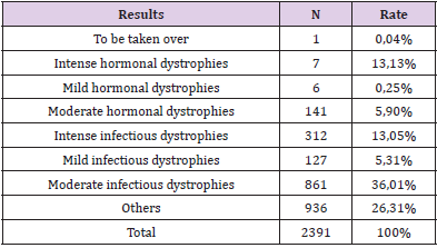
This indicates that surgical patients are usually at a high risk of developing pressure ulcers. The incidence of pressure ulcer leads to greater misuse of nursing resources and high medical costs. Pressure ulcers are not only adverse events in a hospital stay, that have to be treated and justified, but represent also a quantifiable risk in terms of morbidity and mortality. Adjusted for hospital, diagnosis related group (DRG), sex, race and age group, a case-control study revealed 3.98 extra days in hospitals and 7.23% attributable mortality, if a pressure ulcer occurs (Zhan & Miller 2003). From 1990-2001, pressure ulcer was mentioned as one cause among others for 3.79 deaths per 100,000 population in the USA (Redelings et al. 2005) as against the single underlying cause in 18.7%. The authors identified a high concurrence with sepsis (39.7% of all deaths with indication of a pressure ulcer). The risk factors associated with pressure ulcers amount to more than 100 and include medical diagnoses, patients’ demographic characteristics, anthropometrics, physiological status, nutritional status, and hospital environment, among others. The occurrence of pressure sores is a main obstacle to the long term rehabilitation of spinal-cord injured patient [1-3] (Table 1).
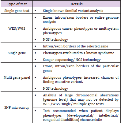
biomedres-openaccess-journal-bjstr

Table 1:

To achieve the best functional result with the most efficient use of resources, a comprehensive treatment plan is needed, that include preoperative workup, physical therapy, nutritional considerations, wound care, treatment of spasticity and reflex spasm, pre and postoperative bowel management, pulmonary consideration, anesthetic consideration, antimicrobial regimen. Surgical patients are prone to developing HAPU. In the United States, patients with HAPU had a longer length of stay, higher total hospitalization costs, and greater odds of readmissions compared with patients with no HAPU. Preventing HAPU involves accurate and ongoing risk assessments so that preventive measures can be implemented as early as possible and carried out throughout the period of immobility. The prevalence of HAPU among surgical patients is about 8.5% or higher depending on the type and the duration of the surgery. Patients with proximal femur fractures or patients after major lower limb amputation, the incidence of pressure ulcers was high (10.4% and 8.8%, respectively). Patients undergoing bowel surgery and peripheral vascular reconstructions are also prone to developing pressure ulcers. Several plausible mechanisms might be accounting for increasing risk of infections in relation to prior pressure sore exposures [4-6]. To start with, pressure sores were perceived to induce impairment of skin protection function by destroying integrity of erythematous skin and prompting reproduction and growth of pathogenic bacteria.

Pressure ulcers might introduce possible resources and entrances of pathogens to human body, which was possible to result in local infections in human body. Secondly, chronic pressure ulcers were supposed to persist chronic inflammation, which might lead to stimulation of cytokines and inflammation factors. An expansive literature suggest that long-term and excessive consumption of cytokines and inflammation factors might be accounting for immune suppression, and in turns led to a weaken ability to prevent invasions and attacks of pathogens. Thirdly, immobilization, as a primary risk factor for the occurrence and development of pressure sores, was considered to be associated with pneumonia. Immobilization might have destructive effects on removing function of bronchial secretions, which was conductive to reproduction of pathogen and help to result in pneumonia. Studies demonstrates an increased risk of several specific infections (surgical incision infection, pneumonia, urinary tract infection) within 14-day after spinal cord operation in patient with pressure sores preoperatively. Among the four stages of pressure ulcer, stage IV has the highest risk of postoperative infections. Intensive care units (ICUs) have the highest PU incidence rates in health care settings, which have been reported as high as 50% .The high rates in the ICUs can be attributed to the high acuity of patients, the nature of their critical illness and the highly invasive nature of the interventions and therapies that critically ill patients receive. Identifying patients at risk for PU development is essential for the effective implementation of PU prevention programs and usage of resources [7-10].

Method

Cochrane Library, MEDLINE/ PubMed, Scopus, CINAHL were used. The search was limited to the English language. In a final search, the reference lists of the included articles were also handsearched to identify further relevant articles.

Eligibility Criteria

Studies met the inclusion criteria if they assessed the effects of preventive measures on surgical patient. If the full text of an article could not be obtained, we included its abstract only when it had sufficient data.

Study Selection

Two independent reviewers screened and selected articles based on the title, subsequently on abstract and finally on full text. Disagreements were resolved via discussion with a third reviewer.

Data Extraction

The following information was extracted from the studies that met the eligibility criteria:

a. first the author’s name and year of publication;

b. participant characteristics (country of origin, sample size and mean age);

c. study characteristics (methods of participant allocation, allocation concealment, blinding, drop-out rates and reasons for drop-outs);

d. nature of aromatherapy intervention (type, dose, duration, route of administration for experimental and control interventions);

e. outcomes (instrument used to assess anxiety and outcome data).

Quality Assessment

The risk of bias of the included studies was assessed independently by two reviewers using the Cochrane Collaboration’s ‘Risk of bias’ tool. The criteria consisted of selection bias (random sequence generation and allocation concealment), performance bias (blinding of participants and personnel), detection bias (blinding of outcome assessment), attrition bias (incomplete outcome data), and reporting bias (selective outcome reporting). Each item was classified as ‘low risk of bias’, ‘high risk of bias’, or ‘unclear risk of bias’. Disagreements were resolved by discussion between two reviewers.

Description

Pressure ulcer development was strongly correlated with indication for admission. In specific patient categories, such as patients with proximal femur fractures or patients after major lower limb amputation, the incidence of pressure ulcers was high (10.4% and 8.8%, respectively). Patients undergoing bowel surgery and peripheral vascular reconstructions are also prone to developing pressure ulcers [11-15].

Preoperative Work-Up

Patient Candidacy: Prior to becoming a candidate for reconstructive surgery, the patient must be prepared physically and mentally endure 6-10 weeks of hospitalization. Patient exhibits selfmotivation since a successful result is obtainable only with patient cooperation. The team approach for spinal cord injured patient. The physical medicine and rehabilitation physician is the primary care doctor for the spinal cord injured patient and intimately involved with perioperative care. Before a procedure the patient must be in optimum physical health, be free of spasms and contractures, be in adequate nutritional status, be able to tolerate the prone position (if needed) and have adequate pulmonary function.

Physical Therapy for SCI Patients: The role of physical therapist have great importance in the care of SCI patient, that prevent contractures. The exercise and proper positioning in bed combat the deforming forces caused by paralysis and spasticity. Custome cut out cushions are useful in proper positioning. The occupational therapy department aids our patient with positioning which causing iatrogenic skin problems. A pressure pad evaluation aids the selection of a cushion with the best pressure distribution properties for the patient with a tendency towards decubitus ulcers.

Preoperative Positioning: Surgical candidates must tolerate prone position for a minimum of 4 weeks postoperatively and must exhibit operatively the ability to maintain the position adequately. Position individualized according to the location of pressure sores. Prone position is mostly applied since the pressure ulcer seen in ischial, sacral, and trochanteric area mainly. The prone position is assumed for 4 week postoperatively with side-side movement and range of motion beginning 6-7 week post operatively.

Nutritional Consideration: Before surgery nutritional status must have priority. The serum albumin level should be greater than 35g/100ml, the lymphocytic count should be greater than 220g%. Delayed cutaneous hypersensitivity is also indicator of decreased visceral protein. Inability to respond to placement of purified protein derivative, mumps, or candida antigens correlate and with higher rates of sepsis and high mortality rates in acute surgical patient.

Wound Care

Preoperative Wound Care: The care of the open ulcer consists of debridement of obviously devitalized tissue. Debridement is done surgically just to the point of bleeding with additional debridement accomplished by frequent dressing changes using mesh gauze sponges/kerlix to debride the wound, decrease the bacterial count. The use of povidone – iodine offer no therapeutic benefit over the use of saline in wound treatment.

Roengenograms: Chest and pelvic roentgenograms are routinely done preoperatively. If there is a large wound with a tracking sinus, a simple sinogram is performed using radio opaque dye injected through a foley catheter to evaluate the extent of the ulcer and help in the preoperatively planning for reconstruction.

Treatment of Spasticity and Reflex Spasms: Excessive reflex activity below the level of the upper motor neuron lesion often exists after a spinal cord injury. The obliteration of supraspinal inhibitory pathways is the reason postulated for this phenomenon. In the immediate postoperative period the spasms can cause bleeding and hematoma that may result in flap necrosis.

Medical Treatment: The drug of choice at the institute for rehabilitation and research is baclofen, which is believed to act centrally and poly synaptically within the spinal cord and brain stem. This begun at a dosage of 5-10mg 4 times a day, not to exceed 100mg daily. Side effects are minimal in patient with SCI, however with a dosage greater than described memory lose and confusion may occur. Diazepam may be used alone or with baclofen. It acts centrally within the brain stem and cerebral cortex to affect spasticity with initial dosage 5mg a day until a total use of 40mg a day is reached. Dantrolene can also be used for the treatment of spasms, it acts on excitation-contraction coupling mechanism of muscle fiber itself. It has a bed effect on liver so that serum glutamic pyruvic transaminase must be monitored monthly. Initial dosage is 25mg twice a day, increased to 25mg 4 times a day then by 25mg increment every third to fifth day to a maximum 200mg daily.

Surgical Management of Spasms

Nerve Blocks: Usefulness of peripheral nerve block is primarily confined to patients who have incomplete lesions and who are not candidates for subarachnoid blocks, since we want to preserve as much sensation and useful motor functions initial attempt with lidocaine, have to repeat as needed.

Epidural Stimulator: Most useful in patients with incomplete lesions but costly.

Subarachnoid Blocks (Phenol Rhizotomy): Useful for patients with complete lesions. Specific for lower extremities and don not affect the trunk and upper extremities.

Urological Management

Initially indwelling catheter is utilized following by intermittent catheterization with or without a condom catheter. Patients who are not voiding or who have high residuals are given methenamine as well as ammonium chloride (1 mg of each every 6 hrs).

Proper Diet and Bowel Management: We have found that even the most complex pressure sores can be successfully managed without a diverting colostomy.The preoperative regimen begun 3 days prior to surgery with a clear liquid diet. The bowel is cleansed with enemas and mild laxatives.

Postoperative Diet and Bowel Management: The clear liquid diet is continued for 5 days postoperatively.1 tab (2.5mg) twice a day is given for 5 days to constipate patient. If contamination does occur the area is thoroughly cleansed and dried immediately. The patient resume their prior bowel program on the 6th day postoperatively.

Antimicrobial Regime for the Decubitus Ulcer: Clinical and experimental studies indicate that anti-microbial agents prevent infection when effective concentrations are prevent infection when effective concentration are present in tissues during or shortly after the surgical procedure. Maximum bacterial shedding occurs during this time. A single parenteral dose of antimicrobial agent is given just prior to surgery, providing adequate tissue levels throughout the procedure. Antibiotics are selected on the basis of preoperative cultures and sometimes modified when the result of intraoperative cultures are known. The duration of administration of antimicrobial agents is individualized, based on the extent inflammation, suppuration, sinus tracts, and bone involvement.

Septic Patient with Decubitus Ulcer: The sin qua none of the treatment of sepsis associated with decubitus ulcer is surgical debridement. The antibiotic regimen is also need for aerobic and anaerobic bacteria.

Pulmonary Consideration: Vital capacity is measured preoperatively in the prone and supine position. After the patient position is changed, he or she is observed for clinical signs of respiratory embarrassment, Arterial blood gas may be obtained 20 minutes after the patient’s position changed.

Sitting Regimen: A pressure pad evaluation must be performed postoperatively to determine new sitting surface requirement. Sitting is begun in the sixth or seventh postoperative week, beginning with 30 minutes of sitting twice during the first day. Lanolin massage of surgical site keeps the area soft and moist. However, pressure ulcer rates can only be used as an indicator of quality of care if certain crucial aspects of data collection and interpretation are taken into account. The VP pad is better than the HDF pad in relieving pressure. However, the measured average pressures for both types of pads were all higher than the threshold value of the capillary closing pressure (32mmHg; Schultz, 2005). Neither of them could reduce the pressure below the capillary closing pressure, which may have contributed to the occurrence of pressure ulcers in both groups. Both the average and peak pressures, measured after applying the VP pads, were significantly lower compared to measurements of the HDF pad; however, there was no significant difference between the two types of pad regarding the prevention of pressure ulcers.

Obese individuals had significantly lower values of surface pressure and shear force than slender individuals at the coccygeal bone site. At the sacral bone site and the thigh site, the values of surface pressure in the obese group were significantly higher than those of the slender group. The reason why obese subjects had higher values was not obvious, but the magnitude of pressure at the sites was low, and so the effect of bony prominences would not be apparent by weight distribution in the obese types. In clinical practice, subjects should be positioned at the bending conformity point or shifted 10cm from that point toward the head of the bed whenever bed operations are performed in order to avoid generating harmful shear forces and surface pressures. There are multiple possible reasons behind the correlation between PCA use and increased pressure ulcer incidence. Frist, the use of a PCA pump can relieve the sense of pain, a decreased sensation and blocked motor nerve owing to a PCA pump might accelerate the development of pressure ulcers. Meanwhile movement actually can be considered self protection for bedridden patients, however PCA can reduce the patient’s sense of pain so that they will reduce their movement, which can reduce the patient’s ability to self protect. Similarly the use of PCA may make patients over emphasize the disease severity so that they may fear moving.

Second because a PCA pump can relieve painful sensations, patients may be less willing to move in order to stay comfortable. Third in order to avoid the events of catheter prolapse and compression, postoperative patients are fearful of moving. Bed- or wheelchair-bound patients tend to suffer from multiple pressure sores and often undergo several operations over the years for reconstruction of the tissue’s integrity. This makes every pressure sore recurrence over previously reconstructed and therefore scarred tissue more complex to treat. Fillet flaps from nonfunctioning body parts not only impose little or no donorsite morbidity but can also prolong pressure sore recurrence by offering robust tissue for padding. In paraplegic patients with massive or bilateral pressure sores, where all other reconstructive measures have been exhausted, fillet flaps offer a last resort option for pressure sore management.

Conclusion

Conscientious effort is needed to prevent the development of HAPU through the early identification of risk and early implementation of preventive measure. So in my opinion when we compare all the points in discussion most applied and applicable method to achieve the best functional result with the most efficient use of resources, a comprehensive treatment plan is needed, that include preoperative workup, physical therapy, nutritional considerations, wound care, treatment of spasticity and reflex spasm, pre and postoperative bowel management, pulmonary consideration, anesthetic consideration, antimicrobial regimen. The majority of MDRPUs were related to poor positioning or fixation of respiratory equipment. Occurrence of these ulcers could be prevented with implementation of preventive strategies such as regular assessment of the area underneath and around medical devices and regular repositioning or securement of devices (45). Using protective dressings to secure and stabilize devices also could reduce the risk for MDRPU development.

For More Articles: Biomedical Journal Impact Factor: https://biomedres.us

Open Access Journals on Surgery

A Rare Case of Ventricular Rupture and Aortic Endocarditis Due to SARS-COV-2 Treated By 2 Step Cardiac Surgery

Introduction

Although SARS-CoV-2 infection primarily targets the respiratory system [1,2], cardiac complication is the most common comorbidity. Several studies from China reported acute cardiac injury in 7.2% to 27.8% among hospitalized patients [3-5]. Some of the cardiovascular complications of COVI- 19 include STelevation myocardial infarction [6], myocarditis [7], cardiogenic shock, arrhythmias, pericardial effusions, cardiac tamponade [8] and infective endocarditis [9]. We report the case of a patient who developed COVID-19 endocarditis and myocarditis with the development of tenacious mediastinal-epicardial adhesions responsible for injury to the right ventricle treated with pericardial patch in two steps.

Case Presentation

Informed consent was obtained by the patient. Consent from the institutional review board was waived, being a clinical case. A 46-year-old male was referred to our hospital with progressive shortening of breath of recent onset. Transthoracic echocardiography revealed bicuspid aortic valve with moderate insufficiency and ascending aortic aneurysm with a maximum diameter of 55mm. No coronary artery disease was found at the angiogram. The patient underwent replacement of the ascending aortic with aortic valve repair (subcommisural annuloplasty). The postoperative course was uneventful and the patient was discharged home. Transthoracic echocardiography showed ejection fraction of 55% and mild aortic valvular insufficiency. After a day the patient performed COVID-19 molecular swab test resulted positive with mild symptoms: fever (37.5°C), mild dyspnea, changes in taste. Two months later the patient was readmitted because of progressive dyspnea (NYHA class III). Transthoracic echocardiogram showed severe aortic valve insufficiency. Transesophageal echocardiogram confirmed severe aortic valve insufficiency with prolapse of the non-coronary cusp and vegetation on both cusps.

Figure 1: Right Ventricular reconstruction without cross clamp, in cardiopulmonary bypass.

Figure 2: Results of right ventricular repair at the time of delay surgical treatment of aortic valve replacement after 10 days.

The patient was referred for redo surgery of aortic valve replacement with mechanical prosthesis. At reoperation, the approach was through median sternotomy after cardiopulmonary bypass instituted by cannulation of the right femoral artery and vein. The surgery was complicated in the first phase by laceration of the right ventricle due to very tenacious mediastinal-epicardial adhesions from pericarditis caused by COVID-19. The patient underwent repair of the right ventricle with bovine pericardium patch (Figures 1 & 2). After discussion in heart team (cardiac surgeon, cardiologist and cardioanesthesiologist) it was decided to postpone the aortic valve replacement. The patient was transferred to intensive care. Ten days later the patient underwent aortic valve replacement with mechanical prosthesis (Sorin Bicarbon 25mm). The postoperative course was uneventful and the patient was discharged home in NYHA class I. Transthoracic echocardiography showed ejection fraction of 45%. After one year the patient was in NYHA class I with aortic prosthesis in place without periprosthetic leaks and vegetations.

Conclusion

This is a rare case reported in the literature of endocarditis and myocarditis caused by COVID-19 characterized by very tenacious mediastinal-epicardial adhesions. More attention is needed during the reopening of patients with previous COVID-19 because, despite the start of CEC and the empting of the right cardiac section, the risk of injury of the right ventricle is very high. When it happens, we suggest to proceed in two steps. In the first step is useful only to repair the right ventricle and in the second step, after 10 days, to proceed with aortic valve replacement/repair. This is important to avoid clamping the aorta which can cause ischemia of the repaired tissue, long CEC times with additional risk for the patient. In conclusion the message to take home is not to underestimate the tenacious adhesions from CVID-19 related myocarditis and, in case of laceration of the right ventricle, proceed in two steps to allow the stabilization of the patient in intensive care.

This is a rare case reported in the literature of endocarditis and myocarditis caused by COVID-19 characterized by very tenacious mediastinal-epicardial adhesions. More attention is needed during the reopening of patients with previous COVID-19 because, despite the start of CEC and the empting of the right cardiac section, the risk of injury of the right ventricle is very high. When it happens, we suggest to proceed in two steps. In the first step is useful only to repair the right ventricle and in the second step, after 10 days, to proceed with aortic valve replacement/repair. This is important to avoid clamping the aorta which can cause ischemia of the repaired tissue, long CEC times with additional risk for the patient. In conclusion the message to take home is not to underestimate the tenacious adhesions from CVID-19 related myocarditis and, in case of laceration of the right ventricle, proceed in two steps to allow the stabilization of the patient in intensive care.

For More Articles: Biomedical Journal Impact Factor: https://biomedres.us

Open Access Journals on Medicine

Genetic Testing and Counselling for Cancer: Evolution, Future and Psychological Impact

Background

The field of cancer screening and testing has come a long way; its evolution from basic biochemical tests screening for particular analytes [1] to the use of gene-based testing and screening models is a definitive example of the evolution of our understanding of cancer syndromes and their underlying genetic basis. Identification of people with cancer predisposition syndromes and germ line pathogenic variants [2] has become easier with the advent and utilization of genome-wide gene sequencing panels [3,4] chromosomal arrays, and whole exome/genome sequencing and, somatic tumor testing (especially for childhood cancers), emphasizing the increasing necessity for both cancer predisposition testing and genetic counselling programs specializing in the testing, management and care of cancer [5].
Genetic counsellors are uniquely qualified to address cancer risk, interpretation of genetic results, and the inevitable complications of the subsequent ethical [6], legal and psychosocial issues that are encountered by individuals dealing with this malady [7,8]. Obtaining informed consent [5,6] from both minors and adults, ascertaining the timing for a genetics referral, providing periodic counselling and the associated aid overtime (especially in the smooth transition of younger patients to adult/ geriatric care) [8,9] and the dearth of childhood cancer predisposition syndromes are some of the unique challenges that plague us today [10]. Incidental findings due to the prevalence of genetic testing are associated with their own set of psychological implications [11].
In countries like India, where there is enormous disparity between socio-economic classes, a genetic counsellor will encounter additional problems including the lack of awareness and education amongst the under-privileged classes, genderand religion-associated disparity in terms of cancer treatment, management and the reality of the influence of political agendas over government health policies.

Entry to Genetic Counselling

Referral to a genetic counselling clinic for cancer evaluation can occur due to the prevalence of cancer syndromes within the family (family history) [12], ethnicity [13,14], phenotypic expressions, high-risk tumour types, non-oncological findings, like the lippigmentation and greater the characteristic macules associated with Peutz-Jeghers syndrome and neurofibromatosis type1 (and bi-allelic mismatch repair) respectively and incidental findings (variances in predisposition genes on chromosomal microarrays and secondary findings detected by exome sequencing) [9]. Family history alone, however, is not a reliable factor when accounting for the necessity of a referral as cancer predisposition has been reported in many cases of absence of said history [10], This can be due to the formation de novo variants, parental germ line mosaicism [15], increasing incidence of smaller nuclear families, low penetrance and recessive inheritance masking an inherited syndrome. Hence, a combination of the aforementioned factors should be taken into account when making a genetics’ referral. Moreover, the future holds the possibility population based genetic testing for cancer predisposition syndromes.
Oncologists are confronted with the possibilities of identifying whether reported variants are indicative of somatic changes or inherent germ line susceptibility [16] and thus subsequent referral to a genetics’ clinic for their particular expertise becomes necessary.

Timing of Genetic Counselling Initiation

The optimal timing of the first genetic counselling session is at the time of tumor diagnosis or after the initiation of treatment in cases of poor prognosis, to ensure that the possibility of prophylactic surgery is considered. The genetic counsellor is qualified to advise the patient on many issues associated with the patient prognosis, associated treatment plans and psychological developments [17,18]. In the case of poor prognosis, DNA banking should be administered, and genetic testing organized in a timely manner in order to assess and counsel their relatives to minimize the economic and psychological burden of post-mortem testing.

Pre-Test Genetic Counselling

Informed consent should be obtained from the patient before they undergo any genetic testing procedure. This involves complete discussion of all aspects related to the test and consequent implications, including information regarding tests most suitable to the patient (based on the various factors that have prompted the referral), the surveillance protocol that may follow (especially in cases of transition from childhood to adulthood), psychosocial [11], ethical and privacy concerns, which all vary in accordance with the laws of different countries, and plans for disclosing results to at-risk relatives [6,19]. Adults are required to provide verbal and written assent towards the procedure; minors should be guided through these discussions in an age-appropriate manner and adolescents should provide verbal and written consent in conjunction with their parents or primary guardians [9]. This allows older children to become involved with and more accustomed to their unique situation and have any fears or misconceptions addressed. The basic tenet of genetic counselling is to give patients the tools required for them to make informed autonomous decisions [20] regarding the testing and management options available to them based on factual (stratified) risk information [21-25]. Despite concerns of exacerbating patients’ anxiety, genetic testing in the absence of prior genetic counselling is linked with negative outcomes including the violation of ethical standards, ordering of unnecessary, perhaps costly, genetic tests, misinterpretation of these results and adverse effects on the patients’ psyche.

Genetic Testing

The invention of Next Generation Sequencing (NGS) technology is the biggest advancement in genetic testing seen in recent years wherein analysis is performed at a multiple gene level, or even whole transcriptomic, genomic and exomic levels, at a relatively lower cost, especially when the increased sensitivity and efficiency of obtaining results are taken into account [16]. A test should be ordered after consultation with a genetic counsellor and the choice of laboratory should be based on the type of test, its level of analysis, methodology and their variant interpretation techniques, expenses involved, time factors and their policies on privacy and ethics. The variants are reported in accordance with the five-tier classification proposed by The American College of Medical Genetics and Genomics (ACMG), namely, pathogenic, likely pathogenic, variant of uncertain significance, likely benign and benign [9,26]. Despite the succinctness of this model, further interpretation by an expert is important especially when deciding the significance/ level of pathogenicity of variants (Table 1).

biomedres-openaccess-journal-bjstr

Table 1: Genetic tests for cancer gene mutation.

Note: {WES – Whole Exome Sequencing; WGS – Whole Genome Sequencing, NGS – Next Generation Sequencing, SNP – Single Nucleotide Polymorphism}

The sensitivity of these tests for identification of pathogenicity varies from greater than 95% to less than 50% and thus, a genetic counsellor must provide counsel on risk perception and management options based on empirical data and in conjunction with the oncologist. Identification of variants without certain significance tailors the advised management options to include surveillance protocols and the possibility of family variant tracking. Relatives can be tested for these only when consent is provided by the patient [6]. The inheritance patterns associated with the identified pathogenic variant determine the relatives who are atrisk. For example, if a syndrome follows an autosomal dominant pattern of inheritance, there is a 50% risk of inheriting a pathogenic variant, and its associated risks, to first-degree relatives of the proband [16,27].

Post- Test Genetic Counselling

For those individuals with a positive diagnosis, post-test counselling involves specific treatment/ management options and the necessity of periodic evaluation. In cases of childhood diagnosis of a hereditary cancer syndrome, reproductive and psychological implications especially, change over increasing age. Hence, lifelong risk management, and understanding of the options for reproductive (post- and pre-natal) counselling [28], testing post-birth, adoption, and/or utilizing donors is disseminated with increasing age of the patient in an appropriate manner suited to the child’s developmental stage8,9. Surveillance options [29] and information regarding the advent of new testing options can only be provided for at-risk individuals/ families if periodic follow-up with the genetic counsellor is maintained.

Psychological and Sociological Implications

Negative effects on the psyche of a patient with a positive diagnosis are primarily concerned with surveillance protocols and the reality of a life-long economic burden8. Invasive medical procedures [30], procedures requiring sedation or general anesthesia, false positive results, incidental findings are implicated by various studies in causing psychological distress to the patient and their families. There arises a general frustration with lack of positive results when it comes to therapies advised, inconclusive outcomes of tests and the delivery of false-positive results (increased in whole-body MRIs) [31], impaired quality of life, reduced satisfaction with care, and worse overall survival. Patients undergoing surveillance may experience anxiety with regards to scanning, referred to as “scanxiety”, and children especially may feel adversely towards repeated blood draws and the claustrophobic confinement experienced during MRI scans [9]. Young adults face an ever-increasing economic burden as they are as of yet not financially secure enough, and may not even possess insurance cover [20], to support life- long management programs.
On the contrary, proactive surveillance and psychological support, from say, cancer survivor groups, may empower the patient and are viable avenues to lead a more well-adjusted life. The patient relies on the care team in such scenarios due to generation of trust and confidence in their counsel [32,33]. Cancer genetic counselling in India. In India, there are about a million cases of cancer diagnosed yearly. When the population of the subcontinent is taken into account, these rates of incidence and mortality, match those seen in high-income countries [34]. These elucidate the lack of early-detection strategies and positive results of treatments within the medical landscape of the country [35].
Studies show that breast, lung, oral, cervical, gastric and colorectal cancers [36] have the highest incidence rates in India. It is evident that most causative agents implicated in cancer in the Indian population are avoidable (tobacco and alcohol consumption, lack of education with respect to female hygiene) [34,37]. There is a general lack of education and support for the low-income, socially underprivileged families owing to the fallacies of an increasingly capitalist state of government [38-40]. Even governmental policies regarding general health are found to be inadequate in the face of growing mortality due to cancer. It is the second-leading cause of death amongst the Indian population.
A patriarchal society has also generated the spread of female reproductive cancers as the importance of feminine hygiene is not acknowledged by many; additionally, a greater economic burden to finance health care is placed on women [41]. In conjunction with the lack of resources and such a mind-set, most evidently seen in under-privileged communities, the incidence of cervical cancers has witnessed an increase over the past 5-10 years. In India, the age of onset of the female reproductive cancers is around a decade earlier than that of developed nations (45-50 years in the former, compared to >60 years of the latter) [42]. Moreover, oncofertility can be improved by developing management, treatment strategies that comprise of professionals from multiple specialties, say a counselling team that includes a reproductive endocrinologist [43] apart from an oncologist, psychologist and the geneticist. Hence, advancement of cancer-associated clinical programs with respect to diagnosis, screening, management/ treatment particular to the Indian race [44] are a necessity in the mission to better the state of women’s health in India [42,45].

Conclusion

Cancer testing and management is an ever-growing field primarily due to our increasing understanding of unknown syndromes and their underlying genetics, and the expansion of available information of known cancer syndromes. In accordance with these basic developments, there should be a correlating evolution in the various genetic testing and counselling practices available for the same. These take the form of improved access to aforementioned resources and formulations of government policies that tackle health care from this perspective [45]. One such advancement can be the necessitation of population-based genetic screening [46,47] and testing for common pathogenic variants of the most abundantly found cancer syndromes, e.g. BRCA 1 and 2 testing for breast cancer [4,42,48]. This type of policy can hold many implications in reducing the burden of cancer in many developing economies, like India. In the same vein, there have been discussions on the use of e-health interventions [49] like psychoeducational interventions delivered via web-based environments with auxiliary methods of communication, which includes support with professionals and peers, offer improved awareness and access to the required support for informed, autonomous decisionmaking. Personalized medicine is an avenue of modern medicine gaining quick popularity as it is tailored to an individual and their needs. Corroborating advancements in cancer genetic testing and counselling to personalized care will initiate the incidence of more positive outcomes as it pertains to lifelong care and management practices [5,11,18,50,51].

For More Articles: Biomedical Journal Impact Factor: https://biomedres.us

Open access Journals on Chemistry

Extractive Values and Antioxidant Properties of Leaves, Seeds, Pods and Coats Moringa Plant

Introduction

Antioxidants are molecules that fight free radicals in human body [1]. Antioxidants cause protective effect by neutralizing free radicals which are toxic byproducts of natural cell metabolism. Research is increasingly showing that antioxidant rich foods and herbs have health benefits. Medicinal herbs are the richest sources of antioxidants compounds [2]. Antioxidants are biomolecules which tackle and destroy free radicals and scavenge diseases [3]. Sagar, et al. and Shao, et al. reported that green plants are chief sources of natural antioxidants, and they are capable of tackling free radicals [3-5]. Moringa oleifera belongs to the single genus family Moringaceae and it is most studied of the thirteen species of Moringa trees [6,7]. It is a deciduous tree that grows up to 12 m tall with an umbrella-shaped crown and grows extremely fast which can reach up to 4 m in its first year. Its leaves are alternate bi or tri-pinnate, 20-70 cm long. Leaflets are usually oval, rounded at the tip, and 1-2cm long and they are dark green in colour with almost whitish in the lower surface [8,9].
Fahey, 2005 reported that the leaves of Moringa oleifera are the most nutritious part of the plant, being a significant source of vitamin B6, vitamin C, pro-vitamin A as beta- carotene, flavonoids, 46 antioxidants, magnesium and protein among other nutrients. The plant is called “Zogale” in Hausa, “ Ewe Igbale” in Yoruba, “Ikwe Oyibo” in Ibo and “Egelengedi” in Idoma while its English name is Bean oil tree or drumstick tree or miracle tree or “Mother’s Best Friend” [6,10,11]. Though a lot of research works have been done on moringa plant [10,12-16]. but there is little, or no work done on the effect of solvents in extracting phytochemicals from the seeds, coats, pods and leaves of the plant. Therefore, the focus of this research work is to evaluate the potency of solvents in extracting bioactive ingredients from seeds, coats, pods and leaves of moringa plant as well as to investigate and compare the antioxidant properties of the first two highest yield solvent-extracts with that of the raw sample with a view of establishing which of the moringa plant parts (seeds, leaves, pods and coats) is richest in phytochemical constituents and antioxidant properties.

Materials and Methods

Source of Materials

The seeds, coats, pods and leaves of Moringa oleifera were collected from a compound of a building at Ajagbale Street, Oka, Ondo City, Ondo State, Nigeria. All chemicals used were of the analytical grade with the highest purity available (<99.5%) and procured from Sigma Aldrich, USA.

Preparation and Extraction of Seeds, Coats, Pods and Leaves of Moringa Plant

The different parts of moringa plant were cut into smaller pieces for easy air-drying. The dried samples were ground separately using electric blending machine (Solitarire Mixer Grinder VTCL Heavy Duty 750 Watts) and each part was sieved with 40 mm mesh size. The powdered samples were divided into portions, packed in air tight containers labelled appropriately prior to extraction. Each sample was extracted separately with each solvent (acetone, chloroform, ethyl acetate, ethanol, methanol and water) at ratio 1:10 for 72 h during which it was intermittently shaken on a shaking orbit machine The resulting mixture was filtered through a 0.45 μm nylon membrane filter. The extracts were desolventised to dryness under reduced pressure at 40 oC by a rotary evaporator (BUCHI Rotavapor, Model R-124, Germany). Weight of extract obtained was used to calculate the percentage yield of extract in each solvent and the dry extracts were stored in a refrigerator (4 0C) prior to analysis [17-19].

Determination of Antioxidant Property

Total Flavonoid

0.1g of extract was weighed into a sample bottle; 10 mL of 80% methanol was added and allowed to soak for 2 hours. 0.4 mL of the solution was measured into a 10 mL volumetric flask, 1.2 mL of 10% sodium hydroxide, 1.2 mL of 0.2 M concentrated sulphuric acid and 3 mL of 3 M sodium nitrate were added. 4.2 mL of distilled water was used to make it up. The absorbance was read using 6850 UV spectrophotometer at wavelength 325 nm [20].

Ferric Reducing Antioxidant Power (FRAP)

0.1g of extract was weighed into a sample bottle; 10 mL of 80% ethanol was added. 2.5 mL sodium phosphate buffer (0.2 M Na2PO3, pH 6.6) and 2.5 mL of 1% potassium ferricyanide were added and incubated at 50˚C for 20 minutes. 2.5 mL of TCA (trichloroacetic acid) was added to stop the reaction. 2.5 mL of the aliquot was taken and diluted with 2.5 mL distilled water and 0.5 mL of 0.1 % ferric chloride was added and allowed to stand for 30 minutes in the dark for color development. The absorbance was read using 6850 UV/Visible spectrophotometer at wavelength 700 nm [21].

DF: Dilution factor. If not diluted, then DF = 1

Total Phenol

0.1g of extract was weighed into a sample bottle; 10 mL of distilled water was added to dissolve. 1 mL of the solution was pipetted into a test tube and 0.5 mL of 2 N Folin-Ciocalteu reagent and 1.5 mL of 20 % sodium carbonate solution was added. The solution was allowed to stand for 2 hours and the absorbance was read using a 6850 UV/Visible spectrophotometer at wavelength 765 nm. Garlic acid solution was used as standard viz 0.5 mg, 1 mg, 2 mg, 4 mg, 6 mg, 8 mg and 10 mg [22].

DF: Dilution factor. If not diluted, then DF = 1

DPPH (2, 2-Diphenyl-1-Picrylhydrazyl) Scavenging

0.1g of extract was weighed into a sample bottle and 10 mL of ethanol was added, stirred for 15 minutes and allowed to stand for 2 hours. 1.5 mL of the extract was pipetted into a test tube and 1.5 mL of DPPH solution was added. The 6850 UV/Visible spectrophotometer was zeroed with ethanol as the blank solution. The absorbance/ optical density of the control (DPPH solution) was read. The absorbance of the test sample was read at 517 nm. [23].

DF: Dilution factor. If not diluted, then DF = 1

Iron (Fe2+) Chelation Assay

0.1g of extract was weighed into a sample bottle, 150 μL of 500 μM FeSO4 was added. 168 μL of 0.1M Tris-HCl (pH 7.4) and 218 μL of saline solution was added. 100 μL of the solution was taken and incubated for 5 minutes, before addition of 13 μL of 0.25% 1, 10-phenanthroline. The absorbance was read using 6850 UV/ Visible spectrophotometer at wavelength 510 nm [24].

Statistical Analysis

Statistical significance tests were performed using SPSS (v. 20, IBM SPSS Statistics, US) at p < 0.05 by means of one-way analysis of variance (ANOVA) followed by LSD post hoc multiple comparison and the experimental results were expressed as mean ± standard mean deviation of three replicates.

Results and Discussion

Extractive Values of Solvent Extracts of Leaves, Seeds Pods and Coats of Moringa Plant

The extractive values (% yield) of leaves, seeds, pods and coat moringa plant using acetone, chloroform, ethanol, ethyl acetate, methanol and water are contained in Table 1. The result showed that the percentage yield of moringa leaf extract was 11.095±0.805 in ethyl acetate, 9.291±2.234 in ethanol, 8.497±0.713 in water, 8.108±2.219 in methanol, 7.039±2.084 in acetone and 3.055 ± 1.735 in chloroform. The percentage yield of moringa pod extract was 2.146±0.003 in methanol, 1.347±0.001 in ethanol, 1.047±0.000 in ethyl acetate, 0.898±0.002 in chloroform, 0.850±0.000 in acetone and 0.199±0.002 in water. The percentage yield of moringa coat extract was 8.709±0.414 in acetone, 3.333±0.108 in ethanol, 3.160±2.731 in methanol, 3.062±0.534 in ethyl acetate, 2.149±1.799 in water and 1.451±0.502 in chloroform. The percentage yield of moringa seed extract was 30.151±1.221 in acetone, 29.997±1.558 in ethyl acetate, 27.725±2.714 in ethanol, 26.190±3.452 in methanol, 23.942±2.955 in chloroform and 21.417±10.455 in water.
In each of the solvent used, there was a significant difference at p<0.05 in the extractive values of the seeds, leaves, pods and coats of moringa plant. In all the solvents used for extraction, it was observed that the extractive value (%) was highest in moringa seed and least in moringa pod. All the solvents used had the first two highest extractive values in moringa seeds and leaves except acetone that had its first two highest extractive values in moringa seeds and coats. According to Alachaher, et al. 2018 [21], there are quite number of factors in which extraction of bioactive compounds depends. The selection of solvent system largely depends on the specific nature of the bioactive compounds being targeted. Also, different solvent systems are available to extract the bioactive compounds from natural products. Extraction efficiency is affected by the chemical nature of phytochemicals, the extraction method used, sample particle size, the solvent used, as well as the presence of interfering substances. Under the same extraction time and temperature, solvent and composition of sample are known as the most important parameters [21].

biomedres-openaccess-journal-bjstr

Table 1: Extractive value of solvent extracts of moringa plant.

Note: * = Result values are expressed as mean value of triplicate determinations ± standard mean deviation

Different letter in the same row showed significant difference (p<0.05).

Antioxidant Properties of Solvent Extracts of Leaves, Seeds, Pods and Coats of Moringa Plant

Antioxidant properties were carried out on the plant raw sample and the first two solvent extracts with the highest extractive values. The antioxidant properties of raw sample, ethanol and ethyl acetate extracts of leaves; acetone and ethyl acetate extracts of seeds; methanol and ethanol extracts of pods as well as acetone and ethanol extracts of coats of moringa plant were examined and these are presented in Table 2 to Table 5. The antioxidant properties of raw sample, ethanol and ethyl acetate extracts of moringa leaves is depicted in Table 2. The concentration (mg/100g) of total flavonoid in moringa leaves ranged between 0.122±0.001 – 0.332±0.001 with ethanol extract had the highest concentration of 0.332±0.001 mg/100g, followed by ethyl acetate extract with concentration of 0.268±0.002 mg/100g while the powdered raw sample has the least concentration of 0.122±0.001 mg/100g. The total phenol concentration (mg/100g) in moringa leaves ranged between 0.181±0.002 – 0.349±0.003 with ethanol extract having the highest concentration of 0.349±0.003 mg/100g, followed by ethyl acetate extract with concentration of 0.251±0.001 mg/100g while the powdered raw sample had the least concentration of 0.181±0.002 mg/100g.

biomedres-openaccess-journal-bjstr

Table 2: Antioxidant properties of moringa leaves.

Note: * = Values are expressed as mean value of triplicate determinations ± standard mean deviation; GAE =Garlic Acid Equivalent Different letter in the same row showed significant difference (p<0.05).

biomedres-openaccess-journal-bjstr

Table 3: Antioxidant properties of moringa seeds.

Note: * = Values are expressed as mean value of triplicate determinations ± standard mean deviation; GAE =Garlic Acid Equivalent

Different letter in the same row showed significant difference (p<0.05).

biomedres-openaccess-journal-bjstr

Table 4: Antioxidant properties of moringa pods.

Note: * = Values are expressed as mean value of triplicate determinations ± standard mean deviation;GAE =Garlic Acid Equivalent

Different letter in the same row showed significant difference (p<0.05).

Table 5: Antioxidant properties of moringa pods.

Note: * = Values are expressed as mean value of triplicate determinations ± standard mean deviation; GAE =Garlic Acid Equivalent

Different letter in the same row showed significant difference (p<0.05)

The percentage DPPH (2,2-diphenyl-1-picrylhydrazyl) scavenging activity of powdered raw sample showed the highest value of 89.25±0.21% followed by ethanol extract of 63.10±0.30% while ethyl acetate extract had the lowest value of 57.45±0.16%. The ethyl acetate extract of moringa leaves exhibited the highest reducing power of concentration of 0.87±0.02 GAE followed by ethanol extract which had concentration of 0.47±0.00 GAE while the powdered sample had the least concentration of 0.32±0.01GAE. There were significant differences at p < 0.05 in the total flavonoid, total phenol, DPPH, iron chelation assay and ferric reducing antioxidant power of raw sample, ethanol extract and ethyl acetate extract of moringa leaves. The antioxidant properties of raw sample, acetone and ethyl acetate extracts of moringa seeds is displayed in Table 3. The total flavonoid concentration (mg/100g) of moringa seeds ranged from 0.363±0.002 – 0.392±0.000 in which the powdered sample had the highest concentration of 0.39±0.000 mg/100g, followed by ethyl acetate extract with concentration of 0.379±0.001 mg/100g while the acetone extract has the least concentration of 0.363±0.002 mg/100g.

There was no significant difference (p < 0.05) in the total flavonoid content of raw sample, acetone extract and ethyl acetate extract of moringa seeds. In moringa seeds, the total phenol concentration (mg/100g) was between 0.059±0.001 – 0.349±0.00. The ethanol extract had the highest concentration while the powdered raw sample had the lowest concentration of total phenol. The ethyl acetate extract of moringa seeds had total phenol concentration of 0.251±0.001 mg/100g. There was significant difference (p < 0.05) in the total phenol concentration of raw sample, acetone extract and ethyl acetate extract of moringa seeds. The DPPH (2,2-diphenyl-1-picrylhydrazyl) radical scavenging activity of powdered raw sample of moringa seeds had the highest activity of 79.81±0.11% followed by ethyl acetate extract with DPPH activity of 77.43±0.16% while acetone extract showed the lowest activity of 71.86±0.30%. There was significant difference (p < 0.05) in the DPPH (2, 2-diphenyl-1-picrylhydrazyl) radical scavenging activity of raw sample, acetone extract and ethyl acetate extract of moringa seeds. The iron chelation activity (%) ranged between 22.36±0.11 – 39.72±0.09 with the lowest and highest concentration in raw sample and acetone extract respectively while the ethyl acetate had iron chelation activity of 38.12a±0.07%.
There was no significant difference (p < 0.05) in iron chelating activity of acetone extract and ethyl acetate extract of moringa seeds. The ethyl acetate extract of moringa seeds exhibited the highest ferric reducing antioxidant power of concentration of 0.55±0.00 GAE followed by acetone extract which had concentration of 0.54±0.01 GAE while the powdered sample had the least concentration of 0.29 ±0.01 GAE. There was no significant difference (p < 0.05) in ferric reducing antioxidant power (FRAP) of acetone extract and ethyl acetate extract of moringa seeds. The antioxidant properties of raw sample, methanol and ethanol extracts of moringa pods is shown in Table 4. The concentration (mg/100g) of total flavonoid of moringa pods ranged between 0.038±0.000 – 0.082±0.002 with methanol extract having the highest concentration and the powdered sample having the least concentration while the total flavonoid concentration of ethanol extract of moringa pod was 0.059±0.001 mg/100g. There was significant difference (p < 0.05) in total flavonoid concentration of raw sample, methanol extract and ethanol extract of moringa pods. The total phenol concentration (mg/100g) of moringa pods was between 0.031±0.000 – 0.131±0.001.

The highest and lowest total phenol concentration were found in ethanol extract and raw sample of moringa pods respectively while methanol extract had total phenol concentration of 0.059±0.000 mg/100g. There was significant difference (p < 0.05) in total phenol concentration of raw sample, methanol extract and ethanol extract of moringa pods. For DPPH (2,2-diphenyl-1-picrylhydrazyl) scavenging activity of moringa pods, it was observed that methanol extract showed the highest value of 96.82±0.20% followed by powdered raw sample which had 83.75±0.11% while ethanol extract showed the lowest value of 81.95±0.10%. There was no significant difference (p < 0.05) in DPPH scavenging activity of raw sample and ethanol extract of moringa pods. The iron chelating activity (%) of moringa pods ranged from 28.14±0.01 – 36.73±0.03 with the lowest and highest concentration in methanol extract and raw sample accordingly and that of ethanol extract was 32.14±0.02%. There was significant difference (p < 0.05) in iron chelating activity of raw sample, methanol extract and ethanol extract of moringa pods.
The ferric reducing antioxidant power (FRAP) of methanol extract of moringa pods exhibited the highest reducing power of 0.53±0.03 GAE, followed by ethanol extract of 0.42±0.01 GAE while the powdered raw sample had least concentration of 0.25±0.00 GAE. There was significant difference (p < 0.05) in ferric reducing antioxidant power of raw sample, methanol extract and ethanol extract of moringa pods. The antioxidant properties of raw sample, acetone extract and ethanol extract of moringa coat is presented in Table 5. The total flavonoid concentration (mg/100g) of moringa coats ranged from 0.049±0.000 – 0.317±0.002 with acetone extract had the highest concentration and the powdered raw sample had the least concentration while ethanol extract had total flavonoid of 0.258±0.001 mg/100g. The concentration (mg/100g) of total phenol of moringa coat was between 0.031±0.000 – 0.118±0.002. The acetone extract and ethanol extract had the lowest and highest concentration while the raw sample of moringa coat had total phenol concentration of 0.063±0.001mg/100g.

The DPPH of ethanol extract moringa coat showed highest activity of 95.20±0.12 % followed by powdered sample which had activity of 85.07±0.09% while acetone extract showed the lowest activity of 72.13±0.10%. The iron chelating activity (%) of moringa coats was between 11.18±0.00 – 44.91±0.04. Ethanol extract had the highest iron chelating activity while acetone extract had the lowest and that of raw sample was 30.34±0.02 %. The ferric reducing antioxidant power of acetone extract of moringa coats exhibited the highest reducing power of 0.85±0.05 GAE followed by ethanol extract of 0.64±0.03 GAE while the powdered raw sample has the least concentration of 0.32±0.01 GAE. There were significant differences (p < 0.05) in all the antioxidant properties considered for raw sample, acetone extract and ethanol extract of moringa coats [25-28].

Conclusion

The moringa seeds are richest in bioactive ingredients and this is followed by moringa leaves while the least bioactive ingredients are found in moringa pods. The solvent extraction efficiency of bioactive ingredients in moringa plant decreases in the order of acetone, ethyl acetate, ethanol, methanol, water and the least is chloroform. The utilization of acetone, ethyl acetate, ethanol and methanol in extracting bioactive ingredients of high antioxidant activities from the seeds and leaves of moringa plant is economical and effective. Further scientific investigation can be conducted using acetone, ethyl acetate, ethanol and methanol extracts of seeds and leaves of moringa plant as antioxidants or preservative in edible oils and their antioxidative potentials can be compared with synthetic antioxidants such as butylated hydroxyl anisole (BHA), butylated hydroxyl toluene (BHT) etc. in edible oils.

For More Articles: Biomedical Journal Impact Factor: https://biomedres.us

Open Access Journal on Medicine

Epidemiological and Cytological Aspects of Cervical Dystrophies in Senegal Using Cervico-Uterine Smears

Introduction

The cervico-uterine smear or cervical smear is a collection of cells from the cervix in order to detect at an early stage any cellular abnormality that may suggest the presence of precancerous or cancerous lesions of the cervix as well as lesions of dystrophy [1]. Dystrophy is an abnormality of cell growth, which is distinct from the metaplastic phenomenon and dysplasia. It consists of mild and limited morphological abnormalities, thought to be of inflammatory or hormonal origin. These abnormalities must be considered according to the context in which they are observed (ectopy metaplasia, atrophy, treatment, irrigative states, and infections) [2].

Methods

It was a retrospective study from August 1, 2014 to June 1, 2016 carried out at the Aristide LE DANTEC hospital in the laboratory of clinical cytology and reproductive biology. The technique used to detect dystrophy lesions was the cervico-vaginal smear

Patients

The study involved 2391 cases of significant cervico-uterine smears in women from various health facilities in the country and sometimes in the sub region. Women in their menstrual period and women in advanced pregnancy were excluded.

Cervico-uterine Sampling

The cervico uterine sampling made in the laboratory of clinical cytology and reproductive biology at Aristide LE DANTEC hospital of Dakar was carried out according to the following procedure: registration of the patient on arrival, the patient was registered in a register with an identifying number, second name, first name, age, and origin and billing number. After this step, a receipt with the registration number was given to the patient to be presented on the day the results are to be retrieved; and interrogation was carried out based on the survey form.

Papanicolaou Coloration

The slides are then dried and stained using the Papanicolaou method.

Interpretation

Satisfactory smears are smears with an abundant epithelial cell population (covering 20% of the slide) of intact morphology, corresponding to squamous (ectocervix), glandular (end cervix) or metaplastic cells without inflammatory reaction or excessive bleeding. These are smears that cannot be reliably analyzed for the following reasons: paucicellar swabs; dense inflammatory or hemorrhagic lesions masking the epithelial elements.

Ethical Consideration

The study protocol was approved by the Ethics Committee of the faculty of medicine, pharmacy and odontology in Cheikh Anta Diop University, Dakar, Senegal. A written informed consent was taken from all participants.

Statistical Analysis

The data was stored and analyzed on the Excel software.

Results

Age

The mean age of the patients was 47.72 years with a standard deviation of 11.37 years. The most represented age (mode) was 40 years (Figure 1).

biomedres-openaccess-journal-bjstr

Figure 1.

Reason of Sending

Routine check-ups with no apparent clinical manifestations represented 43.16% of patients. The other requests were due to various clinical manifestations: metrorrhagia, primary or secondary amenorrhea, pelvic pain, and dysmenorrhea.

Types of Dystrophies

Multiple dystrophic lesions (60.85%) were found of infectious (mild, moderate and intense) and hormonal type. Infectious dystrophies accounted for 54.37%. The infections encountered were bacterial, parasitic (chlamydial), fungal or viral (herpetic) (Table 1).

biomedres-openaccess-journal-bjstr

Table 1.

Discussion

Epidemiological Data

Systematic analysis of the results of our study provided us with important information on the epidemiological data of cervicouterine smears in Senegal. Indeed, after 22 months of collection, the mean age of the patients in our series was 47.72 years with extremes between 15 and 88 years. In Ethiopia, Mesele and his team found the same average age in 2010 (47.7 years), after 6 months of study [3]. This age generally corresponds to the period of genital activity in most women. Epidemiological studies have shown a strong correlation between sexual age and certain HPV infections [4] and patients were sometimes referred by various public (more than 65%) and private health facilities. This can be explained by the rather accessible cost of this examination in our center (less than 10 euros) compared to private structures that perform this same examination. Our countries have very little universal health coverage, unlike several countries in the North where adherence to screening is higher but limited in some areas by disparities [5]. Our patients lived mostly on the outskirts of Dakar, 53.47%. The departments of Pikine and Guédiawaye are home to more than 50% of Dakar’s population [6]. We therefore believe that increasing the number of public screening facilities in the Dakar suburbs would surely increase adherence to systematic screening.
Asymptomatic subjects referred for routine screening represented 43,.17%. These results are in contrast to those obtained by Diallo and his team, who showed 20 years ago that in the absence of functional urogenital signs, women rarely consulted Senegalese health facilities for early detection of cervical lesions [7]. In France, the high health authority recommends systematic screening for precancerous and cancerous cervical lesions by cervico-uterine smears every three years in women aged 25 to 65 [8]. We note that education, information and communication about cervico uterine diseases are fundamental in their prevention.

The Dystrophic Smears

Dystrophic smears with an inflammatory background were founded in 60.73% of patients, 54.37% of which were infectious. Infections are of several types (viral, bacterial, parasitic and mycotic), sometimes intertwined. Cervicitis and cervico-vaginitis are very frequent in developing countries and are characterized by desquamation and ulceration of the surface epithelium with infiltration of neutrophils [9]. A study conducted in the same department found a rate of dystrophy almost similar: 61.33%. The frequency of infections in our countries is explained by several factors including polygamy, poverty and ignorance.

Conclusion

Cervical dystrophy lesions are benign lesions that could, by their persistence, lead to low-grade epithelial lesions.

For More Articles: Biomedical Journal Impact Factor: https://biomedres.us

Open Access Journal on Gastronomy

Evaluation of the Habitual Amaranth (Amaranthus Spp) Consumption Effect on Biochemical and Anthropometric Indicators of a Women’s Group with Diabetes Mellitus 2 in Michoacán, México

Introduction

Diabetes mellitus 2 is a metabolic, chronic, and systemic disease of multifactorial origin, caused by an alteration in insulin production, use, or action. The diabetes risk factors cover advanced age, ethnic minorities, generalized obesity, adiposity, and socioeconomic status [1]. Type 2 Diabetes mellitus (DM2) occurs at any age, generally over 40 years of age, increasing its risk through obesity, lack of physical activity, dyslipidemias, family history of gestational diabetes, hypertension, and age. In most cases, modifying the lifestyle can avoid the disease. The predominance of diabetes is higher in developed countries. However, its proportional increase is higher in developing countries [2]. DM2 is a chronic disease of great epidemiological interest due to its strong relationship with society’s progress. The disease prevalence and incidence have been rising parallel with obesity, sedentary lifestyle, and age [3]. Its primary manifestation is chronic hyperglycemia, which affects the intermediate metabolism of carbohydrates, proteins, and fats [4]. Diabetes is one of the world’s leading health problems, and it is considerably affected by excessive body weight and physical inactivity [5]. This pathology has positioned itself in recent decades as a fundamental cause of premature morbidity and mortality worldwide [4]. It is essential to notice that the pathology mentioned above without adequate control leads to a series of micro and macrovascular comorbidities that significantly affect the patient’s quality of life and life expectancy. On the other hand, specialists diagnose more frequently DM2 and, in most cases, under advanced stages of late complications [6].

It is worth noting that a component of the treatment of this pathology involves lifestyle modifications, including diet and physical activity, and medication, considering diet modification as the fundamental pillar of its treatment [7]. Several studies have shown that interventions based on diet and exercise reduce the risk of developing DM, are helpful for its treatment, and support the delay of its complications by reducing the abdominal circumference, improving insulin sensitivity, decreasing the plasma glucose and triglyceride concentrations, as well as increased HDL8 values [8]. Over more than three decades, DM2 has gradually formed a lousy panorama in Mexico’s public health, frequently associated with obesity and a sedentary lifestyle. The Ministry of Health considers diabetes as the most significant health challenge. This statement represents a clear example of the epidemiological and health care transition that the country is experiencing nowadays. Our country ranks worldwide sixth with people with diabetes [9] (IFD, 2013). During the past few decades, the number of people who have diabetes in Mexico has increased, and it is currently among the leading causes of death in the country [9]. The data identify 6.4 million Mexican adults with diabetes, signifying 9.2% of adults in Mexico have already received a diabetes diagnosis [10]. The national strategy for preventing and controlling diabetes mellitus is creating indicators to improve eating habits and exercise. However, mortality has increased by 17% since its implementation [11]. In Mexico, the average age of people who died from diabetes in 2010 was 66.7 years, suggesting a reduction of 10 years of life expectancy; thus, the seriousness of the diabetes epidemic and the fact that it is a preventable disease call for strengthening strategies to deal with it. Its impact on the quality of life of people who suffer from it makes this disease a priority for the health sector [12].

In Michoacán, according to the Ministry of Health’s data [13], the problems faced by the State in terms of health are characterized by uncontrolled growth of chronic-degenerative diseases. Such as hypertension, obesity, and DM2, for the last one, up to December 2012, was reported 17603 diabetics under treatment in the first level care units. Concerning amaranth, its cultivation antecedents are ancient. Due to its origin and use, it can be considered a native crop of Mesoamerica. Its cultivation dates to approximately 5000 years BC; amaranth or huauhtli was a cereal with high social, religious, and economic importance before the Conquest. Back then, it was as valuable as the cultivation of corn and beans. Amaranth has a high agri-food potential since its seed, plant, and leaves have nutritional values that surpass some commonly used cereals [14]. The analyzes indicate that the contents of crude proteins, lipids, fiber, and ashes of amaranth, in general, are higher than those of grains, while the proportion of carbohydrates is lower. Amaranth seeds contain around 13 to 18% crude protein, although some authors have found 15 to 22% values. Amaranth proteins are mainly found in the embryo (65%), unlike corn, rice, or soybeans, with 80% of their proteins in the endosperm [15]. Amaranth proteins meet the requirements of essential amino acids, according to the standards for adults established in 1985 in the joint report of the Food and Agriculture Organization of the United Nations (FAO), the World Health Organization (WHO), and the United Nations University (UNU). Several studies have focused on evaluating and studying the ability of amaranth to complement the protein quality of other grains, such as corn, wheat, and beans. In combination with roasted amaranth flour, corn constitutes a source of protein that can satisfy the requirements of children and teenagers.

Amaranth, known as kiwicha in some Central and South American countries, has already achieved a renowned and marked interest along with other grains considered “super grains,” such as quinoa, chia, millet, and sorghum, these as an alternative in combating food security and rural development problems [14]. From the perspective of its potential as a functional food, amaranth contains several potentially essential agents in reducing blood cholesterol, such as dietary fiber, squalene, tocotrienols -which are forms of vitamin E- and isoprenoids [16]. Dietary fiber contains indigestible polysaccharides, oligosaccharides, lignin, and substances associated with the plant. The calcium, magnesium boron, iron, and aluminum content are higher than in the seeds of other cereals. Among the most representative bioactive peptides are the Angiotensin-Converting Enzyme (ACE) and Dipeptidyl Peptidase IV Enzyme (DPP IV) inhibitors. DPP IV is responsible for the degradation of insulinotropic hormones related to stimulating the synthesis and promoting insulin secretion by the pancreas’ beta cells, thus lowering blood glucose levels [17]. In the last decades, amaranth has been of great importance due to the functional properties of its macromolecules, mainly proteins. A bioinformatic study showed the existence of encrypted sequences in the reserve proteins of the amaranth grain. Through enzymatic hydrolysis, can be released peptides with various biological functions such as inhibitors of Angiotensin-Converting Enzyme I (iECA-I) and DPP-IV, which are – in that order – the activities of the peptides that occur with the highest frequency of appearance in the proteins of this pseudocereal [18].

The use of DPP-IV inhibitors increases the time of action of these hormones and enhances their effects. There are several types of DPPIV inhibitors that have shown promising results as antidiabetic agents. Besides the hypoglycemic effect, people treated with methanolic extract of amaranth or glibenclamide presented similar cholesterol levels, triglycerides, HDL, LDL, VLDL, and serum phospholipids [19]. Studies in rats fed with diets high in cholesterol demonstrate the positive effect of amaranth consumption on plasma lipid profiles [20]. In contrast, the cholesterol-lowering effects in hamsters produced by consuming protein, whole grain, and amaranth oil isolates [21]. To summarize, apart from its high nutritional value, amaranth is a source of bioactive peptides and a series of phytochemical compounds with beneficial properties for human health and with potential biotechnological development [22]. Moreover, the food product’s multiple nutritional and health-benefit properties from the culinary perspective have great versatility for agribusiness and culinary preparations at the domestic level.

Methodology

The research was conducted among 32 women with DM2, with an average age of 68 years, belonging to the Grupo de Diabéticos of Centro de Salud-Secretaría de Salud of Cojumatlán de Régules, Michoacán. The municipality choice was because DM2 is among the three leading causes of morbidity and mortality between 2000 and 2012, according to the Sanitary Jurisdiction Number 2 of Zamora’s data. Indicating an epidemiological profile in which this pathology represents a significant public health problem, surpassing these data the rest of the municipalities in the Ciénega de Chapala region. The patients received a breakfast enriched with 30 grams of amaranth in grain and 12 grams of amaranth leaf, from Monday to Friday, calculated this mealtime according to a diet that provided 1800 kilocalories. The study ran from March to November 2015. The patients did not present any complications derived from DM2 nor protein restriction by their treating physician. Furthermore, they did not modify the schedule of their pharmacological treatment or their diet at home. Every four months, the patients obtained an evaluation of their biochemical indicators (glucose, cholesterol, and triglycerides) and body weight and body mass index (BMI). Between the second and third quarters, the patients completed a lipid profile. Moreover, a food and nutritional orientation program supplemented the food intervention. Initially, the patients received a diet of 1800 kilocalories according to the patients’ requirements. Since all the patients had some degree of obesity or were overweight, the study restricted to people with not present kidney failure to avoid protein restriction on a patient’s diet. Amaranth enriched their diet (popped grain -30g- and leaf -12g-), considering the regional food culture [23], and given the versatility of this food, local preparations used it at a low cost.’ The study performed a descriptive statistical analysis from the data of 27 remaining patients. It evaluated the differences through non-parametric techniques: Friedman test, followed by Wilcoxon matched-pairs signed rank test (analogous to the analysis of variance and parametric post hoc tests), given the sample size and the non-corroboration of the assumption of normality of the distribution of the variables of interest. The research followed the established in the Reglamento de la Ley General de Salud en Materia de Investigación para la Salud, and it was approved by the Comisión de Bioética del Estado de Michoacán.

Results and Discussion

Depicted below are some general results observed during the study. It is important to remark that there was a shortage of amaranth during December, which impacted the biochemical data of the patients (Chart 1). The decrease in glucose levels is observed in the patients, being more significant during the blood glucose level measurements and the first four months of the amaranth intake regularly (Chart 2). This biochemical indicator increases throughout the study, attributed to the character of amaranth as a cereal, remembering that the usual diet of the patients did not undergo any modification (Chart 3). This indicator behavior in the patients significantly decreased between blood cholesterol samples at the beginning of the study and the first monitoring four months later, with total cholesterol increasing in the following two evaluation periods. However, to better understand this indicator was closely monitored with a lipid profile to characterize the cholesterol type (Chart 4). Throughout characterization of the cholesterol type found in the patients, the study showed a significant increase in HDL between six months and one year after the dietary intervention (Chart 5). Between six months and 12 months after the intervention with amaranth, the research registered an increase in LDL cholesterol levels. Although there was no significant weight loss, this component of body composition remained stable throughout the entire study group. Therefore the Body Mass Index prevailed stable (Table 1). About this indicator, the women in the study had a slight decrease in their hip circumference (Table 2). According to the results, the patients’ blood levels over a year evidenced a decrease in glucose between the initial and final states. Regarding triglycerides, these had an upward behavior in the patients. Concerning total cholesterol, this indicator presented fluctuations, and the initial and final evaluations were practically the same. The determinations of the lipid profile, the HDL of the patients rose significantly; LDL also showed an increase, although in a lesser proportion than HDL. About the levels of VLDL cholesterol, they remained without considerable change. Finally, the patients’ body weight did not increase. To summarize, Table 3 depicts the different variables that were analyzed using the Friedman test. This table shows the initial and final behavior of the parameters that were significant throughout the study: glucose, triglyceride, and cholesterol levels, as well as the HDL, LDL, and VLDL levels of the patients. Meanwhile, Table 4 shows that both cholesterol and VLDL did not change their levels between the initial and final states (Tables 5 & 6). On the other hand, the triglycerides, HDL, and LDL increased significantly compared to the decreased glucose level.

biomedres-openaccess-journal-bjstr

Table 1: Body mass index of the patients over a year.

biomedres-openaccess-journal-bjstr

Table 2: Hip circumference (cm) of patients over a year.

biomedres-openaccess-journal-bjstr

Chart 1: Glucose levels behavior.

biomedres-openaccess-journal-bjstr

Chart 2: Triglyceride levels.

Conclusion

The study evidenced that the habitual consumption of amaranth has a positive effect determined from the amaranth active protein fraction with its inhibitory capacity of DPP-IV related to insulin secretion and the soluble fiber present in amaranth for glycemic control. The presence of polyunsaturated fats allowed an increase in HDL levels. Although the study identified that the amaranth’s behavior could be considered a functional food of habitual consumption for the DM2 patients, further studies among a more significant number of individuals will allow emitting that this cereal contributes to improving their quality of life.

Acknowledgement

This project was funded by por Fundación Mujer Banorte (2015-2016) and México Tierra de Amaranto AC.

For More Articles: Biomedical Journal Impact Factor: https://biomedres.us

Open Access Journals on Physiotherapy

Radial Extracorporeal Shockwave Therapy and A Supervised Exercise Programme in Patients with Lateral Elbow Tendinopathy? A Research Protocol

Background

Lateral elbow tendinopathy (LET), one of the most common lesions of the arm work-related or sport-related pain disorder, is the most appropriate term to use in clinical practice because all the other terms such as lateral epicondylalgia, lateral epicondylosis, lateral epicondylitis and/or tennis elbow make reference to inappropriate pathophysiological, aetiological, and anatomical terms [1]. LET is a syndrome of pain in the area of the lateral epicondyle [2] that may be failed healing tendon response or degenerative rather than inflammatory [3]. Hence, vascular hyperplasia, proteoglycans and glycosaminoglycans, the increased presence of fibroblasts together with disorganized and immature collagen may all take place in the absence of inflammatory cells [4]. The origin of the extensor carpi radialis brevis (ECRB) is the most commonly affected structure is [4]. The dominant arm is commonly affected, between 30 and 60 years of age is the peak prevalence of LET [2,5] and the disorder appears to be severed and of longer duration in women [3,6]. The main complaints of patients with LET are decreased function and pain [2,3]. A therapist should be able to reproduce the symptoms by:
a) Resisted wrist extension and/or resisted middle-finger extension with the elbow in extension,
b) Getting the patient to grip an object and
c) Digital palpation on the facet of the lateral epicondyle [2,3,5].

No ideal treatment has emerged for LET management. A conservative approach is advocated by many clinicians as the treatment of choice for LET [2,3,7,8]. Physiotherapy is usually recommended for LET patients [2-9]. A wide array of physiotherapy treatments has been recommended for the management of LET [10-14]. Such a variety of treatment techniques suggests that the optimal treatment management is not known, and more research is needed to find out the most effective treatment approach in patients with LET [10-14]. An exercise programme is the most common physiotherapy treatment for LET [2-14]. There are two types of exercise programs: home exercise programs and exercise programs carried out in a clinical setting. A home exercise program is commonly advocated for patients with LET because it can be performed any time during the day without requiring supervision by a physiotherapist. Our clinical experience, however, has shown that patients fail to comply with the regimen of home exercise programs [15]. This problem can be solved by exercise programs performed in a clinical setting under the supervision of a physiotherapist. For the purposes of this report, ‘‘supervised exercise program’’ will refer to such programs. Therefore, such a supervised exercise program will be used in the present trial.

Although a supervised exercise program is an effective treatment approach, a supplement to the exercise program should be found to reduce the treatment period. One such modality is Extracorporeal shockwave therapy (ESWT). ESWT is a safe noninvasive procedure [16] in which a device delivers acoustic energy (shockwaves) through the skin surface onto the affected area. Focused shockwaves are typically generated by electromagnetic or piezoelectric techniques [17]. Radial shockwave (RESWT) is nonfocused and generated by a ballistic source [18]. Recently research has started to be published on RESWT, which is relatively new and a promising modality to treat LET. ESWT may promote the release of angiogenetic growth and proliferating factors (e.g. vascular endothelial growth factor and endothelial nitric oxide synthase) that induces neovascularization for tissue regeneration [17]. Furthermore, ESWT may down-regulate inflammatory mediators and directly suppress nociceptors by hyperstimulation analgesia [19].

RESWT is applied only in the area of pain, in the case of LET in the ECRB. I wonder if it can be applied in a different way, like scanning the whole area for example muscles of the forearm or parallel to the elbow joint line. Perhaps, the combination of the above two ways of application, scanning and painful (sensitive) point, leads to a better clinical result. To our knowledge, the effectiveness of the above combination of RESWT in the management of LET has not been investigated. It is possible to combine a supervised exercise programme with RESWT, scanning and painful (sensitive) point, to see if the combination of the above reported therapeutic approaches offers superior results to supervised exercise programme and RESWT in the painful point in LET patients. Therefore, the aim of the present article will be to make a comparison of the effects of an exercise programme consisting of Tendon Neuroplastic Training (TNT) of wrist extensors, static stretching exercises of wrist extensors, isometric of wrist extensors, concentric – eccentric training of wrist extensors and strengthening of supinator, rotator cuff and scapula muscles exercises with RESWT, scanning and painful (sensitive) point and an exercise programme consisting of consisting of Tendon Neuroplastic Training (TNT) of wrist extensors, static stretching exercises of wrist extensors, isometric of wrist extensors, concentric – eccentric training of wrist extensors and strengthening of supinator, rotator cuff and scapula muscles exercises and RESWT in the painful point only for the treatment of LET.

Methods

To assess the effectiveness of an exercise programme with RESWT in the management of LET, a randomized controlled, monocentre trial will be conducted in the in the Laboratory of Neuromuscular and Cardiovascular Study of Motion (LANECASM) for 24 months. Crossover designs are limited in situations where patients are cured by the intervention and do not have the opportunity to receive the other treatments after crossover, therefore a parallel group design will be used [20]. Three investigators will participate in the study: (1) a physiotherapist (DB) who will perform all baseline and follow-up assessments, and will gain informed consent, (2) a physiotherapist, (MS), who will administer the treatments. and (3) the primary investigator, (DS) who will evaluate the patients to confirm the LET diagnosis and will allocate patients to groups. DB will interview each patient to ascertain clinical characteristics, including patient name, sex, age, duration of symptoms, previous treatment, occupation, affected arm and dominant arm and baseline demographic. To demonstrate statistical clinical significance for all outcome measures on LET a sample size of 25 subjects per group is sufficient. Measuring pain relief and functional outcomes in response to physiotherapeutic interventions such as low-power laser light, clinical effects of 20% had been reported as clinically meaningful in placebo-controlled studies. Baseline variance for pain and functional outcomes will be set at 25% in this study. Power calculations will recommend that 25 patients per group is sufficient to detect a 20% change in outcome measures, assuming that variance will be equivalent to 25% with a 5% significant level and 80% of power. The formula that will be used to determine the proper sample size will be:

N=16σ2/d2

where d2 is the effect size and σ2 is the variability of the data. For example, in our trial σ=25 and d=20. Therefore, the above formula is N=16(252)/ (202) =16χ625/400=25

Patients over 18 years old with lateral elbow pain will been evaluated and examined in the LANECASM in Athens between October 2021 and April 2023. In addition, persons with paraplegia, wheeled chair users and athletes with paraplegia as well as, can participate in the study. All patients will live in Greece, will speak Greek and will be either referred by their physician or physiotherapist or self-referred. Patients will participate in the study if, at the time of presentation, they will have been clinically diagnosed LET for at least 4 weeks. Patients will participate in the trial if they report (a) less pain during resistance supination with the elbow in 90∘ of flexion rather than in full extension and (b) pain on the facet of the lateral epicondyle when palpated and (c) pain in at least two of the following four tests [7]:
a) Handgrip dynamometer test.
b) Mill’s test (full passive flexion of the wrist)
c) Tomsen test (resisted wrist extension)
d) Resisted middle finger test
Patients will not participate in the study if they have one or more of the following conditions:
a. Radial nerve entrapment
b. Dysfunction in the shoulder, neck (radiculopathy) and/or thoracic region
c. Neurological deficit
d. Had received any conservative treatment for the management of LET in the 4 weeks before entering the study
e. The affected elbow had been operated on
f. Limitations in arm functions; and
g. Local or generalized arthritis [21-24].

A written explanation of the trial prior to entry into the study will be received by all patients. All patients will sign an informed consent to take part in the study. The study will be approved by the Ethics Committee of the University of West Attica. The allocation of the patients to two groups will be done randomly by drawing lots. Patients in Group A will be treated with a supervised exercise programme and RESWT in the painful point and patients in Group B will be treated with the supervised exercise programme and RESWT, scanning the relative area and painful (sensitive) point. All patients will be instructed to use their arm during the course of the study but to avoid activities that will irritate the elbow such as knitting, lifting, driving a car, using a screwdriver, grasping and handwriting. They will also be informed to refrain from taking pain killer drugs or other conservative treatment throughout the course of the study. A treatment diary will be used to monitor Patient compliance.

Interaction (verbal and non-verbal) and communication between the patient and therapist will be kept to a minimum, and behaviours sometimes used by physiotherapists to facilitate positive treatment outcomes will be purposefully avoided. For example, patients will be given no feedback on their performance in the pre-application and post-application measurements indication or any of the potentially beneficial effects of the treatments [25]. In both groups the elbow will be on the bed extended, the forearm pronated, the wrist in extension (and the hand hanging at the edge of the table. From this position, subjects will flex their wrists and then return to the extension (starting position). In the starting position, subjects will carry out an isometric contraction of wrist extensors. When the isometric contraction finishes the subjects will carry out the eccentric – concentric contraction and so on. The exercise will involve isolated wrist extension and flexion paced to an external audio/visual cue on the patients’ smartphone (PR Metronome; http://eumlab.com/pro-metronome/ ). Subjects will be to track the movement and listen to the sound of the metronome with their eyes. Each beat will be ten seconds apart, so that the pace of the metronome will be setted to 6 beats per minute. This will allow a ten second eccentric, concentric and isometric phase [26]. Both groups will perform three sets of 15 repetitions of slow progressive exercises of the wrist extensors at each treatment, with 1-min rest interval between each set. Subjects will be informed to continue with the exercise even if they complain of mild pain. However, subjects will be informed to stop the exercise if the pain becomes disabling. The disabling and mild pain will be monitored asking the subjects to rate the pain on VAS before and after treatment period. The definition of mild pain will be below 4 on VAS whereas the definition of disabling pain will be above 8 on VAS [21,23]. When subjects are able to carry out the exercise programme without experiencing any discomfort or pain, free weights will be used to increase the load.

Both treatment groups will perform static stretching exercises of the wrist extensors. Three times before and three times after the exercises six times totally will be repeated the static stretching exercises at each treatment session, with a 30 second rest interval between each repetition. The other hand will help to be performed the static stretching exercises of the wrist extensors. The patient’s elbow will be placed in extended position, the forearm in pronated position, and the wrist in ulnar deviation and flexion according to the patient’s tolerance. 30–45 seconds will be holding this position each time and then releases [21-24]. Furthermore, the scapular and rotator cuff muscles will be strengthened. The strengthening exercises will be included
(i) Shoulder lateral rotation and medial with the elbow in 900 and 00 of abduction
(ii) (Shoulder abduction to 900 with flexed elbow:
(iii) Scaption and
(iv) Diagonal pattern from flexion to extension [27].

Upper trapezius, rhomboids, serratus anterior and levator scapulae will be also strengthened [28]. Each exercise will be carried out twice at each treatment with 12 repetitions in each set and 1 min rest interval between each set [29]. Subjects will be informed to continue with the exercise even if they will complain of mild pain. However, subjects will be informed to stop the exercise if the pain became disabling. When subjects will be able to carry out the strengthening exercises without experiencing any discomfort or pain, the load will be increased using therabands or free weights.

Finally, the supinator will be strengthened. Strengthening exercises of the supinator will be carried out with the elbow extended on the table, the forearm pronated, the wrist in mid – position and the hand hanging over the edge of the table. From this position, the patient will supinate their arm slowly while counting to 15 using chronometer, then return to the starting position (pronation) [30]. The above reported exercise programme will be followed five times per week for 4 weeks and will be individualized on the basis of the patient’s description of pain experienced during the process. RESWT will be administered without anaesthesia in the most sensitive point on patient’s lateral epicondyle in Group A. Each patient will receive a total of 2000 shocks per session (8 Hz; 2,5 bars). Each subject will participate in 3 treatment sessions held at weekly intervals. RESWT will be also administered without anaesthesia in Group B. First 2000 shocks (8 Hz, 2.5 bars) will be applied to the most sensitive point of the lateral epicondyle and then 2000 shocks to the dorsal part of the forearm using the same frequency and pressure variables. Each patient will participate in 3 treatment sessions held at weekly intervals. Function, drop-out rate and pain will be measured in the present trial. Each subject will be evaluated at the beginning of the treatment (week 0), at the end of treatment (week 4), at 3 months after the end of the treatment (week 16) and at 6 months (28weeks) after the end of treatment.

A visual analogue scale (VAS) will be used to measure pain. 0 (cm) on VAS means “least pain imaginable” and 10 (cm) on VAS means “worst pain imaginable”. The pain VAS will measure the patient’s worst level of pain over the previous 24 h before each evaluation, and this approach has been shown to be sensitive and valid of the VAS [31]. A visual analogue scale (VAS) will be used to measure function. 0 (cm) on VAS means “no function” and 10 (cm) on VAS means “full function”. Subjects will be informed to report their overall level of elbow function over the previous 24 h before each measurement, and this approach has been shown to be sensitive and valid of the VAS [31]. The Patient-Rated Tennis Elbow Evaluation (PRTEE) Questionnaire will be used to assess pain and function. Patients rate their pain and functional limitation on a scale of 0e10, with 0 being no difficulty and 10 being unable to perform. The scores for the various items are used to calculate an overall scale score ranging from 0 (best score) to 100 (worst score). The scale is scored such that 50% of the score is obtained by summing the five pain items and the remaining 50% is by obtained by summing the 10 functional (specific and usual activity) items and then divided that subtotal by 2. This creates a score out of 100 points where 100 is the highest level of pain and disability. The PRTEE questionnaire, provides a very quick (it takes less than 5 min to complete), easy, and standardized quantitative description of pain and functional disability in patients with LET. The Greek version of PRTEE Questionnaire is a reliable and valid measure when administered to patients with LET [32].

Moreover, pain-free grip strength will be used to measure function. The definition of Pain-free grip strength is the amount of force each patient is able to generate with an isometric gripping action before monitoring pain [25]. A Jamar hand dynamometer that had adjustable handles to accommodate different hand sizes will be used to measure force in pounds. The elbow will be placed in extension, forearm in pronation and internal rotation such that the palmar aspect of the hand faced posteriorly with the arm placed by the patient’s side. Subjects will be then informed to squeeze the dynamometer handles until they will first experience pain and then to release their grip [25]. The attained grip force will be subsequently recorded. The reading will not be visible to the subjects. Three measures of pain-free grip strength will be determined with a 30 seconds rest interval between each effort, and the mean value of these repetitions will be calculated. The rate of drop-out will also be used as an indicator of treatment outcome. Reasons for patient drop out will be categorised as follows:
a) Not returned for follow-up
b) Request for an alternative treatment and
c) A withdraw without reason.

The change from the beginning of the treatment will be calculated for each follow-up. The independent t test will be used to determine the differences between groups. A paired t test will be used to analyze the difference within groups between beginning of the treatment and end of treatment. The level for statistical significance will be adopted as a 5% level of probability. The statistical analysis will be carried out using the SPSS 21.00 statistical software.

Discussion

The main aim of this RCT is to investigate the effectiveness of two physiotherapy treatments in improving function, strength, and pain in LET patients at the end of treatment, at 3 months follow-up and at 6 months follow up. It is expected to examine the following null hypothesis: “there is no difference in function and pain for subjects undergoing physiotherapy intervention with RESWT in the painful point and RESWT, scanning the relative area and painful (sensitive) point”. Many treatments have been recommended for the management of LET. However, there is not the gold standard treatment. Eccentric contraction is recommended [33,34] over other types of contractions for the management of LET. On the other hand, Malliaras and his colleagues [35] proposed that therapists should consider eccentric-concentric loading alongside or instead of eccentric loading. Martinez-Silvestrini et al. [36] concluded that, unlike Achilles’ tendinopathy, LET is often related to forceful grip activities requiring isometric contraction, which would be more beneficial than eccentric contraction in LET. Recently, isometric exercises have been recommended to manage and reduce tendon pain improving the strength at the angle of contraction without producing inflammatory signs [37]. The exercise program in LET should include strengthening exercises for supinator, rotator cuff and scapular muscles [38,39]. Moreover, proprioception is also reduced in LET patients [40]. Techniques to improve the reduced proprioception is also recommended. Furthermore, tendon neuroplastic training (TNT) is needed combining isometric or isotonic strength training with an externally paced audio or visual cue [41]. Finally, stretching has positive effects in the management of tendon injuries such as LET. Τhe aim of stretching is to orientate the new collagen fibres, experience consequently less strain during joint motion and lengthen the muscle-tendon unit, [42-44].

Although a supervised exercise program is an effective treatment approach, a supplement to the exercise program should be found to reduce the treatment period. One such modality is RESWT which is a relatively new treatment approach, but it is reported to be used by clinicians worldwide [45]. Karanasios et al. [46] found in their systematic review that ESWT (radial and focused) offers no benefit in the management of LET patients. On the other hand, two published meta-analyses in 2020 [47,48] reported superior outcomes of the ESWT (radial and focused) compared with sham or other passive treatments in pain reduction and function improvement. The above recommendations should be treated with caution because the three reported mentioned studies examined the effectiveness of ESWT as monotherapy in the management of LET. However, ESWT is rarely applied as a monotherapy in clinical practice. ESWT does not use as a substitute for exercise but as a supplement to exercise program. The present study will be the first trial to examine the effectiveness of RESWT, scanning the relative area and painful (sensitive) point, on chronic LET. A radial shock wave stimulates a much larger area of tissue than a focused shock wave. The effective focal zone of the latter is very small [49], thus, the area of affected tissue that can be treated is also small. The radial shock wave allows treating the original site of the “problem” (e.g. the lateral epicondyle), as well as other affected areas.

One previous pilot study assessed the effectiveness of this RESWT approach on chronic LET [50]. However, the purpose of this article was to evaluate and compare the efficacy of radial and focused shock wave therapies applied to treat LET and no to compare two applications of RESWT in the management of LET, as in our study. The findings of these two trials will encourage the design of future well-designed trials that might produce strong evidence for the effectiveness of RESWT on LET management. RESWT is a dose-response modality and the optimal treatment dose has obviously not yet been discovered. Future studies are needed to standardize RESWR parameters in the management of LET (acute, chronic and calcific).

Conclusion

This is the first trial to examine the efficacy of RESWT, scanning the relative area and painful (sensitive) point, in the treatment of LET. It is expected, the conclusion of the trial to improve the scientific knowledge providing evidence that using RESWT is an effective and safe tool in the treatment of LET symptoms, specifically function and pain.

For More Articles: Biomedical Journal Impact Factor: https://biomedres.us

Open Access Journals on Medicine

Radial Extracorporeal Shockwave Therapy and A Supervised Exercise Programme in Patients with Lateral Elbow Tendinopathy? A Research Protocol

Background

Lateral elbow tendinopathy (LET), one of the most common lesions of the arm work-related or sport-related pain disorder, is the most appropriate term to use in clinical practice because all the other terms such as lateral epicondylalgia, lateral epicondylosis, lateral epicondylitis and/or tennis elbow make reference to inappropriate pathophysiological, aetiological, and anatomical terms [1]. LET is a syndrome of pain in the area of the lateral epicondyle [2] that may be failed healing tendon response or degenerative rather than inflammatory [3]. Hence, vascular hyperplasia, proteoglycans and glycosaminoglycans, the increased presence of fibroblasts together with disorganized and immature collagen may all take place in the absence of inflammatory cells [4]. The origin of the extensor carpi radialis brevis (ECRB) is the most commonly affected structure is [4]. The dominant arm is commonly affected, between 30 and 60 years of age is the peak prevalence of LET [2,5] and the disorder appears to be severed and of longer duration in women [3,6]. The main complaints of patients with LET are decreased function and pain [2,3]. A therapist should be able to reproduce the symptoms by:
a) Resisted wrist extension and/or resisted middle-finger extension with the elbow in extension,
b) Getting the patient to grip an object and
c) Digital palpation on the facet of the lateral epicondyle [2,3,5].

No ideal treatment has emerged for LET management. A conservative approach is advocated by many clinicians as the treatment of choice for LET [2,3,7,8]. Physiotherapy is usually recommended for LET patients [2-9]. A wide array of physiotherapy treatments has been recommended for the management of LET [10-14]. Such a variety of treatment techniques suggests that the optimal treatment management is not known, and more research is needed to find out the most effective treatment approach in patients with LET [10-14]. An exercise programme is the most common physiotherapy treatment for LET [2-14]. There are two types of exercise programs: home exercise programs and exercise programs carried out in a clinical setting. A home exercise program is commonly advocated for patients with LET because it can be performed any time during the day without requiring supervision by a physiotherapist. Our clinical experience, however, has shown that patients fail to comply with the regimen of home exercise programs [15]. This problem can be solved by exercise programs performed in a clinical setting under the supervision of a physiotherapist. For the purposes of this report, ‘‘supervised exercise program’’ will refer to such programs. Therefore, such a supervised exercise program will be used in the present trial.

Although a supervised exercise program is an effective treatment approach, a supplement to the exercise program should be found to reduce the treatment period. One such modality is Extracorporeal shockwave therapy (ESWT). ESWT is a safe noninvasive procedure [16] in which a device delivers acoustic energy (shockwaves) through the skin surface onto the affected area. Focused shockwaves are typically generated by electromagnetic or piezoelectric techniques [17]. Radial shockwave (RESWT) is nonfocused and generated by a ballistic source [18]. Recently research has started to be published on RESWT, which is relatively new and a promising modality to treat LET. ESWT may promote the release of angiogenetic growth and proliferating factors (e.g. vascular endothelial growth factor and endothelial nitric oxide synthase) that induces neovascularization for tissue regeneration [17]. Furthermore, ESWT may down-regulate inflammatory mediators and directly suppress nociceptors by hyperstimulation analgesia [19].

RESWT is applied only in the area of pain, in the case of LET in the ECRB. I wonder if it can be applied in a different way, like scanning the whole area for example muscles of the forearm or parallel to the elbow joint line. Perhaps, the combination of the above two ways of application, scanning and painful (sensitive) point, leads to a better clinical result. To our knowledge, the effectiveness of the above combination of RESWT in the management of LET has not been investigated. It is possible to combine a supervised exercise programme with RESWT, scanning and painful (sensitive) point, to see if the combination of the above reported therapeutic approaches offers superior results to supervised exercise programme and RESWT in the painful point in LET patients. Therefore, the aim of the present article will be to make a comparison of the effects of an exercise programme consisting of Tendon Neuroplastic Training (TNT) of wrist extensors, static stretching exercises of wrist extensors, isometric of wrist extensors, concentric – eccentric training of wrist extensors and strengthening of supinator, rotator cuff and scapula muscles exercises with RESWT, scanning and painful (sensitive) point and an exercise programme consisting of consisting of Tendon Neuroplastic Training (TNT) of wrist extensors, static stretching exercises of wrist extensors, isometric of wrist extensors, concentric – eccentric training of wrist extensors and strengthening of supinator, rotator cuff and scapula muscles exercises and RESWT in the painful point only for the treatment of LET.

Methods

To assess the effectiveness of an exercise programme with RESWT in the management of LET, a randomized controlled, monocentre trial will be conducted in the in the Laboratory of Neuromuscular and Cardiovascular Study of Motion (LANECASM) for 24 months. Crossover designs are limited in situations where patients are cured by the intervention and do not have the opportunity to receive the other treatments after crossover, therefore a parallel group design will be used [20]. Three investigators will participate in the study: (1) a physiotherapist (DB) who will perform all baseline and follow-up assessments, and will gain informed consent, (2) a physiotherapist, (MS), who will administer the treatments. and (3) the primary investigator, (DS) who will evaluate the patients to confirm the LET diagnosis and will allocate patients to groups. DB will interview each patient to ascertain clinical characteristics, including patient name, sex, age, duration of symptoms, previous treatment, occupation, affected arm and dominant arm and baseline demographic. To demonstrate statistical clinical significance for all outcome measures on LET a sample size of 25 subjects per group is sufficient. Measuring pain relief and functional outcomes in response to physiotherapeutic interventions such as low-power laser light, clinical effects of 20% had been reported as clinically meaningful in placebo-controlled studies. Baseline variance for pain and functional outcomes will be set at 25% in this study. Power calculations will recommend that 25 patients per group is sufficient to detect a 20% change in outcome measures, assuming that variance will be equivalent to 25% with a 5% significant level and 80% of power. The formula that will be used to determine the proper sample size will be:

N=16σ2/d2

where d2 is the effect size and σ2 is the variability of the data. For example, in our trial σ=25 and d=20. Therefore, the above formula is N=16(252)/ (202) =16χ625/400=25

Patients over 18 years old with lateral elbow pain will been evaluated and examined in the LANECASM in Athens between October 2021 and April 2023. In addition, persons with paraplegia, wheeled chair users and athletes with paraplegia as well as, can participate in the study. All patients will live in Greece, will speak Greek and will be either referred by their physician or physiotherapist or self-referred. Patients will participate in the study if, at the time of presentation, they will have been clinically diagnosed LET for at least 4 weeks. Patients will participate in the trial if they report (a) less pain during resistance supination with the elbow in 90∘ of flexion rather than in full extension and (b) pain on the facet of the lateral epicondyle when palpated and (c) pain in at least two of the following four tests [7]:
a) Handgrip dynamometer test.
b) Mill’s test (full passive flexion of the wrist)
c) Tomsen test (resisted wrist extension)
d) Resisted middle finger test
Patients will not participate in the study if they have one or more of the following conditions:
a. Radial nerve entrapment
b. Dysfunction in the shoulder, neck (radiculopathy) and/or thoracic region
c. Neurological deficit
d. Had received any conservative treatment for the management of LET in the 4 weeks before entering the study
e. The affected elbow had been operated on
f. Limitations in arm functions; and
g. Local or generalized arthritis [21-24].

A written explanation of the trial prior to entry into the study will be received by all patients. All patients will sign an informed consent to take part in the study. The study will be approved by the Ethics Committee of the University of West Attica. The allocation of the patients to two groups will be done randomly by drawing lots. Patients in Group A will be treated with a supervised exercise programme and RESWT in the painful point and patients in Group B will be treated with the supervised exercise programme and RESWT, scanning the relative area and painful (sensitive) point. All patients will be instructed to use their arm during the course of the study but to avoid activities that will irritate the elbow such as knitting, lifting, driving a car, using a screwdriver, grasping and handwriting. They will also be informed to refrain from taking pain killer drugs or other conservative treatment throughout the course of the study. A treatment diary will be used to monitor Patient compliance.

Interaction (verbal and non-verbal) and communication between the patient and therapist will be kept to a minimum, and behaviours sometimes used by physiotherapists to facilitate positive treatment outcomes will be purposefully avoided. For example, patients will be given no feedback on their performance in the pre-application and post-application measurements indication or any of the potentially beneficial effects of the treatments [25]. In both groups the elbow will be on the bed extended, the forearm pronated, the wrist in extension (and the hand hanging at the edge of the table. From this position, subjects will flex their wrists and then return to the extension (starting position). In the starting position, subjects will carry out an isometric contraction of wrist extensors. When the isometric contraction finishes the subjects will carry out the eccentric – concentric contraction and so on. The exercise will involve isolated wrist extension and flexion paced to an external audio/visual cue on the patients’ smartphone (PR Metronome; http://eumlab.com/pro-metronome/ ). Subjects will be to track the movement and listen to the sound of the metronome with their eyes. Each beat will be ten seconds apart, so that the pace of the metronome will be setted to 6 beats per minute. This will allow a ten second eccentric, concentric and isometric phase [26]. Both groups will perform three sets of 15 repetitions of slow progressive exercises of the wrist extensors at each treatment, with 1-min rest interval between each set. Subjects will be informed to continue with the exercise even if they complain of mild pain. However, subjects will be informed to stop the exercise if the pain becomes disabling. The disabling and mild pain will be monitored asking the subjects to rate the pain on VAS before and after treatment period. The definition of mild pain will be below 4 on VAS whereas the definition of disabling pain will be above 8 on VAS [21,23]. When subjects are able to carry out the exercise programme without experiencing any discomfort or pain, free weights will be used to increase the load.

Both treatment groups will perform static stretching exercises of the wrist extensors. Three times before and three times after the exercises six times totally will be repeated the static stretching exercises at each treatment session, with a 30 second rest interval between each repetition. The other hand will help to be performed the static stretching exercises of the wrist extensors. The patient’s elbow will be placed in extended position, the forearm in pronated position, and the wrist in ulnar deviation and flexion according to the patient’s tolerance. 30–45 seconds will be holding this position each time and then releases [21-24]. Furthermore, the scapular and rotator cuff muscles will be strengthened. The strengthening exercises will be included
(i) Shoulder lateral rotation and medial with the elbow in 900 and 00 of abduction
(ii) (Shoulder abduction to 900 with flexed elbow:
(iii) Scaption and
(iv) Diagonal pattern from flexion to extension [27].

Upper trapezius, rhomboids, serratus anterior and levator scapulae will be also strengthened [28]. Each exercise will be carried out twice at each treatment with 12 repetitions in each set and 1 min rest interval between each set [29]. Subjects will be informed to continue with the exercise even if they will complain of mild pain. However, subjects will be informed to stop the exercise if the pain became disabling. When subjects will be able to carry out the strengthening exercises without experiencing any discomfort or pain, the load will be increased using therabands or free weights.

Finally, the supinator will be strengthened. Strengthening exercises of the supinator will be carried out with the elbow extended on the table, the forearm pronated, the wrist in mid – position and the hand hanging over the edge of the table. From this position, the patient will supinate their arm slowly while counting to 15 using chronometer, then return to the starting position (pronation) [30]. The above reported exercise programme will be followed five times per week for 4 weeks and will be individualized on the basis of the patient’s description of pain experienced during the process. RESWT will be administered without anaesthesia in the most sensitive point on patient’s lateral epicondyle in Group A. Each patient will receive a total of 2000 shocks per session (8 Hz; 2,5 bars). Each subject will participate in 3 treatment sessions held at weekly intervals. RESWT will be also administered without anaesthesia in Group B. First 2000 shocks (8 Hz, 2.5 bars) will be applied to the most sensitive point of the lateral epicondyle and then 2000 shocks to the dorsal part of the forearm using the same frequency and pressure variables. Each patient will participate in 3 treatment sessions held at weekly intervals. Function, drop-out rate and pain will be measured in the present trial. Each subject will be evaluated at the beginning of the treatment (week 0), at the end of treatment (week 4), at 3 months after the end of the treatment (week 16) and at 6 months (28weeks) after the end of treatment.

A visual analogue scale (VAS) will be used to measure pain. 0 (cm) on VAS means “least pain imaginable” and 10 (cm) on VAS means “worst pain imaginable”. The pain VAS will measure the patient’s worst level of pain over the previous 24 h before each evaluation, and this approach has been shown to be sensitive and valid of the VAS [31]. A visual analogue scale (VAS) will be used to measure function. 0 (cm) on VAS means “no function” and 10 (cm) on VAS means “full function”. Subjects will be informed to report their overall level of elbow function over the previous 24 h before each measurement, and this approach has been shown to be sensitive and valid of the VAS [31]. The Patient-Rated Tennis Elbow Evaluation (PRTEE) Questionnaire will be used to assess pain and function. Patients rate their pain and functional limitation on a scale of 0e10, with 0 being no difficulty and 10 being unable to perform. The scores for the various items are used to calculate an overall scale score ranging from 0 (best score) to 100 (worst score). The scale is scored such that 50% of the score is obtained by summing the five pain items and the remaining 50% is by obtained by summing the 10 functional (specific and usual activity) items and then divided that subtotal by 2. This creates a score out of 100 points where 100 is the highest level of pain and disability. The PRTEE questionnaire, provides a very quick (it takes less than 5 min to complete), easy, and standardized quantitative description of pain and functional disability in patients with LET. The Greek version of PRTEE Questionnaire is a reliable and valid measure when administered to patients with LET [32].

Moreover, pain-free grip strength will be used to measure function. The definition of Pain-free grip strength is the amount of force each patient is able to generate with an isometric gripping action before monitoring pain [25]. A Jamar hand dynamometer that had adjustable handles to accommodate different hand sizes will be used to measure force in pounds. The elbow will be placed in extension, forearm in pronation and internal rotation such that the palmar aspect of the hand faced posteriorly with the arm placed by the patient’s side. Subjects will be then informed to squeeze the dynamometer handles until they will first experience pain and then to release their grip [25]. The attained grip force will be subsequently recorded. The reading will not be visible to the subjects. Three measures of pain-free grip strength will be determined with a 30 seconds rest interval between each effort, and the mean value of these repetitions will be calculated. The rate of drop-out will also be used as an indicator of treatment outcome. Reasons for patient drop out will be categorised as follows:
a) Not returned for follow-up
b) Request for an alternative treatment and
c) A withdraw without reason.

The change from the beginning of the treatment will be calculated for each follow-up. The independent t test will be used to determine the differences between groups. A paired t test will be used to analyze the difference within groups between beginning of the treatment and end of treatment. The level for statistical significance will be adopted as a 5% level of probability. The statistical analysis will be carried out using the SPSS 21.00 statistical software.

Discussion

The main aim of this RCT is to investigate the effectiveness of two physiotherapy treatments in improving function, strength, and pain in LET patients at the end of treatment, at 3 months follow-up and at 6 months follow up. It is expected to examine the following null hypothesis: “there is no difference in function and pain for subjects undergoing physiotherapy intervention with RESWT in the painful point and RESWT, scanning the relative area and painful (sensitive) point”. Many treatments have been recommended for the management of LET. However, there is not the gold standard treatment. Eccentric contraction is recommended [33,34] over other types of contractions for the management of LET. On the other hand, Malliaras and his colleagues [35] proposed that therapists should consider eccentric-concentric loading alongside or instead of eccentric loading. Martinez-Silvestrini et al. [36] concluded that, unlike Achilles’ tendinopathy, LET is often related to forceful grip activities requiring isometric contraction, which would be more beneficial than eccentric contraction in LET. Recently, isometric exercises have been recommended to manage and reduce tendon pain improving the strength at the angle of contraction without producing inflammatory signs [37]. The exercise program in LET should include strengthening exercises for supinator, rotator cuff and scapular muscles [38,39]. Moreover, proprioception is also reduced in LET patients [40]. Techniques to improve the reduced proprioception is also recommended. Furthermore, tendon neuroplastic training (TNT) is needed combining isometric or isotonic strength training with an externally paced audio or visual cue [41]. Finally, stretching has positive effects in the management of tendon injuries such as LET. Τhe aim of stretching is to orientate the new collagen fibres, experience consequently less strain during joint motion and lengthen the muscle-tendon unit, [42-44].

Although a supervised exercise program is an effective treatment approach, a supplement to the exercise program should be found to reduce the treatment period. One such modality is RESWT which is a relatively new treatment approach, but it is reported to be used by clinicians worldwide [45]. Karanasios et al. [46] found in their systematic review that ESWT (radial and focused) offers no benefit in the management of LET patients. On the other hand, two published meta-analyses in 2020 [47,48] reported superior outcomes of the ESWT (radial and focused) compared with sham or other passive treatments in pain reduction and function improvement. The above recommendations should be treated with caution because the three reported mentioned studies examined the effectiveness of ESWT as monotherapy in the management of LET. However, ESWT is rarely applied as a monotherapy in clinical practice. ESWT does not use as a substitute for exercise but as a supplement to exercise program. The present study will be the first trial to examine the effectiveness of RESWT, scanning the relative area and painful (sensitive) point, on chronic LET. A radial shock wave stimulates a much larger area of tissue than a focused shock wave. The effective focal zone of the latter is very small [49], thus, the area of affected tissue that can be treated is also small. The radial shock wave allows treating the original site of the “problem” (e.g. the lateral epicondyle), as well as other affected areas.

One previous pilot study assessed the effectiveness of this RESWT approach on chronic LET [50]. However, the purpose of this article was to evaluate and compare the efficacy of radial and focused shock wave therapies applied to treat LET and no to compare two applications of RESWT in the management of LET, as in our study. The findings of these two trials will encourage the design of future well-designed trials that might produce strong evidence for the effectiveness of RESWT on LET management. RESWT is a dose-response modality and the optimal treatment dose has obviously not yet been discovered. Future studies are needed to standardize RESWR parameters in the management of LET (acute, chronic and calcific).

Conclusion

This is the first trial to examine the efficacy of RESWT, scanning the relative area and painful (sensitive) point, in the treatment of LET. It is expected, the conclusion of the trial to improve the scientific knowledge providing evidence that using RESWT is an effective and safe tool in the treatment of LET symptoms, specifically function and pain.

For More Articles: Biomedical Journal Impact Factor: https://biomedres.us

Open Access Journals on Ophthalmology

Intraoperative Anterior Segment Ocular Coherence Tomography in Ophthalmic Surgery

Introduction

Optical Coherence Tomography (OCT) is a non-contact imaging technology that produces detailed cross-sectional images, using low-coherence interferometry in biological tissues. [1] Anterior Segment OCT (AS-OCT) is an innovative tool for evaluation of the cornea, conjunctiva, sclera, anterior chamber (AC), anterior chamber angle, intraocular lens, and adjacent anterior segment structures. It has been helpful for the diagnosis and management of conjunctival diseases, anterior segment tumors, Corneal diseases, and AC inflammation. [2] Standard clinical OCT systems are large and stationary. In the past few years, the OCT became an important tool for selected ophthalmic surgeries, and adaptations had to be made to convert the OCT to a tabletop system. Today there are several commercial systems of microscope-integrated intraoperative OCT (iOCT), which focuses on OCT video visualization in highresolution, maximum integration with the microscope, and some of them integrate a three-dimensional heads-up display system for maximum convenience of the surgeon while visualizing the surgical field. [3]

Intra-Operative Oct

The iOCT is a rather new technology and it is currently applied in a wide variety of ophthalmic surgeries. The AS-OCT is used in lamellar keratoplasty, Penetrating keratoplasty (PKP), cataract surgeries, glaucoma surgeries, and other corneal procedures (post-trauma, epithelial/fibrous growth). The posterior segment OCT aids in cases of optic pit-related maculopathy, retinopathy of premature, macular hole, retinal detachment, proliferative diabetic retinopathy, epiretinal membrane, and posterior uveitis. [3] Over the years, two major studies were conducted to test the benefits of the intra-operative OCT (iOCT)- the PIONEER study and the DISCOVER study. Both studies collected Clinical characteristics and iOCT imaging was obtained during surgical milestones as directed by the operating surgeon. A surgeon questionnaire was issued to each surgeon and completed after each case. The PIONEER study was published in 2014 and enrolled 531 eyes- 275 anterior segment cases and 256 posterior segment cases. In the anterior segment surgeries, the most common procedure was Descemet stripping automated endothelial keratoplasty (DSAEK, n=135), and the second most common was cataract extraction with an intraocular lens implant.
Immediately after the surgery, the surgeon was required to fill a feedback form. Overall following iOCT, 48% of the eyes revealed persistent interface fluid requiring additional manipulations. In deep anterior lamellar keratoplasty (DALK), 1 of 3 cases where the surgeon did not believe the trephination was deep enough, iOCT revealed the depth was optimal and did not require further deepening. In 56% of the cases of DALK, iOCT prompted further manual dissection to deepen the initial trephination. [4] The DISCOVER study was published in 2015, enrolled 227 eyes, 91 of them were anterior segment cases. The most common procedure was DSAEK (43%), following DALK (9%). In this study, 8% of the cases were Descemet endothelial keratoplasty (DMEK) cases (in the PIONEER study there were no DMEK cases at all). According to surgeons in the study, 44% of the total anterior segment cases were changed or modified due to the iOCT findings. [5] Regarding the influence of iOCT on surgical time, the DISCOVER study concluded that in 47% of the cases, the iOCT minimized the surgical time by eliminating unnecessary manipulations but did not measure the minutes that were spared. Recent studies of iOCT use in anterior segment procedures (Figure 1).

biomedres-openaccess-journal-bjstr

Figure 1: Corneal staff in Hadassah Hospital during DMEK procedure, using iOCT, ARTEVO 800 of produced by ZIESS.

DALK

The DALK procedure gained popularity as an alternative to PKP. The “big-bubble” technique uses a forceful injection of air into the deep stroma, to create cleavage and to separate the Descemet membrane (DM) from the overlying stroma. The reported rate of successfully achieving a big bubble is 66% to 90%. For novice surgeons, the step of big-bubble generation is the most common surgical step at which perforation of DM occurs. [6] According to Myerscough et al, there are two main advantages of iOCT in DALK procedure – the first is assessing the depth of the cannula tip before performing pneumatic dissection, and the second is confirming that pneumatic dissection has indeed occurred. [7] Liu et al presented a new approach for DALK surgery in which a low-energy FSL created an anterior stromal dissection and a pre-Descemet intrastromal tunnel for the air injection in one step, to a preprogrammed depth, with the guidance of the iOCT and to facilitate big-bubble creation. 14 eyes were included: 11 eyes with keratoconus and no evidence of a history of acute hydrops and 3 eyes with corneal scars. In all cases, a big bubble was successfully achieved without intraoperative complications related to lasers (Figure 2).

biomedres-openaccess-journal-bjstr

Figure 2: A 30-year-old woman with advanced keratoconus, in DALK procedure using iOCT.
1. Picture number 1: separating the stroma layer from the DM, with the cannula corneal plane clearly visible.
2. Picture number 2: During air injection (Big bubble), the air reached the stroma, and caused an emphysema.
3. Picture number 3: The emphysema in the stroma is seen, and there is an additional air bubble underneath the stroma, separating it from the pre-Descemet. This separation succeeded after six attempted injections of air. The iOCT was a crucial factor is the success of this procedure.
4. Picture number 4: Removing the stroma. The pre-Descemet is exposed.
5. Picture number 5: after full removal of the stroma.
6. Picture number 6: After suturing the graft- it is visible in the OCT that it is attached properly to the DM

DSAEK

Most of the data about the DSAEK procedure still relies on the data drawn from the PIONEER study, due to its relatively large cohort. The two main surgical techniques of DSAEK differ in the air infused to the anterior chamber- one uses an active air infusion system while the other manually introduce air into the anterior chamber for graft positioning. Hallahan, et al analysed the fluid dynamics and clinical outcomes for iOCT-assisted DSAEK from the PIONEER study. They used a few features measured with the iOCT and discovered that high amounts of interface fluid significantly correlated with graft non-adherence rates within the first postoperative week, following placement and optimization of intraoperative lenticule adherence. Also, the iOCT revealed a significant difference between the area, volume, and thickness of maximum fluid pockets between the two surgical techniques- the manual technique had higher values, but in both techniques, there was a significant reduction of interface fluid during the procedure [8].

DMEK

DMEK is currently the preferred procedure to replace diseased endothelium and is superior to DSAEK and PKP for visual recovery and a lower graft rejection rate. On the other hand, the surgeon must be skilled to perform this procedure. The difficult steps are preparation of the donor lamella, transfer of the graft into the AC, unfolding and orientation of the graft, and successful attachment after air filling. Another major challenge is operating through a hazy and/or scarred cornea, which makes the visibility obscure. [9-10] Muijzer, et al. examined 38 eyes which underwent DMEK procedure. iOCT was used during the surgery and its main steps. In 42% of the cases, the iOCT altered the surgical decision-making process. In 21% of the surgeries, OCT revealed interface fluid or a minor detachment of the graft, findings that were not noted using the en face surgical microscope view; In 32% of surgeries, the iOCT image provided crucial information regarding the graft orientation [10].

Traumatic/Post-Operative Damage

Epithelial downgrowth is a rare complication of intraocular surgery or trauma characterized by the invasion of surface epithelial cells into the AC of the eye. Fibrous downgrowth is a similar but somewhat less aggressive condition characterized by fibrovascular connective tissue invading into the eye. Those pathologies may have a devastating sequela. There are a few case reports published that emphasized the utility of iOCT in those uneventful cases. Shazly, et al. discussed a case of a woman that underwent a DSAEK procedure in an eye with fibrous ingrowth and 2 glaucoma shunt devices. The iOCT provided a clear dissection plane of the fibrous membranes and a clear view of their relation to the iris and corneal endothelium. This proved to be valuable given poor visibility through the opacified cornea. In addition, it allowed determining the extent and location of the interface fluid gap between the DSAEK graft and the host cornea. [11] Ruland, et al. published a case of a woman with primary open angle glaucoma and corneal decompensation of the right eye secondary to tube shunt presented for a 3-month followup of PKP. The patient had a membrane connecting the iris to the host cornea and underwent a biopsy and excision of the membrane assisted by iOCT. The poor view of the peripheral anterior chamber secondary to recent corneal transplantation was aided using iOCT, especially in the manipulation and acquisition of material for biopsy [12] (Figure 3).

biomedres-openaccess-journal-bjstr

Figure 3: A 23-year-old patient who suffered an eye trauma 10 years ago. He underwent vitrectomy with intraocular lens implantation successfully. After 10 stable years, his intra-ocular pressure increased uncontrollably, and an Ahmed valve was eventually implanted with a suspicion of epithelial downgrowth. Later, the patient underwent an ingrowth membrane removal with 5-Fluorouracyl treatment and followed by eight Methotrexate injections to the AC twice a month. Then he was referred to remove the remained tissue, opening the Ahmed valve and go through a DMEK procedure.
1. Picture number 1: During the insertion of the maintainer the surgeon came across a tissue which cover the whole corneal endothelium and the Ahmed valve opening.
2. Picture number 2: The tissue is demonstrated in the iOCT
3. Picture number 3: Excision of the tissue by pulling it through a limbal incision.
4. Picture number 4: The epithelial downgrowth is demonstrated in the iOCT- a bright finding appears in the center of the cornea from the outer layer to the innermost layer and into the AC.
5. Picture number 5: Removing the hidden epithelial downgrowth tissue with a vitrectome.
6. Picture number 6: An additional cryotherapy was conducted in the area to prevent further growth of the epithelium. 7. Picture number 7: A DMEK procedure was conducted, the graft seals the limbal opening.
A year and a half after the surgery, there is no return of the epithelial growth, and the patient best corrected visual acuity is 0.5 (decimal).

Conclusion

Anterior segment IOCtl in ophthalmic surgeries is proven to be an efficient and important tool, which can impact surgeon decision in many cases, and perhaps surgical outcomes. Cases of opaque corneas which were almost impossible to operate are given hope due to this technology. Further prospective studies should be conducted on this issue.

For More Articles: Biomedical Journal Impact Factor: https://biomedres.us

Open Access Journals on Biotechnology

Health Informatics: A Vital Strategy to Tackle Pandemic Diseases

Mini Review

Medical records for a patient’s medical information were written on paper with the details of their medical history and care, but they were not in widespread use until 1900-1920 [1]. Physicians have to go through all paper charts to search for relevant information for the patients. They may end up not finding any information linked to the disease because these records on paper have restrictions on retrieving information and being limited with information. The writings of medical records have evolved to be maintained in a computer system to make it convenient for physicians [2]. Data is a valuable asset in the calibration, validation, and evaluation of any condition, and it plays a critical role in comprehending the disease. As of now, we are aware of the critical significance of health informatics, particularly in the maintenance of medical records. Medical records and health-related data play an important part in many disease outbreaks, the secondary disease approaching after these diseases, one of them the whole world faced recently is COVID-19.
According to statistics, the rising number of people diagnosed with COVID-19 as a tragedy can provide a wealth of information for measuring and studying these types of ailments in the future, allowing for early detection and treatment. The term “health informatics” refers to the use of information technology and modern computer software to maintain medical records that contain not only episodic medical interactions but also health and lifestyle data with information on the effectiveness of drugs and therapeutic strategies in the form of Electronic Health Records (EHR), has become popular in recent years [1]. When we talk about Health Informatics (HI), the discussion is about the multidisciplinary field encompassing a wide range of disciplines that one conceptualizes, constructs, develops, implements, and evaluates. The assessment is based on related methods, tools, and concepts for clinical care and research support [3]. Due to the virulence and transmissibility of the causative virus, SARS-CoV-2, the pandemic coronavirus outbreak of 2019 has piqued the interest of many researchers and medics throughout the world [2]. This pandemic has had an impact on the global economy and healthcare system. Even in an era when information technology reigns supreme, exact information about the number of cases, the severity of disease, mortality rate, and clinical predictions lags [4]. Applying digital technologies such as big data analytics, next-generation communications networks, and artificial intelligence could solve this fundamental difficulty connected to pandemic management and containment. Collaborative data infrastructures, databases, and digital technologies are some of the existing health informatics solutions that have the potential to speed up COVID-19 epidemiology, pathophysiology, and healthcare system dynamics discoveries. There are issues with data sharing and governance and the near-term directions for improving and supporting clinical research in the COVID-19 pandemic [5].
Public health authorities must be able to access the data shared globally to monitor the COVID-19 outbreak. The ‘Worldometer,’ which offers a real-time update of the actual number of individuals suffering from the covid-19 disease worldwide, daily new cases of the disease [6], disease distribution by nations, and disease severity, are just a few of the initiatives taken by the organizations [7]. Artificial Intelligence (AI) and Deep Learning techniques can help to improve COVID-19 detection and diagnosis. These algorithms can be used as a primary screening tool for suspected infections, and those who are at a higher risk of disease can be tested for confirmation or quarantined. Although most patients with coronavirus infection exhibit minor symptoms, clinicians are using the same amount of isolation, treatment, and monitoring techniques on all of them [7]. By automating various processes such as determining the role of treatment and care by analyzing clinical data with the use of pattern recognition approaches, and digitalization of patient’s reports in terms of medical records for future reference, AI and machine learning-based systems can be used to reduce the burden of work for health care professionals and medical staff [8]. Furthermore, this massive data can be utilized to train multiple machine learning algorithms to classify patients as patients with mild, moderate, or severe disease, particularly those at high risk of mortality, based on the severity of the infection, to treat the patient most effectively accordingly [7]. The patient data can be utilized as a training dataset for predicting other patients’ mortality risks using a prognostic prediction algorithm based on machine learning approaches [8].

For More Articles: Biomedical Journal Impact Factor: https://biomedres.us