Open access clinical and medical journal |BJSTR

Frequency of Urinary Tract Infection in Females with Preterm Labor

Introduction

Preterm labor is defined as the onset of labor before 37 completed weeks of pregnancy and is a leading cause of neonatal morbidity and mortality worldwide [1]. The substantial increase in shortened gestations noted in the last decades occurred late in the preterm period, between 34 and 36 weeks (late preterm), and in deliveries in weeks 37 and 38 (early term) [2]. Preterm birth is the main cause of morbidity and mortality during the perinatal period. Classical risk factors are held responsible for only 1/3 of preterm births and no current intervention has produced an appreciable reduction of this event [3]. Various maternal demographic, behavioral, and clinical characteristics have been associated with PTB including maternal race/ethnicity, maternal age at either extreme, cigarette smoking, low pre-pregnancy weight, psychosocial stress, previous PTB, and maternal intrauterine infections [4]. Pregnancy is a state associated with physiological, structural and functional urinary tract changes which promote ascending infections from the urethra [5].

The urinary tract is a common site of infection in humans. During pregnancy, urinary tract infection (UTI) is associated with increased risks of maternal and neonatal morbidity and mortality, even when the infection is asymptomatic [6]. Urinary tract infections (UTI) are among the most common bacterial infections in humans. UTI is commonly diagnosed based on clinical findings of bacteriuria (bacteria in midstream urine) counts of > 105colony forming units (cfu)/mL along with patient-reported symptoms. Lower bacterial counts are considered clinically significant when urine is collected by catheterization. Cystitis, or infection of the bladder, is typically accompanied by painful urination (dysuria), urgency, and frequent urination. A more severe infection of one or both kidneys, called pyelonephritis, is often accompanied by fever and flank pain, often in addition to symptoms of cystitis [6].

A study reported urogenital infections in the preterm labor included urinary tract infection in 36.7% of women [7]. Another study reported urogenital infection was seen in 19 women in PTB as 36.54 % [8]. Another study reported that frequency of UTI in females with preterm birth was 6.2% [4]. The rationale of this study is to determine frequency of urinary tract infection in females with preterm labor. The study is planned on local population as no local data is available and existing global data has no consensus i.e. 6.2%.[4] – 36.7% [7].Through this study if we find higher frequency then in future, it can be recommended that unlike the general population, all pregnant women was screened for UTI and asymptomatic bacteriuria should be treated in every case that is diagnosed to minimize the risk of preterm birth especially that is a leading cause of neonatal morbidity and mortality.

Review of Literature

Pregnancy causes numerous changes in the woman’s body that increase the likelihood of urinary tract infections (UTIs). Hormonal and mechanical changes can promote urinary stasis and vesicoureteral reflux. These changes, along with an already short urethra (approximately 3-4 cm in females) and difficulty with hygiene due to a distended pregnant belly, help make UTIs the most common bacterial infections during pregnancy. UTIs during pregnancy are associated with risks to both the fetus and the mother, including pyelonephritis, preterm birth, low birth weight, and increased perinatal mortality. In general, pregnant patients are considered immunocompromised UTI hosts because of the physiologic changes associated with pregnancy. These changes increase the risk of serious infectious complications from symptomatic and asymptomatic urinary infections even in healthy pregnant women. Oral antibiotics are the treatment of choice for asymptomatic bacteriuria and cystitis. The standard course of treatment for pyelonephritis is hospital admission and intravenous antibiotics. Antibiotic prophylaxis is indicated in some cases. Patients treated for symptomatic UTI during pregnancy should be continued on daily prophylactic antibiotics for the duration of their pregnancy. Annual health costs for UTI exceed $1 billion. Although the condition- specific cost of asymptomatic bacteriuria or UTI in pregnancy is unknown, screening for these conditions in pregnant women is cost-effective, compared with treating UTI and pyelonephritis without screening. Goals for future research include targeting low-income groups and women in developing countries for screening and early treatment, as well as determining whether a causal relation exists between maternal UTI and childhood neurologic consequences.

Definitions of Key Terms

Urinary Tract Infection

UTI is defined as the presence of at least 100,000 organisms per milliliter of urine in an asymptomatic patient, or as more than 100 organisms/mL of urine with accompanying pyuria (> 7 white blood cells [WBCs]/mL) in a symptomatic patient. A diagnosis of UTI should be supported by a positive culture for a uropathogen, particularly in patients with vague symptoms [9].

Asymptomatic Bacteriuria

Asymptomatic bacteriuria is commonly defined as the presence of more than 100,000 organisms/mL in 2 consecutive urine samples in the absence of declared symptoms. Untreated asymptomatic bacteriuria is a risk factor for acute cystitis (40%) and pyelonephritis (25-30%) in pregnancy. These cases account for 70% of all cases of symptomatic UTI in unscreened pregnant women.

Acute Cystitis

Acute cystitis involves only the lower urinary tract; it is characterized by inflammation of the bladder as a result of bacterial or nonbacterial causes (eg, radiation or viral infection). Acute cystitis develops in approximately 1% of pregnant patients, of whom 60% have a negative result on initial screening. Signs and symptoms include hematuria, dysuria, suprapubic discomfort, frequency, urgency, and nocturia. These symptoms are often difficult to distinguish from those due to pregnancy itself. Acute cystitis is complicated by upper urinary tract disease (ie, pyelonephritis) in 15 50% of cases.

Acute Pyelonephritis

Pyelonephritis is the most common urinary tract complication in pregnant women, occurring in approximately 2% of all pregnancies. Acute pyelonephritis is characterized by fever, flank pain, and tenderness in addition to significant bacteriuria. Other symptoms may include nausea, vomiting, frequency, urgency, and dysuria. Furthermore, women with additional risk factors (eg, immunosuppression, diabetes, sickle cell anemia, neurogenic bladder, recurrent or persistent UTIs before pregnancy) are at an increased risk for a complicated UTI.

Pathophysiology

Infections result from ascending colonization of the urinary tract, primarily by existing vaginal, perineal, and fecal flora. Various maternal physiologic and anatomic factors predispose to ascending infection. Such factors include urinary retention caused by the weight of the enlarging uterus and urinary stasis due to progesterone-induced ureteral smooth muscle relaxation. Blood-volume expansion is accompanied by increases in the glomerular filtration rate and urinary output. Loss of ureteral tone combined with increased urinary tract volume results in urinary stasis, which can lead to dilatation of the ureters, renal pelvis, and calyces. Urinary stasis and the presence of vesicoureteral reflux predispose some women to upper urinary tract infections (UTIs) and acute pyelonephritis. Calyceal and ureteral dilatation are more common on the right side; in 86% of cases, the dilatation is localized to the right. The degree of calyceal dilatation is also more pronounced on the right than the left (average 15 mm vs 5 mm). This dilatation appears to begin by about 10 weeks’ gestation and worsens throughout pregnancy. This is underscored by the distribution of cases of pyelonephritis during pregnancy: 2% during the first trimester, 52% during the second trimester, and 46% in the third trimester. [10] Although the influence of progesterone causes relative dilatation of the ureters, ureteral tone progressively increases above the pelvic brim during pregnancy. However, whether bladder pressure increases or decreases during pregnancy is controversial. Glycosuria and an increase in levels of urinary amino acids (aminoaciduria) during pregnancy are additional factors that lead to UTI. In many cases, glucose excretion increases during pregnancy over non pregnant values of 100 mg/day. Glycosuria is due to impaired resorption by the collecting tubule and loop of Henle of the 5% of the filtered glucose, which escapes proximal convoluted tubular resorption.

The fractional excretion of alanine, glycine, histidine, serine, and threonine is increased throughout pregnancy. levels of cystine, leucine, lysine, phenylalanine, taurine, and tyrosine are elevated in the first half of pregnancy but return to reference range levels by the second half. The mechanism of selective aminoaciduria is unknown, although its presence has been postulated to affect the adherence of Escherichia coli to the urothelium [11].

Etiology

Infection

E coli is the most common cause of urinary tract infection (UTI), accounting for approximately 80-90% of cases. It originates from fecal flora colonizing the periurethral area, causing an ascending infection. Other pathogens include the following [12]

• Klebsiella pneumoniae (5%)
• Proteus mirabilis (5%)
• Enterobacter species (3%)
• Staphylococcus saprophyticus (2%)
• Group B beta-hemolytic Streptococcus (GBS; 1%)
• Proteus species (2%)

Gram-positive organisms, particularly Enterococcus faecalis and GBS, are clinically important pathogens. Infection with S saprophyticus, an aggressive community-acquired organism, can cause upper urinary tract disease, and this infection is more likely to be persistent or recurrent. Urea-splitting bacteria, including Proteus, Klebsiella, Pseudomonas, and coagulase-negative Staphylococcus, alkalinize the urine and may be associated with struvite stones. Chlamydial infections are associated with sterile pyuria and account for more than 30% of atypical pathogens. GBS colonization has important implications during pregnancy. Intrapartum transmission that leads to neonatal GBS infection can cause pneumonia, meningitis, sepsis, and death. Current guidelines recommend universal vaginal and rectal screening in all pregnant women at 35-37 weeks’ gestation rather than treatment based on risk factors. Incidental documentation of GBS bacteriuria suggests a higher colonization count than is revealed by a screening vaginal or rectal culture. Beta-streptococcal colonization in the urine warrants immediate treatment and antibiotic prophylaxis when the patient presents in labor. Whether beta streptococci are associated with preterm labor is controversial. In a prospective study, McKenzie et al found no relation between beta-streptococcal bacteriuria and preterm labor, but they described the use of urinary antibodies to identify at-risk women [13]. In 2043 consecutive women, those with E coli antibodies at the initial visit and at 28 weeks’ gestation and women with beta-streptococcal antibodies at 28 weeks’ gestation had a significantly higher chance of preterm delivery.

Cesarean Delivery

Cesarean delivery is associated with UTI (increasing the likelihood 2.7-fold), but this association may be confounded by bladder catheterization or prolonged rupture of membranes (PROM). The incidence of symptomatic UTI is 9.3%, and that of asymptomatic bacteriuria is 7.6%.

Orthotopic Continent Urinary Diversion

Many women who, in the past, would have been counseled against pregnancy are now attempting pregnancy. In orthotopic continent diversion (OCD), an ileal-ascending colon conduit is made (OCD, Kock pouch) and reattached to the in-situ urethra (OCD) or a continent abdominal stoma (Kock pouch). Typical candidates are patients born with congenital exstrophy of the bladder in whom primary reconstruction has failed. Recurrent UTI and hydronephrosis are common because of outflow obstruction of the orthotopic stoma secondary to uterine compression or uterine prolapse. Indwelling catheterization of the urethra or continent stoma may be necessary, particularly during the later stages of pregnancy. In rare cases, a percutaneous nephrostomy tube or antegrade passage of a ureteral stent may be indicated.

Epidemiology

United States Statistics

The frequency of urinary tract infection (UTI) in pregnant women (0.3-1.3%) is similar to that in nonpregnant women [14]. Changes in coital patterns (eg, position, frequency, postcoital antibiotics) can offset recurrence in at-risk individuals. Overall, UTIs are 14 times more frequent in women than in men. This difference is attributed to the following factors:

• The urethra is shorter in women
• In women, the lower third of the urethra is continually contaminated with pathogens from the vagina and the rectum
• Women tend not to empty their bladders as completely as men do
• The female urogenital system is exposed to bacteria during intercourse

A difference between pregnant and non-pregnant women is that the prevalence of asymptomatic bacteriuria in pregnant women is 2.5-11%, as opposed to 3-8% in non-pregnant women. In as many as 40% of these cases, bacteriuria may progress to symptomatic upper UTI or pyelonephritis; this rate is significantly higher than that seen in non-pregnant women [15]. Several patient-level factors are associated with an increased frequency of bacteriuria during pregnancy. Compared with non-indigent patients, indigent patients have a 5-fold increased incidence of bacteriuria. The risk is doubled in women with sickle cell trait. Other risk factors for bacteriuria include diabetes mellitus, [1] neurogenic bladder retention, history of vesicoureteral reflux (treated or untreated), previous renal transplantation, [16] and a history of previous UTIs.

International Statistics

Versi et al described a higher prevalence of bacteriuria in pregnant white women (6.3%) than in pregnant Bangladeshi women (2%). Pregnancies that resulted in preterm deliveries were strongly associated with bacteriuria in white women; this association was not observed in Bangladeshi women. The authors hypothesized that the difference could be due to variation in hygiene practices and clothing. A large population-based study of nearly 200,000 pregnant Israeli women demonstrated a 2.5% rate of asymptomatic bacteriuria and a 2.3% rate of symptomatic UTI. [17] In this population, asymptomatic bacteriuria was found to have an association with multiple pregnancy complications, including hypertension, diabetes, intrauterine growth retardation, prolonged hospitalization, and preterm labor. The authors suggested that these findings may be a marker for intensity of prenatal care rather than a specific causal effect of the urinary infection. Additionally, their follow-up study examining women with symptomatic UTI showed a clear association between UTI and low birth weight and preterm delivery, a finding consistent with those of multiple previous investigations [18].

Age- and Race-Related Demographics

The prevalence of UTI during pregnancy increases with maternal age. A retrospective analysis of 24,000 births found the prevalence of UTI during pregnancy to be 28.7% in whites and Asians, 30.1% in blacks, and 41.1% in Hispanics. When socioeconomic status is controlled for, no significant interracial differences seem to exist. A survey-based analysis of self-reported UTI found similar trends. This study also considered Native American women and found the highest prevalence of UTI in this population (24.2%) as compared with Asian (10.3%), white (16.6%), Hispanic (18.3%), and black (20.3%) women [19]. UTI is associated with preterm delivery in persons of all races. The adjusted odds ratio in infants with very low birth weight is 2.8 in blacks and 5.6 in whites, adjusted for parity, body mass index, maternal age, marital status, cigarette smoking, education, and prenatal care. The overall relative risk of bacteriuria in blacks or whites is estimated at 1.5-5, and the relative risk of preterm birth in women with bacteriuria is 1.8-2.3.

Prognosis

In most cases of bacteriuria and urinary tract infection (UTI) in pregnancy, the prognosis is excellent. The majority of long-term sequelae are due to complications associated with septic shock, respiratory failure, and hypotensive hypoxia (ie, extremity gangrene). Maternal UTI has few direct fetal sequelae because fetal bloodstream infection is rare; however, uterine hypoperfusion due to maternal dehydration, maternal anemia, and direct bacterial endotoxin damage to the placental vasculature may cause fetal cerebral hypoperfusion. Untreated upper UTIs are associated with low birth weight, prematurity, premature labor, hypertension, preeclampsia, maternal anemia, and amnionitis [9]. A retrospective population-based study by Mazor- Dray et al showed that UTI during pregnancy is independently associated with intrauterine growth restriction, preeclampsia, preterm delivery, and cesarean delivery [20]. A prospective cohort study of pregnant patients also suggested an association between maternal UTI and childhood asthma.

Preeclampsia

A case-control study demonstrated an increased odds (1.22-fold) of preeclampsia in women with any UTI during pregnancy versus those without UTI [21]. A multicenter retrospective study found that the presence of UTI in pregnancy, particularly in the third trimester, is strongly associated with preeclampsia. Rates of preeclampsia in patients with UTI compared with those without reported UTI were 31.1% vs 7.8%, respectively (P < 0.001). The authors hypothesize that the increased maternal inflammatory burden from UTI enhances the risk of preeclampsia.

History

The presentation varies according to whether the patient has asymptomatic bacteriuria, a lower urinary tract infection (UTI; ie, cystitis) or an upper UTI (ie, pyelonephritis). Burning with urination (dysuria) is the most significant symptom in pregnant women with symptomatic cystitis. Other symptoms include frequency, urgency, suprapubic pain, and hematuria in the absence of systemic symptoms. The usual complaints of increased frequency, nocturia, and suprapubic pressure are not particularly helpful, because most pregnant women experience these as a result of increased pressure from the growing uterus, expanding blood volume, increased glomerular filtration rate, and increased renal blood flow [22]. Pyelonephritis symptoms on presentation vary. They often include fever (>38°C), shaking chills, costovertebral angle tenderness, anorexia, nausea, and vomiting. Right-side flank pain is more common than left-side or bilateral flank pain. Patients may also present with hypothermia (as low as 34°C). Lower UTI symptoms are common but not universal.

Physical Examination

During the physical examination, the findings should be considered in relation to the duration of pregnancy. The differential diagnoses may change from one trimester to the next, and the increasing size of the gravid uterus may mask or mimic disease findings. A thorough physical examination is recommended, with particular attention to the abdomen. Suprapubic or costovertebral tenderness may be present. In asymptomatic bacteriuria, no physical findings are typically present. Symptoms may arise intermittently, only to be overlooked because of lack of persistence or severity. Pelvic examination is recommended in all symptomatic patients (with the exception of third-trimester patients with bleeding) to rule out vaginitis or cervicitis. In patients with cystitis, tenderness can often be elicited with isolation of the bladder on pelvic examination. Patients with pyelonephritis have fever (usually >38°C), flank tenderness upon palpation, and an ill appearance. Flank tenderness occurs on the right side in more than half of patients, bilaterally in one fourth, and on the left side in one fourth. Pain may also be found suprapubically with palpation. Assessment of the fetal heart rate on the basis of gestational age should be included as part of the evaluation. Often, owing to maternal fever, the fetal heart rate is elevated to more than 160 beats/min.

Complications

The primary complication of bacteriuria during pregnancy is cystitis, though the primary morbidity is due to pyelonephritis. Other complications may include the following:

• Perinephric cellulitis and abscess
• Septic shock (rare)
• Renal dysfunction (usually transient, but as many as 25% of pregnant women with pyelonephritis have a decreased glomerular filtration rate)
• Hematologic dysfunction (common but seldom of clinical importance)
• Hypoxic fetal events due to maternal complications of infection that lead to hypoperfusion of the placenta
• Preeclampsia [23]
• Premature delivery leading to increased infant morbidity and mortality

Pulmonary injury may also complicate UTI in pregnancy. Approximately 2% of women with severe pyelonephritis during pregnancy have evidence of pulmonary injury due to systemic inflammatory response syndrome and respiratory insufficiency. Endotoxins that alter alveolar-capillary membrane permeability are produced; subsequently, pulmonary edema and acute respiratory distress syndrome develop.

Diagnostic Considerations

The differential diagnosis of urinary tract infection (UTI) in pregnancy includes the following:

• Cervicitis
• Chlamydial Genitourinary Infections
• Cystitis, Nonbacterial
• Ectopic Pregnancy
• Interstitial Cystitis
• Nephrolithiasis
• Trichomoniasis
• Trigonitis
• Urethritis
• Vaginitis

Other disorders to consider include the following:

• Glomerulonephritis
• Group B streptococcal colonization
• Sexually transmitted infection (eg, gonorrhea, nongonococcal urethritis)
• Threatened or incomplete miscarriage
• Urge incontinence
Vaginal infections can cause or mimic UTIs, which are common in women of reproductive age, affecting 25-35% of women aged 20-40 years. Discriminating between the 2 depends on the results of vaginal and urinary cultures.

Cervicitis

Cervicitis is an inflammation of the uterine cervix, characteristically diagnosed by: [1] a visible, purulent or mucopurulent endocervical exudate in the endocervical canal or on an endocervical swab specimen and/or [2] sustained, easily induced endocervical bleeding when a cotton swab is gently passed through the cervical os. [24].

Non-Infectious Cervicitis

Non-infectious cervicitis can be caused by the following:

• Local trauma – eg, cervical irritation caused by tampons, a cervical cap, the string from an intrauterine contraceptive device, a pessary, or a diaphragm
• Radiation
• Chemical irritation – eg, vaginal douches, latex exposure, or contraceptive creams
• Systemic inflammation – eg, Behçet syndrome
• Malignancy

Infectious Cervicitis

The infectious etiologies of cervicitis, all of which are sexually transmitted infections (STIs), are significantly more common than the noninfectious causes. This article focuses on the infectious etiologies of cervicitis. Infectious cervicitis may be caused by Chlamydia trachomatis, Neisseria gonorrhoeae, or herpes simplex virus (HSV). In most cases of cervicitis, however, lab tests fail to isolate an organism; this is particularly true in women with low risk factors.

Chlamydia (Chlamydial Genitourinary Infections)

Chlamydial infection can cause disease in many organ systems, including the genitourinary tract. Chlamydiae are small gram-negative obligate intracellular microorganisms that preferentially infect squamocolumnar epithelial cells. They include the genera Chlamydia (of which the type species is Chlamydia trachomatis) and Chlamydophila (eg, Chlamydophila pneumoniaeand Chlamydophila psittaci). C trachomatis can be differentiated into 18 serovars (serologically variant strains) on the basis of monoclonal antibody–based typing assays. These serovars are associated with different medical conditions, as follows:

• Serovars A, B, Ba, and C – Trachoma, a serious eye disease endemic in Africa and Asia that is characterized by chronic conjunctivitis and can lead to blindness
• Serovars D-K – Genital tract infections
• Serovars L1-L3 – Lymphogranuloma venereum (LGV), which is associated with genital ulcer disease in tropical countries.

C trachomatis infection affects the cervix, urethra, salpinges, uterus, nasopharynx, and epididymis [25,26]; it is the most commonly reported bacterial sexually transmitted disease (STD) in the United States and a leading cause of infertility in women. C trachomatis infection causes other diseases as well, including conjunctivitis, pneumonia or pneumonitis, afebrile pneumonia syndrome (in infants born vaginally to infected mothers), Fitz-Hugh-Curtis syndrome, and trachoma (the world’s leading cause of acquired blindness). C pneumoniae infection is spread via respiratory droplets and causes pharyngitis, bronchitis, and pneumonia. C psittaci infection is spread by bird droppings and aerosols and causes psittacosis. These infections are not discussed in this article. At present, fewer than 50% of sexually active young females in the United States are screened for the presence of chlamydiae. Nationally, the annual screening rate increased from 25.3% in 2000 to 43.6% in 2006, then decreased slightly to 41.6% in 2007. [5] The US Preventive Services Task Force recommends routine screening for chlamydial infections. The USPSTF recommends screening for chlamydia in sexually active females aged 24 years or younger and in older women who are at increased risk for infection. Routine Chlamydiascreening of sexually active young women is recommended to prevent consequences of untreated chlamydial infection (eg, pelvic inflammatory disease (PID), infertility, ectopic pregnancy, and chronic pelvic pain) [27]. A guideline synthesis is also available from the National Guideline Clearinghouse.

Nonbacterial and Noninfectious Cystitis

Nonbacterial cystitis is a catchall term that encompasses various medical disorders, including infectious and noninfectious cystitis, as well as painful bladder syndrome/interstitial cystitis (PBS/IC). PBS/ IC describes a syndrome of pain and genitourinary symptoms (eg, frequency, urgency, pain, dysuria, nocturia) for which no etiology can be found. There are many controversies regarding nonbacterial cystitis, including possible etiologic agents, methods of diagnosis, and treatment, especially for noninfectious causes.

Infectious nonbacterial cystitis includes the following forms of the disease:

• Viral
• Mycobacterial
• Chlamydial
• Fungal
• Schistosomal

Non-infectious nonbacterial cystitis includes the following forms of the disease:

• Radiation induced
• Chemical
• Autoimmune
• Hypersensitivity

General symptoms of cystitis include urgency, frequency, dysuria, and, occasionally, hematuria, dyspareunia, abdominal cramps, and/or bladder pain and spasms. Establishing or excluding a specific diagnosis often requires repeated cultures and various urologic procedures, including cystoscopy with bladder biopsies, various bladder tests, and immune system function examinations. Some conditions, such as carcinoma in situ, bladder calculi, and urethral foreign bodies, may result in symptoms that mimic those of nonbacterial cystitis.

Ectopic Pregnancy

Overview

Ectopic pregnancy continues to be the leading cause of first-trimester maternal death. Although diagnosis and management have improved, the incidence of this disease has continued to climb since the US Centers for Disease Control and Prevention (CDC) started collecting data in 1970 [28]. This increase is likely due to a continued rise in the prevalence of predisposing risk factors. Ectopic pregnancy has additional significance in that the associated mortality with this condition usually affects an otherwise healthy segment of the population.

Preferred Examination

To diagnose an ectopic pregnancy, beta-HCG tests are required. A negative beta-HCG result effectively excludes the diagnosis of an ectopic or intrauterine pregnancy. In unstable patients, surgical evaluation and/or laparoscopy should be performed with or without culdocentesis. In patients with a stable clinical condition, transabdominal (TA) and endovaginal (EV) ultrasonography are performed [29]. The demonstration of an intrauterine gestational sac effectively excludes the diagnosis of an ectopic pregnancy. A search for a possible ectopic pregnancy as part of a heterotopic pregnancy should be attempted. Medical management is often associated with follow-up imaging [30,31]. Follow-up ultrasonography, along with follow-up beta-HCG levels, can be helpful if the diagnosis is unclear. A normal intrauterine pregnancy should demonstrate a 48-hour beta-HCG doubling time. Magnetic resonance imaging (MRI) has been used as a problem-solving tool in patients in stable condition and with special circumstances [32-35]. Active research continues in an attempt to elucidate an ectopic- specific serum marker. Multiple markers show some diagnostic benefit in attempting to discriminate an ectopic pregnancy from a normal intrauterine gestation; however, their use is still widely in the investigative stage. Some of the many markers investigated include progesterone, cancer antigen-125 (CA-125), pregnancy-associated plasma protein A (PAPP-A), and activin A.

Limitations of Techniques

TA and EV ultrasonography are recommended in all studies. In a patient in stable condition, a full bladder should be present as a proper TA ultrasonography window. In unstable patients in whom an expeditious diagnosis is needed, the time delay for the bladder to fill may be undesirable. TA and/or EV ultrasonography may be performed in these patients with an empty bladder. Both TA and EV examinations should still be performed with the acknowledgment of the limited, yet important, aspects of the TA portion of the examination. TA examination enables better evaluation of the superior uterus and superiorly positioned adnexa. It may aid detection of free peritoneal fluid and/ or hemorrhage beyond the cul-de-sac. Transvaginal examination provides a detailed evaluation of the endometrial cavity and ovaries, but the high-frequency transducer that allows improved near-field resolution compared with TA examinations suffers from limited sound penetration (far-field imaging). MRI examination is time consuming and costly. Computed tomography (CT) scan findings are nonspecific in ectopic pregnancies and pose a hazard of ionizing radiation, which may be harmful to normal pregnancies.

Interstitial Cystitis

Interstitial cystitis is a clinical syndrome characterized by daytime and nighttime urinary frequency, urgency, and pelvic pain. Interstitial cystitis has no clear etiology or pathophysiology, and diagnostic criteria for the syndrome remain undefined. Despite considerable research, universally effective treatments do not exist; therapy usually consists of various supportive, behavioral, and pharmacologic measures. Surgical intervention is rarely indicated [36]. The International Continence Society has coined the term painful bladder syndrome (suprapubic pain with bladder filling associated with increased daytime and nighttime frequency, in the absence of proven urinary infection or other obvious pathology) and reserves the diagnosis of interstitial cystitis for patients with characteristic cystoscopic and histologic features of the condition [37]. An international consensus panel was able to generally agree on the following definition of interstitial cystitis/ bladder pain syndrome (IC/BPS): unpleasant sensation (pain, pressure, discomfort) perceived to be related to the urinary bladder and associated with lower urinary tract symptoms of more than 6 weeks’ duration, in the absence of infection or other identifiable causes. American Urological Association (AUA) guidelines published in 2011 and amended in 2014 use an evidence-based approach to provide a clinical framework for the diagnosis and management of this condition. [3] The Canadian Urological Association issued a guideline on diagnosis and treatment of IC/BPS in 2016.

Despite years of intensive research, there are no specific clinical or urinary markers currently clinically available; no absolutely specific radiographic, laboratory, or serologic findings; and no biopsy patterns that are pathognomonic for interstitial cystitis. The syndrome remains a diagnosis of exclusion. Intensive study has been done to attempt to identify biomarkers for IC/BPS. Some interesting studies have shown that bladder nitric oxide is an accurate marker for Hunner lesions, but these are not present in all patients, and the test requires specific equipment, which has limited widespread clinical use. Differences in levels of cytokines and chemokines, specifically CXCL- 10, have shown some ability to differentiate patients with and without Hunner lesions. Other studies of ulcerative IC/BPS have shown that numerous other cytokines and chemokines are up-regulated as well, heralding a possible urinary test to identify patients. Perhaps the most promising urinary biomarker for IC/BPS is antiproliferative factor (APF). This small 8–amino-acid peptide has been associated with suppression of cell growth, increases in transcellular permeability, and lowering of levels of proteins that form intercellular junctional complexes. APF is synthesized and secreted by bladder epithelial cells from patients with IC/BPS and may play a key role in pathophysiology. In vitro studies have shown that removal of APF from cell culture media restored cell proliferation and membrane integrity. Studies have also suggested a role for APF in the therapeutic effect of hydrodistension in patients with IC/BPS, although further confirmatory studies are necessary [38].

Treatment

The most important element in treating patients with interstitial cystitis is education and emotional support. Periodic exacerbations are managed as they occur because no long-term therapy has been shown to prevent or delay recurrent episodes. Therefore, the purpose of treatment is to palliate and alleviate symptoms. Because no discrete pathognomonic pathologic criteria exist for assessing and monitoring disease severity, indications and goals for treatment are based on the degree of patient symptoms. Assessing patient response to treatment is also complicated because of the subjective nature of symptoms; the waxing and waning nature of symptoms without treatment; and the lack of objective serologic, physical, or histopathologic findings. Conservative measures and oral or intravesical treatments are considered first-line treatment

Nephrolithiasis

Nephrolithiasis specifically refers to calculi in the kidneys, but renal calculi and ureteral calculi (ureterolithiasis) are often discussed in conjunction. The majority of renal calculi contain calcium. The pain generated by renal colic is primarily caused by dilation, stretching, and spasm because of the acute ureteral obstruction.

Signs and Symptoms

The classic presentation for a patient with acute renal colic is the sudden onset of severe pain originating in the flank and radiating inferiorly and anteriorly; at least 50% of patients will also have nausea and vomiting. Patients with urinary calculi may report pain, infection, or hematuria. Patients with small, non-obstructing stones or those with staghorn calculi may be asymptomatic or experience moderate and easily controlled symptoms.

The location and characteristics of pain in nephrolithiasis include the following:

• Stones obstructing ureteropelvic junction: Mild to severe deep flank pain without radiation to the groin; irritative voiding symptoms (eg, frequency, dysuria); suprapubic pain, urinary frequency/ urgency, dysuria, stranguria, bowel symptoms
• Stones within ureter: Abrupt, severe, colicky pain in the flank and ipsilateral lower abdomen; radiation to testicles or vulvar area; intense nausea with or without vomiting
• Upper ureteral stones: Radiate to flank or lumbar areas
• Midureteral calculi: Radiate anteriorly and caudally
• Distal ureteral stones: Radiate into groin or testicle (men) or labia majora (women)
• Stones passed into bladder: Mostly asymptomatic; rarely, positional urinary retention

Diagnosis

The diagnosis of nephrolithiasis is often made on the basis of clinical symptoms alone, although confirmatory tests are usually performed. Examination in patients with nephrolithiasis includes the following findings:

• Dramatic costovertebral angle tenderness; pain can move to upper/lower abdominal quadrant with migration of ureteral stone
• Generally unremarkable abdominal evaluation: Possibly hypoactive bowel sounds; usually absence of peritoneal signs; possibly painful testicles but normal-appearing
• Constant body positional movements (eg, writhing, pacing)
• Tachycardia
• Hypertension
• Microscopic hematuria

Testing

The European Association of Urology (EAU) recommends the following laboratory tests in all patients with an acute stone episode [39]

• Urinary sediment/dipstick test: To demonstrate blood cells, with a test for bacteriuria (nitrite) and urine culture in case of a positive reaction
• Serum creatinine level: To measure renal function
Other laboratory tests that may be helpful include the following: • CBC with differential in febrile patients
• Serum electrolyte assessment in vomiting patients (eg, sodium, potassium, calcium, PTH, phosphorus)
• Serum and urinary pH level: May provide insight regarding patient’s renal function and type of calculus (eg, calcium oxalate, uric acid, cystine), respectively
• Microscopic urinalysis
• 24-Hour urine profile
Imaging Studies
The following imaging studies are used in the evaluation of nephrolithiasis: • Noncontrast abdominopelvic CT scan: The imaging modality of choice for assessment of urinary tract disease, especially acute renal colic
• Renal ultrasonography: To determine presence of a renal stone and the presence of hydronephrosis or ureteral dilation; used alone or in combination with plain abdominal radiography
• Plain abdominal radiograph (flat plate or KUB): To assess total stone burden, as well as size, shape, composition, location of urinary calculi; often used in conjunction with renal ultrasonography or CT scanning
• IVP (urography) (historically, the criterion standard): For clear visualization of entire urinary system, identification of specific problematic stone among many pelvic calcifications, demonstration of affected and contralateral kidney function
• Plain renal tomography: For monitoring a difficult-to-observe stone after therapy, clarifying stones not clearly detected or identified with other studies, finding small renal calculi, and determining number of renal calculi present before instituting a stone-prevention program
• Retrograde pyelography: Most precise imaging method for determining the anatomy of the ureter and renal pelvis; for making definitive diagnosis of any ureteral calculus
• Nuclear renal scanning: To objectively measure differential renal function, especially in a dilated system for which the degree of obstruction is in question; reasonable study in pregnant patients, in whom radiation exposure must be limited [40-42].

Management

Supportive Care and Pharmacotherapy

Medical treatment of nephrolithiasis involves supportive care and administration of agents, such as the following:

Acute Stone Attacks (Renal Colic):

• IV hydration
• NSAIDS (eg, ketorolac, ketorolac intranasal, ibuprofen)
• Nonnarcotic analgesics (eg, APAP)
• PO/IV narcotic analgesics (eg, codeine, morphine sulfate, oxycodone/APAP, hydrocodone/APAP, dilaudid, fentanyl)
• Alpha blockers (eg, tamsulosin, terazosin)
• Antiemetics (eg, metoclopramide, ondansetron)
• Antibiotics (eg, ampicillin, gentamicin, trimethoprim-sulfamethoxazole, ciprofloxacin, levofloxacin, ofloxacin)

Stone Prevention/Chemolysis

• Uricosuric agents (eg, allopurinol)
• Alkalinizing agents (eg, potassium citrate, sodium bicarbonate): For uric acid and cysteine calculi
• Thiazide diuretics- helps treat hypercalcicuria

Surgical Options

Stones that are 7 mm and larger are unlikely to pass spontaneously and require some type of surgical procedure, such as the following:

• Stent placement
• Percutaneous nephrostomy
• Extracorporeal shockwave lithotripsy (ESWL)
• Ureteroscopy
• Percutaneous nephrostolithotomy (PCNL) or mini PNCL
• Open nephrostomy- more historical
• Anatrophic nephrolithotomy- for large complex stag horn calculi that cannot be cleared by an acceptable number of PCNLs.Typically now done via laparoscopic or robotic approach

Trichomoniasis

Trichomoniasis is a sexually transmitted infection (STI) caused by the motile parasitic protozoan Trichomonas vaginalis. It is one of the most common STIs, both in the United States and worldwide. [6,43,44] The high prevalence of T vaginalis infection worldwide and the frequency of coinfection with other STIs make trichomoniasis a compelling public health concern. Notably, research has shown that infection with T vaginalis increases the risk of HIV transmission in both men and women. Trichomoniasis is also associated with adverse pregnancy outcomes, infertility, postoperative infections, and cervical neoplasia [45]. Humans are the only known host of T vaginalis. Transmission occurs predominantly via sexual intercourse. The organism is most commonly isolated from vaginal secretions in women and urethral secretions in men. It has not been isolated from oral sites, and rectal prevalence appears to be low in men who have sex with men [46]. Women with trichomoniasis may be asymptomatic or may experience various symptoms, including a frothy yellow-green vaginal discharge and vulvar irritation. Men with trichomoniasis may experience nongonococcal urethritis but are frequently asymptomatic.

Trichomoniasis is thought to be widely underdiagnosed due to a variety of factors, including a lack of routine testing, the low sensitivity of a commonly used diagnostic technique (wet mount microscopy), [47,48] and nonspecific symptomatology. Self-diagnosis and self-treatment or diagnosis by practitioners without adequate laboratory testing may also contribute to misdiagnosis. Testing is recommended for T vaginalis in all women seeking care for vaginal discharge and screening for T vaginalis in women at high risk of STI. Sex partners of infected women should also be treated. Both patient and partner should abstain from sex until pharmacological treatment has been completed and they have no symptoms. Infected women who are sexually active have a high rate of reinfection; thus, rescreening at 3 months post treatment should be considered. Currently, no data are available on rescreening men. Oral metronidazole (Flagyl) remains the treatment of choice for trichomoniasis. In cases in which the firstline agent is ineffective, other nitroimidazoles or high doses of metronidazole may be used. Topical metronidazole and other antimicrobials are not efficacious and should not be used to treat trichomoniasis.

Trigonitis

Trigonitis refers to the nonkeratinizing squamous metaplastic changes in the bladder trigone. The trigone is the triangular area of the bladder bound by the ureteral orifices and the internal urethral sphincter, which is normally lined by urothelium, a type of transitional epithelial tissue. This entity was first described by Heymann in 1905 as cystitis trigoni and was subsequently described by Cifuentes as a true trigonal membrane. [49] It is also referred to in the literature as pseudomembranous trigonitis or vaginal metaplasia. Squamous metaplasia of the trigone occurs almost exclusively in women of childbearing age. It is almost nonexistent in children. Although the exact cause of trigonitis is unknown, the condition usually occurs in response to an irritative (eg, chronic indwelling catheter) or infectious process. Trigonitis is a benign lesion without malignant potential. It does not require follow-up cystoscopy. However, it should be distinguished from keratinizing squamous metaplasia (leukoplakia), which does require follow-up.

Urethritis

Urethritis is defined as infection-induced inflammation of the urethra. The term is typically reserved to describe urethral inflammation caused by a sexually transmitted disease (STD), and the condition is normally categorized as either gonococcal urethritis (GU) or nongonococcal urethritis (NGU).

Signs and Symptoms

Many patients with urethritis, including approximately 25% of those with NGU, are asymptomatic and present to a clinician following partner screening. Up to 75% of women with Chlamydia trachomatis infection are asymptomatic.

Signs and symptoms in patients with urethritis may include the following:

• Urethral discharge: May be yellow, green, brown, or tinged with blood; production unrelated to sexual activity
• Dysuria (in men): Usually localized to the meatus or distal penis, worst during the first morning void, and made worse by alcohol consumption; typically, not present are urinary frequency and urgency
• Itching: Sensation of urethral itching or irritation between voids
• Orchalgia: Heaviness in the male genitals
• Worsens during menstrual cycle (occasionally).
• Systemic symptoms (eg, fever, chills, sweats, nausea): Typically, absent

Diagnosis

Most patients with urethritis do not appear ill and do not present with signs of sepsis. The primary focus of the examination is on the genitalia. Examination in male patients with urethritis includes the following:

• Inspect the underwear for secretions
• Penis: Examine for skin lesions that may indicate other STDs (eg, condyloma acuminatum, herpes simplex, syphilis); in uncircumcised men, retract the foreskin to assess for lesions and exudate
• Urethra: Examine lumen of the distal urethral meatus for lesions, stricture, or obvious urethral discharge; palpate along urethra for areas of fluctuance, tenderness, or warmth suggestive of abscess or for firmness suggesting foreign body
• Testes: Examine for evidence of mass or inflammation; palpate the spermatic cord, looking for swelling, tenderness, or warmth suggestive of orchitis or epididymitis
• Lymphatics: Check for inguinal adenopathy
• Prostate: Palpate for tenderness or bogginess suggestive of prostatitis
• Rectal: During the digital rectal examination, note any perianal lesions

Examine female patients in the lithotomy position. Include the following evaluation:

• Skin: Assess for lesions that may indicate other STDs • Urethra: Strip the urethra for any discharge • Pelvis: Complete pelvic examination, including the cervix

Testing

Urethritis can be diagnosed based on the presence of one or more of the following:

• A mucopurulent or purulent urethral discharge
• Urethral smear that demonstrates at least 5 leukocytes per oil immersion field on microscopy
• First-voided urine specimen that demonstrates leukocyte esterase on dipstick test or at least 10 WBCs/hpf on microscopy All patients with urethritis should be tested for Neisseria gonorrhoeae and C trachomatis. Laboratory studies may include the following:

• Gram stain
• Endourethral and/or endocervical culture for N gonorrhoeae and C trachomatis
• Urinalysis: Not useful test in urethritis, except to help exclude cystitis or pyelonephritis
• Nucleic acid–based tests: For C trachomatisand N gonorrhoeae (urine specimens) and other Chlamydia species (endourethral samples)
• Nucleic acid amplification tests (eg, PCR for N gonorrhoeae, Chlamydia species)
• KOH preparation: to evaluate for fungal organisms
• Wet mount preparation: To detect the movement/presence of Trichomonas
• STD testing for syphilis serology (VDRL) and HIV serology
• Nasopharyngeal and/or rectal swabs: For gonorrhea screening in men who have sex with men
• Pregnancy testing: In women who have had unprotected intercourse.

Imaging Studies

Imaging studies, specifically retrograde urethrography, are unnecessary in patients with urethritis, except in cases of trauma or possible foreign body insertion.

Procedures

Patients with urethritis may undergo the following procedures:
• Catherization: In cases of urethral trauma; to avoid urinary retention and tamponade urethral bleeding
• Cystoscopy: In cases when catherization is not possible, for placement of a catheter; to remove foreign body or stone in the urethra
• Dilation of urethral strictures with filiforms and followers
• Placement of suprapubic tube: In severe cases of urethral trauma that prevent placement of urethral catheters or in the absence of adequate facilities for emergent cystoscopy; temporizing measure to divert urine and relieve patient discomfort [50-53].

Management

Symptoms of urethritis spontaneously resolve over time, regardless of treatment. Administer antibiotics that cover both GU and NGU. Regardless of symptoms, administer antibiotics to the following individuals:

  • Patients with positive Gram stain or culture results
  • All sexual partners of the above patients
  • Patients with negative Gram stain results and a history consistent with urethritis who are not likely to return for follow-up and/ or are likely to continue transmitting infection
    Antibiotics used in the treatment of urethritis include the following:
  • Azithromycin
  • Ceftriaxone
  • Cefixime
  • Ciprofloxacin
  • Ofloxacin
  • Doxycycline
  • Moxifloxacin

Vaginitis

Vaginitis (inflammation of the vagina) is the most common gynecologic condition encountered in the office. It is a diagnosis based on the presence of symptoms of abnormal discharge, vulvovaginal discomfort, or both. Cervicitis may also cause a discharge and sometimes occurs with vaginitis. Discharge flows from the vagina daily as the body’s way of maintaining a normal healthy environment. Normal discharge is usually clear or milky with no malodor. A change in the amount, color, or smell; irritation; or itching or burning could be due to an imbalance of healthy bacteria in the vagina, leading to vaginitis. The most common causes of vaginitis in symptomatic women are bacterial vaginosis (40-45%), vaginal candidiasis (20-25%), and trichomoniasis (15-20%); yet 7-72% of women with vaginitis may remain undiagnosed. [54-57] The workup for patients with vaginitis depends on the risk factors for infection and the age of the patient. Accurate diagnosis may be elusive, and care must be taken to distinguish vaginitis from other infectious and noninfectious causes of symptoms. All women presenting with abnormal vaginal discharge should have a careful pelvic examination. Condition-specific tests (ie, colposcopy and cervical biopsies) are indicated for suspected cervical cancer. Studies that may be performed in cases of suspected vaginitis include saline wet mount, the so-called whiff test, pH testing, culture, nucleic acid amplification testing, and a number of other second-line tests. Treatment of vaginitis varies by cause and is directed at the relevant pathogen. Inpatient care usually is not indicated, unless serious pelvic infections arise or evidence of systemic infection in an immunocompromised host is present.

Urinary Tract Infections in Pregnancy Treatment & Management

Because of the dangers of maternal and fetal complications, acute care (eg, in the emergency department [ED]) should focus on identifying and treating asymptomatic and symptomatic bacteriuria, along with ensuring that an alternate process is not the cause of the symptoms. Treatment of asymptomatic bacteriuria in pregnant patients is important because of the increased risk of urinary tract infection (UTI) and its associated sequelae. [22] ED care may involve the following:

  • Administration of appropriate antibiotics
  • Administration of fluid if the patient is dehydrated
  • Admission if any indication of complicated UTI exists

Behavioral Methods

Any discussion of treatment should be prefaced with a discussion of behavioral methods that may be used to ensure good hygiene and reduce bacterial contamination of the urethral meatus, thereby preventing inadequate treatment and recurrent infection.

Behavioral methods include the following:

  • Avoid baths
  • Wipe front-to-back after urinating or defecating
  • Wash hands before using the toilet
  • Use washcloths to clean the perineum
  • Use liquid soap to prevent colonization from bar soap
  • Clean the urethral meatus first when bathing.

Antibiotic Therapy

Oral antibiotics are the treatment of choice for asymptomatic bacteriuria and cystitis. Appropriate oral regimens include the following:

  • Cephalexin 500 mg 4 times daily
  • Ampicillin 500 mg 4 times daily
  • Nitrofurantoin 100 mg twice daily
  • Sulfisoxazole 1 g 4 times daily

The resistance of Escherichia coli to ampicillin and amoxicillin is 20-40%; accordingly, these agents are no longer considered optimal for treatment of UTIs caused by this organism. Fosfomycin, a phosphonic acid derivative, is useful in the treatment of uncomplicated UTIs caused by susceptible strains of E coli and Enterococcus species. Fosfomycin is a US Food and Drug Administration (FDA) category B agent in pregnancy (ie, animal studies have not demonstrated a risk to the fetus and there are no adequate and well-controlled studies in pregnant women). Although 1-, 3-, and 7-day antibiotic courses have been evaluated, 10-14 days of treatment is usually recommended to eradicate the offending bacteria. For example, studies with cephalexin, trimethoprim-sulfamethoxazole, and amoxicillin have indicated that a single dose is as effective as a 3- to 7-day course of therapy, but the cure rate is only 70%.

A systematic review that compared single-dose antibiotic treatment with 4- to 7-day treatments concluded that single-dose regimens may be less effective than a short-course regimen, but until more data become available from large trials, pregnant women with asymptomatic bacteriuria should be treated with the standard regimen [58-62]. Treatment success depends on eradication of the bacteria rather than on the duration of therapy. A test-for-cure urine culture should show negative findings 1-2 weeks after therapy. A nonnegative culture result is an indication for a 10- to 14-day course of a different antibiotic, followed by suppressive therapy (eg, nitrofurantoin 50 mg at bedtime) until 6 weeks postpartum. Mathai et al suggest the need for disseminated guidelines for practitioners in developing countries such as India. [24] Their study documented inappropriate use of antibiotics in terms of safety, cost, susceptibility, and threat for developing resistance.

Treatment of Pyelonephritis

The standard course of treatment for pyelonephritis consists of hospital admission and intravenous (IV) administration of cephalosporins or gentamicin. IV fluids must be administered with caution. Patients with pyelonephritis can become dehydrated because of nausea and vomiting and need IV hydration. However, they are at high risk for the development of pulmonary edema and acute respiratory distress syndrome (ARDS). Fever should be managed with antipyretics (preferably, acetaminophen) and nausea and vomiting with antiemetics. Most antiemetics can be used for adverse effects caused by antibiotics, but doxylamine, Emetrol (Wellspring, Sarasota, FL; pregnancy class A), dimenhydrinate, and metoclopramide (pregnancy class B) are preferred. Preterm labor and delivery are additional risks associated with pyelonephritis. These risks must be evaluated and treated early in the course of admission. In a randomized, controlled trial of outpatient treatment of pyelonephritis in pregnancy, Millar et al concluded that outpatient therapy is as safe and effective as inpatient care in the treatment of pyelonephritis before 24 weeks’ gestation. However, the prevailing view is still that aggressive inpatient hydration and parenteral antibiotics are necessary. Pyelonephritis places the patient at risk for spontaneous abortion in early pregnancy and for preterm labor after 24 weeks’ gestation.

In their study, Millar et al treated outpatients with 2 doses of intramuscular (IM) ceftriaxone and 10 days of oral cephalexin [63]. Initial outpatient therapy and traditional inpatient therapy failed to cure equal numbers of patients. Benefits include the obvious cost savings and the psychosocial benefits for the patient. Risks include septic shock and respiratory insufficiency at home during outpatient therapy. Strict guidelines for an observation period before ED discharge, patient education, and home nursing have been discussed. In addition, approximately two thirds of the outpatient treatment group did not complete the study because the subjects developed 1 or more complications. If outpatient therapy is considered, only selected patients in their second trimester should be considered. More study is necessary before a change in the physician’s practice pattern is considered.

Antibiotic Selection

Antibiotic selection should be based on urine culture sensitivities, if known. Often, therapy must be initiated on an empirical basis, before culture results are available. This requires clinical knowledge of the most common organisms and their practice-specific or hospital-specific sensitivities to medications. Institution-specific drug resistances should also be considered before a treatment antibiotic is chosen. For instance, with E coli infection alone, resistance to ampicillin can be as high as 28-39%. Resistance to trimethoprim-sulfamethoxazole has been described as 31%, and resistance to first-generation cephalosporins may be as high as 9-19%. Maternal physiologic changes that influence pharmacokinetics include increased glomerular filtration rate (GFR) and renal plasma flow, increased volume of distribution, decreased gastric motility and emptying, and decreased albumin levels. Serum levels of antibiotics are lower in pregnancy because of the gross increase in blood volume and the increased GFR.

Some antibiotics should not be used during pregnancy, because of their effects on the fetus. These include the following:

  • Tetracyclines (adverse effects on fetal teeth and bones and congenital defects)
  • Trimethoprim in the first trimester (facial defects and cardiac abnormalities)
  • Chloramphenicol (gray syndrome)
  • Sulfonamides in the third trimester (hemolytic anemia in mothers with glucose-6-phosphate dehydrogenase [G6PD] deficiency, jaundice, and kernicterus) Fluoroquinolones are to be used with caution in pregnancy.

Both ciprofloxacin and levofloxacin have been assigned pregnancy category C by the FDA (fetal risk is not confirmed by human studies but has been shown in some animal studies). Although not a first-line option for treatment of UTI in pregnancy, in certain clinical situations the benefits of using a fluoroquinolone may outweigh the risks to the developing fetus. Risks to the mother from fluoroquinolones must also be considered. In May 2016 the FDA advised that the risks of fluoroquinolones generally outweigh the benefits for patients with uncomplicated infections, including UTIs, and recommended reserving fluoroquinolones for patients who do not have alternative treatment options. Disabling and potentially permanent serious adverse effects associated with fluoroquinolones have involved tendons, muscles, joints, nerves, and the central nervous system [64-67]. Nitrofurantoin is safe and effective; however, poor tissue penetration has limited its use in pyelonephritis. In the past, nitrofurantoin was completely avoided in the third trimester because of hemolytic effects on the newborn.

Currently, restriction of this agent is limited to the last several weeks of pregnancy. Use during this period can cause hemolytic anemia in the fetus or neonate as a consequence of their immature erythrocyte enzyme systems (glutathione instability). Nitrofurantoin is also safe and effective for once-daily prophylactic therapy during pregnancy. Macrolides are not first-line agents for UTI in pregnancy. However, they are well tolerated by mother and fetus. A meta-analysis concluded that although antibiotic treatment is effective in patients with UTIs, the data are insufficient to recommend any specific regimen for treatment of symptomatic UTIs during pregnancy [68-71]. All of the antibiotics studied were effective in terms of both increasing cure rates of UTI in pregnancy and decreasing the incidence of associated adverse outcomes.

Current regimens are summarized below.

First-line therapy

  • Nitrofurantoin monohydrate/macrocrystals 100 mg orally twice daily for 5-7 days or
  • Amoxicillin 500 mg orally twice daily (alternative: 250 mg orally three times daily) for 5-7 days or
  • Amoxicillin-clavulanate 500/125 mg orally twice daily for 3-7 days (alternative: 250/125 mg orally three times daily for 5-7 days) or
  • Cephalexin 500 mg orally twice daily for 3-7 days
    Second-line therapy
  • Fosfomycin 3 g orally as single dose with 3-4 oz. of water

Surgical Treatment

Surgical care is rarely indicated, unless one of the pathologic causes listed in the differential diagnoses is suspected. In patients with urethral or bladder diverticulum, bladder stones, urethral syndrome, lower urinary tract trauma, interstitial cystitis, or bladder cancer, cystoscopy may aid in establishing the diagnosis. A retrograde stent or a percutaneous nephrostomy tube should be placed to relieve ureteral colic or decompress an obstructed infected collecting system. More invasive procedures, such as ureteroscopic stone extraction, [29] are rarely indicated. Extracorporeal shock wave lithotripsy (ESWL) is contraindicated in pregnancy. In the rare patient for whom invasive surgical therapy is indicated, the operation should be planned for the second trimester. Surgical intervention during the first trimester is associated with miscarriage; surgery in the third trimester is associated with preterm labor. Urgent surgical intervention in the third trimester should coincide with delivery of the fetus.

Preterm Labor

Preterm labor is defined as the presence of uterine contractions of sufficient frequency and intensity to effect progressive effacement and dilation of the cervix prior to term gestation. Occurring at 20- 37 weeks’ gestation, preterm labor precedes almost half of preterm births and is the leading cause of neonatal mortality in the United States.

Risk of Preterm Labor

The exact mechanisms of preterm labor are largely unknown but are believed to include the following:

  • Decidual hemorrhage such as abruption and mechanical factors such as uterine overdistention from multiple gestation or polyhydramnios
  • Cervical incompetence (eg, trauma, cone biopsy)
  • Uterine distortion (eg, müllerian duct abnormalities, fibroid uterus)
  • Cervical inflammation as a result of, for example, bacterial vaginosis (BV) or trichomonas
  • Maternal inflammation/fever (eg, urinary tract infection)
  • Hormonal changes (eg, mediated by maternal or fetal stress)
  • Uteroplacental insufficiency (eg, hypertension, insulin-dependent diabetes, drug abuse, smoking, alcohol consumption) [72-76].

Risk Assessment During Pregnancy

Physical Assessment

The integrity of the cervix and the extent of any prior injury to the cervix may be assessed by speculum and digital examination. The presence of asymptomatic bacteriuria, sexually transmitted disease (STD), and symptomatic BV may be investigated

History

A history of prior preterm deliveries places the patient in the high-risk category. Of the predictors of preterm birth, past obstetric history may be one of the strongest predictors of recurrent preterm birth.

Cervical Length

A short cervical length in the early or late second trimester has been associated with a markedly increased risk of preterm labor and delivery. In a study, a cervical length of 25 mm or less at 28 weeks had a 49% sensitivity for prediction of preterm delivery at less than 35 weeks [77-79].

Laboratory Tests

In patients with a history of midtrimester loss, laboratory tests for risk assessment include the following:

  • Rapid plasma reagin test
  • Gonorrheal and chlamydial screening
  • Vaginal pH/wet smear/whiff test
  • Anticardiolipin antibody (eg, anticardiolipin immunoglobulin [Ig] G and IgM, anti-beta2 microglobulin)
  • Lupus anticoagulant antibody
  • Activated partial thromboplastin time
  • One-hour glucose challenge test
    In addition, one should consider TORCH (toxoplasmosis, other infections, rubella, cytomegalovirus infection, herpes simplex), immunoglobulin G, and immunoglobulin M screening whenever the historical or clinical suspicion is present.

Diagnosis

Contractions of sufficient frequency and intensity to effect progressive effacement and dilation of the cervix at 24-37 weeks’ gestation are indicative of active preterm labor. If the diagnosis of preterm labor is suspected, but not confirmed, it may be prudent to first obtain a vaginal fetal fibronectin (FFN) sample before pelvic cervical examination. If the diagnosis remains in doubt after the exam, the FFN specimen can be sent to the lab for analysis [80-82].

Management

Progesterone

Studies support the use of progesterone supplementation to reduce preterm birth in patients at high risk for recurrent preterm delivery.

Tocolytic Agents

Criteria that indicate consideration of tocolytic therapy include more than 6 contractions per hour resulting in a demonstrated cervical change or presumed prior cervical change (transvaginal cervical length < 25 mm, >50% cervical effacement, or cervical dilation ≥20mm). If contractions are present without cervical change, management options include continued observation or therapeutic sleep for the patient (eg, morphine sulphate 10-15 mg subcutaneous).

The most common tocolytic agents used to treat preterm labor include the following:

  • Magnesium sulfate (MgSO4): Widely used as the primary tocolytic agent because it has similar efficacy to terbutaline (one of the previous agents of choice), with far better tolerance
  • Indomethacin: An appropriate first-line tocolytic for early preterm labor (< 30 wk) or preterm labor associated with polyhydramnios
  • Nifedipine: Despite its unlabeled status, several randomized studies have found nifedipine to be associated with a more frequent successful prolongation of pregnancy than other tocolytics [83-87].

Overview

Preterm labor is defined as the presence of uterine contractions of sufficient frequency and intensity to effect progressive effacement and dilation of the cervix prior to term gestation (between 20 and 37 wk). Preterm labor precedes almost half of preterm births and preterm birth occurs in approximately 12% of pregnancies and is the leading cause of neonatal mortality in the United States. In addition, preterm birth accounts for 70% of neonatal morbidity, mortality, and health care dollars spent on the neonate, largely due to the 2% of American women delivering very premature infants (< 32 wk). [88- 90] Despite the current use of material, effort, and money in perinatal medical technology, neonatal mortality rates for newborns born in the United States (5 per 1,000 babies) may rank as low as 32nd among the 33 industrialized nations, superior only to Latvia. [91,92] Successful reduction of perinatal morbidity and mortality associated with prematurity may require the implementation of effective risk identification and behavioral modification programs for the prevention of preterm labor; these in turn require both an improved understanding of the psychosocial risk factors, etiology, and mechanisms of preterm labor and programs for accurate identification of pregnant women at risk for premature labor and delivery. In fact, recent evidence suggests that early identification of at-risk gravidas with timely referral for subspecialized obstetrical care may help identify women at risk for preterm labor and delivery and decrease the extreme prematurity (< 32 wk) rate, thereby reducing the morbidity, mortality, and expense associated with prematurity.

Goals of Management

The focus of this article is the prevention, diagnosis, and treatment of preterm labor with intact membranes. The management of preterm labor associated with ruptured membranes is reviewed in Premature Rupture of Membranes; however, the overall goals of both management schemes are similar. Goals of obstetric patient management of preterm labor should include

(1) Early identification of risk factors associated with preterm birth,
(2) Timely diagnosis of preterm labor,
(3) Identifying the etiology of preterm labor,
(4) Evaluating fetal well-being,
(5) Providing prophylactic pharmacologic therapy to prolong gestation and reduce the incidence of respiratory distress syndrome (RDS) and intra-amniotic infection (IAI),
(6) Initiating tocolytic therapy when indicated, and
(7) Establishing a plan of maternal and fetal surveillance with patient/provider education to improve neonatal outcome.

Risk of Preterm Labor

The exact mechanism(s) of preterm labor is largely unknown but is believed to include decidual hemorrhage, (eg, abruption, mechanical factors such as uterine overdistension from multiple gestation or polyhydramnios), cervical incompetence (eg, trauma, cone biopsy), uterine distortion (eg, müllerian duct abnormalities, fibroid uterus), cervical inflammation (eg, resulting from bacterial vaginosis [BV], trichomonas), maternal inflammation/fever (eg, urinary tract infection), hormonal changes (eg, mediated by maternal or fetal stress), and uteroplacental insufficiency (eg, hypertension, insulin-dependent diabetes, drug abuse, smoking, alcohol consumption) [93-96]. A genome wide association study that included 43,568 European women identified six genes (BF1, EEFSEC, AGTR2, WNT4, ADCY5, and RAP2C) that were associated with gestational duration, of which, three genes were associated with preterm birth (EBF1, EEFSEC, and AGTR2). Although prediction of preterm delivery remains inexact, a variety of maternal and obstetric characteristics are known to increase the risk, presumably via one of these mechanisms. Finally, the fetus plays a role in the initiation of labor. In a simplistic sense, the fetus recognizes a hostile intrauterine environment and precipitates labor by premature activation of a fetal-placental parturition pathway. Risk factors for preterm birth include demographic characteristics, behavioral factors, and aspects of obstetric history such as previous preterm birth. Demographic factors for preterm labor include nonwhite race, extremes of maternal age (< 17 y or >35 y), low socioeconomic status, and low pre-pregnancy weight.

Preterm labor and birth can be associated with stressful life situations (eg, domestic violence; close family death; insecurity over food, home, or partner; work and home environment) either indirectly by associated risk behaviors or directly by mechanisms not completely understood. Many risk factors may manifest in the same gravida. Methods used for predicting preterm birth include home uterine activity monitoring (HUAM), assessments of salivary estriol, fetal fibronectin (FFN), the presence of BV, and cervical length assessment.

  • While hospital tocodynamometry has been effective for monitoring uterine contractions to evaluate preterm labor, HUAM has not been proven valuable in detecting or preventing preterm birth and is not currently recommended for use.
  • The proposed use of salivary estriol measurements in detecting preterm labor was based on the belief that the adrenal gland production of dehydroepiandrosterone increases before the onset of labor, which results in an increase of maternal estriol. Unfortunately, maternal estriol levels show diurnal variation, peaking at night, and are suppressed by betamethasone administration, thereby decreasing the predictive value of salivary estriol in the detection of preterm delivery risk.
  • FFN is a basement membrane protein that helps bind placental membranes to the decidua. While a negative FFN is helpful in predicting women who are not destined to deliver preterm, a positive FFN has limited value in predicting women who will deliver preterm. Nevertheless, FFN has a predictive value in identifying patients who will or will not deliver within the subsequent 1-2 weeks.
  • While the presence of BV has been associated with the risk of preterm delivery, prospective treatment trials eradicating asymptomatic BV failed to reduce the risk of preterm delivery.
  • Longer term prediction of the risk of preterm delivery is achieved by cervical length measurements. A short cervical length in the early or late second trimester has been associated with a markedly increased risk of preterm labor and delivery. The prediction of preterm delivery may potentially be improved by combining FFN testing with measurements of cervical length.

Pre-Conceptual Evaluation

While the risk for preterm birth in nulliparous patients is hard to determine, past obstetric experience and personal behavior may provide significant insight into future pregnancy outcome in multiparous women. Identifying at-risk patients pre-conceptually may allow additional treatment options. Women who seek birth control have a 30% chance of becoming pregnant in the next 2 years, suggesting that these women represent one potential opportunity for intervention. The presence of the following risk factors should be addressed prior to pregnancy.

Cervical Trauma

The most common etiologies for cervical injury are elective abortion, surgeries to treat cervical dysplasia, and injury occurring at delivery. A single uncomplicated elective abortion at less than 10 weeks’ gestation does not increase the risk of mid trimester loss or preterm birth unless the cervix has been forcibly dilated to more than 10 mm at the time of the abortion. However, patients with a history of multiple first-trimester elective terminations or one or more second-trimester elective abortions may be at increased risk for preterm delivery. Cervical dilatation with laminaria or cervical ripening agents, such as misoprostol, appears to be less traumatizing to the cervix than mechanical dilation. Cervical dysplasia should be treated appropriately whenever diagnosed. However the incidence of preterm birth and cervical incompetence may be increased 200-300% after preconceptual surgical treatment (eg, cold knife cone, cryoconization, laser cone, LEEP) of cervical intraepithelial neoplasia (CIN). The risk of subsequent preterm delivery may be proportional to the amount of cervical tissue removed during surgery. Surprisingly, the ease of performing LEEP for relatively minor abnormalities may have paradoxically led to more cervical injury than was observed with the relatively more invasive cone biopsy. Obstetric trauma may be underestimated as a risk for mid trimester loss or preterm birth. While women may relate a history of cervical laceration, often they are unaware of the injury and the obstetric records of the previous delivery may be misleading as to the extent of the cervical injury. Therefore, visual inspection of the cervix is important to assess the degree of injury and risk. Defects that involve more than 50% of the cervical length may indicate a higher risk for mid trimester loss [97]. The accuracy of transvaginal ultrasonic measurements to determine risk of cervical incompetence, specifically in the presence of a history of cervical trauma, has yet to be determined.

Genital Tract Infection

The young gynecology patient diagnosed with gonorrhea, chlamydia, or trichomoniasis has an approximate 25% risk of reinfection during the subsequent 12 months, but a clear association between these organisms and preterm delivery has not been established. BV is a vaginal syndrome associated with an alteration of the normal vaginal flora rather than an infection specific to any one organism and a lack of vaginal inflammation is evident when compared with vaginitis. The diagnosis of BV should be suspected with a positive Gram stain result or the presence of 3 of 4 traditional diagnostic signs (homogenous gray-white discharge, >20% clue cells on saline wet smear, positive whiff test, and a vaginal pH >4.50) Patients should be treated per the US Centers for Disease Control and Prevention guidelines, with test-of-cure sampling and subsequent treatment if necessary.

Preterm Labor/Birth History

A history of prior preterm deliveries places the patient in the high-risk category. Of the predictors of preterm birth, past obstetric history may be one of the strongest predictors of recurrent preterm birth. Given a baseline risk of 10-12%, the risk of recurrent preterm birth after 1, 2, and 3 consecutive preterm births may be increased to approximately 15%, 30%, and 45%, respectively. Pre-conceptual counseling should help encourage patients to make informed decisions concerning future pregnancy in light of prematurity risk in the presence of previous preterm delivery. Often the best time to counsel the patient is at her 4- to 6-week postpartum check after a preterm delivery. Lykke et al found that spontaneous preterm delivery, preeclampsia, or fetal growth deviation in a first singleton pregnancy predisposes women to those complications in their second pregnancy, especially if the complications were severe. In a registry-based cohort study of 536,419 Danish women, delivery between 32 and 36 weeks of gestation increased the risk of preterm delivery in the second pregnancy from 2.7% to 14.7% (odds ratio [OR] 6.12; 95% confidence interval [CI], 5.84-6.42) and increased the risk of preeclampsia from 1.1% to 1.8% (OR 1.60; 95% CI, 1.41-1.81). A first delivery before 28 weeks increased the risk of a second preterm delivery to 26.0% (OR 13.1; 95% CI, 10.8-15.9) and increased the risk of preeclampsia to 3.2% (OR 2.96; 95% CI, 1.80-4.88). The optimal method of preventing preterm birth in multiple gestations has yet to be proven. Cervical cerclage, prophylactic bed rest, and empiric use of tocolytics have not been successful.

Most recently, a randomized controlled trial by Lim et al suggests that the use of 17α-hydroxyprogesterone caproate does not prevent neonatal morbidity or preterm birth in multiple pregnancies. Preeclampsia in a first pregnancy with delivery between 32 and 36 weeks increased the risk of preeclampsia in a second pregnancy from 14.1% to 25.3% (OR 2.08; 95% CI, 1.87-2.31) and increased the risk for a small for gestational age infant from 3.1% to 9.6% (OR 2.82; 95% CI, 2.38-3.35). Fetal growth 2 to 3 standard deviations below the mean in a first pregnancy increased the risk of preeclampsia from 1.1% to 1.8% (OR 1.62; 95% CI, 1.34-1.96) in the second pregnancy.

Mid-trimester Loss

Mid-trimester loss has many etiologies, including infection (eg, syphilis), anti-phospholipid syndrome, diabetes, substance abuse, genetic disorders, congenital müllerian abnormalities, cervical trauma, and cervical incompetence. Unfortunately, many mid-trimester losses remain unexplained. A complete workup may be of value in selected patients following a mid-trimester loss [98-100].

Objective

To determine the frequency of urinary tract infection in females with preterm labor.

Operational Definitions

Preterm Labor

It was labeled if females present as four uterine contractions in 20 min or eight in 60 min plus progressive change in the cervix; cervical dilatation greater than 1 cm; and cervical effacement 80% or greater at gestation <37 completed weeks.

UTI

UTI is commonly diagnosed based on clinical findings of bacteriuria (bacteria in midstream urine) counts of >105colony forming units (cfu)/mL.

Materials and Methods

Study Setting

This study was conducted at Emergency Department of Gynecology, Lady Atchison Hospital, Lahore.

Duration of Study

May 25, 2018 to November 25, 2018.

Study Design

It was a Cross-Sectional Study.

Sampling Technique

Non-Probability Consecutive Sampling.

Sample Size

A total of 357 females were estimated using an expected percentage of UTI in preterm labor as 36.7%.(7) We used 95% confidence level and 5% margin of error.

Sample Selection

Inclusion Criteria

  • Pregnant females aged 18-40 years
  • Gestational age above 22 weeks and < 37 weeks (was estimated on USG)
  • Any parity
  • Patients presenting with pre-term labor (as per operational definition).

xclusion Criteria

  • Cases with history of Rh- isoimmunization (on clinical record)
  • Multiple gestations (on USG)
  • Structural uterine abnormalities (on clinical examination)
  • Pregnancies complicated with medical disorders like hypertension (BP > 80 / 140)
  • Chronic renal disorders (on history).

Data Collection Procedure

A total of 357 cases fulfilling inclusion criteria was taken from Emergency Department of Gynecology, Lady Atchison Hospital, Lahore. After taking informed consent form all subjects or attendants they was taken in this study. Their basic demographic information (like age), contact details, gestational age, parity was taken. From each female, urine sample was taken in sterilized container and was sent to hospital Laboratory for complete urinalysis and culture/sensitivity. UTI was diagnosed as per operational definition. All females was managed according to standard protocol to lengthen their gestational age.

Data Analysis Plan

SPSS v22.0 was used to enter and analyze data using descriptive statistics like Mean±S.D and frequency and percentage. Mean±S.D was used for quantitative data like age, gestational age and parity. Frequency and percentage was used for categorical data like UTI. Data was stratified for age, gestational age and parity to address effect modifiers. Post-stratification, Chi-Square test was used and p-value ≤0.05 was considered as significant.

Results

This study was carried out to determine frequency of urinary tract infection in females with preterm labor. The total patients of our study was 357, information was collected and analyzed the results of analysis are as follows. The mean age of patients was 28.45 ± 4.44 years in our study. Total patients of our study was 357 with minimum age in years was 18 and the maximum age in year was 40. The mean gestational age of patients was 28.80 ± 4.32 weeks in our study. Total patients of our study was 357 with minimum gestational age was 22 weeks and the maximum gestational was 36 weeks. The mean value of parity in our patients was 3.36 ± 1.76. The minimum value of parity was 1 in our study and the maximum value of parity was 6 in our study. The results of urinary tract infection of our study patients are as, the frequency distribution showed that out of 357 patients 115 (32,2%) have urinary tract infection and 242 (67.8%) have not urinary tract infection. After stratification of age the frequency distribution results showed that 228 (63.9%) was from 18-30 years age group and 129 (36.1%) was from 31-40 years age group. More was belongs to 18-30 years age group.

Similarly after stratification of gestational age the results of frequency distribution showed that out of 357 patients 170 (47.6%) was from 22-30 weeks gestational age group and 187 (52.4%) was from 31-37 weeks gestational age group. More was from 31-37 weeks gestational age group. In parity the results of frequency distribution showed that out of 357 patients 205 (57.4%) was from less than 3 group and 152 (42.6%) was from more than three groups. More patients was from less than 3 group. When urinary tract infection compare with the age 57 having urinary tract infection was from 18-30 years age group and 58 having urinary tract infection was from 31-40 years age group. The p value of chi square showed a significant results between age and urinary tract infection as the value is less than level of significance. In gestational age the 58 patients having urinary tract infection was from 22-30 weeks gestational age group and 57 having urinary tract infection was from 31-37 weeks gestational age group. The p value of chi square showed non-significant results as the p value is greater than the level of significance. The parity results was as 57 patients having urinary tract infection was from less than 3 group and 58 was from the more than 3 groups. The p value of the chi square showed significant results between urinary tract infection and parity as the value is less than the level of significance (Tables 1-7).

Table 1: Frequency distribution of urinary tract infection.

biomedres-openaccess-journal-bjstr

Table 2: Frequency distribution of age.

biomedres-openaccess-journal-bjstr

Table 3: Frequency distribution of gestational age.

biomedres-openaccess-journal-bjstr

Table 4: Frequency distribution of Parity

biomedres-openaccess-journal-bjstr

Table 5: Stratification of UTI with respect to age.

biomedres-openaccess-journal-bjstr

Table 6: Stratification of UTI with respect to gestational age.

biomedres-openaccess-journal-bjstr

Table 7: Stratification of UTI with respect to parity.

biomedres-openaccess-journal-bjstr


Discussion

As the main objective of our study was to determine frequency of urinary tract infection in females with preterm labor. Urinary tract infection is the common of all bacterial infections, affecting human beings throughout their life span especially in women. Around 20% women in the age range 20-65 years suffer from at least one episode per year, while 50% develop urinary tract infections within their lifetime.( 69) Not surprisingly infections of the urinary tract are the most common bacterial infections encountered during pregnancy. Plausible explanation of increasing risk of UTI and peaking during weeks 22 to 24, was due to uretheral dilatation, decreased tone, decreased urine concentration and increased stasis, plus hormonal changes, all these factors contribute to the increased risk with increased pregnancy duration.( 79) The mean age of patients was 28.45 ± 4.44 years in our study. The mean gestational age of patients was 28.80 ± 4.32 weeks in our study. The mean value of parity in our patients was 3.36 ± 1.76. In urinary tract infection of our study patients out of 357 patients 115 (32.2%) have urinary tract infection and 242 (67.8%) have not urinary tract infection. Results are matched with a study.(90) The prevalence of UTI increases with age by 1% to 1.5% per decade and that the frequency of UTI is directly proportional to gravidity. The numbers of patients were not sufficiently large to determine which effect that of age or that of gravidity was greater. In other population-based study, on non-pregnant women, however, age was found to have a greater effect than gravidity on the prevalence of bacteriuria (36).

Urinary tract infections (UTIs) are common in pregnant women and pose a great therapeutic challenge, since the risk of serious complications in both the mother and her child is high. Pregnancy is a state associated with physiological, structural and functional urinary tract changes which promote ascending infections from the urethra. Unlike the general population, all pregnant women should be screened for bacteriuria with urine culture, and asymptomatic bacteriuria must be treated in every case that is diagnosed, as it is an important risk factor for pyelonephritis in this population. The antibiotic chosen should have a good maternal and fetal safety profile The age frequency distribution results showed that 228 (63.9%) was from 18-30 years age group and 129 (36.1%) was from 31-40 years age group. More was belongs to 18-30 years age group. Similar results also find in a study.(89) The gestational age showed that out of 357 patients 170 (47.6%) was from 22-30 weeks gestational age group and 187 (52.4%) was from 31-37 weeks gestational age group. More was from 31-37 weeks gestational age group. The parity showed that out of 357 patients 205 (57.4%) was from less than 3 group and 152 (42.6%) was from more than three groups. More patients was from less than 3 group. The urinary tract is a common site of infection in humans. During pregnancy, urinary tract infection (UTI) is associated with increased risks of maternal and neonatal morbidity and mortality, even when the infection is asymptomatic. By mapping available rates of UTI in pregnancy across different populations, we emphasize this as a problem of global significance.

Many countries with high rates of preterm birth and neonatal mortality also have rates of UTI in pregnancy that exceed rates seen in more developed countries. A global analysis of the etiologies of UTI revealed familiar culprits as well as emerging threats. Screening and treatment of UTI have improved birth outcomes in several more developed countries and would likely improve maternal and neonatal health worldwide. However, challenges of implementation in resource-poor settings must be overcome. Our study furthermore strengthens the association between urinary tract infections with adverse fetal outcomes. Health education about personal hygiene, urine cultures early in pregnancy and during the third trimesters especially for low socioeconomic pregnant women must be considered in routine clinical practice.

Conclusion

UTI in pregnancy (whether symptomatic or asymptomatic) is a risk factor for adverse outcomes that endanger the health of both mother and fetus. Multiple sources of evidence strongly support screening and treatment of UTI as a valuable approach for improving birth outcomes. Resource availability appears to be the primary hurdle for eventual reductions in adverse pregnancy outcomes associated with UTI. Where the resources are available, we encourage maternal and child health organizations and local governments to consider, or reconsider ASB screening and treatment as part of a package of infection prevention strategies (including sexually transmitted infections) to reduce pregnancy complications. It is our hope that the information compiled here will increase awareness of the global significance of UTI in maternal and neonatal health and embolden governments, nongovernmental organizations, and researchers to do their part to make urine screening and UTI treatment a reality for all pregnant women.

Follow us on: Biomedical Journal Impact Factor: https://biomedres.us

https://scholar.google.com/citations?user=OFtUOZYAAAAJ&hl=en
https://www.base-search.net/Search/Results?lookfor=Biomed+Journal+of+Scientific+%26+Technical+Research&l=en&refid=dcsuggesten
https://citefactor.org/journal/2574-1241/biomedical-journal-of-scientific-technical-research
https://ideas.repec.org/s/abf/journl.html
https://econpapers.repec.org/article/abfjournl/
https://www.ncbi.nlm.nih.gov/nlmcatalog/101723284
https://bjstr.org/
https://bjstr.org/about.php
https://biomedres.us/reprints.php
https://biomedres.net/submit-manuscript.php
Top medical open access journal |BJSTR

Ultrastructure of the Mitochondrial Membrane and Respiratory Ensembles

Introduction

The mitochondrial membrane system serves not only as a structural case, but also contains highly organized ensembles of respiratory enzymes that participate in ATP synthesis, in the osmotic work of active transport and in the mechanical coupling underlying changes in the shape and volume of mitochondria. Now we will consider the ultrastructure of the mitochondrial membrane according to chemical analysis and electron microscopy and other physical studies, as well as the molecular and enzymatic architecture of respiratory ensembles, which are believed to be structural units of the mitochondrial membrane [1].

The Chemical Composition of the Mitochondrial Membrane

A number of preparations of mitochondrial membranes have been subjected to detailed analysis; these include Keilin—Hartree particles, electron-carrying Green particles and “holate” Ball and Cooper particles. All these drugs are derived from the mitochondria of the heart. In addition, the composition of digitonin fragments of rat liver mitochondria obtained [2] was studied. As already noted, such membrane preparations are largely similar in their enzymatic composition. They usually contain 35-40% lipids, mainly phosphatides, and 60-65% protein. The slight differences between the drugs, which are sometimes observed, seem to be due to different production conditions using various physical and chemical methods of destroying the structure of mitochondria [3]. The composition of lipids. The composition of mitochondrial lipids from rat liver and bovine heart has been studied in several laboratory studies. In general, almost all mitochondrial lipids, especially in preparations of bovine heart mitochondria, are found in the membrane fraction. Over 90% of membrane lipids are phospholipids; the rest are mainly composed of triglycerides, diglycerides [4] and cholesterol. Rat liver mitochondria contain significant amounts of phosphatidylethanolamine, phosphatidylcholine [5], inositphosphatides, cardiolipin and phosphatidylserine; the content of plasmalogen and sphingomyelin is low, sometimes they are completely absent.

The mitochondria of the bovine heart, on the contrary, are very rich in plasmalogens; they contain derivatives of both ethanolamine and choline. Mitochondria of both types contain a lot of cardiolipin; almostall of the cardiolipin of intact cells is found in mitochondria [6]; in microsomes, on the contrary, the content of cardiolipin is very low, and sometimes it is not detected at all. Phosphatidine acid in most preparations is contained in vanishingly small amounts. Lysophosphatides are found in extracts of intact mitochondria and digitopine fragments obtained using a chloroform—methanol mixture, but it is quite possible that they are formed as a result of hydrolysis of the initial lipids during mitochondrial extraction. Indeed, lysophosphatides are known to cause mitochondrial swelling and the uncoupling of oxidative phosphorylation [7]. The composition of fatty acids in lipids of mitochondria and microsomes of rat liver was also studied. Lipids of liver mitochondria contain palmitic (17.8%), stearic (14.9%), oleic (11.9%), linoleic (23.4%) and arachidonic (17.0%) acids, which are the main components. In general, compared with unfractionated liver tissue, mitochondria contain less palmitic and oleic acid, but are characterized by a significantly higher relative content of linoleic, arachidonic and docosahexaenoic acids [8].

The fatty acids of cardiolipin are almost entirely represented by linoleic acid. However, the mitochondria of other tissues and other animal species do not always contain such large amounts of linoleic acid. Undoubtedly, the bulk of the membrane lipids performs an exclusively structural function, forming a two-layered lipid core of the membrane. However, it has been shown that some membrane lipids perform one or another specific function in the transfer of electrons, the movement of ions and in the processes of swelling and contraction. For example, phosphatidic acid [9] and (or) cardiolipin, as well as phosphatidylinosite, are involved in the mechanism of mitochondrial contraction, as already shown in Chapter IX. Lecithin, apparently, performs a specific function, participating in the action of cytochrome oxidase and dehydrogenase of p-hydroxybutyric acid, as shown by the work of Green’s laboratory. Of some interest is the possible presence of small amounts of lipoaminoacids and lipopeptides in mitochondria, since such lipids can participate in the biosynthesis of membrane proteins. However, they have not yet been discovered [10].

The characteristic content and quantitative ratio of lipids in the mitochondrial membrane are probably due to the need to maintain a thermodynamically stable double layer of lipids forming the backbone of the membrane, which serves as a support for respiratory ensembles. Apparently, the fact that almost all lipids of the mitochondrial membrane are extracted with a mixture of chloroform [11] and methanol is of great importance. This indicates the presence of only a small number of covalent bonds between lipids and protein elements, or even their complete absence. This fact indicates a high degree of stabilization of lipids and proteins in membrane structures. The work of Green’s laboratory shows that the added labeled phospholipids of the medium can significantly. the degree of exchange with phospholipids of the mitochondrial membrane, but further studies are needed to clarify this effect and its specificity, since lipids of mitochondria and hyaloplasm differ from each other in composition and rate of renewal [12].

Squirrels

It is well known that proteins and enzymes of the mitochondrial membrane are very difficult to solubilize in their native form and are difficult to get rid of lipids. This significantly delayed the study of electron transfer enzymes and oxidative phosphorylation. There is some evidence that it is the lipids of the membrane that give proteins insolubility and cause their strong fixation. Thus, the extraction of membrane fragments with acetone or detergents such as cholate [13] often causes a weakening of such fixation or solubilization of specific membrane enzymes. The method of solubilization and fractionation of membrane proteins with cholate was carefully developed in Green’s laboratory, which made it possible to isolate cytochromes a, B and c, as well as the so-called structural membrane protein. When treated with cholate, there is no true extraction of proteins in a stable water-soluble mono-dispersed form, since after removal of the cholate, the proteins soon become insoluble again, as is typical for polymers [14].

Criddle postulated that hydrophobic bonds determine the ability of membrane proteins and enzymes to polymerize and form insoluble polymolecular complexes with each other and with some lipids. Crane showed that cytochrome c combines with phosphatidylethanolamine to form a stable complex soluble in ether. This complex is called lipocytochrome C [15]. In it, the 2-amino groups of cytochrome c lysine residues form electrostatic bonds with lipid molecules. It is possible that this interaction of lipid—protein together with hydrophobic bonds provides stabilization of the membrane structure.A structural protein. Criddle highlighted from bovine heart mitochondria treated with a mixture of cholate, deoxycholate and sodium dodecyl sulfate, a monomeric form, which they called the structural protein of the mitochondrial membrane [16]. This drug did not contain cytochromes and flavoproteins. Since this protein makes up 55% of the total membrane protein mass, they assumed that it is the main structural protein of the membrane; therefore, the mitochondrial membrane, like the protein envelope of a virus, consists of repeating identical protein molecules.

At a neutral pH, the structural protein is in polymer form and insoluble in water, but with the help of anionic detergents and alkalis, it can be converted into a soluble form. Monomeric forms predominate in such solutions, but when neutral pH is restored or detergents are removed, insoluble polymers and aggregates are formed. The monomeric form has a molecular weight of about 22,000, but the tendency to polymerization violates the accuracy of sedimentation and electrophoretic studies [17]. The study of the amino acid composition showed that about 41% of the residues are represented by non-polar amino acids; the total content of aspartic and glutamic acids is 16%. These data show that the structural protein is only slightly more hydrophobic than most other proteins, but perhaps the sequence of amino acids and the features of the tertiary structure ensure the predominance of nonpolar groups in certain areas of the surface.

In viral proteins consisting of repeating identical monomers, the Y-terminal and C-terminal amino acid residues of the units are identical. Such an analysis of the structural protein of mitochondria is complicated by the incompleteness of its reaction with specific substances used for the analysis of end groups. Carboxypeptidase has been shown to cleave off leucine and smaller amounts of tyrosine [18]; attempts to identify the I-terminal amino groups have not been successful. It turned out that the structural protein combines with pure cytochromes a, B and c to form water-soluble complexes in a molar ratio of 1: 1, and the conditions of this interaction are different for each case. The combination of a structural protein with cytochrome B causes a significant change in the redox potential of the latter. It is assumed that predominantly hydrophobic bonds are formed in such complexes.

Further, it turned out that the structural protein combines with phospholipids. Thus, the structural protein is capable of interacting with two other basic molecular elements of the membrane — with electron carriers and with phospholipids.The research of Criddle and his collaborators has not yet resolved all the questions regarding the homogeneity and identity of the structural protein, however, this is currently impossible. Nevertheless, the data they have obtained seems to be very significant and promising. The tendency of cytochromes [19], flavoproteins and structural protein to exist in monomeric and polymeric forms indicates a pronounced tendency of these molecules to form very stable macromolecular ensembles with a lamellar structure.

Other Protein Components of the Membrane

We have already mitochondrial actomyosin was mentioned, which can be extracted from intact mitochondria of 0.6 M KS1 and precipitated by dilution to 0.15 LT KS1 [20]. Data on the size, shape and homogeneity of this protein or protein complex are still insufficient, but of great importance is the fact that its content in mitochondria is quite high and that it, at least according to According to preliminary data, it has many properties of myofibrillar actomyosin. This protein also acts as a mitochondrial contraction factor. It is probably one of the components of the mitochondrial membrane. Another protein component of the mitochondrial membrane is the phosphoprotein [21] described earlier. To date, it has not been isolated in its native form, but the work of Kennedy, Boyer and Wadkins shows that it contains phosphorylated residues of histidine and serine. Since the phosphorus content decreases during mitochondrial swelling, increases with ATP-induced contraction, and since phosphoproteins apparently participate in the mechanisms of ion transport in mitochondria, it is possible that the specific function of the mitochondrial membrane phosphoprotein is related to its properties of polyelectrolyte, capable of enzymatic attachment and release of charged groups.

The existing data do not exclude the possible identity of the structural protein and the mitochondrial phosphoprotein; it is known that the structural protein contains histidine and serine residues [22]. Finally, it should be noted that in mitochondria, as in other membrane systems, sialic acid was found, apparently bound to a protein. Its significance has not yet been clarified, but Thomson and McLees showed that groups carrying a negative charge and determining the electrophoretic properties of the mitochondrial membrane are characterized by a low value of pK in those areas where sialic acid is detected. Thomson andMcLees also showed that the charge density of the membrane does not change with swelling, therefore, the membrane does not stretch, but straightens with an increase in the total surface.

Electron Carriers and Breathing Ensembles

The chemical, physical and catalytic properties of flavoproteins and cytochromes strongly bound to the mitochondrial membrane were described in detail above [23], in particular, their tendency to form insoluble polymers or complexes with each other and with a structural protein was mentioned. Simple molar ratios between electron carriers indicate that they exist in the form of specifically organized repeating “ensembles” with the properties of biochemical and molecular “machines”. It is quite possible that respiratory ensembles are located in the membrane structure more or less regularly, since with the gradual destruction of membranes (under the influence of ultrasound) fragments are formed in which the ensembles themselves, the number of electron carriers per 1 mg of protein, and the lipid content remain almost constant [24].

It is now quite clear that electron transporters with catalytic activity and conjugating enzymes should make up a significant proportion of the total membrane protein mass. Spectroscopic and chemical analysis conducted in the laboratories of Ball, Green, Chance and Leninger showed that over 25% of the total membrane protein consists of these enzymes, which are part of respiratory ensembles. This fact convincingly shows that the mitochondrial membrane is not an inert shell, but an extremely complex structure consisting of repetitive proliferative systems. This pattern also indicates that the physical and chemical properties of the membrane, such as permeability and conformation, are determined by complex enzymatic reactions [25] and equilibria occurring in the membrane. This assumption arose on the basis of the above data on the movement of water and ions in mitochondria. Therefore, the specific location and spatial orientation of individual enzyme molecules in this continuous highly ordered structure is of paramount importance for the molecular architecture and function of the membrane.

Membrane Ultrastructure

Even before the advent of the electron microscope [26], convincing data were obtained indicating that most biological membranes are largely similar in their chemical and physical properties and that the backbone of these membranes is represented by a double lipid layer. These data, summarized by Dawson and Danielli [27], were confirmed by subsequent experiments. The modern concepts of membrane ultrastructure are based on the following considerations. Most biological membranes contain 30-40% lipids (mainly phospholipids) and 60-70% protein; These stoichiometric ratios are consistent with the assumption of a structure consisting of a double lipid layer approximately 60 A thick, which is covered on each side with at least one layer of protein molecules.

It is assumed that in the double lipid layer, the hydrocarbon chains are oriented inward and form a continuous phase, and the polar groups of phospholipids are located on the outer surfaces. Calculations have shown that the phospholipid content [28] in the erythrocyte membrane is sufficient to form a double layer of lipid molecules over the entire cell surface. Another significant point is that the permeability coefficient of natural membranes (i.e., the amount of substance penetrating through the membrane per unit surface) for various nonelectrolytes is proportional to the olive oil—water distribution coefficient for these substances. Indeed, lipid-soluble substances are characterized by the fastest penetration, which is consistent with the presence of a continuous lipid phase in the membrane. Tedeschi and Harris have convincingly shown that the permeability of the mitochondrial membrane also follows these patterns. Biological membranes are characterized by very high electrical resistance and capacitance; this implies the presence of structures containing a continuous dielectric layer, which can be created by a continuous lipid phase [29]. Pauli has recently established that the mitochondrial membrane is also characterized by high electrical resistance. At the same time, the results of measuring the surface tension of biological membranes are consistent with the assumption of the presence of a protein layer.

It is also worth mentioning the experiments of Schmidt, Finean, Robertson and others on myelin sheaths. These shells consist of membranes of Schwann cells surrounding a nerve fiber concentrically; as a result, a unique conformation of the membrane with repeating layers appears, which is available for study by X-ray diffraction methods, as well as using polarization and electron microscopes. Relevant studies have convincingly shown the presence of a double lipid layer with a thickness of about 55 A, forming the backbone of the myelin sheath, and two layers of protein on both sides of this backbone.

Electron Microscopy of Mitochondrial Membranes Using the ‘Positive Contrast’ Method

For more than ten years, osmium tetrachloride and potassium permanganate have been widely used as fixators in electron microscopic examination of the cell ultrastructure, in particular membranes and granules. These agents have a high density relative to the electron beam and are specifically deposited in cell structures, creating a so-called “positive contrast”[30]; the surrounding substance does not capture the fixator and remains transparent to electrons. This positive contrast is probably due to the chemical or physical affinity of the fixative to the specific chemical or physical structures of the object. Each of these fixators creates a positive contrast, but they act somewhat differently. There is still no complete consensus on the interpretation of electronic microphotographs obtained using this method. Shestrand [31] believes that under normal conditions, there is a kind of fusion of two membrane elements.

When mitochondria swell, stratification occurs along the middle of the three lines detected by permanganate. Robertson and other researchers, on the contrary, believe that fusion in the region of this line is rarely observed and that there is a space between the elements of a complex membrane that can “shrink” when mitochondria are placed into a hypertonic sucrose solution [32]. The work of Robertson, Finean and Steckenius allows us to conclude that osmium and permanganate are deposited in a specific way in the mitochondrial membrane and reveal the relative location of its elements. The question of the true location of protein and lipid molecules in the membrane has not been completely resolved. If Robertson suggested that all biological membrane systems consist of so-called elementary membranes characterized by a certain and constant thickness; in each of these elementary units, two thin dense lines separated by a light gap, 25 A thick, are revealed during permanganate treatment [33]. As the Phinean diagram shows, the total thickness of each pair of lines is 75 A. Robertson believes that each elementary membrane contains a double layer of lipid molecules with a thickness of 50 A, covered on each side with a monolayer of unfolded protein molecules with a thickness of 10-15 A, which in total amounts to about 75 A. Robertson believes that such elementary membranes can exist in isolation, for example in a cell membrane, or they can connect in pairs, forming complex membranes for example, mitochondrial membranes and crystals.

This concept has played a positive role in the analysis and systematic study of complex membrane structures, however, recent work by Shestrand and his school has allowed us to establish the existence of significant differences between biological membranes of different types; these differences are revealed on osmium-treated preparations using high-resolution electron micrography. Electron micrographs of mitochondrial crysts [34] made by Shestrand show that when treated with osmium, significantly thicker dense lines are revealed in each of the paired membranes than in the plasma membrane, and a thin granular structure of these lines is detected. This is to be expected, since the mitochondrial crystals contain highly organized respiratory ensembles that are not present in the plasma membrane. It is likely that the plasma membrane contains some other types of organized enzyme systems that are less studied. Robertson showed that elementary membranes can differ in thickness in different cell membranes, and suggested that these differences are due to the uniqueness of lipids and proteins in a membrane of this type [35].

Negative Contrast

Recently, when studying membrane structures, and in particular mitochondrial membranes, the method of negative contrast has been increasingly resorted to. In negative contrast, a substance with a high electron density, chemically inert, is used, which does not connect to any components of membrane structures, but only surrounds the structure and reveals its outlines. Phosphowolframate and uranium acetate are usually used for this purpose. Negative contrast was particularly useful in identifying the structure of isolated viral particles [36]. Fernandez-Moran recently applied the negative contrast technique in combination with high resolution to study the ultrastructure of the mitochondrial membranes of the bovine heart. On the micrographs of mitochondrial membranes he obtained, he found spherical or ellipsoidal particles about 70-90 A in diameter, which he called “elementary particles”. These particles are connected by a narrow “leg” to the transparent backbone of the membrane [37]. Most of the Preparations show that both surfaces of this skeleton are dotted with elementary particles. In the first works, Fernandez-Moran noted that these particles are found both on outer membranes and on crystals, but in later works he focused on the latter. Fernandez-Moran also reported the presence of elementary particles in the membranes of erythrocytes and in the myelin sheath [38].

The Fernandez-Moran data were then confirmed in a number of laboratories. Stackenius observed such particles in the mitochondria of the neurospore, Parsons Greenwalt — in the mitochondria of the rat liver, A. D. Smith — in the mitochondrial crystals of insect flying muscles [39]. Stackenius and Parsons failed to establish a definite pattern in the distribution of particles, while Smith showed that elementary particles are collected on the surface of mitochondria in the form of characteristic repeating clusters or rosettes.Smith also noted that negative contrast can reveal the characteristic thin membrane structure. He suggested that an elementary particle and its leg represent half of a symmetrical structure shaped like a dumbbell,while the other half is “walled up” in a membrane. Smith determined that there are 400 such particles per 1 mk2 of the membrane surface. The method of negative contrast also revealed other aspects of the structure of mitochondria. Horn and Whittaker showed that with negative contrast, the crystals of the mitochondria of the brain are revealed in the form of worm-like structures, which are indentations of the inner surface of the mitochondrial membrane; in some photographs, the lumen of the crystals is visible. Parsons and Greenwalt observed long, narrow ribbon-like structures covered with elementary particles in lysed mitochondria [40].

Parsons suggested that these are crystals, but Greenwalt and other researchers believe that these structures are too long, narrow and numerous to be mistaken for crystals. Recently, Shestrand suggested that the elementary particles detected by negative contrast are an artifact. However, he still supported the view that the membrane surface is covered with particles. Using the freeze—drying method [41] to study cytoplasmic membranes, Shestrand found naturally arranged particles with an average diameter of 40-45 A. He suggested that these particles represent the globular micelle of lipids.

The Structure of the Double Lipid Layer

It is likely that in the not too distant future we will learn how to reconstruct membrane systems with an ordered arrangement of enzymes and enzyme systems; this is confirmed by recent successes in creating stable two-layer lipid structures and in understanding the physical principles of their formation and stabilization. Radin, Muller and their collaborators have recently shown that stable two-layered lipid structures, resembling in their physical properties the lipid layers of natural membranes, can be reconstructed from crude extracts of brain lipids. A drop of brain lipid solution in a chloroform—methanol mixture [42] was placed in a narrow slit in a plastic partition between chambers containing a solution of KS1. As the solvents diffuse from the droplet into the aqueous phase, the oriented phospholipid molecules at the boundary of the two solvents in the droplet approach each other and eventually combine into a fairly stable double layer located across the gap.

The thickness of such a double layer corresponds to the thickness of the double lipid layer of natural membranes; it has high electrical resistance and other properties characteristic of these membranes.

Thompson and Huang improved the Radin and Muller method and developed more precise methods for studying the optical, electrical, mechanical properties and permeability of double lipid layers. They studied the formation of double layers of highly purified lipids of known composition in order to determine the basic prerequisites for the formation of a self-stabilizing two-layer system at the molecular level. Thompson and his collaborators found that such stable double layers are not formed from any phospholipid or neutral lipid; To obtain a stable system, a certain combination of specific lipids is necessary [43]. The simplest stable system is formed by the interaction of egg lecithin and “-tetradecane, which can be replaced by methyloleate, or cholesterol with “-decane. Contrary to expectation, it turned out that such a simple system is very similar in properties to natural membranes.

Thompson and his collaborators believe that the structure and stability of the double lipid layers may eventually be determined by the same factors that determine the formation of a stable secondary, tertiary and quaternary protein structure. Their stability is determined by the nature and ratio of their lipid molecules, their electrical charges and spatial packaging. The stabilization of double lipid layers is mainly determined by dispersion van der Waals forces acting between methylene groups [44] of adjacent hydrocarbon chains. In each individual case, these forces are very weak, but when repeated repeatedly in three directions along tightly packed hydrocarbon chains, they become irresistibly strong.Thompson emphasized that different types of lipids can have the same importance for the structure of membranes as amino acids for the structure of protein, it’s secondary, tertiary and quaternary structure of membranes is determined by the structure of its constituent lipid molecules, their spatial packaging, their quantitative ratio and flat shape. Indeed, such double layers of lipids represent one of those two or three thermodynamically stable conformations of phospholipid—water systems [45] described by Lusatti and Hasson. In addition to the lamellar arrangement, rectangular and inverted micellar shapes are also possible. The latter do not create a continuous hydrocarbon phase and are unlikely to occur in natural membranes.

Ultrastructure of the Respiratory Ensemble

We have already seen that respiratory ensembles consisting of a complete set of electron carriers and conjugating enzymes are located in the mitochondrial membrane and are separated by equal intervals. These ensembles make up 25% (or more) of the total membrane protein mass, which is released by destroying membranes by physical or chemical methods. Based on the known carrier content, we can calculate how many ensembles there are per mitochondria [46]. Estabrook and Golovinsky determined that one liver mitochondria contains about 17,000 cytochrome a molecules, and one heart mitochondria contains 50,000. If each respiratory ensemble contains one cytochrome a molecule, then there should be about 17,000 respiratory ensembles in each liver mitochondria; if the ensembles are not monomers, then their number will be correspondingly smaller. It should be remembered that cytochrome oxidase can exist as a complex containing 6 cytochrome a molecules.

Apparently, the ensembles are localized mainly in crist membranes, the surface area of which is much larger than the area of the outer membrane. Moreover, it turned out that the mitochondrial respiration rate correlates with the number of crysts and is maximal in the mitochondria of the insect flying muscle. The components included in the minimal respiratory ensemble and their molecular weights are listed below. It is assumed that such a minimal ensemble contains one molecule of NAD * H2 dehydrogenase, succinate dehydrogenase [47] and cytochromes a, b, c and C4. It should be noted that the molecular weight of the largest component of NAD * H2 dehydrogenase is not precisely established, but the table shows data for the Synger enzyme, which, apparently, it is the most “native”. Based on these data, the molecular weight of the particle representing the minimum respiratory ensemble turned out to be 1,350,000. The location in the membrane, put forward on the basis of Robertson’s data on the structure of the elementary membrane of the cell. It was assumed that the ensemble is an integral component of the protein monolayer of the membrane. An alternative assumption is that the ensemble is not part of the main monolayer, but is laterally attached to the membrane. This assumption is probably in better agreement with the data of Shestrade, who showed that the crist membranes are significantly thicker than the outer membrane [48].

The Leninger scheme has some advantages, since, in particular, it allows for a simple orientation of the active centers of the enzymes included in the ensemble with respect to the membrane plane, which is consistent with the vector properties of ion transfer processes and mechanochemical phenomena in the membrane. Recently, after the discovery of the so-called elementary particles, Green, Fernandez- Moran and their collaborators suggested that these particles are respiratory ensembles in which the carriers are arranged in the form of a spherical particle connected to the membrane by means of a leg. In confirmation of this, they showed, that the respiratory ensembles isolated from the mitochondria of the heart are about the same size as elementary particles and have a spherical shape when contrasted negatively. The diameter of such isolated particles, however, is significantly larger than the particle sizes observed in intact membranes, so final conclusions about the identity of these formations are hardly possible yet. Indeed, there are discrepancies between the size of respiratory ensembles and the weight of elementary particles. If the latter have a spherical shape and a diameter of 90 A, then their weight is significantly less than the molecular weight of the minimum respiratory ensemble (1,400,000). In addition, the calculations of Green and his collaborators used the molar ratios of carriers and the values of molecular weights, which are not completely consistent with the values obtained in other laboratories.

In such calculations, it is important to take into account that the respiratory chain does not necessarily represent a monomer, if only because the active cytochrome oxidase [49] is considered a pentamer or hexamer. But then the number of other cytochromes and flavoproteins should also be higher. Moreover, other enzymes strongly associated with respiratory particles are the same as O-p-hydroxybutyrate dehydrogenase, acyl dehydrogenase (fatty acid)-CoA and a-glycerophosphate dehydrogenase may also be part of the main respiratory ensemble; in this case, the weight of the respiratory ensemble should be higher than the weight of the elementary particle observed with negative contrast. Of great interest are Green’s further experiments, which may make it possible to more accurately assess the correctness of his assumptions. Recently, Chance has put forward a slightly different assumption about the meaning of elementary particles. He believes that each elementary particle contains one type of electron carrier molecules and probably some additional protein components, rather than representing a complete ensemble. In his opinion, adjacent elementary particles, each of which contains a specific carrier, interact with each other through oscillatory movements limited by the length and flexibility of the leg. These movements are similar to the hinge movements of the lipoic acid group in a-ketoacid dehydrogenases [50] (see Chapter III).

Vector Parameters of the Structure of the Respiratory Ensemble

An ideal model of the structure of the mitochondrial membrane and the arrangement of enzyme molecules in the respiratory ensemble should take into account not only the size and shape of the enzymes, but also two other properties of the respiratory ensemble [51]. In previous chapters, it was emphasized that the ensemble is a powerful machine that is capable not only of forming ATP, but also of moving ions and of conformational changes in membranes during swelling and contraction of mitochondria. Indeed, there is evidence that all these three types of activity are “vector” and are characterized by a certain direction. This property should have a structural basis, i.e. the asymmetric organization of individual enzymes or entire ensembles in the membrane.

The vector properties of respiratory ensembles [52] formed the basis of Mitchell’s concept of the anisotropic localization of respiratory enzymes in the mitochondrial membrane and the vector action of membrane enzymes, which is opposed to the scalar (direction-independent) action of soluble enzymes. Many, if not all, enzymatic reactions are asymmetric with respect to the active center of the enzyme, but the vector characteristic cannot be expressed as movement in one direction unless some barrier restricts the rotational and vibrational diffusion of enzyme molecules [53].

Follow us on: Biomedical Journal Impact Factor: https://biomedres.us

https://scholar.google.com/citations?user=OFtUOZYAAAAJ&hl=en
https://www.base-search.net/Search/Results?lookfor=Biomed+Journal+of+Scientific+%26+Technical+Research&l=en&refid=dcsuggesten
https://citefactor.org/journal/2574-1241/biomedical-journal-of-scientific-technical-research
https://ideas.repec.org/s/abf/journl.html
https://econpapers.repec.org/article/abfjournl/
https://www.ncbi.nlm.nih.gov/nlmcatalog/101723284
https://bjstr.org/
https://bjstr.org/about.php
https://biomedres.us/reprints.php
https://biomedres.net/submit-manuscript.php
Journals on medical research |BJSTR

Predicting the Body Weight of Indigenous Sheep from Linear Body Measurement Traits Using Classification and Regression Tree Data Mining Algorithm

Introduction

Ethiopia harbours a diverse population of sheep across various production systems and agro ecologies [1,2]. Small ruminants play a significant role in the livelihoods of resource-poor communities [3,4]. The small ruminant population in Ethiopia has been increasing due to factors such as population growth, urbanization, and climate change. This growth, coupled with the rising demand for meat in domestic and international markets, has elevated the importance of small ruminants in the country’s livestock sector [3,5]. According [5], Ethiopia possesses the largest populations of small ruminants (42.9 million sheep and 52.5 million goats) in the world. Almost the entire population (99.6% and 99.9%) respectively consists of native sheep and goats, with only a small number of crossbred sheep found mainly in research stations or their surrounding area. Despite the large size and wide distribution of sheep in Ethiopia, their productivity remains relatively low. This can be attributed to various factors, including inadequate nutrition, disease prevalence, insufficient breeding strategies, and a limited understanding of the production system. Body weight (BW) is an important economic trait in livestock production. Linear body measurements (LBMs) can be utilized by farmers to predict the BW of their animals. Predicting BW assists farmers in making informed decisions regarding breeding, feeding, medication, and pricing [6]. However, smallholder farmers in rural areas often face challenges in estimating the BW of their animals due to the lack of weighing scales, which are expensive to acquire.

Previous efforts to predict BW using multiple linear regression equations have been limited by poor predictions caused by multicollinearity issues [7-9]. To address this problem, some researchers have employed principal component and factor analyses in multiple linear regression analysis [9,10]. However, data mining algorithms such as classification and regression tree (CART) offer more robust and accurate alternatives to traditional methods [6-13]. These non-parametric algorithms can handle large datasets without requiring any ambiguous parametric structure [14]. They have been successfully used in predicting BW, growth responses, age, milk yield, nutritional efficiency, energy expenditure, and clinical conditions in livestock [15-19]. However, there is a lack of information on the use CART analytical technique for BW prediction in Ethiopia. The objectives of the current study were thus twofold: i. determine the association between BW and LBMs of indigenous sheep. ii. establish a model to predict BW from LBMs using the CART data mining algorithm.

Materials and Methods

Description of the Study Area

The study was conducted in three districts (Damote-Sore, Damote- Gale, and Sodo-Zuria) of the Wolaita Zone of Ethiopia. Wolaita Soddo is located 330 km away from Addis Ababa and is situated at 6.51 – 7.35 N latitude and 37.23 – 38.14 E longitudes. The three districts were purposively selected based on sheep population density (Figure 1).

Figure 1

biomedres-openaccess-journal-bjstr

Data Collection

A total of 347 ewes (148 from Damote-Sore, 64 from Damote-Gale and 135 from Sodo-Zuuria) were randomly selected and used for physical measurements. In livestock, such studies are mainly carried out on females due to their larger numbers as compared to males [20-22]. Body weight (BW) and linear body measurements (LBMs) namely height at whither (HW), body length (BL), chest depth (CD), chest girth (CG), rump length (RL), rump height (RH), pelvic width (PW), shoulder width (SW), head width (HW), head length (HL), cannonbonelength (CBL), cannonbone circumference (CBC), ear length (EL), horn length (HL), tail length (TL), and tail circumference (TC) were recorded following the recommended FAO descriptors for sheep genetic resources [23]. Measuring tapes and a suspended spring balance were used to measure the respective LBMs and BW of sheep.

Statistical Data Analysis

For all statistical analyses in this study, JMP 17 Pro [24] was used.

Exploratory Data Analysis

The BW and LBMs were subjected to exploratory data analysis to get results of descriptive statistics and correlation coefficients. Descriptive statistics such as average, standard deviation, standard error, and coefficient of variation were calculated. Pearson correlation coefficient was used to estimate the degree of the linear relationship between BW and LBMs.

Classification and Regression Tree (CART) Analysis

In this study, the impact of LBMs on sheep BW was assessed using a model based on the CART algorithm [25,26]. CART is a tree-based model that identifies optimal cut-off values for LBMs that significantly influence BW. By establishing prediction rules based on the values of LBMs, CART aims to create homogeneous subgroups, referred to as “terminal nodes,” by reducing variation in the BW. In CART, the initial node is known as the “root node,” and continuous LBMs are transformed into categorical variables by applying appropriate cut-off values. Initially, all observations are placed at the impure or heterogeneous root node. The objective is to devise a rule that divides these observations and generates binary nodes that are more internally homogeneous than the root node. The tree-building process continues until it becomes infeasible, with the maximum tree size reached. To determine the most effective regression tree with appropriate complexity and fit to the training data, a 10-fold cross-validation with a one-standard error rule was employed as an error estimation method, and the corrected Akaike information criterion (AICc) value was calculated [27]. The model with the lowest root average squared error, coefficient of variation, and corrected Akaike information criteria values but the highest R2 values were selected as the optimum. The Bonferroni method is used to obtain adjusted significance values for merging and splitting criteria.

Results and Discussion

Exploratory Data Analysis

Descriptive statistics results for BW and LBMs namely heightat- whither, body length, chest depth, chest girth, rump length, rump height, pelvic width, shoulder width, head width, head length, cannon bone length, cannon bone circumference, ear length, horn length, tail length, and tail circumference are expressed as mean, standard deviation, coefficient of variation, minimum, and maximum in the Table 1 below. The degree of linear association among the LBMs measured by the Pearson correlation coefficient (r) and their statistical significance are presented in Table 2. The correlation coefficients varied from 0.11 (between RL and PW) to 0.97 (between HG and BL and between BW and HG). Among 107 possible pairs of correlations, all pairs of correlations were found significant. Such positive and significant correlation coefficient values have also been reported in sheep by the studies of [28,29]. BW was positively correlated, with a statistical significance at p < 0.05, with all the LBM traits. In line with the current study, previous studies also reported a positive and statistically significant correlation between BW and body length (BL) [26]. Additionally, heart girth (HG) and BWT were observed to have a statistically significant correlation by [30]. Positive correlations suggest that selection for a trait also leads to a correlated response in the other trait, thus providing a basis for the genetic manipulation and improvement of animals.

Table 1: Descriptive statistics of BW and LBMs (n = 347).

biomedres-openaccess-journal-bjstr

Note: SE-standard error of mean; STD-standard deviation; CV- coefficient of variation; Min.- Minimum; Max.-Maximum. The mean body weight was 25.2 kg while for the LBM traits average values were 70.5 cm (HG), 63.3 cm (HW), 63.6 cm (BL), 17.0 cm (PW), 16.5 cm (SW), 29.7 cm (CD), 9.2 cm (HdW), 18.0 cm (HdL), 16.0 cm (CbL), 9.7 cm (CbC), 29.7 cm (TL), 17.2 cm (TC), 65.4 cm (RH), 19.7 cm (RL), and 10.5 cm (EL) respectively. Rump length varied the most (CV = 40.4 %) while height at withers (CV = 5.8 %) varied the least. The descriptive statistics results found in this study agree with earlier reports by [28] and [29].

Table 2: Correlation coefficients and their statistical significance levels among BW and LBMs (n = 347).

biomedres-openaccess-journal-bjstr

Note: significant at p<0.05 for all correlation coefficients; Bodyweight (BW); heart girth (HG); height at whither (HW); body length (BL); chest depth (CD); heart girth (HG); rump length (RL); rump height (RH); pelvic width (PW); shoulder width (SW); head width (HdW); head length (HdL); cannon bone length (CbL); cannon bone circumference (CbC); ear length (EL); horn length (HL); tail length (TL); and tail circumference (TC).

Classification and Regression Tree (CART) Analysis

LBMs are alternatives which can be used to predict BW mostly in rural areas where weighing scale is not available [6,31,32]. CART is the best decision tree technique for recognizing LBMs playing a critical role in the BW of animals [6,33]. In the current study, CART analysis was conducted to predict BW based on LBMs.The CART model’s performance was evaluated on both training and validation datasets Table 3. The training dataset consisted of 260 observations, and the regression tree had 8 splits. For the training dataset, the model explained R2 = 95% of the variance in BW, indicating that 95% of the variability in BW was accounted for by the LBMs. The logworth value for the partition was 68.73, indicating a significant relationship between the predictors and body weight. The root average squared error (RASE) was found to be 1.04, suggesting that, on average, the model’s predictions deviated from the actual BWs by approximately 1.04 units. The corrected Akaike Information Criterion (AICc) value was 780.59 for the training dataset. In a similar study conducted by [34], they compared the predictive performance of different data mining algorithms in predicting the BW of Mengali rams in Pakistan. They estimated R2 (0.90) which is comparable with the present study. However, [35] and [36] obtained a lower value of R2 (72% and 85%) respectively as compared to the current study. Such differences may be ascribed to wide variations in animal ages, type of breed, managerial conditions, and environmental factors.

Table 3: CART Model’s Performance.

biomedres-openaccess-journal-bjstr

The validation partition, comprising 87 observations, yielded an R2 = 94%, indicating a good fit between the LBMs and BW weight. The RASE for this partition was 1.28, suggesting a slightly higher average prediction error compared to the training partition. The CART diagram (Figure 2) constructed result has 16 Nodes and it provides a visual representation of the relationships between the LBMs and BW.The CART analysis identified distinct nodes and partitions based on different LBMs: heart girth (HG), body length (BL), rump height (RH), and shoulder width (SW). This result was in agreement with those reported by several authors [37,38]. These partitions allowed for the identification of subgroups within the population that exhibited variations in average BW. The root node (Node 0) represented the entire dataset, consisting of 260 observations. The mean BW of sheep in Node 0 was 25.35 kg, with a standard deviation (SD) of 4.80 kg. The initial partition based on HG resulted in two distinct nodes: Node 1 (HG < 72 cm) and Node 2 (HG ≥ 72 cm). Similarly, [13,39,40] reported that heart girth had the largest influence on the BW of Dorper rams. In disagreement with the current study, [33] reported that body length had the highest remarkable role in BW prediction followed by heart girth. Node 1 had a predicted average BW of 21.15 kg, which was lower than that of Node 2 (29.18 kg). Node 1, representing animals with HG < 72 cm, was further divided based on BL into Node 3 (BL < 61 cm) and Node 4 (BL ≥ 61 cm). In Node 3 (BL < 61 cm), the average BW was 18.95 kg (SD =1.70).

Figure 2

biomedres-openaccess-journal-bjstr

This partition comprised 62 observations. In Node 4(BL ≥ 61 cm), the average BW increased to 23.35 kg (SD = 1.33 kg). Within the BL < 61 group (Node 3), a further split was made based on RH. For RH values < 60 cm (Node 7), there were 19 observations with a mean body weight of 16.95 kg (SD = 1.03). For RH values ≥ 60 (Node 8), there were 43 observations with a mean body weight of 19.84 kg (SD = 1.07).For the BL ≥ 61 cm (Node 4) group, there were 62 observations with a mean body weight of 23.35 kg (SD = 1.33). Within the BL ≥ 61 cm (Node 4) group, a further split was made based on BL values. When BL was < 64 (Node 9), there were 37 observations with a mean body weight of 22.49 kg (SD = 0.93). For BL values ≥ 64 cm (Node 10), there were 25 observations with a mean body weight of 24.64 kg (SD = 0.57). Returning to the initial split, when HG was ≥ 72 cm (Node 2), there were 136 observations with a mean body weight of 29.18 kg (SD = 2.59). Within the HG ≥ 72 cm (Node 2) group, a further split was made based on RH. For RH values < 69 cm (Node 5), there were 81 observations with a mean body weight of 27.41 kg (SD = 1.46). For the RH ≥ 69 cm (Node 6) group, a subsequent split was made based on SW. When SW was < 19 cm (Node 11), there were 24 observations with a mean body weight of 30.54 kg (SD = 0.78). Within the SW < 19 cm (Node 11) group, a further split was made based on EL. For EL values < 12 cm (Node 13), there were 15 observations with a mean body weight of 30 kg (SD = 0). For EL values ≥ 12 cm (Node 14), there were 9 observations with a mean body weight of 31.44 kg (SD = 0.53).

For the SW ≥ 19 cm (Node 12) group, a subsequent split was made based on CD. When CD was < 36 cm (Node 15), there were 23 observations with a mean body weight of 32.35 kg (SD = 0.49). For CD values ≥ 36 cm (Node 16), there were 8 observations with a mean body weight of 34 kg (SD = 0). The CART analysis results demonstrate the hierarchical structure of the relationship between LBMs and BW in the sheep population. The identified nodes and partitions provide a clear understanding of how different LBMs contribute to variations in BW. The average BW and standard deviation values within each partition provide insights into the range and dispersion of BW within the identified subgroups. These findings have important implications for animal management and breeding programs. By considering LBMs such as HG, BL, RH, and SW, it is possible to predict the BW of animals and identify specific subgroups with different BW characteristics. This information can be utilized to optimize feeding strategies, monitor growth patterns, and identify potential outliers or sheep with exceptional BW.

Variable Importance

The importance of each EV in predicting the BW of sheep is presented in descending order in Table 4. The table shows the number of splits associated with each trait, their respective sum of squares (SS), and the contribution of contributions to the model.The results indicate that heart girth (HG) has the highest importance in predicting BW, as it accounts for approximately 75% of the overall contribution in the regression tree model. Body length (BL) is the second most important predictor, with a contribution of approximately 12% to the overall prediction of BW. Rump height (RH) follows in importance, by accounting for approximately 11% of the contribution in the regression tree model. Shoulder width (SW) demonstrates a relatively lower importance compared to the other predictors. It contributes approximately 1% to the overall prediction of body weight. The remaining predictor variables (i.e., HW, PW, CD, HdW, HdL, CL, CC, TL, TC, RL, and EL) do not contribute to the prediction of BW, as they have zero contribution values. These findings suggest that heart girth (HG), body length (BL), and rump height (RH) are the most important LBM traits for predicting BW in the studied sheep population. Researchers should focus on these traits when assessing the relationship between LBMs and BW. In agreement with the current study, [35] specified the CART algorithm to predict BW from chest girth and height at withers at yearling age in the indigenous sheep breeds of Pakistan.

Table 4: Linear body measurements important in predicting body weight.

biomedres-openaccess-journal-bjstr

Note: Others = HW, PW, CD, HdW, HdL, CL, CC, TL, TC, RL, and EL.

Terminal Leaf Report

Table 5 The terminal leaf report provides information about the mean body weight and the count of observations for each terminal leaf node of the regression tree. The terminal leaf labels are based on the conditions for chest girth (CG). The terminal leaf report provides information on the mean BW and the number of observations within each terminal leaf of the regression tree. The report revealed nine distinct terminal leaves in the CART (Nodes 5, 7, 8, 9, 10, 13, 14, 15, and 16), representing different combinations of LBMs. Each terminal leaf is identified by the condition that categorizes the observations falling within it. In leaf 1, individuals with HG < 72, BL < 61, and RH < 60 had an average body weight of 17 kg. While in leaf 2, individuals with HG < 72, BL < 61, and RH ≥ 60 had an average body weight of 20 kg. In leaf 3, individuals with HG < 72, BL ≥ 61, and BL < 64 had an average body weight of 22 kg. While in leaf 4, individuals with HG < 72, BL ≥ 61, and BL ≥ 64 had an average body weight of 25 kg. In leaf 5, individuals with HG ≥ 72 and RH < 69 had an average body weight of 27 kg; while in leaf 6, individuals with HG ≥ 72, RH ≥ to 69, SW < 19, and EL < than 12 had an average body weight of 30 kg. In leaf 7, individuals with HG ≥ 72, RH ≥ 69, SW < 19, and EL ≥ 12 had an average body weight of 31 kg; while in leaf 8, individuals with HG ≥ 72, RH ≥ 69, SW ≥, and CD < 36 had an average body weight of 32 kg. Finally, in leaf 9, individuals with HG ≥ 72, RH ≥ 69, SW ≥ 19, and CD ≥ 36 had the highest average body weight of 34 kg. These findings demonstrate the specific combinations of LBM traits that are associated with varying body weights. The regression tree analysis provides a comprehensive understanding of the relationships between these predictors and body weight, shedding light on the significant influence of particular LBM traits on weight variations [41-43].

Table 5: Terminal leaf report with mean body weight and count.

biomedres-openaccess-journal-bjstr

Conclusion

The current study used Pearson correlation coefficient to determine the association between body weight (BW) and LBMs of sheep with the use of CART algorithms to identify LBMs that can be used to estimate BW. Results of the correlation analysis indicated that there is a relationship between BW and LBMs in the studied sheep population. BW was significantly correlated with all LBM traits (p < 0.05). The CART analysis indicated that BW can be effectively predicted using a combination of LBMs. The predictors including heart girth (HG), body length (BL), rump height (RH), and shoulder width (SW) exhibited varying levels of contribution to the model. HG was the most important predictor, followed by BL and RH, while SW contributed relatively less to the model. The current study will help resource-limited farmers, researchers, and extension officers in determining the feed amount, drug dose, and market price of an animal and in improving the profitability of animal farms. It is recommended that the prediction of BW using LBMs, especially at rural areas might save farmers expenses for scales and help in decision-making for breeding purposes. However, further research and validation using larger datasets and diverse sheep breeds are recommended to confirm the generalizability of these findings. Additionally, other factors such as age, sex, and breed effects were not considered in this analysis, and their potential influence on BW should be explored in future.

Follow us on: Biomedical Journal Impact Factor: https://biomedres.us

https://scholar.google.com/citations?user=OFtUOZYAAAAJ&hl=en
https://www.base-search.net/Search/Results?lookfor=Biomed+Journal+of+Scientific+%26+Technical+Research&l=en&refid=dcsuggesten
https://citefactor.org/journal/2574-1241/biomedical-journal-of-scientific-technical-research
https://ideas.repec.org/s/abf/journl.html
https://econpapers.repec.org/article/abfjournl/
https://www.ncbi.nlm.nih.gov/nlmcatalog/101723284
https://bjstr.org/
https://bjstr.org/about.php
https://biomedres.us/reprints.php
https://biomedres.net/submit-manuscript.php
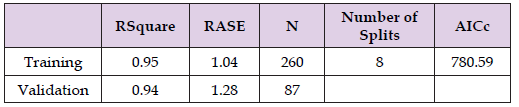
Open access clinical and medical journal |BJSTR

Neuron Regeneration After Intracerebral Haemorrhage by Transplantation of the Oligodendrocyte Progenitors Derived from Neural Stem Cells

Introduction

Spontaneous intracerebral haemorrhage (ICH) is a catastrophic disease with a 30-50% mortality, and survivors are often live with long-term neurological deficits [1]. After blood vessels corrupted, blood clot formation could cause the occurrence of brain edema [2]. The hematoma mass effect and tissue disruption result in primary damage, while inflammatory reaction and erythrocyte lysis cause secondary damage [3]. Although it is difficult to reduce the primary injury, treatments could help to suppress the secondary brain damage. For the current treatment methods, no effective treatment has been used on this devastating disease. Some studies indicated that a better understanding of the white matter injury may lead to a new way for treating ICH [4]. White matter contains axons, myelin sheaths that surround them, as well as oligodendrocytes which produce myelin [5]. Integrated axons are important for central nervous system communication, and large abundant of afferent and efferent pathways gathered in white matter. In human, around 80% of ICH patients were reported with white-matter damage, due to its essential role, even small lesion in white matter may lead to the impairment of neuronal function [6]. It has been known that oligodendrocytes regulate the neuronal function recovery and white matter injury [7]. In the central nerves system (CNS), oligodendrocytes originate from oligodendrocyte progenitor cells (OPCs), which derived from neuroepithelial progenitor cells (NPCs) in the embryonic stage [8]. OPCs secrete a series of growth factor such as transforming growth factor (TGF) to stimulate endothelial cells to express tight junction protein [9]. In addition, OPCs also secrete growth factors that promote the proliferation of endothelial cells and enter regions that lack of oxygen [4].

Evidence showed that adult human brain contains NSCs/NPCs in the periventricular area provides a promising approach to the CNS regeneration after injury by using grafted NSCs through the activation of endogenous regenerative capacity of damaged cells [10]. However, after CNS damage, reactive endogenous astrocytes form a glial scar and are considered to be detrimental to the axonal regeneration [11,12]. Previous studies have found that transplanted NSCs give rise almost exclusively to astrocytes and to relatively few oligodendrocytes and neurons [13,14]. Suppression of astroglial differentiation could effectively reduce the graft-induced aberrant axonal sprouting and promote further sensory and motor recovery [15]. We hypothesized that OPCs transplantation could promote the neuron regeneration after ICH in mice. After transplanting the OPCs into the experimental ICH animals, the correlation between functional and histological recovery were assessed for the control groups and the OPC-treated groups, and our results suggested that OPCs transplantation could help the recovery of ICH mice and promoted the neuron regeneration.

Materials and Methods

Immortalization of NSCs

The human neural stem cells (Gibco, N7800-100) were immortalized by transfecting TERT and BMI1 gene which carry GFP gene (SBI, CD513B-1). Firstly, we cultured the neural stem cells with EGF and bFGF in knockout DMEM basic medium in suspension and replaced with new medium every 2 days, thus then the cells will be passaged every 4 days. Plate the neural stem cells in poly-L-lysine and laminin coated dishes for two days, and add lentivirus containing TERT and BMI1 genes into the attachment culture NSCs. Large numbers of GFP positive cells grow rapidly as the cell proliferation.

Neuron, Astrocyte, and Oligodendrocyte Differentiation from NSCs

For neuron differentiation, the neural stem cells were cultured in Neuron induction medium-1 containing Neurobasal Medium and DMEM/F12 (1:1) supplemented with N2, B27, 10 ng/ml BDNF, and 200 mM ascorbic acid. Four days later, the culture medium was replaced with Neuron induction medium-2 containing Neurobasal Medium and DMEM/F12 (3:1) supplemented with 0.5% N2, B27, 20 ng/ ml BDNF, and 200 mM ascorbic acid. 7 days later, the cells were collected and stained with anti-III tubulin antibody. For astrocyte differentiation, the neural stem cells were cultured in Astrocyte induction medium containing neurobasal medium, 10% FBS, 1× NEAA and 2 mM L-glutamine. 7 days later, the cells will be collected and stained with anti-GFAP antibody.

For OPC differentiation from NSCs, the neural stem cells were differentiated into OPCs with OPC induction medium containing DMEM/ F12 supplemented with 20 ng/ml FGF2, 20 ng/ml PDGF-AA and 100 ng/ml SHH for 5 days. We used the Oligodendrocyte induction medium containing neural base medium supplemented with 0.4 ng/ml T3, 200 ng/ml SHH, 100 ng/ml noggin, 10 μM dibutyryl cyclic-AMP sodium salt, 100 ng/ml IGF-1 and 10 ng/ml NT3, to culture the OPCs for generating pre-oligodendrocytes and mature oligodendrocytes.

Animal Preparation and ICH Model Establishment

Our animal experiment protocols were approved by the Animal Ethics Committee of Shenzhen Second People’s Hospital. A total of 60 adult male C57/BL6 (20-25g) mice were included in this study. The mouse was anesthetized with intraperitoneal administration of a mixed solution containing 50 mg/kg ketamine and 5 mg/kg xylazine and was placed in a prone position on a stereotaxic frame, then an incision was made over the scalp. A borehole was drilled 2.5 mm lateral to bregma, and 0.5 μl of the collagenase solution (0.075 Units, Sigma, C5138) was injected 3 mm deep into the mouse brain by using a 26-gauge Hamilton syringe. The syringe was left in place for 10 minutes to prevent back-leakage before being withdrawn, than withdraw needle at a rate of 1 mm/min.

Cell Transplantation

7 days after ICH surgery, mice were anesthetized using same methods of modeling and fixed on stereotaxic frame. Two boreholes were drilled anteriorly and posteriorly with 1mm distance to the ICH borehole respectively. 25,000 OPCs in 1μl of saline were injected 3 mm into the brain at each borehole. The whole procedure was limited to 10 minutes, then wait for 5 minutes to withdraw the syringe at a rate of 1mm/min.

Behavior Test

Mice gaits were recorded and analyzed by the Cat-Walk XT system (Noldus Information Technology, Wageningen, Netherlands). Cat Walk XT contained hardware part of a glass walkway plate with green light, an inverted high-speed video camera, and a software package for quantitative gait assessment. The footprint area was illuminated and scattered when mice walked through the glass plate and captured by the inverted camera.

Mice were trained to walk fluently through the glass walkway for 5 days before collagenase injection. Each mouse needed to complete at least 3 runs. A typical run contained at least 3 step cycles, and we calculated the average data of the parameters from all step cycles. Western-Blot Analysis

The mice were overdose anesthetized and sacrificed via decapitation. Right striatum was collected and well mixed with sufficient volume of cold RIPA lysis buffer that containing protease inhibitor cocktail. Protein concentration was then measured with a BCA kit after the extraction. Proteins (10μg-20μg) were later loaded into 8 to 10% SDS-polyacrylamide gel. The proteins were then transferred to nitrate cellulose membrane, and blocking by 5% non-fat milk, further incubated at 4°C overnight with primary antibody. Washed membrane with TBST for three times with 10 mins each, and then incubated with secondary antibodies for another 2 h at room temperature, finally detected signals by using enhanced chemiluminescent (Thermos Fisher Scientific).

Statistical Analysis

All data were presented as mean ± SEM. Statistical analyses were conducted by IBM SPSS 22.0 software. For behavior and molecular tests, statistical analyses were processed by one-way ANOVA followed by post hoc tests for between-group comparisons. P < 0.05 after Tukey adjustment for multiple comparisons was considered statistically significant.

Results

Immortalization of Human Neural Stem Cells

Previous literature described that TERT and BMI1 could be used to immortalize kinds of stem cells [16]. We immortalized the human neural stem cells by transfecting TERT and BMI1 gene through lentivirus which carry GFP gene (Figure 1A). The immortalized neural stem cells sustained a strong proliferation ability and expressed GFP protein (Figure 1B). We use a TRAP assay kit (Millipore Catalog No. S7700) to detect the telomerase activity of the immortalized neural stem cells. In Figure 1C, we know that the telomerase activity of immortalized neural stem cells was significantly increased, and the 293T cell is positive control, while the 293T cell heat treatment is negative control for the activity loss when heat. The two templates are positive control, and the two immortalized NSCs groups have strong signal, while the two groups of heat treatment for the immortalized NSCs are negative (Figure 1C). These results suggested that the neural stem cells were immortalized successfully.

Figure 1

biomedres-openaccess-journal-bjstr

Oligodendrocyte Progenitor Cells Were Produced from Human Neural Stem Cells

Neural stem cells could differentiate to several kinds of neural cell types with conditional culture medium [17]. We have a try to differentiate the neural stem cells to neurons, astrocytes, and oligodendrocytes. The neuron differentiation protocols were described in a Figure 2A. We used neuron induction medium to generate neurons from the neural stem cells, and the neuron marker, a-III tubulin, was highly expressed in the differentiated neurons (Figure 2B). Meanwhile, we generated astrocytes with conditional culture medium, and the astrocytes could express the astrocyte specific marker, GFAP (Figures 2C & 2D). We also developed a OPC differentiation protocol to generate OPCs (Figure 3A). We can see the OPC morphology after differentiation (Figure 3B), and a series of OPC markers were expressed in the induced OPCs (Figure 3C).

Figure 2

biomedres-openaccess-journal-bjstr

Figure 3

biomedres-openaccess-journal-bjstr

Locomotors Function Detection for Ich Mice Transplanted with Opcs by Catwalk System

To study the influence of OPCs transplantation on neuron regeneration, we have a try to establish mouse ICH model. Bacteria collagenase was injected in the right side of striatum, and the hematoma formation will be in the right striatum. Three days after ICH surgery, the mice were given an overdose of phenobarbital and perfused. Brain corona dissection showed the different extents of hemorrhage from anterior-posterior coordinates in the right striatum, and it confirmed that we established the ICH mouse model successfully (Figure 4A). Cat Walk gait analysis system is a highly sensitive tool for assessing gait and locomotion [18]. The mouse traverses a glass plate voluntarily (towards a goal box), and its footprints are captured (Figure 4B). CatWalk visualizes the prints and calculates data related to print dimensions and the relationship of the time and distance relationships between footfalls. The parameters we selected to assess the mice locomotor function are Stand time, Swing speed, and Step cycle. Swing Speed is calculated for the normal groups and the OPCs treated groups, and the statistic result showed a similar gait style in the mice treated with OPCs to the Normal group (Figure 4C). Stand time describes the duration of contact of an individual paw with the glass plate. The bar chart illustrates ICH mice treated with OPCs performed the similar locomotor function compared to the normal mice, while ICH mice take a longer duration of stand (Figure 4D). Step cycle describes the duration of two consecutive initial steps by the same paw, and the OPC treated mice take a short time, closed to the level of normal mice (Figure 4E). According to the gait analysis results, we concluded that OPC transplantation can improve the mice locomotor function after ICH.

Figure 4

biomedres-openaccess-journal-bjstr

Transplantation of Opcs Promoted the Expression of Neuron Markers

Striatum Neurofilament-L (NF-L), Neurofilament-M (NF-M), and Microtubule associated protein 2 (MAP2) was investigated by Western blot analysis after behavior test, in order to evaluate the difference on neuron regeneration introduced by the different treatments for the ICH mice. Neurofilaments is necessary for the maturation of regenerating myelinated axons in mice [19]. For evaluating the axon regeneration of ICH model injected with OPCs, we assessed the expression of neurofilament in the corticospinal tract tissue of Pons for the surgical control (ICH group), ICH mice injected with medium (medium groups), and ICH mice injected with OPCs (OPC groups). We found that the expression of neurofilament-L was increased in the OPC-treated groups compared to the ICH groups (Figures 5A & 5B). Microtubule-associated protein 2 (MAP2) is a sensitive marker of seizure- related brain damage [20]. In our result, OPCs transplantation induced the expression of MAP2 compared to the ICH groups (Figures 5C & 5D). Thus, then we concluded that OPCs transplantation could promote the neuron regeneration of ICH mice.

Figure 5

biomedres-openaccess-journal-bjstr

Discussion

Grafting of neural stem cells into experimental ICH in mice has been shown to promote neurological recovery, [13,14] and the mechanism could be that of neuro-regeneration. [21] More recently, oligodendrocyte progenitor cells (OPCs) can be reprogrammed from fibroblasts, using iPSC (induced pluripotent stem cell) technology, by over-expression of several transcription factors, such as Sox 10, Oligo2 and Zfp536. [22,23] These OPCs derived from human iPSC has been shown to promote myelination and axon regeneration in a murine model of congenital hypomyelination. [24] This concept of utilizing a specialized OPCs generated from neural stem cells (NSCs) has inspired us to develop the topical OPCs treatment for murine ICH. In our study, we have generated OPCs from immortalized human NSCs and demonstrated that their transplantation in murine ICH improves neurological outcome neuro-regeneration, although the underlying mechanism remains unclear. MicroRNAs networks are dynamically regulated in developing oligodendrocytes, and microRNAs modulates oligodendrocyte progenitor cell development and differentiation. [25] During the induction of OPCs from NSCs, micro RNAsmay play essential roles in regulating the expression of OPC marker and mediate the proliferation and survival, and the regulation mechanism is worth further exploring. In the current study, we have established some preliminary data that the murine ICH model treated with NSCs-induced OPCs could promote axon regeneration after ICH as reflected by the statistically increased levels of Neurofilament-L (NF-L), MAP2, and an improved performance in three Catwalk parameters.

Ethical Approval

The procedures involving animals and their care were conducted under the approval of the Animal Ethics Committee of the Second People’s Hospital of Shenzhen.

Conflicts of Interest

There are no conflicts of interest.

Declarations of Interest

NO.

Ethics Declarations

The procedures involving animals and their care were conducted under the approval of the Animal Ethics Committee of the Second People’s Hospital of Shenzhen.

Follow us on: Biomedical Journal Impact Factor: https://biomedres.us

https://scholar.google.com/citations?user=OFtUOZYAAAAJ&hl=en
https://www.base-search.net/Search/Results?lookfor=Biomed+Journal+of+Scientific+%26+Technical+Research&l=en&refid=dcsuggesten
https://citefactor.org/journal/2574-1241/biomedical-journal-of-scientific-technical-research
https://ideas.repec.org/s/abf/journl.html
https://econpapers.repec.org/article/abfjournl/
https://www.ncbi.nlm.nih.gov/nlmcatalog/101723284
https://bjstr.org/
https://bjstr.org/about.php
https://biomedres.us/reprints.php
https://biomedres.net/submit-manuscript.php

Journals on Medical Casereports |BJSTR

Novel Spectrophotometric Determination of Isopropamide Iodide in Pharmaceutical Formulations and Environmental Wastewater Samples: Application to Content Uniformity Testing

Introduction

Isopropamide iodide, (3-carbamoyl-3,3- diphenylpropyl) di- isopropylmethyl ammonium iodide (Figure 1), is a quaternary ammonium antimuscarinic with peripheral effects similar to those of atropine. It is used as an adjunct in the treatment of peptic ulcer disease, in the relief of gastrointestinal and urinary-tract disorders associated with smooth muscle spasm [1]. The literature survey reveals that several methods have been reported for determination of isopropamide iodide in pure form and in pharmaceutical formulations. Official method includes potantiometric titration for pure form and Uv spectrophotometric method for tablets [2]. The spectrophotometric method include second derivative and ion-pair complexation reaction [3-8]. Other methods reported for determination of isopropamide iodide like high-performance liquid chromatography [9], and with other drugs by LC methods [10-12]. Spectrophotometry is by far the instrumental technique of choice in the laboratories of underdeveloped and developing nations for the quantification of drugs, owing mainly to its simplicity, high sensitivity and selectivity and often demanding low-cost equipment. The objective of the present work is to develop and validate a simple, sensitive and economically viable spectrophotometric method for the determination of isopropamide iodide in bulk and in their pharmaceutical formulations. The proposed method was based on the extraction of isopropamide iodide into chloroform as blue colored ion pair with bromophenol blue. The blue colored ion pair was quantitated spectrophotometrically at 600 nm. The proposed method has been validated for linearity, sensitivity, precision, accuracy, and selectivity.

Figure 1

biomedres-openaccess-journal-bjstr

Apparatus

Spectro-scan 50 UV- visible (double beam) spectrophotometer with 1.0 cm quartz cells was used for absorption measurements.

Reagents

All chemical used were of analytical or pharmaceutical grade and distilled water was used throughout. Standard materials and pharmaceutical preparations (isopropamide iodide tablets) were provided from AL-Hokamaa company for pharmaceutical industries (HPI) Mosul-Iraq.

Isopropamide Iodide Solution (0 .01%)

This solution was prepared by dissolving 0.01 gm of isopropamide iodide in 100 ml distilled water in a volumetric flask.

Buffer Solution (pH10)

This solution contains 50 ml of 0.05 M sodium bicarbonate and 10.7 ml of 0.1M sodium hydroxide, it was then diluted to100 ml by distilled water in a volumetric flask [13].

Bromo Phenol Blue (0.1%) [BDH, UK]

This solution was prepared by dissolving 0.1 gm of Bromophenol blue in 100 ml distilled water in a volumetric flask.

Recommended Procedure

Aliquots of standard drug solution of isopropamide iodide 0.5- 5 ml were taken and transferred into a series of 50 ml of separating funnels. To each funnel 3.0 ml of buffer solution(pH10) and 2 ml of 0.1% Bromophenol blue was added. Reaction mixture was shaken gently for 5 min. Then 10 ml of chloroform was added to each of them. The contents are shaken thoroughly for 5 min and allowed to stand, so as to separate the aqueous and chloroform layer. Colored chloroform layer was separated out in dry test tubes containing anhydrous sodium sulfate. The absorbance was measured at 600 nm against reagent blank.

Procedure for Pharmaceutical Preparations (tablets)

Weight and powder 10 tablets. Dissolve a quantity of the powdered tablets containing 0.01 gm of isopropamide iodide in about 50ml distilled water and mixed for 20 mint and then filtered. The filtrate was mad up to 100ml with distilled water. Treat 3ml of this solution as mentioned under recommended procedure.

Procedure for Industrial Wastewater

To demonstrate the practical applicability of the proposed method, industrial waste water sample from ALhokamaa company for pharmaceutical industries (HPI) Mosul-Iraq were analyzed by spiked with the concentrations (5, 10, 15 μg.ml-1) of isopropamide iodide and aliquot of this solution was treated as described above for recommended procedures.

Results and Discussion

The ion-pair complex is a special form of molecular complex resulting from two oppositely charged ions extractable into organic solvents from aqueous phase at suitable ph. In the recent years ion pair extraction spectrophotometry has received substantial significance for the quantification of many pharmaceutical compounds [14-17]. A new method has been developed for the spectrophotometric determination of isopropamide iodide. The method was based on the ion transfer complex formed by BPB, an acidic dye, and isopropamide iodide that have basic nitrogen as electron donor to yield ion-pair salts which form color compound extractable from the aqueous solution to chloroform organic phase. The absorbance spectra and proposed reaction mechanism are shown in Figure 1. The reaction variables were optimized by varying each variable while keeping others constant for obtaining maximum absorbance. The reaction was found to be quantitative in alkaline medium (pH 10). It was found that 3 ml of pH10 solution give high sensitivity and this amount has been used for subsequent experiments. The effect of the amount of BPB on the absorbance was investigated. A maximum and constant absorbance was found with 1 to 5 ml of 0.1% BPB solution and 2.0 ml has been used for subsequent experiments. The effect of extracting solvent different organic solvents (chloroform, benzene, toluene, cyclohexane, and carbon tetrachloride have been tried in order to investigate their ability to extract the isopropamide iodide -BPB ion pair complex.

Among the various organic solvents tried, chloroform was found to be the most appropriate solvent. Hence it was chosen for extraction of the isopropamide iodide -BPB ion pair complex from the aqueous phase. The color reaction occurred at room temperature immediately and remained stable for at least 6 h., and a reaction time of 5 min was selected for reproducible results. Under the experimental conditions described, Beer ‘s law is obeyed over the concentration range 2-20 μg /ml (Figure 2). Calibration curve was prepared from absorbance values so obtained.

Figure 2

biomedres-openaccess-journal-bjstr

Accuracy and Precision

The accuracy and precision of the method was established by analyzing the pure drug solution at three different concentrations, each determination being repeated six times. The relative error (%) and relative standard deviation values are summarized in Table 1. From Table 1 the values of standard deviation were satisfactory and the recovery studies were close to 100%. The RSD% value is less than 1.5 indicative of accuracy of the method.

Table 1: Accuracy and precision of the proposed method.

biomedres-openaccess-journal-bjstr

Note: a: Mean of six determinations.

Analytical Application

The proposed method was satisfactorily applied to the determination of isopropamide iodide in its pharmaceutical preparations tablets and wastewater samples, the results of the assay of the pharmaceutical preparations revels that there is close agreement between the results obtained by the proposed method and the lable claim (Table 2), and the results of water samples Table 3 show that the recovery values obtained were closed to 100%.

Table 2: Determination of isopropamide iodide in pharmaceutical formulations.

biomedres-openaccess-journal-bjstr

Note: *mean value of ten determinations.

Table 3: Determination of isopropamide iodide in wastewater samples.

biomedres-openaccess-journal-bjstr

Note: *mean value of ten determinations.

Application of the Method to Content Uniformity [18,19]

The proposed method proved to be suitable for the content uniformity test, where a great number of assays on individual tablets are required. Data presented in Table 4 indicate that the proposed method can accurately and precisely quantitate isopropamide iodide in its commercially available tablets. The mean percentage (with RSD) of the labeled claim found in ten tablets was (0.518%) which fall within the content uniformity limits specified by the USP 30 [20].

Table 4: Content uniformity testing of isopropamide iodide tablets using the proposed method.

biomedres-openaccess-journal-bjstr

Conclusion

The developed method is found to be highly sensitive, accurate, simple, precise and economical, and can be used for routine quality control analysis of isopropamide iodide in pure form, bulk, pharmaceutical formulations and environmental wastewater samples.

Acknowledgments

The first author wishes to express gratitude to Al-Hokama company for pharmaceutical industry (HPI) (Nineveh – Iraq.) for providing gift samples of isopropamide iodide standard material and for permission and facilities to carry out the research work.

Follow us on: Biomedical Journal Impact Factor: https://biomedres.us

https://scholar.google.com/citations?user=OFtUOZYAAAAJ&hl=en
https://www.base-search.net/Search/Results?lookfor=Biomed+Journal+of+Scientific+%26+Technical+Research&l=en&refid=dcsuggesten
https://citefactor.org/journal/2574-1241/biomedical-journal-of-scientific-technical-research
https://ideas.repec.org/s/abf/journl.html
https://econpapers.repec.org/article/abfjournl/
https://www.ncbi.nlm.nih.gov/nlmcatalog/101723284
https://bjstr.org/
https://bjstr.org/about.php
https://biomedres.us/reprints.php
https://biomedres.net/submit-manuscript.php
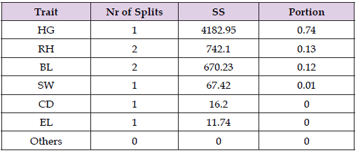
Open Access journals on surgery |BJSTR

Prevalence and Outcomes of Acute Respiratory Tract Infections Among Infants in a Nigerian Tertiary Health Care Institution

Introduction

Respiratory tract infections (RTIs) refer to any infectious illness affecting the upper, lower, or both respiratory tracts. Upper respiratory tract infections (URTIs) encompass conditions like laryngitis, the common cold, pharyngitis, acute rhinitis, tonsillitis, acute rhinosinusitis, and otitis media [1]. Lower respiratory tract infections (LRTIs) include acute bronchiolitis, bronchitis, tracheitis, and pneumonia. In 2017, approximately 5.4 million children under the age of five died globally, with about half of these deaths occurring in sub-Saharan Africa [2]. Acute Respiratory Tract infections were the leading cause of mortality among this demographic [2].

Acute respiratory infection refers to an infectious condition that can disrupt normal breathing patterns. It may specifically target either the upper respiratory tract, encompassing the area from the sinuses to the vocal cords, or the lower respiratory system, extending from the vocal cords to the lungs. This ailment poses significant risks, especially to children, older individuals, and those with compromised immune systems [3-6]. Symptoms vary depending on whether the upper or lower respiratory tract is affected, and can include congestion in the nasal sinuses or lungs, a runny nose, coughing, sore throat, body aches, and fatigue [3].

On a global scale, respiratory diseases stand as the leading cause of illness and death, particularly affecting infants and young children [4]. Pneumonia continues to represent a significant contributor to childhood mortality, resulting in nearly 1.3 million deaths annually, a substantial portion of which could be prevented. Paediatric tuberculosis accounts for as much as 20% of the total tuberculosis burden in countries with high incidence rates [5,7-9]. Despite advancements in modern medicine, Acute Respiratory Infections (ARIs) persist as the most prevalent type of infectious disease, even in many developed nations such as the USA. ARIs, collectively comprising a range of diseases and symptoms, rank among the primary causes of morbidity and mortality in pediatric populations across sub-Saharan Africa, where more than 10% of children succumb before their fifth birthday [10- 13]. In Nigeria alone, the World Health Organization (WHO) estimated that 868,000 children under the age of five died from pneumonia in 2010. With an annual toll surpassing 140,000 children, Nigeria holds the highest ARI-related childhood mortality rate in Africa and the second-highest globally [14,15].

ARIs are affected by socio-demographic and socio-cultural risk factors, which can be modified with simple strategies. It is recommended that control programs for ARIs should be multifaceted with a strong political will [16].

Acute respiratory tract infection in children remains a killer disease in both low and middle-income countries and high-income countries. Infants are highly susceptible to these infections because of their low immune system and dependence on adults who may end up transmitting the infectious agent to them [17]. In the present study, we aimed to assess the prevalence and outcome of ARIs among infants in a Nigerian tertiary health care institution.

Methods

Study Area

The study was done in the Children Emergency Room of Nnamdi Azikiwe University Teaching Hospital, Nnewi, south-east Nigeria.

Study Design

This study is a 2-year retrospective cross sectional study of the prevalence and outcome of respiratory tract infection in infants.

Study Population

The study included infants, who visited the children’s emergency room in NAUTH, Nnewi to access care from January 2021 through December 2022.

Inclusion Criteria

Children, less than a year were managed for ARIs in CHER, NAUTH, Nnewi.

Exclusion Criteria

Children greater than one year. Children less than a year who accessed care in the children’s emergency room, NAUTH Nnewi, whose data could not be obtained from the Nursing register.

Sample Size Determination

This was calculated using the Cochrane formula, which is given as;

Where n is the minimum sample size
Z is standard normal deviate = 1.96
P is the population proportion = 9.7 percent.47
q=(1-p); 100-9.7= 90.3 percent
d is the degree of precision = 0.05

Sampling Technique

A convenience sampling technique was used to obtain data of infants who met the inclusion criteria from the nursing register, and from the records department of NAUTH, Nnewi.

Study Instruments

A proforma was designed to record the patients’ profile which included: age, sex, diagnosis, year, season of diagnosis, and outcome of the diagnosis.

Data Analysis

All available data were analyzed using a statistical package for social sciences (SPSS Version 25.0). Variables analyzed included age, (which is continuous data), sex, season, and year of diagnosis of the disease. Categorical variables were analyzed using percentages and proportions while continuous variables were analyzed using mean and standard deviation and the association between categorical variables was analyzed using the chi-square test. The level of significance was set at p< 0.005.

Ethical Considerations

This research work was done with approval from the Nnamdi Azikiwe University Ethics Committee with approval number of NAU/ HREC/28/02/12/2023/05. Permission was also gotten from Records Department of Nnamdi Azilkiwe University Teaching Hospital, Nnewi. Information obtained from case notes was used only for this project and the patient’s identity was kept strictly confidential.

Results

The data used for analysis was obtained from records of patients admitted at CHER. The mean age and SD were calculated as 5.79 ± 3.51 months. The majority were under 4 months (42.3%). There were more males (55.6%) than females (44.4%) as shown in Table 1. Table 2 shows the prevalence of acute respiratory infection. A total of six hundred and ninety four infants were admitted during the period of study. Among these, one hundred and forty-two infants had acute respiratory infections. The prevalence of acute respiratory infection was therefore determined as 20.5%. Table 3 shows the distribution of cases according to year and month, while Figure 1 shows the graphical presentation of Case distribution according to month and year. A higher occurrence of ARI was recorded in the year 2022 (70.4%) when compared to that of 2021 (29.6%). In 2022, the prevalence of ARI was higher in November (27.0%) and December (24.0). But in 2021, ARI was recorded higher in March (23.8%) and June (26.2%). Table 4 shows the distribution of acute respiratory tract infection according to specific diagnosis. Bronchopneumonia ranked highest amongst ARIs seen in these children while tonsillitis was the least. The majority of the infants were discharged (75.4%). It was noted that 4.2% were discharged against medical advice.

Figure 1

biomedres-openaccess-journal-bjstr

Table 1: Socio-demographic status of the respondents.

biomedres-openaccess-journal-bjstr

Table 2: Prevalence of acute respiratory infection.

biomedres-openaccess-journal-bjstr

Table 3: Distribution of cases according to year and month.

biomedres-openaccess-journal-bjstr

Table 4: Distribution of acute respiratory tract infection according to specific diagnosis.

biomedres-openaccess-journal-bjstr

Mortality was seen in 20.4% of cases. Figure 2 shows the association of outcome with some variables while Table 5 shows the association of treatment outcome with some variables. The relationship between treatment outcome and variables (age, sex, gender and year) is presented in Table 5. The outcome of treatment showed a significant association with age (X2 = 13.836; p = 0.006). The number of patients discharged was significantly higher among the 0-4 months age category. Mortality was recorded highest among the 9-12 months age group. Treatment outcome showed no significant association with sex and year (p > 0.05 in these cases). Table 6 shows the distribution according to comorbidities and causes of Mortality. Sepsis with Bronchopneumonia was the highest cause of mortality among infants with ARIs, contributing about 75.8%. There is also a significant relationship between mortality and protein energy malnutrition.

Figure 2

biomedres-openaccess-journal-bjstr

Table 5: Association of treatment outcome with some variables.

biomedres-openaccess-journal-bjstr

Table 6: Distribution according to Comorbidities and causes of Mortality.

biomedres-openaccess-journal-bjstr

Discussion

The aim of this study was to determine how common acute respiratory infections (ARIs) are among infants at a tertiary healthcare facility and what outcomes result from them. Analysis of the data revealed that out of 694 infants admitted during the study period, 142 were diagnosed with ARIs, indicating a prevalence rate of 20.5%. This underscores the substantial burden of ARIs within this age group, emphasizing the urgency for prompt interventions to reduce the associated illness and mortality rates. The prevalence identified in this study (20.5%) aligns closely with findings from Memeli et al, whose study on preschool children showed a prevalence of 24.9% [21]. Similarly, it corresponds well with Eberechukwu et al’s study at the University of Port-Harcourt, Nigeria, which documented a prevalence of 27.1% [18]. However, Abayneh et al and Kumar et al, in India, recorded higher prevalence rates of 40.7% [19] and 59.1% [20], respectively. These variations in prevalence may stem from differences in sampling techniques; while our study employed a retrospective approach, Kumar et al conducted a cross-sectional prospective study [21]. Additionally, the significant overcrowding in India, recognized as a risk factor for acute respiratory infections (ARIs), likely contributes to the elevated prevalence observed in their research [21]. Conversely, Ofaeli et al’s study at NAUTH, Nnewi, Nigeria reported a lower incidence of 14.45%, possibly influenced by their inclusion of patients spanning ages 0-17 years, making it challenging to determine the exact prevalence among infants [22].

Bronchopneumonia stands out as the primary culprit behind acute respiratory infections (ARIs) in infants, constituting 83.5% of cases, with bronchiolitis trailing behind at 9.5%. This finding echoes Rashmi et al’s findings, where bronchopneumonia accounted for 72.5% of ARIs among children [23]. The heightened prevalence of ARIs raises significant public health concerns, as it imposes strains on healthcare systems, depletes medical resources, and raises the risk of potential outbreaks. Given the ease with which ARIs can spread, particularly among vulnerable populations like children, effective prevention and management strategies are essential for curbing hospitalizations, averting complications, and mitigating overall healthcare expenditures. In our investigation, we delved into the impact of monthly and seasonal fluctuations on the prevalence of acute respiratory infections (ARIs) among infants. Out of the total 142 ARI cases examined, 81 occurred notably between November and March (dry/ harmattan season), while 61 were recorded between April and October (rainy season).

Noteworthy increases in ARI incidence were observed in June (19%), November (32%), and December (28%), coinciding with the onset of the rainy and dry seasons. This phenomenon could be attributed to the observed weakening of the immune system during seasonal transitions. These findings align with Abayneh et al’s study, which similarly highlighted peaks in ARI prevalence among under- fives in June (23.8%), November (15.9%), and December (14.7%) [24]. The public health implication of these trends is significant: during the onset of the rainy season, increased indoor activities may facilitate disease transmission, while the early dry season’s dry air may exacerbate respiratory tract irritation, heightening susceptibility to severe forms of ARIs. The study observed a slightly higher prevalence of acute respiratory infections (ARIs) among males (55.6%) compared to females (44.4%), consistent with findings by Ujunwa et al, who reported a male prevalence of 51% and female prevalence of 49% [9]. This trend also aligns with research conducted by Gabriel et al, which found a prevalence of 50.2% in males and 49.8% in females [24].

This disparity may stem from the greater activity levels of male infants, leading to increased environmental exposure. The slight male predominance in ARI prevalence holds potential public health implications, suggesting possible physiological differences between the sexes in susceptibility to respiratory infections. Tailored public health strategies might be necessary to address these differences, including enhancing healthcare access for male infants and educating caregivers on infection prevention. Further research is warranted to delve deeper into the underlying factors and to develop effective interventions. The study also examined the discharge outcomes of acute respiratory infections (ARIs) among infants. Out of the total 142 cases analyzed, 75% were discharged, while mortality accounted for 20.4%, and 4% were discharged against medical advice. This finding contrasts with a similar study conducted by Alok et al, which reported a lower mortality rate of 9% [25].

The variance could be attributed to the inclusion of both in-patients and outpatients in their study, whereas our research focused solely on in-patients in the emergency room, where mortality rates tend to be higher. The elevated mortality rate from ARIs in infants raises significant public health concerns due to its impact on a vulnerable population, imposing substantial economic burdens and potentially leading to long-term health implications. Implementing preventive measures and interventions is imperative for reducing this mortality rate and promoting the overall health and well-being of infants. The primary cause of mortality observed in this study was sepsis accompanied by bronchopneumonia, accounting for 75.3% of cases, followed by sepsis with bronchiolitis and acute respiratory infections (ARIs) with protein energy malnutrition, each contributing to 10.3% of mortality cases. Consequently, sepsis alone was responsible for the mortality of 86.6% of infants admitted to the Children’s emergency ward. These findings align with research conducted by Trenton et al, which similarly found that sepsis alone accounted for approximately 80% of mortality cases [26].

Sepsis presents a critical public health concern due to its potential to lead to high mortality rates among infants. Timely and appropriate medical care, along with preventive measures, are essential to address this issue and safeguard the well-being of infants. The major strength of this study lies in the currency of its findings and its exclusive focus on infant data. However, there are several limitations to our study. First, due to the small sample size, we cannot conclude significant differences in the association between various sociodemographic variables and cute respiratory infections. Due to the retrospective cross-sectional design, we lacked information regarding causality in relation to mortality [27-31].

Conclusion

Acute respiratory infections (ARIs) pose a significant burden in our community, evident by a prevalence of 20.5% among infants admitted to a tertiary healthcare facility. This raises concerns and underscores the need for additional interventions to alleviate the impact of this disease on the population. Seasonal variations also influence the prevalence of ARIs, with notable increases observed during the peak of both the rainy and dry seasons. Males exhibit a slightly higher susceptibility to ARIs compared to females. Bronchopneumonia remains the most common cause of ARIs among infants in our study, with most mortality cases complicated by sepsis. Given that respiratory tract infections in children are highly preventable, it is essential to implement measures to mitigate the rising incidence and associated complications. Recommendations include ongoing training and retraining of healthcare personnel by hospital management to recognize early signs of sepsis, aiming to prevent mortality. Additionally, healthcare staff should prioritize the early diagnosis of congenital anomalies and promptly refer affected infants to specialists for appropriate management. Ensuring timely diagnosis and effective management of respiratory illnesses is crucial to prevent the development of life-threatening complications.

Conflict of Interest Statement

The authors report no conflicts of interest concerning this work.

Ethical Statement

Ethical approval was obtained from the Nnamdi Azikiwe University Ethics Committee with approval number of NAU/ HREC/2S/02/12/2023/05. This study was conducted in accordance with the Declaration of Helsinki.

Acknowledgments

We are grateful to the Medical record department and department of Paediatrics for their cooperation during this study.

Author Contributions

Design: EAE, AUW, ACV and ENU. Analyses: AUW and STE. Writing: CIO, AAO, THE, OCE, NGU, and ESE. Supervision: EAE, GUE and JEM. CIO, AAO, THE, OCE, NGU, AUW, ACV, STE, CTE, KCN and GUE contributed to the interpretation of the analyses and writing of the manuscript. All authors approved the final manuscript.

Follow us on: Biomedical Journal Impact Factor: https://biomedres.us

https://scholar.google.com/citations?user=OFtUOZYAAAAJ&hl=en
https://www.base-search.net/Search/Results?lookfor=Biomed+Journal+of+Scientific+%26+Technical+Research&l=en&refid=dcsuggesten
https://citefactor.org/journal/2574-1241/biomedical-journal-of-scientific-technical-research
https://ideas.repec.org/s/abf/journl.html
https://econpapers.repec.org/article/abfjournl/
https://www.ncbi.nlm.nih.gov/nlmcatalog/101723284
https://bjstr.org/
https://bjstr.org/about.php
https://biomedres.us/reprints.php
https://biomedres.net/submit-manuscript.php

Journals on Biomedical Imaging |BJSTR

An Evaluation of the Interventions for Management of Breast Cancer Among Women in Nigeria: A Narrative Review

Introduction

Around the world, breast cancer is recognized as the single biggest cause of cancer deaths in women [1]. Even more so, there is an established correlation between the breast cancer outcomes in women from richer countries and those from poorer countries, referred to as ‘lower and middle-income countries’ (LMICs), with more than half of the breast cancer cases and two-third of the deaths occurring in this LMICs [1-3]. According to Agha et al, there were 266,120 new cases of breast cancer among women in the United States and 41,400 breast cancer deaths reported by the American Cancer Society in 2018 [4]. However, by comparison, Chidebe et al reported that new cancer cases among women in Nigeria per year stood at 102,000 with 72,000 deaths in the same period [5]. In LMICs like Nigeria, the burden of breast cancer is higher because of late presentation of most cases and the reasons for this include misconceptions about the disease from personal, cultural, or religious ideologies, fear of breast removal, poverty [6]. In addition, other social determinants of late presentation of breast cancer in women of sub-Saharan African countries such as Nigeria include poor access to healthcare, lower levels of education, and lack of health insurance [7]. There are several risk factors associated with breast cancer and they can be categorized as demographic factors such as gender and age; reproductive factors that include late menopause; hormonal factors such as use of oral contraceptives; hereditary factors which include genetics and history of breast cancer within family [8].

Cancer Research UK added a few more risk factors such as being overweight, consumption of alcohol, smoking and being inactive. In addition, however, there are a few unpopular risk factors associated with breast cancer, the likes of air pollution, diabetes, night work, and socio-economic status [9]. The interventions for women with breast cancer in Nigeria can depend on the histological characteristics of the cancer such as the grade of the tumour using Notttingham Grading System, pathological diagnosis, as well as immunohistochemistry (IHC) [10]. Other factors that determine interventions are oestrogen and progesterone receptor status, human epidermal growth factor receptor II (HER 2) expression, triple negativity, etc. [8]. Interestingly, several interventions for breast cancer among women in Nigeria include surgical intervention; or systemic treatments like chemotherapy, targeted therapy, hormonal treatment, and in some rare cases, radiotherapy [7,10]. In addition to the already mentioned above, there are a few other factors that can determine the choice of these interventions for Nigerian women with breast cancer, and they include: availability of relevant skills (especially in case of surgery), cost and accessibility to the intervention (in cases of systemic therapy and radiotherapy), and compliance to therapy [8].

Breast cancer among Nigerian women is as unique as the available interventions, from late presentations to financial challenges, cultural, personal, and even marital doubts, as well as limited available professional know-how. Therefore, it will be very interesting to critically research the latest available interventions, how they have improved the grim public health outcomes of this breast cancer in Nigeria, and what options there are for improvement. The overall aim of this work is to critically evaluate the available interventions for breast cancer among women in Nigeria to assess their suitability, impacts, challenges, and how best they can be improved to increase chances of better outcomes and reduce breast cancer mortality.

Methods

Research Philosophy

The research was based on interpretivism, also known as the interpretivist, research philosophy. This philosophy consists of interpretation of elements of a study by the researcher, thereby incorporating a human element to the study [11,12].

Research Approach and Conceptual Research Design

This research was done using the mixed method study, which is a study method that merges both the qualitative and quantitative methods into one study so as achieve a wider, more holistic assessment of the research problem [13]. The primary aim of mixed study research method is to take advantage of the strengths and then cut down on the limitations of both quantitative and qualitative designs [14]. Interestingly, due to lingering effects of the Covid-19 pandemic, the research adopted a secondary or desktop research approach, whereby no new or raw data was collected in the process. According to Aven, secondary or desktop research is a research methodology that involves collating and analyzing already-existing bodies of information on the research topic, and therefore no form of experimentation is required [15]. This is opposed to empirical research that involves experimentation then gathering, analysis and interpretation of raw data from the field [16].

Literature Search Strategy

According to Aromatases and Rianto, constructing an effective search strategy is a significant but repetitive component of research, involving regular modifications and re-evaluation [17]. The basis of an excellent search strategy is in a review protocol which has succinct review questions and inclusion criteria [17]. An effective search strategy should exhibit sensitivity, which is, being able to pick up relevant information; as well as specificity, which is the ability to exclude irrelevant information [18]. The study would utilize a keyword search using research databases such as Medline, Summons, CINAHL, EMBASE, PubMed, Google Scholar, and Cochrane to identify appropriate articles. The categories of articles reviewed would include full text published articles and even abstracts, in so far as a minimum of one outcome of interest such as ‘interventions’, ‘breast cancer’, ‘women’, or ‘Nigeria’, were mentioned in the abstract. Afterwards, these search strings would be modified to incorporate combinations of these keywords such as ‘evaluation’ OR ‘assessment’ AND ‘management’ OR ‘treatment’ AND ‘breast carcinoma’ OR ‘breast malignancy’ AND ‘women’ OR ‘girls’ AND ‘Nigeria’ OR ‘West Africa’.

Therefore, with these search strings, articles that evaluated the interventions for breast cancer among women in Nigeria were identified. In addition to the electronic database searches, the search strategy would involve a manual review of the appropriate references and studies. Importantly, the electronic database searches would be limited to a twenty-year study period, which would span from April 2002 to April 2022, to easily distinguish articles with appropriate and upto- date information published within the stipulated interval. Moreover, other search strings such as ‘breast cancer policies’ OR ‘breast cancer statistics’ were used to identify grey literature which could be published from government website or other reputable organizations such as the World Health Organization, etc (Table 1).

Table 1: Search keywords.

biomedres-openaccess-journal-bjstr

Inclusion and Exclusion Criteria

In this research, the articles which were selected for the study included peer-reviewed, qualitative, and quantitative journals on the interventions for breast cancer among women in Nigeria or Africa. Conversely, the study excluded journal publications on breast cancer interventions for women outside of sub-Saharan Africa. In addition, any sub-Saharan African studies on breast cancer which did not include interventions or management were excluded. In the same vein, the government or WHO publications that explored management of breast cancer among women in Nigeria were considered. Notably, the study also rejected any research publications that did not discuss the various intervention options for breast cancer among Nigerian women. However, the exclusion criteria, interestingly, also included research publications not published in English. To this end, a series of electronic original literature searches produced 1261 papers from seven databases. A total of about 634 articles were recovered then filtered through the inclusion and exclusion criteria. Subsequently, 7 research articles were accepted as having met the set standards.

Reliability and Validity

To ensure the reliability and validity of the data, the discussion was written as a systemic review of relevant research on breast cancer conducted no more than five years ago. Like earlier stated in the course of the work, the research utilized the mixed method study, which combines both the qualitative and quantitative methods into one study so as achieve a more holistic assessment and minimize the limitations of each study design. Importantly, as earlier stated, this was desktop/conceptual research, with no new or raw data collected in the process, and the literature strategy utilized was as documented in the Methodology segment.

Ethical Approval

The ethical approval for this project was gotten from the Ethics Panel of the Faculty of Health Studies, University of Bradford, before the commencement of this research project.

Results

Among other databases, the PubMed searches yielded 95 papers. The results were reported using the following themes. Common Interventions for Breast Cancer Management in Nigeria

Mastectomy: Found to be one of the primary interventions for breast cancer management in Nigeria. This surgical procedure involves the removal of one or both breasts and is often employed in cases where the cancer has spread extensively within the breast tissue.

Chemotherapy: Identified as a common treatment modality, chemotherapy involves the administration of drugs to kill cancer cells. It is often used in conjunction with other treatments such as surgery or radiation therapy.

Radiotherapy: Another prevalent intervention, radiotherapy utilizes high-energy radiation to target and destroy cancer cells. It is typically employed post-surgery to eradicate any remaining cancer cells and reduce the risk of recurrence.

Hormonal Therapy (Tamoxifen): Hormonal therapy, particularly using drugs like tamoxifen, is recognized as an effective approach for certain types of breast cancer. It works by blocking the effects of estrogen in breast tissue, thereby inhibiting the growth of hormone- sensitive tumors.

HER2-Positive Breast Cancer Therapy (Trastuzumab):

HER2-positive breast cancer, characterized by the overexpression of the HER2 protein, is often treated with targeted therapies such as trastuzumab. This medication specifically targets HER2-positive cancer cells, impeding their growth and proliferation.

Impacts of Interventions

Financial Challenges: Many women in Nigeria face significant financial burdens associated with breast cancer treatment, including the cost of surgery, chemotherapy, and medications. These expenses can place a considerable strain on individuals and families, often leading to financial distress and even bankruptcy.

Sexual and Marital Challenges: Breast cancer and its treatments can have profound effects on women’s sexual health and intimate relationships. Physical changes resulting from surgery or chemotherapy may affect body image and self-esteem, leading to challenges in sexual intimacy and marital relationships.

Psychosocial Challenges: Women with breast cancer often experience psychosocial difficulties, including negative body image, anxiety, depression, and fear of social stigma. These challenges can significantly impact quality of life and overall well-being, affecting not only the patients themselves but also their families and caregivers. Potential Solutions to Address Impacts

Improved Health Insurance Scheme: Enhancing access to comprehensive health insurance coverage can help alleviate the financial burden associated with breast cancer treatment. By covering the costs of surgeries, medications, and supportive care services, individuals can receive timely and adequate treatment without incurring exorbitant out-of-pocket expenses.

Subsidization of Chemotherapy: Implementing subsidies or cost-sharing programs for chemotherapy and other essential treatments can make them more affordable and accessible to women from diverse socioeconomic backgrounds.

Provision of Psychosocial Support: Establishing psychosocial support programs and counseling services can assist women and their families in coping with the emotional and psychological challenges of breast cancer. These initiatives can provide a safe space for individuals to express their feelings, share experiences, and receive guidance and support from trained professionals.

Support for Affected Male Partners: Recognizing the impact of breast cancer on intimate relationships, efforts should also be made to support male partners and caregivers. Providing education, counseling, and resources tailored to their needs can help strengthen familial support networks and improve overall coping mechanisms.

Discussion

The motivation for this study was that breast cancer presents a significant public health concern across Africa, comprising 34% of new cancer diagnoses and contributing to 22% of cancer-related fatalities among women in 2020. Over the last two decades, the region has experienced a nearly twofold increase in new breast cancer cases annually. While healthcare systems grapple with the rising number of patients, there exists a unique opportunity to improve patient care, thereby extending survival rates and fostering recovery prospects. Regrettably, a substantial proportion of individuals in Africa lack access to adequate healthcare services, placing them at a disadvantage compared to patients in more developed regions like Europe or North America. This disparity in access significantly impacts their chances of survival. To confront this escalating challenge, the World Health Organization (WHO) launched the Global Breast Cancer Initiative (GBCI) in 2021. This initiative aims to provide strategic guidance and coordination to enhance breast cancer care, particularly in low- and middle-income nations. The GBCI sets forth the ambitious goal of annually reducing breast cancer mortality rates by 2.5%, potentially saving 2.5 million lives by 2040. As per a retrospective qualitative cohort study conducted by Olasehinde et al., titled “Contemporary Management of Breast Cancer in Nigeria: Insights from an Institutional Database,” an examination of interventions for breast cancer was undertaken among 607 women in a hospital situated in southwestern Nigeria spanning the years 2010 to 2018 [10].

The study analyzed data extracted from a six-year electronic database covering the period from 2010 to 2016. The findings revealed that 70.2% of the participants underwent chemotherapy, with only approximately 40% completing the regimen. Notably, 14% of the cohort received Trastuzumab for HER2-positive breast cancer. Furthermore, half of the patients underwent surgery, with 92% opting for radical mastectomy, 3.3% undergoing simple mastectomy, and 0.2% undergoing lumpectomy or breast conservation surgery. The proportion of patients receiving radiotherapy was 7.9%. This study, akin to a retrospective cohort investigation conducted by Knapp et al., examined the correlation between geographical accessibility and cancer stage at diagnosis, as well as outcomes for individuals with breast cancer in southwest Nigeria [19]. The research spanned from 2009 to 2019 and relied on data extracted from an electronic database of a single government hospital within the region. The findings indicated that the interventions administered to breast cancer patients encompassed chemotherapy (70.8%), surgery (46.5%), and radiotherapy (8.5%). However, there was limited documentation regarding the hormone receptor status of the patients, which was not incorporated into the analysis. Additionally, individuals receiving treatment at other healthcare facilities within the catchment area were excluded from the study, thereby constituting a limitation. Regarding the effects of these treatments on the holistic health and welfare of Nigerian women, several studies were pertinent.

For instance, Olasehinde et al. conducted an additional investigation delving into the quality of life and challenges experienced by young Nigerian women who underwent mastectomy following a breast cancer diagnosis [10]. The study methodology involved conducting one-on-one, in-depth narrative interviews with 15 post-mastectomy patients under the age of 45. Analytical frameworks and codes were developed utilizing ATLAS.ti7 software. The findings revealed that mastectomy had various impacts on these women, including alterations in body image, decreased libido, strained marital relationships, and concerns about social stigma. The study also highlighted depression as the primary psychosocial consequence of both the disease and its treatments. Notably, the study faced limitations due to its small sample size of 15 and the exclusion of post-mastectomy patients older than 45 years [10]. Interestingly, the results align with those of a study conducted by Maleki et al [20]. The researchers undertook qualitative inquiry to assess the sexual relationships of breast cancer survivors of reproductive age in Iran. Their approach involved conducting in-depth semi-structured interviews with 21 female survivors who had undergone various interventions including chemotherapy, mastectomy, and radiotherapy. Data analysis was carried out utilizing the content analysis method. The findings revealed that the interventions predominantly impacted these women in terms of sexual and psychosocial aspects, such as decreased frequency of sexual activity with their partners, difficulties achieving orgasm, emotional disturbances, and negative body image perceptions.

However, the study was constrained by its small sample size and the exclusive recruitment of patients from urban centers, thus presenting an unbalanced representation by neglecting rural communities [20]. These two observations regarding the impacts of breast cancer interventions on Nigerian women are consistent with the findings of a study conducted by Adetunji, et al. [21]. The researchers investigated the relevance of body image, physical attractiveness, sexual satisfaction, and self-esteem in influencing the psychological adjustment of patients undergoing mastectomy in southwestern Nigeria. Their study employed descriptive quantitative research methods with a sample size of 120 participants. Patients were surveyed using a structured questionnaire. The results revealed that mastectomy had adverse effects on the body image, sexual desire, and physical attractiveness of the women, subsequently impacting their psychosocial adjustment within society. However, the study faced limitations due to its small sample size and the homogeneity of the study population, which comprised individuals solely from southwestern Nigeria. Consequently, these findings may not fully represent the experiences of women from diverse regions across Nigeria. [21] Regarding the strategies to address the significant impacts of breast cancer interventions among Nigerian women, several studies offer valuable insights. Olaogun, et al. conducted research aimed at evaluating the socio-demographic patterns of presentation, management outcomes, and potential solutions for breast cancer challenges among women in Nigeria [22].

Their study adopted a descriptive retrospective approach, analyzing data from 82 histologically confirmed breast cancer patients treated at a hospital in southwestern Nigeria between 2011 and 2015, along with pertinent solutions to their challenges. The findings indicated that major challenges included late presentation and inadequate diagnostic facilities, which impeded timely interventions. Consequently, proposed solutions encompassed initiatives such as raising awareness about breast cancer and promoting breast self-examination (BSE) to enhance early diagnosis. Additionally, there was a pressing need for government intervention to provide sufficient human and equipment resources for early diagnosis, including CT scanners and MRI machines, as well as affordable chemotherapy medications. However, it is important to note that this study had limitations, particularly in its exclusion criteria, which omitted histologically confirmed breast cancer patients who defaulted in follow-up [22]. The above findings were corroborated by the outcomes of a study conducted by Adejoh et al. focusing on “Social Role Performance and Self-Identity among Breast Cancer Patients in Lagos, Nigeria [23].” The researchers utilized a qualitative approach, conducting face-to-face, semi-structured interviews with 15 women diagnosed with breast cancer and treated with surgery, chemotherapy, or radiotherapy. Their aim was to explore the impact of the disease on the patients’ social lives and to identify potential solutions.

The findings revealed that recommended solutions to mitigate the effects of the disease included enhanced moral support from religious leaders and the broader religious community, as well as improved marital support from husbands who often grapple with the burden of the illness. Additionally, the study proposed better governmental support, particularly for female civil servants diagnosed with the disease, in the form of extended sick leave and improved financial assistance packages. Furthermore, the study underscored the necessity for the government to establish well-funded breast cancer support networks nationwide, aimed at providing psychotherapy and mutual emotional support to affected patients. These networks would serve as crucial platforms for addressing the psychosocial needs of breast cancer patients across the country [23]. Evaluation of interventions for the management of breast cancer among women in Nigeria highlights the importance of addressing not only the medical aspects of treatment but also the broader socio-economic and psychosocial challenges faced by patients and their families. By implementing comprehensive strategies that encompass financial support, psychosocial care, and community engagement, it is possible to enhance the quality of life and well-being of individuals affected by breast cancer in Nigeria. As a theoretical or literature-based investigation devoid of experimentation and direct observation, this study has limitations since it relies solely on existing literature findings, like previous systematic reviews in Nigeria [24-28]. Consequently, it may not have accessed the most dependable information typically derived from primary research studies.

Conclusion

The first key finding from the study is that women with breast cancer in Nigeria present very late to the hospital with advanced disease and this thereby means that surgical removal of the breast (mastectomy), as unwelcome as it is, becomes the most likely intervention. Regarding the impacts of these breast cancer interventions on Nigerian women, key findings from the study were mostly financial constraints and psychosocial challenges such social stigma, poor body image, reduced sexual libido, and depression. The main interventions for breast cancer in Nigeria, which are surgery, chemotherapy, and radiotherapy, are similar to those obtainable in high-income countries. However, the inherent governmental and healthcare challenges in Nigeria mean that these interventions are not optimal and therefore have huge, long-lasting impacts on the affected women and their families. The Nigerian government has a huge role to play in ameliorating these negative impacts through establishment of a deep and all-encompassing health insurance scheme as well as support networks, which will cushion these impacts and help these women live as near-normal lives as possible. Also, beyond a discovery of the main three already-mentioned interventions for breast cancer in Nigeria, the study uncovered that hormonal therapy with Tamoxifen, and HER2-positive therapy with trastuzumab, were equally very common forms of breast cancer chemotherapy interventions in Nigeria, depending on the immunochemical properties of the cancer.

In like manner, the study found out, rather unfortunately, that radiotherapy interventions in Nigeria are still rudimentary with very few centres and poorly functioning equipment. Interestingly, this research uncovered several viable solutions to these impacts such as an establishment of better and more robust health insurance scheme by the government; subsidization of chemotherapy medications; establishment and equipment of more radiotherapy centres across the country, as well as improving the diverse categories of support for breast cancer patients such as support groups, religious and family support. More frequent breast cancer awareness programs and increased publicity on breast self-examination can go a long way in preventing late presentations and improve chances of life-saving interventions. From the above discussions, it has become apparent that the inherent challenges and the impacts of the several interventions available for breast cancer among women in Nigeria should reshape the approach of all stakeholders involved in the care of breast cancer patients, including the doctors, care professionals, policy makers, non-governmental organizations and ultimately, the Nigerian government at all levels. This research has a number of policy and practice implications. Firstly, there needs to be a change in government policy towards improving the scope and depth of health insurance in Nigeria to ameliorate the financial strain from breast cancer treatment.

The present scheme, the National Health Insurance Scheme (NHIS), is not well-funded and as such covers less than 5% of the over 200 million Nigerians, according to Alawode and Adewole [29]. Secondly, due to paucity of functional radiotherapy centres, the Nigerian government needs to establish new centres, refurbish the few existing ones, and hire more skilled labour in to meet the radiotherapy needs of breast cancer patients in Nigeria. Also, the current government policies and system of healthcare practice in Nigeria do not provide enough psychosocial and emotional support for breast cancer patients in Nigeria [10]. This needs to change. The government, through the ministry of health – and non-governmental organizations – should establish more breast cancer support groups, networks, and forums across Nigeria. This will provide the much-needed psychosocial support to these women and enable them to adapt to life afterwards. In addition, there is a huge gap in health practice in Nigeria that utterly neglects the emotional stress of men whose partners undergo mastectomy [30-32]. Therefore, a key part of future breast cancer management guidelines should be to tackle this and establish viable systems that provide emotional support for these affected male partners and the need for prophylactic surgeries [33].

Conflict of Interest Statement

The authors report no conflicts of interest concerning this work.

Ethical Approval

Ethical approval was not required for this systematic review as the research was based on information retrieved from published studies.

Data Sharing

All datasets generated and analysed, including the study protocol, search strategy, list of included and excluded studies, and data extracted, are available in the article and upon request from the corresponding author.

Follow us on: Biomedical Journal Impact Factor: https://biomedres.us

https://scholar.google.com/citations?user=OFtUOZYAAAAJ&hl=en
https://www.base-search.net/Search/Results?lookfor=Biomed+Journal+of+Scientific+%26+Technical+Research&l=en&refid=dcsuggesten
https://citefactor.org/journal/2574-1241/biomedical-journal-of-scientific-technical-research
https://ideas.repec.org/s/abf/journl.html
https://econpapers.repec.org/article/abfjournl/
https://www.ncbi.nlm.nih.gov/nlmcatalog/101723284
https://bjstr.org/
https://bjstr.org/about.php
https://biomedres.us/reprints.php
https://biomedres.net/submit-manuscript.php

Open access medical journal |BJSTR

New Insights for Alzheimer Disease and other Neurodegenerative Diseases

Cholinesterase Inhibitors

Herbs are currently one of the most effective medications for treating and slowing the progression of a variety of ailments, including diabetes, cancer, and neurological disorders such as Alzheimer’s disease [2]. Natural products have recently gained popularity as supplements or alternative medicine due to their efficacy and low risk of negative effects [3]. It has been demonstrated that therapeutic interventions can delay or prevent the progression of age-related neurocognitive deterioration. Grape is one of the most widely farmed traditional fruits in the world, and grape-derived extracts have been shown to have a variety of biological actions that prevent the neurological damage caused by AD. Grape-derived extracts are natural sources of polyphenols that may promote healthy brain aging by exerting anti-oxidative, anti-inflammatory, anti-acetylcholinesterase, and anti-amyloidogenic properties [3]. According to Araya-Quintanilla [4], there is no consistent evidence that omega-3 supplementation improves cognitive function in AD patients in the short to medium term. It is widely recognized that coenzyme Q10 (CoQ10) has significant antioxidant capabilities.

Because one of the main mechanisms involved in the pathogenesis of Alzheimer’s disease (AD) and other neurodegenerative diseases is oxidative stress, analysis of the concentrations of CoQ10 in different tissues of AD patients and with other dementia syndromes, as well as the potential therapeutic role of CoQ10 in AD, have been addressed in several studies. We conducted a comprehensive review and meta-analysis of these studies evaluating tissue CoQ10 levels in patients with dementia and controls, and found that AD patients had similar serum/plasma CoQ10 levels as controls. We also reviewed the potential therapeutic effects of CoQ10 in AD and other dementia models (which revealed significant neuroprotective effects of coenzyme Q10) and in patients with AD, other dementias, and moderate cognitive impairment (with ambiguous results). The possible involvement of CoQ10 treatment in AD and improving memory in elderly rodents demonstrated in laboratory models warrants further investigation in individuals with AD, other forms of dementia, and mild cognitive impairment [5].

Acetylcholinesterase Inhibitor for the Treatment of Alzheimer’s Disease

The cholinergic hypothesis is an essential hypothesis for understanding the pathophysiology of Alzheimer’s disease. This concept describes how AD is caused by a reduction in the neurotransmitter acetylcholine. Many medications used to treat Alzheimer’s disease are based on the cholinergic theory [6]. Cell death causes neurotransmitter deficits in the brain, including acetylcholine (Ach), serotonin, and norepinephrine. Many studies have found that a low level of neurotransmitters in the cholinergic system causes cognitive decline and memory loss in Alzheimer’s patients [6-8]. Acetylcholinesterase (AChE) is a key enzyme that breaks down the ACh neurotransmitter. Several treatment techniques raise ACh neurotransmitter levels before increasing cholinergic transmission by inhibiting ACh hydrolysis with AChE inhibitors, activating nicotinic and muscarinic receptors, or administering cholinomimetic drugs [8]. Drugs including galantamine, tacrine, donepezil, metrifonate, and rivastigmine block AChE, increase Ach levels, and improve cholinergic transmission. These medications have been used to treat the symptoms of Alzheimer’s disease, which are caused by cholinergic neuron degeneration and impaired transmission. However, inhibiting AChE has not proven to be highly beneficial in Alzheimer’s treatment [8]. Side effects of AChE inhibitors include nausea, vomiting, diarrhea, stomach pain, dyspepsia, and skin rash [9]. Tacrine has also been linked to hepatotoxicity in clinical trials [10]. Physostigmine was removed from the drug market due to numerous downsides. As a result, it is critical to discover new AChE inhibitors, such as those found in medicinal plant resources, that have less side effects.

Antioxidants for the Treatment of Alzheimer’s Disease

Many research have found that oxidative stress affects brain tissues in Alzheimer’s disease patients. Oxidative stress causes protein, lipid, and DNA oxidation, as well as glycoxidation [11]. Oxidative stress is defined as an imbalance between the formation of reactive oxygen species (ROS) and the antioxidative defense mechanism, which is responsible for ROS elimination. It causes age-related neurodegeneration and cognitive deterioration [12]. ROS, including superoxide anion radical (O2˙−), hydrogen peroxide (H2O2), hydroxyl radical (˙OH), singlet oxygen (1O2), alkoxyl radicals (RO˙), peroxyl radicals (ROO˙), and reactive nitrogen species (RNS), such peroxynitrites (ONOO−), contribute to many human degenerative illnesses [13]. Oxidative stress arises when ROS and RNS levels exceed the antioxidant system’s clearance capacity, causing cell metabolic malfunction and other pathological diseases or aging [14]. The major signs of Alzheimer’s disease include increased protein oxidation, lipid oxidation, DNA oxidation, and glycoxidation [15]. In Alzheimer’s patients, oxidative stress is frequently induced by an imbalance between ROS levels and the antioxidative defense system. As a result, antioxidant treatment is a promising way to prevent disease development.

A recent study found a relationship between antioxidant intake and decreased dementia symptoms [16]. Antioxidants, such as vitamin E, vitamin C, selegiline, estrogen, and Ginkgo biloba, may slow the progression of Alzheimer’s disease [17]. Furthermore, several medicinal herbs that contain antioxidants have the ability to treat Alzheimer’s disease. Jung et al. found that three primary alkaloids in Coptidis rhizoma, groenlandicine, berberine, and palmatine, may have anti-AD effects by decreasing AChE activity and Aβ buildup, as well as antioxidant capacity to lower ROS and RNS levels [13]. Silibinin, a flavonoid isolated from Silybum marianum, possesses antioxidant properties. Silibinin can reduce memory impairment and oxidative damage caused by Aβ in mice, making it a possible therapeutic for Alzheimer’s disease [18]. As a result, more research into medicinal plants with antioxidant qualities for Alzheimer’s disease treatment is needed.

Amyloid Hypothesis and Alzheimer Drugs Targeting Β-Amyloid

The amyloid theory was developed in 1991. Extracellular β-amyloid deposits are believed to be the primary etiology of Alzheimer’s disease [19]. β-amyloid, a small fragment of the amyloid precursor protein (APP), results from a processing mistake in the brain. Amyloid plaques refer to aggregated clumps of β-amyloid proteins. Amyloid plaques in the brain cause neuronal cell death, which leads to Alzheimer’s disease. APP is a lengthy protein that has up to 771 amino acids. Two enzymes cleave APP, producing β-amyloid [20]. The APP is initially cut by β-secretase, followed by another cut by gamma-secretase to produce β-amyloid, which can be 38, 40, or 42 amino acids long. β-amyloid’s length of 42 amino acids makes it more chemically sticky than other lengths, causing clumps and plaque development. β-Amyloid plaques can cause tau protein tangles. These tangles also harm brain cells, resulting in dementia [21]. The creation of β-amyloid plaques causes oxidative stress in neurons, accelerating the progression of Alzheimer’s disease [22].

Amyloid plaques primarily include the 42 amino acid type of Aβ, one of multiple isoforms present. Other isoforms reported include Aβ1−40 and Aβ25−35. Several studies have employed Aβ1−42 and Aβ25−35 to induce oxidative stress and investigate the neuroprotective effects of natural products on various cell lines [23]. Aβ exposure causes increased ROS generation, mitochondria malfunction, apoptosis, and downregulation of antioxidant genes, resulting in neuronal cell dysfunction and worsening AD symptoms [24]. The APOE4 isoform of apolipoprotein is an important familial risk factor for Alzheimer’s disease. This APOE4 may lead to β-amyloid accumulation in the brain [19]. Alzheimer’s disease patients have more amyloid plaques. Researchers suggest that medications that suppress β-amyloid accumulation could potentially treat Alzheimer’s disease. PBT2, a therapeutic candidate, is the second generation of eight-hydroxyquinoline analogs for Alzheimer’s disease. In animal models, PBT2 effectively detoxifies Aβ and increases β-amyloid clearance [25]. A clinical investigation found that a 250 mg dosage of PBT2 lowered Aβ1−42 levels in the brain [26]. Drugs that decrease β-amyloid buildup could be a potential treatment for Alzheimer’s disease. β-amyloid plaques in the brain cause inflammation and damage to neurons. Neuroinflammation is another hallmark of Alzheimer’s disease. Cytokines, including interleukin-1, interleukin-6 (IL-6), and tumor necrosis factor alpha, have been linked to neuroinflammation. β-amyloid peptide increases the expression of several cytokines.

These cytokines measure the buildup of β-amyloid peptide [27]. Some studies hypothesized that these cytokines play a significant role in the course of Alzheimer’s disease [28]. Long-term usage of nonsteroidal anti-inflammatory medicines (NSAIDs) has been shown to slow the progression of Alzheimer’s disease [29]. NSAIDs may help reduce Alzheimer’s symptoms by inhibiting COX enzymes and activating PPARγ [29]. NSAIDs have been demonstrated to reduce COX expression, which enhances prostaglandin synthesis while decreasing cytokine production [30]. Transgenic mice with Alzheimer’s disease and cell cultures of peripheral, glial, and neuronal NSAIDs, including ibuprofen, indomethacin, and sulindac, have been shown to reduce Aβ levels [29]. However, the favorable effects of NSAIDs in Alzheimer’s disease treatment are still being debated due to the little evidence offered [31].

Herbal Medicine for the Treatment of Alzheimer’s Disease

Medicinal plants have been demonstrated to slow the progression and symptoms of a number of disorders, including Alzheimer’s [32]. Many studies have been undertaken to look into the effects of total medicinal plant extracts on Alzheimer’s disease, as well as to isolate and identify the active ingredients [33]. Many substances, including lignans, flavonoids, tannins, polyphenols, triterpenes, sterols, and alkaloids, have been demonstrated to have diverse positive pharmacological effects, such as anti-inflammatory, anti-amyloidogenic, anticholinesterase, and antioxidant properties [32]. Some substances, such as aged garlic extract, curcumin, melatonin, resveratrol, Ginkgo biloba extract, green tea, and vitamins C and E, have been utilized in Alzheimer’s patients and have shown promising benefits [34]. Below is a summary of several medicinal plants and isolated chemicals used to treat Alzheimer’s disease.

Natural Product-Based Therapeutics for Alzheimer’s Disease

Natural products can prevent the aggregation of Aβ and tau peptides and improve cholinergic transmission. Natural compounds that target many pathogenic pathways may be able to slow or even prevent the onset and progression of Alzheimer’s disease [35]. Because there are no effective pharmacological therapies for Alzheimer’s disease, alternative activities have concentrated on preventing the illness through diet changes, the use of dietary supplements, the intake of functional food ingredients, and organic products [36]. Novel medications are critical for enhancing patient care and alleviating symptoms. There are several pharmacological therapies for diagnosis and control Alzheimer’s disease are presented in recent times, but none have yielded optimistic results in clinical studies. Recent research has shown that certain dietary variables reduce the risk of Alzheimer’s disease, prompting scientists to study the benefits of phytoconstituents and extracted bioactive compounds [37]. Natural remedies have drug-like properties that allow them to penetrate across cellular membranes and interfere with protein-protein interactions [37,38]. Chemicals produced from various plant parts, such as roots, bulbs, tubers, rhizomes, foliage, pods, seeds, and buds, suppress the formation of damaging amyloid plaques while increasing cholinergic signaling [39].

Antioxidant-rich diets have been demonstrated to reduce oxidative stress in the CNS. As a result, natural chemicals have a wide range of pharmacological properties, piqueing the interest of scientists seeking to use them in the development of therapeutic molecules to treat a number of disorders [40,41]. The use of food and other natural sources as supplements to treat a variety of illnesses is a common practice in Ayurveda, Siddha, Unani, Chinese Herbal Medicines, and other traditional therapies. The strategy of harnessing these bioactive phytocompounds has evolved throughout the decades. Previously, research was mostly focused on showing the pharmacological effects of distinct plant components or the entire extract itself [42,43]. With the advent of nutraceuticals, the uses of bioactive phytocompounds have expanded beyond the consumption of natural products as dietary supplements to include the investigation of their unique medicinal qualities in order to produce prospective medications.

Medicinal Plants Used for the Treatment of Alzheimer’s Disease

Curcuma Longa

Curcuma longa is a rhizomatous perennial plant of the ginger family, Zingiberaceae. The active molecules are water-insoluble curcuminoids such as curcumin, demethoxycurcumin, and bis-demethoxycurcumin. Curcumin is the primary curcuminoid that gives turmeric its yellow color [44]. Curcumin exhibits anti-inflammatory, antioxidant, anticancer, and antibacterial properties, among others [45]. A earlier analysis found that curcumin could be a potential chemical for the treatment of Alzheimer’s disease [46]. Another study found that feeding curcumin to aged animals with β-amyloid plaque accumulation reduced plaque deposition [47]. Curcumin can reduce oxidative damage in the brain [48]. Curcumin can cure β-amyloid pathology in an Alzheimer’s transgenic mice model [49]. Curcumin’s antioxidant and anti-inflammatory qualities also helped to lessen some Alzheimer’s symptoms [49]. In vitro, curcumin may reduce lipid peroxidation and neutralize ROS levels, making it more powerful than vitamin E [50]. Curcumin (5-10 μM) rescued PC12 cells from Aβ-induced neurotoxicity by reducing oxidative damage and tau hyperphosphorylation [47]. Curcumin has been shown in a clinical trial to benefit healthy middle-aged individuals by reducing plasma β-amyloid protein levels. More tests are required to determine the precise mechanism of C. longa.

Bacopa Monnieri

Bacopa monnieri is widely used in traditional medicine as a nerve tonic, diuretic, and cardiotonic agent, as well as to treat asthma and rheumatism. It also applies to epilepsy and insomnia [51]. B. monnieri is mostly composed of saponins and triterpenoids. These chemicals, including bacopasides III, IV, and V, as well as bacosides A, B, bacosaponins A, B, and C, were isolated from B. monnieri. B. monnieri has been found to contain several saponin glycosides, including jujubogenin bisdesmosides, bacopasaponins D, E, and F. B. monnieri also contains additional antioxidant-active chemicals such as alkaloids, sterols, betulic acid, polyphenols, and sulfhydryl [52]. Traditional medicine has employed B. monnieri to improve memory and cognitive function [53]. Many research on the neuropharmacological effects and nootropic activity of B. monnieri extracts have been undertaken extensively [54]. B. monnieri increases protein kinase activity in the hippocampus, providing nootropic effects [55]. In an experimental Alzheimer model, rats administered B. monnieri extract demonstrated reduced cholinergic degradation and improved cognition [56]. Another investigation found that B. monnieri reduced AChE activity and elevated ACh levels [56]. Furthermore, B. monnieri extracts rescued neurons from β-amyloid damage. Furthermore, neuronal cells treated with B. monnieri extract had lower levels of ROS, implying that it reduced intracellular oxidative stress [56]. A clinical investigation of AD patients found that a polyherbal formulation comprising B. monnieri extract successfully enhanced cognitive functioning while decreasing inflammation and oxidative stress [56]. Nonetheless, more research is needed to determine the possible neuroprotective activity of B. monnieri against AD.

Convolvulus Pluricaulis

The phytochemicals of Convolvulus pluricaulis suggest that it may include active components such as triterpenoids, flavonol glycosides, anthocyanins, and steroids. They provide nootropic and memory-enhancing action to C. pluricaulis [56]. Another study found that C. pluricaulis can soothe nerves by controlling stress hormone, adrenaline, and cortisol levels in the body [56]. C. pluricaulis has traditionally been used to treat nervous system disorders such as stress, anxiety, mental tiredness, and sleeplessness [57]. Rats’ learning and memory were greatly improved after being fed an ethanolic extract of C. pluricaulis, as well as its ethyl acetate and water fractions [58]. Oral treatment of C. pluricaulis reduced scopolamine’s neurotoxic effects by lowering tau and AβPP protein and mRNA levels [59]. Unfortunately, there is no clinical data on the effects of C. pluricaulis on Alzheimer’s patients. Further research is needed to study the potential beneficial effects of C. pluricaulis on AD treatment.

Centella Asiatica

Traditional treatments have employed Centella asiatica to rejuvenate neural cells. It has also been shown to improve IQ, lifespan, and memory [56]. Asiatic acid and asiaticoside found in C. asiatica have been proven to lower H2O2-induced cell cytotoxicity, free radical levels, and prevent β-amyloid cell damage in vitro. C. asiatica extracts reduced β-amyloid pathology and oxidative stress in a mouse model of Alzheimer’s disease [60]. C. asiatica ethanol extracts have been shown to protect neurons from Aβ1-40-induced neurodegeneration. C. asiatica can also reduce ROS production and alter the antioxidative defense system in cells by increasing the activity of superoxide dismutase, catalase, glutathione peroxidase, glutathione reductase, and glutathione and glutathione disulfide. These activities show that C. asiatica plays a significant role in the prevention and treatment of Alzheimer’s disease [56].

Ginkgo Biloba

Ginkgo biloba leaves have been used in traditional medicine to improve cognition and prevent age-related deteriorations. Ginkgo biloba leaf extracts contain flavonoids, organic acids, and terpenoids, which have neuroprotective properties. Ginkgo biloba leaf extracts may inhibit neuronal death by scavenging free radicals, reducing mitochondrial dysfunction, activating JNK and ERK pathways, and inducing cytotoxicity against β-amyloid. Bilobalide, a key terpenoid found in Ginkgo biloba leaves, exhibits significant neuroprotective properties [61]. Bilobalide has been demonstrated to lower the expression of p53, Bax, and caspase-3 proteins in PC12 cells while also inhibiting ROS-induced apoptosis [62]. Bilobalide also promoted neurogenesis and synaptogenesis in neural cells by raising the amounts of phosphorylated transcription factor CREB and neurotrophin BDNF [56]. Furthermore, a clinical trial of selling a dietary supplement containing Ginkgo biloba extract showed that it was effective in treating cognitive impairment associated with aging and the early stages of Alzheimer’s disease [56].

Zingiber Officinaleis

Zingiber officinaleis is commonly used in dietary supplements as extracts or as ginger tea ingredients. Gingerols, shagols, bisabolene, zingiberene, and monoterpenes have all been identified from Z. officinaleis [57]. In vitro studies have demonstrated that Z. officinaleis inhibits AChE. Inhibiting the AChE enzyme increases ACh levels in synapses, boosts cholinergic pathway activity, and improves cognitive performance in Alzheimer’s disease patients. Furthermore, Z. officinaleis has the ability to reduce lipid peroxidation, providing a protective effect against AD. In rats fed with Z. officinaleis extract, lipid peroxidation levels were lowered, according to an in vivo study. The potential of Z. officinaleis extract to minimize overstimulation of N-methyl-D-aspartate (NMDA) receptors and prevent free radicals formation [63].

Allium Sativum

Allium sativum has been utilized as a dietary and medical supplement. Bui and Nguyen found that aged garlic extract had a neuroprotective impact in a Tg2576 mouse model, with mice fed old garlic extracts showing improved memory in the hippocampus region [56]. The process has scavenged free radicals, boosted enzyme antioxidants such superoxide dismutase, catalase, glutathione peroxidase, and glutathione, inhibited lipid peroxidation, and lowered inflammatory prostaglandins [64]. Furthermore, aged garlic extracts inhibited 3-hydroxy- 3-methylglutaryl-CoA reductase, resulting in lower cholesterol production. Aged garlic extracts can protect neurons from β-amyloid neurotoxicity and apoptosis, reducing cognitive decline, ischemia or reperfusion-related neuronal death, and improving learning and memory retention [56]. A recent study found that aged garlic extracts were useful in improving short-term memory and reduce neuroinflammation in Aβ-induced rats [65] and microglial cells [56].

Others

Some natural product compounds have also been isolated from medicinal plants for the treatment of AD.

Quercetin

Quercetin is a flavonoid component present in a wide range of therapeutic plants, including apples, onions, berries, green tea and red wine. Quercetin is a potent antioxidant that scavenges ROS [66]. Additionally, it has anticancer, antiviral, antiinflammatory, and antiamyloidogenic effects [67]. Quercetin at a concentration of 10 μM inhibits the accumulation of β-amyloids, showed antiamyloidogenic action. Quercetin inhibited Aβ-induced apoptosis in neuronal cells. However, at higher doses (40 μM), quercetin may cause cytotoxicity [56]. A recent study found that nanoencapsulated quercetin in zein nanoparticles effectively corrected cognitive and memory impairments in senescence-accelerated P8 mouse models. The mechanism could be connected to decreased expression of the hippocampus astrocyte marker.

Epigallocatechin-3-Gallate

Epigallocatechin-3-gallate is a flavonoid catechin found in Camellia sinensis. Epigallocatechin-3-gallate has strong antioxidant properties, and numerous research have looked at its effects on a variety of disorders, including cancer, cardiovascular disease, and neurological disease [68]. Epigallocatechin-3-gallate has been demonstrated to improve glutathione peroxidase activity, limit AChE activity, and reduce NO metabolite production and ROS generation in mice with streptozotocin- induced dementia [69]. In mutant PS2 Alzheimer mice, epigallocatechin-3-gallate decreased γ-secretase enzyme activity while increasing memory formation [70]. Epigallocatechin-3-gallate reduced LPS-induced memory loss and death by decreasing amyloid precursor protein expression, suppressing beta-site APP cleaving enzyme 1 activity, and lowering β-amyloid levels. Furthermore, it has been shown to reduce the expression of inflammatory factors, such as tumor necrosis factor-α (TNF-α), interleukin 1β, IL-6, inducible nitric oxide synthase (iNOS), cyclooxygenase-2 (COX-2), soluble intracellular adhesion, and molecule-1 macrophage colony-stimulating factor, and to prevent astrocyte activation in neuronal cells [56]. Epigallocatechin- 3-gallate has been demonstrated to diminish Aβ buildup and increase neprilysin enzyme expression, which is the rate-limiting enzyme for Aβ breakdown in senescence-accelerated P8 mice [56].

Berberine

Berberine is a quaternary ammonium salt and isoquinoline alkaloids derived from Coptis chinensis [56]. Berberine has numerous biological effects, including antioxidant activity, inhibition of AChE and butyrylcholinesterase, monoamine oxidase, and cholesterol-lowering activity (58). Tg mice administered 100 mg/kg of berberine orally exhibited significant increases in learning and spatial memory [71]. Berberine treatment of BV2 microglia cells significantly reduced β-amyloid-induced expressions of IL-6, COX-2, and iNOS [72]. Berberine inhibits the PI3K/protein kinase B and MAPK pathways, reducing NF-κB expression significantly [56]. A study found that berberine can improve memory, lower Aβ and APP levels, inhibit Aβ plaque deposition in the hippocampus [56], and prevent the rise.

Resveratrol

Resveratrol is a stilbene-type polyphenolic molecule. Resveratrol is present in red wine, almonds, grape and fruit skins [73]. Many studies have demonstrated that it possesses anti-cancer, anti-inflammatory, antioxidant, and cardiovascular protective qualities, as well as the ability to reduce blood glucose levels and provide neuroprotection [74]. Resveratrol has strong antioxidant properties, scavenging ROS, boosting glutathione levels, and enhancing endogenous antioxidants. Resveratrol can lower β-amyloid levels by promoting non-amyloidogenic cleavage of APP and improving β-amyloid clearance [56]. Resveratrol (15, 45, and 135 mg/kg) also reduced AChE activity in neural cells [56]. A randomized, double-blind, placebo-controlled trial of resveratrol for Alzheimer’s disease found that it is safe, well-tolerated, and can reduce CSF and plasma Aβ40 levels [56]. He et al. [75] has demonstrated that resveratrol protects against hyperphosphorylation and/or facilitates tau protein dephosphorylation. Furthermore, they discovered that resveratrol mitigated the negative effects of Alzheimer’s disease in a rat model by inhibiting cholinergic pathways, lowering oxidative stress and increasing spatial memory. Resveratrol may control neuroinflammation and generate adaptive immunity by activating SIRT1 [56].

Huperzine A

Huperzine A is a sesquiterpene alkaloid discovered in Huperzia serrata. Traditional medicine has utilized Huperzine A to alleviate fever and edema. H. serrate extract can also be taken as a dietary supplement to improve memory [76]. Huperzine A has a significant anti-AChE activity. Its mechanism is similar to that of rivastigmine, donepezil, and galantamine, which are used to treat Alzheimer’s disease. Huprines, a novel combination of tacrine and Huperzine A, are effective AChE inhibitors that can dramatically attenuate Aβ-induced memory damage [56]. Clinical research have indicated that huperzine A has very few side effects, including gastroenteric symptoms, dizziness, headaches, nausea, and a slower heart rate. This is an advantage of huperzine A over other AChE inhibitors for Alzheimer’s disease treatment [76]. Huperzine A may inhibit various apoptotic factors, including caspase-3, Bax, and p53. Huperzine A also controlled the expression and secretion of nerve growth factor. Huperzine A improved Tg mice’s learning capacity and memory in a Morris water maze test. The method involves inhibiting PKC/MAPK, γ-secretases, and BACE, while also increasing phospho GSK-3 [77].

Huperzine A reduces β-amyloid plaques and oligomeric A levels in the cortex and hippocampus [78]. Moreover, huperzine A can inhibit the NMDA receptor and potassium chanel in the brain [56]. A phase II trial of huperzine A in mild to moderate AD found that huperzine A 200 μg did not exhibit cognitive improvements; however, huperzine A 400 μg improved the Alzheimer’s Disease Assessment Scale-cognitive [56]. However, more research is needed to properly understand the efficacy of huperzine A against AD.

Rosmarinic Acid

Rosmarinic acid is a polyphenolic carboxylic acid found in numerous Lamiaceae species [56]. Rosmarinic acid contains numerous pharmacological activity, including antioxidant, antibacterial, anti- inflammatory, anticancer, antiviral, and neuroprotective properties [79]. Rosmarinic acid can effectively prevent β-amyloid-induced memory loss by inhibiting NF-κB and TNF-α expressions [80]. Rosmarinic acid has also been found to protect neuronal PC12 cells from beta-amyloid-induced cytotoxicity. Furthermore, it may reduce the hyperphosphorylation of tau proteins. Rosmarinic acid’s ability to prevent ROS production, caspase-3 activation, and DNA fragmentation may explain why it inhibits apoptotic pathways [81]. Rosmarinic acid may also prevent locomotor activity, short-term spatial memory, and biochemical changes in brain tissue in a rat model of Alzheimer’s disease by lowering lipid peroxidation and inflammation [56]. More clinical trials are needed to demonstrate rosmarinic acid’s efficacy against Alzheimer’s disease.

Luteolin

Luteolin is a flavonoid found in numerous medicinal plants, including Magnoliophyta, Pteridophyta, Bryophyta, and Pinophyta. Luteolin has been demonstrated to exhibit a variety of biological activities, including anti-inflammatory, antioxidant, anticancer, antibacterial, and neuroprotective properties [56]. Luteolin can also inhibit zinc-induced tau protein hyperphosphorylation, which could be explained by its antioxidant properties and capacity to modulate the tau phosphatase/ kinase system. Luteolin can diminish amyloid precursor protein expression and β-amyloid production [82]. Luteolin can also reduce apoptosis by lowering intracellular ROS levels, boosting the antioxidant endogenous system by increasing SOD, CAT, and GPx activity, and activating the NRF2 pathway [56]. Luteolin has also been demonstrated to alleviate cognitive dysfunction, considerably boost the antioxidant system, reduce lipid peroxide formation, and regulate inflammatory responses in rats’ brain tissue caused by chronic cerebral hypoperfusion [83]. Another study found that luteolin improved cognitive function and memory in a streptozotocin-induced AD rat model [56]. Despite these findings, more clinical trials are needed to validate luteolin’s protective properties against Alzheimer’s disease.

Alkaloids

Alkaloids can relieve the pathogenesis of Alzheimer’s disease by acting as muscarinic receptor agonists, anti-oxidants, anti-amyloid inhibitors, AChE and BuChE inhibitors, α-synuclein agglomeration inhibitors, and dopaminergic and nicotine agonists [84]. In biomedicine, alkaloids have a wide range of therapeutic efficacy, including analgesics (e.g., morphine), anti-diabetic (e.g., piperine), anti-tumor (e.g., berberine), and anti-microbial properties (e.g., ciprofloxacin). Certain alkaloids, such as cocaine, caffeine, and nicotine, have both stimulating and neuropsychiatric effects on the central nervous system (for example, psilocin). Although alkaloids have a long history and a wide spectrum of qualities, few are offered as useful and effective medicines. They have a wide range of protective effects in situations such as seizures, psychological difficulties, cerebral ischemia, Alzheimer’s disease, memory lapses, anxiety, stress, and many more. Alkaloids inhibit the development of neurodegenerative illnesses by a variety of mechanisms, including inhibiting AChE, increasing GABA levels, and functioning as NMDA antagonists [84,85].

Terpenoids

Multiple studies and clinical trials have confirmed that essential oils have favorable effects on Alzheimer’s disease patients. Plant essential oils and particular terpenes have been shown to exhibit antioxidant and AChEI activity [86]. Terpenoids like ginsenosides and ginkgolides, as well as cannabinoids, are effective anti-AD substances. Ginsenoside Rg3 reduced Aβ generation by 84% in CHO-2B7 cells and 31% in Tg2576 transgenic mice [87]. Ginsenoside Rg3 reduces Aβ levels via increasing Aβ breakdown and the synthesis of neprilysin, a key enzyme in Aβ degradation. Ginsenoside Re protects PC12 cells from neurotoxicity caused by Aβ. Ginsenoside Rb1 lowers neuroinflammatory indicators in hippocampus cells and reverses Aβ-induced cognitive impairment in mice. Ginsenoside Rb1 improves spatial working memory by increasing brain synaptic plasticity [88]. Ginkgolides, a labdane-form of cyclic diterpenes, are commonly isolated from Ginkgo biloba. Ginkgolide A and B therapy protects nerve cells from synaptic damage as determined by synaptophysin loss, a presynaptic synaptic biomarker, and improves nerve cell survival despite A-induced toxicity. Ginkgolide B protects nerve cells in the hippocampus from Aβ-induced cell death by increasing brain-derived neurotrophic factors are synthesized, and nerve cell death is reduced in hemorrhaging rat brain cells [89].

Phenols

Resveratrol has been shown to reduce the expression of pro-inflammatory molecules like NF-kB and TNF-α in glial cells while raising the quantity of anti-inflammatory cytokine IL-10, which is connected to Alzheimer’s disease. Resveratrol enhances spatial cognitive ability in Alzheimer’s disease rats by improving antioxidant activity. Resveratrol promotes SIRT1 expression, protecting neuronal cells from ROS, free radicals, and Aβ-induced inflammation [90]. Oxyresveratrol, a substance produced from the Morus alba tree, decreases iNOS molecule synthesis in LPS-mediated macrophages, reducing NO generation. Oxyresveratrol possesses neuroprotective benefits against Aβ protein-mediated neurotoxicity in cortical nerve cells, as well as anti-inflammatory and anti-apoptotic capabilities by lowering TNF-α, IL-1β, and IL-6 production and suppressing caspase-1 and NF-kB expression [91]. Quercetin has been proven to have neuroprotective characteristics due to its antioxidant activity and ROS (OH, superoxide anions) scavenging activities across the BBB. Quercetin’s neuroprotective qualities are mostly evidenced by dysregulation of cytokines via MAPK signaling pathways and p13K/Akt networks. Quercetin has also been shown to block the LOX and COX proteases, which are involved in the production of eicosanoids and the activation of NF-kB [92].

New Promising Synthetic Compounds

Theories such as excitatory neurotransmitter neurotoxicity, altered insulin signaling, oxidative stress, and inflammation can all help to explain the causes of dementia and provide a new theoretical foundation and therapeutic target for the development of new AD medicines. 5 Acetylcholine esterase (AChE) is the most important enzyme that regulates acetylcholine levels. Numerous studies have revealed a link between learning and memory functions and AChE activity in experimental animals.6 Thus, the use of acetylcholinesterase (AChE) inhibitors like as rivastigmine or dopenzil, which increase the availability of acetylcholine at cholinergic synapses, constitutes a significant therapeutic strategy for the condition.7 A literature review revealed that the coumarin in nucleus, also known as 2H-1-benzopyran-2-one, is the primary structural component of several natural products and synthetic compounds with a wide range of biological activities . Coumarins are fascinating molecules for drug discovery in the field of AChEIs due to the possibility of chemical substitutions at multiple locations within this core structure. Recent in vitro studies have shown that the coumarin moiety can inhibit cholinesterase enzymes.

In addition to AChE suppression, the coumarin nucleus has a number of biological effects on Alzheimer’s disease, including inhibition of the enzyme secretase-1 (BACE-1), cyclooxygenase (COX)/ lipoxygenase (LOX) antagonists, cannabinoid receptor 2 (CB2) antagonists, gamma amino butyric acid (GABA) receptor agonists, NMDA receptor antagonists, and monoamineoxidase (MAO) inhibitors. Many coumarin natural compounds, including esculetin I, decursinol II, scopoletin III, and mesuagenin IV, exhibit anti-Alzheimer properties. [13,93]. N′ – (2,3-Dihydro-1H-inden-1-ylidene)2-((4-methyl-2- oxo- 2H-chromen-7-yl)oxy)acetohydrazide N′- (2,3-Dihydro-1H-inden-1- ylidene)2-((4-methyl-2-oxo-2H-chromen-7-yl)oxy)acetohydrazide design and synthetic route for the manufacture of novel 2-oxo-coumarin- 7-oxymethylene acetohydrazide derivatives 4a–d. The chemical structures of the novel analogues were investigated using IR, 1H-NMR, 13C-NMR, and mass spectra. The newly synthesized compounds were tested as acetylcholinesterase (AChE) inhibitors and antioxidants in comparison to donepezil and ascorbic acid, which served as reference medications. Compound 4c was the most effective AChE inhibitor, with an IC50 of 0.802 mM and DPPH scavenging activity of 57.14 ± 2.77.

Furthermore, the T-maze test revealed that AD groups treated with 4c had a significant reduction in the time it took rats to approach food when compared to donepezil, with percentages of improvement reaching 148.37% versus the usual treatment (181.70%). Furthermore, compound 4c enhanced the expression of the Bcl-2 gene while decreasing the expression of the Bax and Tau genes in the brain samples of AD-induced rats treated with compound 4c in a way similar to that found with the reference drug donepezil. In silico investigations revealed that compound 4c has a promising binding mechanism within the active site of acetylcholine esterase and is regarded as the most stable binding mode among compounds 4a, 4b, and 4d. Also, compound 4c had the lowest binding energy score. Furthermore, the pharmacokinetic profile revealed that compound 4c followed Lipinski’s rule, with good oral bioavailability, high human intestine absorption, and inactivation by the CYP3A4 enzyme. Compound 4c is not mutagenic or tumorigenic and not an annoyance. Fortunately, it indicated a safe toxicity profile.

As a result, the coumarin analogue 4c may be considered a basic nucleus in the yield of drug development for new multi-targeted directed ligands against Alzheimer [93]. Abd El-Karim, et al. [94], Based on molecular docking studies, the anticholinesterase activity of novel benzofuran compounds was investigated, and the compounds demonstrated varying inhibitory effects with IC 50 values that were very near to donepezil levels.

Stem Cells

The standard mediations tested provided no therapeutic advantages for AD. As a result, there is a significant unmet demand among Alzheimer’s disease patients. Stem cells have recently acquired popularity as a viable alternative to conventional treatments and surgery. Several attempts have been made to comprehend the clinical applications of stem cells in terms of sophisticated cellular and molecular mechanisms of neuroregeneration and neurodegeneration [95]. Stem cell-based therapy has the potential to be a promising technique for treating a variety of neurologic conditions that now lack effective treatments, such as stroke, Parkinson’s disease, Huntington’s disease, amyotrophic lateral sclerosis, and Alzheimer’s disease. This article examines the current literature by stem cell type and considers the future of stem cell-based therapy in Alzheimer’s disease.

Expected Mode of Action

Stem cells can integrate with existing brain networks [95]. They also produce a number of neurotrophic factors to regulate neuroplasticity and neurogenesis [95], which appear to boost brain acetylcholine levels, resulting in better memory and cognitive performance in an animal model. Stem cell-based therapy has two basic modes of action: endogenous and exogenous, depending on the mechanisms of action [95]. Traditionally, cell-based therapies have attempted to repair injured tissue through tissue repopulation, either via transdifferentiation or direct participation of infused stem cells. However, current evidence suggests that engrafted stem cells are not the primary source of freshly produced neurons. Furthermore, unlike Parkinson’s disease, AD is distinguished by the death of multiple unique nerve cell types. Because of this diversity, transplanting certain mature cell types is not feasible [95]. Rather than employing the cell replacement paradigm, there is a rising interest in stimulating endogenous repair through paracrine mechanisms.

Transplanted stem cells offer trophic support to the microenvironment, which enhances the survival of affected/remaining nerve cells [96]. Using this method, the primary goal is to increase hippocampus neoneurogenesis (to compensate for neurodegeneration) by increasing the number of resident neural stem cell niches. Hippocampal neoneurogenesis is thought to play an important function in memory and learning. Neurotrophic factor (BDNF), nerve growth factor (NGF), insulin growth factor-1 (IGF-1), and vascular endothelial growth factor (VEGF) have been proposed as paracrine mediators from transplanted stem cells [97]. Unfortunately, in humans, the potential for neurogenesis declines significantly with age, which is primarily when AD occurs [98]. Furthermore, regulation of inflammation has been hypothesized as additional mode of action [99].

Nanoparticles

BBB: A Limiting Factor in Drug Delivery to Brain

The blood-brain barrier serves an important function in transporting biomolecules into and out of the brain neuronal system. Therefore, knowing the structural and functional properties of the BBB is critical for optimizing medication transport to the brain. This protective unit factor, which is made up of vascular endothelial cell layers tied back by tight junctions and other supportive structures, helps to prevent chemicals from being shuttled between the blood and the brain [100]. The endothelial cells are bordered by a basement membrane covered with astrocyte end-feet and are constantly watched by microglial cells [100]. Cohesive domains, which are coupled to endothelial cells, allow for the selective transit of tiny molecules across the BBB [100]. Transcytosis is a regulated intracellular transport mechanism that meets the protein and peptide requirements for brain homeostasis. Depending on the type of the molecules (hydrophilic or hydrophobic), endothelial cells may aid transport via a variety of unique transporting proteins. Preclinical research have shown that various nanocarriers can be used to treat brain diseases such as Alzheimer’s. These carriers encapsulate anti-AD medications as cargo and transport them across the blood-brain barrier. Recent reviews on transcytosis and nanocarrier transport over the BBB are recommended for a more in-depth understanding [101].

Nanomedicines: A Promising Approach for Drug Delivery Through BBB

Several small-sized nanocarriers have been used to treat brain disorders such as brain cancer and Alzheimer’s disease in order to distribute FDA-approved [102], commercially available medications in a safe and effective manner. These nanocarriers carrying specific pharmaceuticals are classified as nanomedicines. While there is no treatment for Alzheimer’s disease, it demonstrates how currently FDA-approved medications address its symptoms. Current anti-AD medications can only treat clinical symptoms, not reverse or prevent disease progression. To deliver these indicated medications to the afflicted area of the AD brain, NP-functionalized nanomedicine is regarded as the most practical technique. Nanomedicines have special features that allow them to deliver anti-AD pharmaceuticals to specific regions in the brain. Nanomedicines have the advantages of smaller size and enhanced biocompatibility, making it easier to deliver therapeutic chemicals into the brain [103].

Nanomedicines are small (about 100-10,000 times smaller than a human cell) and can easily interact with proteins and chemicals both on the cell surface and inside the cell [100]. NP-functionalized nanomedicines contain central core structures that ensure drug encapsulation or conjugation while also providing protection and sustained blood circulation [104]. Nanomedicines can transport drugs directly to specific cells or intracellular compartments, such as Aβ, at predetermined dosages [105]. Nanomedicines can reduce dose and frequency, hence improving patient compliance [106]. Regardless of some clinical issues, nanomedicines may have advantages over other conventional methods of drug delivery to the brain in terms of brain favorability, greater stability, biocompatibility and biodegradability, protection from enzymatic degradation, increased half-life, improved bioavailability, and controlled release to cure Alzheimer’s disease. NPs were used to traverse the BBB, utilizing various transport pathways to achieve anti-AD effects from the delivered payloads.

Nanomedicines to Manage AD

Nanomaterials are being extensively studied for their potential to treat Alzheimer’s disease pathology. The following sections discuss how nanostructure-based delivery systems are being used to diagnose and treat Alzheimer’s disease [100].

Lipid-Based NPs for AD Therapy

Many studies have shown the amazing importance of lipid-based nanocarriers for use in drug delivery systems to treat CNS illnesses such as Alzheimer’s. Lipid nanoparticles have great promise for delivering anti-AD medicines via nasal methods to treat AD [107].

NP-Chelation-Based AD Therapy

Neuronal degeneration is a significant pathology in Alzheimer’s disease, and oxidative stress has been identified as one of the primary risk factors for initiating and promoting neurodegeneration. Compared to the normal brain, the AD brain has dysregulated metal levels such as iron, aluminum, zinc, and copper, which may enhance oxidative stress, toxic radical production, and disruption in DNA activity, as well as mediate the onset of AD symptoms [108]. Nanotechnology successfully contributes to chelation therapy in the inhibition of oxidative stress in dealing with metal accumulation and the resulting oxidative stress in the brain. To lower the levels of corresponding metals in the AD brain, Fe and Cu metal nanoparticles in the form of chelators were used. These chelators are intended for safe administration with minimal damage to healthy brain tissues [109].

Chelation treatment effectively dissolves Aβ plaques in Alzheimer’s disease brains. Copper is the most important trace element in metal chelation, with 54 copper-binding proteins discovered in human proteomes [110]. Copper ions modulate the production of amyloid precursor proteins, and chelators can lower Aβ buildup by up to 50% in AD transgenic mice. Cu-conjugated NP formulation in the form of chelator clioquinol (CQ) has the ability to reverse metal precipitation of amyloid protein in Alzheimer’s patients [100]. NP-chelator conjugates suppress Aβ aggregation and protect neurons from neurotoxicity, without altering proliferation [100]. To improve intake and concentration, NP-iron chelator conjugates are coated with polysorbate 80, which mimics low-density lipid (LDL) receptors on brain cells and facilitates passage across the BBB [111].

In vitro investigations have revealed that 8-hydroxyquinoline derivatives, particularly compound-5b chelation, significantly prevent self-induced Aβ aggregation in AD. Furthermore, they have minimal toxic effects and good penetrative ability across the BBB [112]. Similarly, xanthone derivatives in the chelation form block acetylcholinesterase, a neuronal enzyme. These compounds have the potential to cure Alzheimer’s disease due to their cholinergic inhibitory and antioxidant activities [113]. Another study develops deferasirox and tacrine chelators and investigates their supportive roles in the therapy of Alzheimer’s disease. The compounds exhibit good multifunctional activity in inhibiting acetylcholinesterase [114]. Nano-N2PY, an NP-chelator conjugate, helps protect cortical neurons from Aβ-related damage [100]. Similarly, other metal chelators, such as ethylene diamine tetraacetic acid (EDTA), iodochlorhydroxyquin (clioquinol), and deferoxamine, are being utilized to treat AD pathologies and show encouraging outcomes [100].

Nanomedicine–Theranostics Formulations

Gold (Au) NPs
The process for treating Aβ fibrils with gold-containing NPs (AuNPs) is similar to treating cancer cells with metallic NPs [115]. Molecular docking, system biology, and time course simulation studies confirm AuNPs’ synergistic function in preventing Aβ production in the brain [115]. AuNPs show promise in identifying Alzheimer’s disease both in their naked form and when conjugated with other chemicals. AuNP exposure can reduce brain damage in the AD model due to its antioxidant and anti-inflammatory properties [116]. AuNPs combined with decreased graphene or coated with anti-tau antibodies can function as neuroprobes, detecting Tau-441 target proteins in both blood and cerebrospinal fluid [117]. AuNPs coupled with Co2+ can be employed to study Aβ peptide aggregation kinetics and self-assembly in MRI images [102]. AuNPs use biosensory techniques to diagnose Alzheimer’s disease pathology. AuNPs, for example, are employed to create an electrochemical immunosensor capable of detecting tau proteins [118]. Chiral identification of stable AuNPs improves their ability to prevent Aβ aggregation [119].

Protein-Coated NPs
The use of protein-coated NPs in multifunctional therapeutic techniques is very important in the treatment of Alzheimer’s disease [120]. SA-NP formulation improves the efficacy of R-flurbiprofen, an anti-AD medication, in lowering Aβ peptide toxicity in the brain [121]. SA-NPs, including R-flurbiprofen, are employed to transport tacrine and can sustain bioavailability while causing minimal hepatotoxicity [100]. In addition, NPs combined with BSA and sialic acid have been employed to identify early Aβ production in the start of AD [122]. Furthermore, administered protein-based NPs act as contrast agents in the brain, allowing for improved imaging to examine Aβ plaques [122]. Antibody (Ab)-Decorated NPs. To cure Alzheimer’s disease, using immunotherapy dosages against amyloid aggregates causes major adverse effects such as meningoencephalitis [123]. To reduce the negative effects of immunotherapy, the use of NPs coated with antibodies for specific target proteins may be the most effective way to identify and dissolve protein aggregations in brain cells. Secondary ion mass spectrometry is used to image AD-associated proteins in the brain using antibodies coated with metal oxide nanoparticles [124].

Chitosan-based nano-vehicles coated with modified Ab fragments are employed to target Aβ amyloids in AD cells. NP-Ab formulation is used with contrast agents such as FITC and Alexa Fluor [100] to improve absorption across the BBB and diagnostic accuracy. NP-Ab formulations are used with contrast agents as FITC and Alexa Fluor [100]. W20/XD4-SPIONs, a combination of super paramagnetic iron oxide NPs and Aβ oligomer-specific scFv-AbW20 and class A scavenger receptor activator XD4, show promise for treating Alzheimer’s disease [125]. Another study found that superparamagnetic iron oxide NPs coupled with Aβ oligomer-specific scFv-antibody and class A scavenger receptor activator have promising early diagnostic potential for Alzheimer’s disease [125]. PEG-NPs coated with particular Abs can breakdown Aβ-42 [126]. Decorating the surface of PLGA NPs with 83-14 monoclonal Ab could substantially attenuate the neurotoxicity generated by Aβ fibrils in AD brain [127-130].

Conclusion

AD is a progressive neurological disease that currently lacks an effective treatment. Current approved pharmacological treatments are exclusively focused on symptom control and slowing disease progression. Obstacles to understanding the etiology of the disease, the development of medications that target a specific therapeutic target, and the following design of clinical trials are all reasons for the failure of available therapies.

Follow us on: Biomedical Journal Impact Factor: https://biomedres.us

https://scholar.google.com/citations?user=OFtUOZYAAAAJ&hl=en
https://www.base-search.net/Search/Results?lookfor=Biomed+Journal+of+Scientific+%26+Technical+Research&l=en&refid=dcsuggesten
https://citefactor.org/journal/2574-1241/biomedical-journal-of-scientific-technical-research
https://ideas.repec.org/s/abf/journl.html
https://econpapers.repec.org/article/abfjournl/
https://www.ncbi.nlm.nih.gov/nlmcatalog/101723284
https://bjstr.org/
https://bjstr.org/about.php
https://biomedres.us/reprints.php
https://biomedres.net/submit-manuscript.php

medical and medicinal journal |BJSTR

Characterization of Capacitance-Voltage and Current- Voltage Behavior in TiN/Al2O3/p-Si MIS Structures

Introduction

An MIS capacitor, also called MOS in the case where the insulating layer is oxide-based, is a structure formed by the superposition of Metal – Insulator – Semiconductor heterojunctions. It is one of the most important elementary components in microelectronic technology, it provides the conduction channel for MISFET transistors (MOSFET), it is also an essential element for the characterization of epitaxial layers, or to evaluate the potential of a technology. This structure has been widely studied in recent decades. The study of the electrical characteristics of MIS devices is of great importance for understanding the physical characteristics, such as the nature of the interface between the semiconductor and the insulator. This structure can be compared to a capacitor made up of two conductive electrodes separated by an insulating material [1]. The MIS diode is the most useful device in the study of semiconductor surfaces. Indeed, the reliability and stability of all semiconductor devices are extremely linked to their surface conditions.

Following the increasing miniaturization of MOS devices, silicon oxide (SiO2) has reached these limits where it is necessary to use dielectrics other than SiO2 (𝜀𝑟 = 3.9) to be able to amplify the capacitance per unit of surface. The use of high permittivity dielectrics and a solution allowing a capacitance value equivalent to SiO2 with larger films. In this case, we can limit the leakage current through the gate oxide (insulator) and reduce the value of the electric field which is the cause of electrostatic breakdown as well as leaks in the structure. For gate contacts it is preferable to use metals whose output work is high so as to increase the height of the potential barrier at the interface which leads to the reduction of leakage current and thus avoid problems of poly-depletion. The introduction of new metallic materials and High-K materials (strong insulators Permittivity) becomes mandatory in future generations of MIS capabilities, in order to achieve the desired performance. These materials have very good intrinsic properties but certain problems appear from their integration into technological sectors.

The Objectives Pursued in this Work are Divided into Two Parts

The first will concern the study of capacitance as a function of voltage for different frequencies and for different surfaces. Subsequently, we will study the effect of the thickness of the oxide, the doping of the substrate as well as the effect of the temperature variation on the capacitance of our structure. The second objective of this report will focus on the study of current density as a function of voltage while varying certain parameters such as the effect of oxide thickness, doping and the temperature of the MIS structure. We used the TCAD “Computer-Aided Design Technology” to make a comparative study of the electrical behavior of the metal-insulator-Semiconductor Schottky diode with different parameters such as doping, thickness of the insulator, temperature, etc. TCAD contains physical models for the description and simulation of semiconductor devices. It completely imitates the technological processes of Semiconductor. Essentially it acts as a virtual fabrication. The structure of a device is approximated by a finite number of discrete elements called “MESH”, which can be one-dimensional, two-dimensional or three-dimensional.

Structure and Geometry of the MIS Capacity

Silvaco TCAD software is used to simulate the electrical characteristics of the two-dimensional MIS structure. This structure has a surface area of 100*100 um2 manufactured on P-doped silicon with a value of 1e16 cm-3, and then a thickness of 5 nm of Al2O3 is deposited just below a TiN metal grid.

The essential properties to consider for the gate oxide are:
1. The value of the permittivity
2. Band structure
3. The discontinuity of the bands for the transport of loads
4. Thermodynamic stability
5. The quality of the interface with the Si
6. The morphology of the film
7. Compatibility with the gate electrode and with the technological process
8. Reliability [2,3].

The main “high-κ” materials and their properties are presented in Table 1. Al2O3 oxide is widely studied as a dielectric of capacitive structures, its thermal capacity is equal to 1.97 J/K.cm3 and its thermal conductivity is equal to 33W/K.cm3.

Table 1: Properties of high-κ materials (according to [4- 8]).biomedres-openaccess-journal-bjstr

Figure 1

biomedres-openaccess-journal-bjstr

In this part we will study the variation of the capacitance as a function of the voltage of the MIS structure and have the effect of different variables (like temperature, doping and etc.) on the capacitance curve. The curve in Figure 2 represents the variation of the capacitance as a function of the voltage of the MIS structure: TiN / Al2O3 (5nm) / p-Si with a surface area of 100*100 um2 and at high frequency and for a p-doped silicon substrate of value 1e16 cm-3. The C-V curve is very close to reality, which validates the simulation results. The characteristic of the capacitance as a function of the gate voltage makes it possible to give the capacitance of the Cox oxide and to approximately determine the operating regimes of the structure: the accumulation regime, the depletion regime and the inversion regime. The accumulation and depletion regimes appear clearly, the inversion regime is generally more difficult to observe on silicon-based MIS structures at room temperature, due to the low generation rate of minority carriers, the deep depletion regime is more often reported in the literature [5,6]. The accumulation capacity is equal to 21nF. From the curve of (1/C2) – V presented in Figure 3, it is possible to determine the flat band voltage (VFB) which corresponds to the intersection of the tangent to the curve (1/C2) – V in depletion regime and the axis of tensions. The flat band voltage obtained from curve 3 is VFB = – 0.5V and the capacitance CFB ≈ 12 nThe shift of the flat band voltage (VFB = – 0.5V) is due to the difference in the output work Фms between the metal and the semiconductor, to the fixed and moving charges present in the insulating layer and to the charges of interface to the Al2O3/Si interface. VFB can be written in the form

Where Qf fixed charges, Qit the interface charges and Ci the capacity of the insulating layer.

Figure 2

biomedres-openaccess-journal-bjstr

Figure 3

biomedres-openaccess-journal-bjstr

We notice by continuing the polarization, the majority carriers are increasingly pushed back from the insulator-semiconductor interface, the thickness of the ZCE is then maximum W=Wmax, such that

With ε_scthe dielectric permittivity of the semiconductor and Ф_ Fi given by the Eq. (4):

The curvature of the energy bands increases, and for a certain voltage, the intrinsic Fermi level E_Fi falls below the Fermi level E_ Fsc of the doped semiconductor. The surface potential Vs reaches a threshold for which the majority carrier density at the interface is equal to the minority carrier density in the semiconductor.

The threshold corresponding to the transition between the depletion regime and the inversion regime is defined by Eq. (5):

In the inversion regime, the charge at the interface is conditioned by the depletion charges because the minority carriers are in negligible quantity compared to the density of the dopants. On the contrary, in the strong inversion regime, the charge at the interface is conditioned by the minority carriers whose density, on the surface, is much greater than the density of the dopants. The threshold Vs, for which the density of minority carriers is equivalent to the density of the dopants (p=Nd for type N or n=Na for type P), is defined by Eq. (6):

The gate voltage corresponding to this condition is called threshold voltage VTH [7]:

With Cox the surface capacity of the oxide expressed in F.m-2.

Figure 4 presents the energy band diagram, the charge distribution and the electric field distribution in an ideal MOS structure on a P-type substrate in the inversion regime [8]. The total charge in the substrate is the sum of the charges from the dopants of the QW semiconductor in the ZCE and the minority carriers accumulated at the Qinv semiconductor insulating interface. In inversion regime, the charge in the semiconductor is written by Eq. (8):

The capacity of the Csc interface is maximum in inversion mode, because the ZCE is maximum (W=Wmax). The total capacity of the structure is then written by Eq. (11):

With Cox the capacitance of the oxide, Cw capacitance due to the ZCE and Cinv capacitance due to the minority carriers accumulated at the insulator-semiconductor interface as shown in Figure 5.

Figure 4

biomedres-openaccess-journal-bjstr

Figure 5

biomedres-openaccess-journal-bjstr

Study of the Capacity for Different Values of Oxide Thickness

In this part, we vary the thickness of Al2O3 to show the influence of the thickness of the oxide on the C-V curve while retaining the doping at 1e 16 cm-3 and the frequency at 1KHz. Figure 6 gives the simulated C-V characteristics for different Al2O3 thicknesses over five 100*100 μm² MIS capacitors comprising TAl2O3 oxide thicknesses of 1, 2, 3.4 and 5 nm. From this curve we notice that the variation in the thickness of the oxide acts on the C-V characteristic at the level of accumulation and depletion regime, while at the inversion regime no variation is observed. Indeed, for semiconductors whose majority carriers are holes (P-type semiconductor), an accumulation of majority carriers is formed at the insulator-semiconductor interface for Vg&less;VFB. In a first approximation, the capacity of the interface can be negligible and the equivalent capacity of the structure is then written by Eq. (12):

Indeed, in accumulation, the MIS capacitance is equivalent to a plane capacitor with a thickness equal to the thickness of the Tox insulation and whose capacitance of the insulating layer is given by Eq. (13):

Figure 6

biomedres-openaccess-journal-bjstr

From this last equation and Figure 6, we can see that the difference between the C-V curves increases with decreasing oxide thickness. Indeed, the more the thickness of the oxide decreases, the greater the quantum confinement effects, thus leading to a strong overestimation of the capacity (Figure 7). The work output of the TiN electrode will be determined from the value of the flat band voltage using the relationship Eq. (14):

Figure 7

biomedres-openaccess-journal-bjstr

Figure 8

biomedres-openaccess-journal-bjstr

Determining the output work of the electrode requires a priori knowing, in addition to the flat band voltage, the density of fixed charges in the insulator Qf. However, it is possible to overcome this parameter by defining the flat band voltage that we would have in the absence of oxide. This is obtained by extrapolating the VFB-tox characteristic presented in Figure 8 for a zero Al2O3 thickness. The flat band voltage obtained in this case is equal to -0.64 V. This value then corresponds to the difference between the output work of the electrode and the semiconductor 𝜙M – 𝜙S. Depending on the characteristics of the silicon substrate (NA= 1e16 cm-3 and Ni= 1010 cm-3 gives 𝜙S = 5eV) we obtain for the TiN electrode deposited on Al2O3 an output work equal to 4.36 eV. Extrapolation of the curve for zero Al2O3 thickness makes it possible to overcome fixed charges in the oxide.

Study of the C-V Characteristic for Different Surfaces of the MIS Structure

The surface of the structure having great importance in the field of nanoelectronics, for this the study of the capacitance as a function of the voltage for different surfaces is given in Figure 9. This figure shows the proportionality relationship between the capacitance and its surface as shown by the Eq. (16):

Figure 9

biomedres-openaccess-journal-bjstr

We notice from this curve that the value of the capacity in the accumulation regime increases with the increase in the surface area of the structure. In the inversion regime we notice that there is superposition of the capacity curves for different surface values. We therefore deduce that the surface of the MIS structure acts on the C-V characteristic only at the level of the accumulation and depletion regime.

Study of the C-V Characteristic for Different Doping Values of the Substrate

In this part, we present a modeling to simulate the effect of substrate doping on the C-V characteristic of our TiN / Al2O3 (5nm) / p-Si structure for a surface of 100*100um2 and at high frequency 1KHz. The deformations of the C-V curves generated by various doping values are presented in Figure 10. The uniformity of the insulation thickness over the entire surface of a wafer (silicon wafer) is a key parameter to guarantee the same characteristics electrical connections from one component to another. This curve shows the evolution of the C-V curve as a function of the doping of the substrate. The parts of the C-V curve that depend only on substrate doping correspond to the substrate depletion and inversion regime. On the other hand, the accumulation regime is not affected by this variation in the doping profile. The voltage of flat bands varies with the variation of the doping value on the C-V characteristic of the MIS structure. The Nsub doping concentration of the substrate is related to the inverse of the slope of the 1/C2 curve as a function of the VG voltage. The doping concentration is calculated by [9] (Eq. (17)):

Figure 10

biomedres-openaccess-journal-bjstr

Study of Capacity as a Function of Frequency

In this part we studied the variation of the capacitance as a function of frequency for our MIS structure with a surface area of 100*100 um2 and for a doping value equal to 1e16 cm-3. Figure 11 shows the simulated C-V characteristics for varying ac signal frequencies from 1 kHz to 1 GHz. The high frequency C-V characteristics show a strong decrease in capacitance for frequencies above 1 MHz, this demonstrates the presence of a high density of states in the oxide, and these states can follow the low frequency ac signal, but cannot follow the AC signal for high measurement frequencies, which causes the capacitance (dispersion) to drop. Dispersion can also result from series resistance created by the lightly doped semiconductor [10]. We deduce from this curve that the effect of frequency on the C-V characteristic only appears for the 1 GHz frequency whereas for the 1 KHz, 10 KHz, 100 KHz and 1 MHz frequencies the C-V characteristics do not change and are superimposed on each other, on the other. The capacity decreases for the 1 GHz frequency and reaches a value of around 1.4e-10 F. We also note that the variation in frequency only acts on the accumulation regime of the C-V curve.

Study of Capacity as a Function of Temperature

Figure 12 represents the variation of the capacitance as a function of the voltage for different temperature values ranging from 77 K to 800 K and for a frequency of 1 KHz. The variation of the temperature for the MIS TiN / Al2O3 (5nm) / p-Si structure leads to the variation of the C-V characteristic for the three operating regimes (accumulation, depletion and inversion). We also note that the increase in temperature is accompanied by the decrease in the flat band voltage, and this may be due to the reduction of the total charge density in the oxide and the defect density at the interface [11]. At low temperature 77 K up to ambient temperature 300 K, the C-V characteristic has a weak inversion zone while beyond 300 K we find a strong inversion zone; here the holes are pushed back with the appearance of an electron- rich layer near the semiconductor interface.

Figure 11

biomedres-openaccess-journal-bjstr

Figure 12

biomedres-openaccess-journal-bjstr

Current Study of TiN/Al2O3 (5nm)/p-Si Structure

Grid Current

The search for a minimum gate current density is a determining element in the integration process of high-K oxides. In Table 2 we find some examples of current density reported on Al2O3 by different authors. Compared to SiO2, the Al2O3 oxide leads to a reduced current density of the order of 2 to 3 decades [12].

Table 2: Current density reported for Al2O3 oxide by several authors.biomedres-openaccess-journal-bjstr

Current-Voltage Characteristics

Figure 13 shows the variation of the absolute log of the current density as a function of the voltage Vg applied to the MIS structure at ambient temperature. From this graph, we see that:

a. Current density increases for voltages above and below 0.3V
b. The current characteristic in the case of SiO2 is the same as that of Al2O3, which shows that the injection mechanism is the same for both structures.
c. At the same oxide thickness of 5nm, we notice that the current density of the High-K oxide Al2O3 is greater than that of the standard oxide SiO2 (shift of two decades).
d. The rapid increase in current density observed in the case of the metal grid structure suggests a Fowler-Nordheim type conduction mechanism. The current-voltage characteristic was modeled by a Fowler-Nordheim model in the accumulation regime (forward polarization regime) and inversion (reverse polarization regime) of silicon.
e. The shift from zero in the current density is explained mathematically by the following equation Eq. (18):

Figure 13

biomedres-openaccess-journal-bjstr

Current Versus Voltage for Different Thickness of Al2O3 Oxide

In this paragraph we will see the effect of the Al2O3 oxide thickness on the current behavior of the MIS TiN/ Al2O3 / P-Si structure with a surface area of 100×100 μm². In Figure 14 we represent the simulated leakage currents in negative polarization (injection of electrons through the upper interface) and in positive polarization (injection of holes through the lower interface) in seven TiN/ Al2O3 / P-Si capacitors, whose dielectric thickness varies from 4 to 10 nm each time. In this case, the curves overlap with a shift of approximately 3 decades by a variation of 1nm in the thickness of Al2O3 oxide. It can be noted that the current density is higher for injection from the gate than from the substrate. This phenomenon is due to the asymmetry of the band diagram of the structure [13-18]. The position of the charge does not have the same impact on the two currents because the probability of passage of electrons is different depending on the injecting electrode (grid or substrate). The asymmetry, observed when the current decreases, suggests a non-uniform distribution of charges trapped in the insulation. In this case the leakage current through the oxide increases with decreasing oxide thickness.

Figure 14

biomedres-openaccess-journal-bjstr

Current Versus Voltage for Different Substrate Doping Values

For the same surface S = 100*100 um2, the TiN/ Al2O3 / P-Si structure was simulated for different doping values ranging from an undoped substrate to a doped substrate 1e16 cm-3 as shown in Figure 15. We note that the current density is minimal in the negative voltage regime for the undoped MIS structure, whereas in the positive voltage regime the current density does not depend on the substrate doping values. In the negative voltage regime the current density increases with increasing doping values.

Current Versus Voltage for Different Temperature Values

The temperature dependence with leakage current is studied to understand the current transport mechanism. This current is simulated in a temperature range of 280K up to 440K with a step of 20K as shown in Figure 16.

Figure 15

biomedres-openaccess-journal-bjstr

Figure 16

biomedres-openaccess-journal-bjstr

Depending on the direction of injection of the carriers, we can observe a significant asymmetry in terms of temperature activation.

1. Injection of electrons from the grid (VG < 0): In the MIS structure, very weak temperature dependence is observed and this confirms that the predominant conduction mechanism is of the Fowler Nordheim type.
2. Injection of electrons from the substrate (VG > 0): In the regime of injection of carriers from the substrate, the temperature dependence is greater, which indicates that the conduction is assisted by traps (notably in the Al2O3 dielectric of 5 nm), unlike Fowler Nordheim type conduction.

Conclusion

The research focuses on exploring the electrical characteristics C-V and I-V of the MIS structure TiN/Al2O3/P-Si, simulated using Silvaco TCAD software. The analysis showcases proficiency in simulation techniques, with numerical simulations aligning well with theoretical curves from literature. Notably, asymmetry in temperature activation based on carrier injection direction is observed, with weak temperature dependence in electron injection from the grid indicating Fowler Nordheim conduction and greater temperature dependence in electron injection from the substrate suggesting trap-assisted conduction. Moreover, increasing doping values lead to higher current density in the negative voltage regime. The study also highlights that the surface and oxide thickness of the MIS structure significantly influence the C-V characteristic during accumulation and depletion regimes. Additionally, the temperature-dependent analysis of the MIS TiN/ Al2O3/P-Si structure reveals variations in the C-V characteristic across accumulation, depletion, and inversion regimes. As temperature increases, there is a decrease in flat band voltage possibly due to reduced total charge density in the oxide and interface defect density. Notably, a weak inversion zone is observed at temperatures between 77 K and 300 K, transitioning to a strong inversion zone beyond 300 K where an electron-rich layer near the semiconductor interface repels holes. Frequency also plays a significant role in the C-V characteristic, particularly at 1 GHz, impacting the accumulation regime notably.

Acknowledgment

Dr. Slah Hlali would like to express his gratitude and thanks for his funding by the Tunisian Ministry of Higher Education and Scientific Research, which enabled him to carry out his research and provided him with all the necessary assistance.

Conflict of Interest

The authors (Slah Hlali, Neila Hizem and Adel Kalboussi) declare that they have no conflict of interest.

Thanks

Thanks to microelectronic and instrument laboratory (μEi)group

Follow us on: Biomedical Journal Impact Factor: https://biomedres.us

https://scholar.google.com/citations?user=OFtUOZYAAAAJ&hl=en
https://www.base-search.net/Search/Results?lookfor=Biomed+Journal+of+Scientific+%26+Technical+Research&l=en&refid=dcsuggesten
https://citefactor.org/journal/2574-1241/biomedical-journal-of-scientific-technical-research
https://ideas.repec.org/s/abf/journl.html
https://econpapers.repec.org/article/abfjournl/
https://www.ncbi.nlm.nih.gov/nlmcatalog/101723284
https://bjstr.org/
https://bjstr.org/about.php
https://biomedres.us/reprints.php
https://biomedres.net/submit-manuscript.php

List of open access medical journal |BJSTR

Nature’s Remedies: Medicinal Plant Flowers and their Role in Health and Sustainability

Background

The doctor treats, “Nature Heals”, said the famous Hippocrates. By medicinal plant flowers we mean those plants or parts of the plant that, through the substances they contain, can be used to cure or alleviate some diseases. Not all plants have medicinal value. For this they must contain one or more active substances that exert a certain beneficial action on the body. These substances are formed in plants as a result of metabolic processes that take place at the cellular level, located inside specialized tissues. Substances with a well-defined chemical structure are known as active principles [1]. The MPFs, for thousands of years, represent the raw material for the ancient and ever-present phytotherapy. Man since the dawn of his existence, at first from instinct, then from intuition and experience, and later through a scientific, rational approach, has used and continues to use, for the purpose of curing or ameliorating diseases, MPFs [2]. Ancient human civilizations each contributed to the knowledge and use of a growing number of MPFs. Numerous ancient and medieval scholars knew and recorded in their writings the medicinal properties and the way to use many plants. Nowadays, around half of the pharmaceutical products in the world are based on MPFs or have in their composition extracted active principles from plants (alkaloids, glycosides, volatile oils, etc.), being used in the treatment of serious diseases of the heart, stomach, etc. Added to this are the products used in the preparation of teas and in various industries (food, perfumery, cosmetics). Flowers, with their incredible diversity and amazing beauty, hide behind their colorful petals much more than a simple aesthetic appeal.

Throughout history, people discovered and began to harness their therapeutic properties, turning them into essential components in traditional and, later, modern medicine. Today, the pharmaceutical industry actively recognizes and integrates floral extracts to develop innovative and effective treatments. The use of MPFs dates back to ancient times, when healers and shamans of local communities identified through empirical methods plants that could relieve pain or treat various ailments. This ancestral wisdom has been passed down from generation to generation, forming the basis of naturopathic medicine. Nowadays, science has begun to decipher the mechanisms by which these plants act at the cellular and molecular level, thus validating many of the traditional uses and opening the way to new pharmaceutical discoveries [3]. It is estimated that on a global scale, around 20,000 species of medicinal and aromatic plants are used today in popular and “cult” phytotherapy, of which the most used are around 300 plants. All over the world, the enrichment of the assortment of medicinal and aromatic plants and the diversification of their use is in full swing. The concern regarding the valorization of medicinal and aromatic plants in our country has gained a recognized scientific support since the beginning of this century. It is worth mentioning that the first experimental station in the world, specialized in the study of medicinal plants, was established in Cluj (Romania), in 1904, thus laying the foundations for experimental research in the field of medicinal and aromatic plants from our flora. [1].

Flowering herbs have been an essential component in traditional medicine and continue to be valued for their therapeutic properties in modern medicine as well. These plants not only enrich the natural landscape, but are also an invaluable source of active ingredients that can prevent and treat various ailments. From pain relief to wellness enhancement, flowering herbs have served humanity throughout history. The history of the use of MPFs dates back to ancient times, being documented in various cultures around the globe. Ancient Egyptians, Greeks, Romans and Asian civilizations recognized their value and used them wisely to treat disease and maintain health [4,5]. Flowering herbs are not only a source of beauty and calm, but also a storehouse of benefits for our health. Their use spans a wide spectrum of applications, from relieving minor symptoms to supporting healing processes in more serious conditions. Plants that can be used in phytotherapy:

  • Extracts to improve digestion: artichoke, plantain, ash, fennel, garlic, dandelion, nettle, oats.
  • Extracts with antibacterial effect: pomegranate, sage, echinacea.
  • Extracts with anti-inflammatory effect: chamomile, dandelion, sage.
  • Extracts for cardiovascular conditions: linden, artichoke, hawthorn, lily, mistletoe.
  • Extracts for diabetes: artichoke, dandelion, pomegranate.
  • Extracts for immunity: acerola, garlic, thyme, echinacea, sage, nettle, parsley.
  • Extracts for gynecological conditions: raspberry, mouse tail, St. John’s wort,
  • Extracts for allergies: dandelion, St. John’s wort, nettle.
  • Extracts for liver disorders: artichoke, dandelion, sage, St. John’s wort.
  • Extracts for emotional imbalances: St. John’s wort, ginko-biloba, nightshade, hawthorn.
  • Extracts for skin conditions: potato, thyme.
  • Herbs for migraine: St. John’s wort, captalan or sweet burdock, mint or isma, willow extract, ginger, green tea/caffeine, valerian, coriander seeds, lavender oil, rosemary, horseradish. Plants and extracts for atherosclerosis: artichoke extract, garlic, niacin, red rice yeast, hawthorn.
  • Plants, supplements for rheumatoid arthritis: lamb’s tongue oil, cayenne pepper, cat’s claw or cat’s claw, evening primrose oil, fish oil, turmeric, ginger, green tea extract.
  • Supplements for Crohn’s disease: Boswellia serrata, turmeric, green tea, elder mallow.
    Plants for gastroesophageal reflux disease: peppermint oil, ginger root, cumin, angelica, German chamomile, rostopasca, licorice root, armor, turmeric.
  • Essential oils/volatile compounds to treat depression: lavender, wild ginger, bergamot, rose oil, ylang-ylang oil, chamomile oil, sweet orange oil, grapefruit oil, jasmine oil [6,7];

There are a multitude of flowering and non-flowering medicinal plants that are valued for their therapeutic properties. Each of these herbs offers unique benefits, being used in various ways to improve health and treat specific conditions [8]. The MPFs are an important source of treatment for various diseases and health conditions. Due to their effectiveness and low cost, many people have turned to these herbs as an alternative to conventional drugs. However, it is important to remember that the use of MPFs must be done in a responsible and sustainable way, to avoid the extinction of species and to ensure the preservation of the environment. The sustainable use of MPFs involves a range of practices, such as responsible collection, cultivation in agroforestry systems and conservation of natural areas. Moreover, it is necessary to have a thorough knowledge of the properties and indications of each plant, so that its use is safe and effective. To achieve this, it is important to have qualified professionals, such as herbalists and biologists, who can advise you on the correct use of herbs [9].

Concepts and Definitions

Phytotherapy is the use of MPFs for therapeutic purposes, which can be used to prevent, treat and cure diseases. This practice is widely used throughout the world, being one of the oldest forms of medicine. Phytotherapy is considered a complementary therapy and is used in conjunction with other forms of treatment. Phytotherapy is a branch of therapy that deals with the means of preventing and curing diseases, through plants. The field of pharmacognosy has seen great progress that has increased the use of MPFs by practitioners of alternative medicine. This systematic review explored the history and current application of phytotherapeutic drugs, as there are currently many therapeutic possibilities in the use of phytotherapeutic drugs and MPFs. What exactly is phytotherapy and how does it work in the human body [10]. Ethnobotany is the study of the relationships between humans and plants, especially as they relate to medicinal, dietary, and cultural use. This science studies the diversity of plants used by different cultures and peoples, as well as the methods of using and preparing these plants [11]. Pharmacognosy is the science that studies the chemicals present in MPFs and their use in the production of medicines. This science is responsible for identifying, isolating and characterizing active substances present in MPFs, as well as studying their pharmacological effects [12].

Challenges and Future Prospects

Sustainable use of MPFs can bring socioeconomic benefits to local communities. Collecting and cultivating these plants can generate income for families and promote the economic development of the region. However, it is important that this use is done responsibly, avoiding overexploitation of the plants and ensuring that the income generated is distributed fairly among community members. It is also important that producers have access to information on the medicinal plant market and sustainable cultivation techniques to ensure product quality and market competitiveness [13]. For the sustainable use of MPFs, it is important to recognize that there are many challenges to face. Here are some questions that need to be addressed:

Preservation of Biodiversity

Excessive collection of MPFs can lead to the disappearance of some species. It is important to ensure that collection is done sustainably and that plant populations are monitored to ensure their survival [14].

Quality Control

The MPFs must be harvested and processed properly to ensure their effectiveness and safety. It is important to establish quality standards and ensure that plants are grown, harvested and processed properly [15].

Access to Resources

Communities that depend on MPFs for their livelihoods must have access to the resources needed to grow and harvest plants sustainably. This can include training, equipment and funding [16].

Regulation

It is important to establish clear regulations for the use of MPFs. This may include creating laws to protect plants, regulating the collection and trade of MPFs, and ensuring that harvesting and processing practices are safe and sustainable [17].

Research and Development

Research is essential for understanding the medicinal properties of plants and developing new herbal medicines. It is important to support research and development of new products to ensure that MPFs remain a viable option for treating disease [18]. Although there are many challenges to face, there are also many promising future prospects for the sustainable use of MPFs. With the right support, communities can continue to use MPFs safely and sustainably, while protecting biodiversity and promoting research and development of new products [19].

Why People Choose Herbal Medicine Over Science- Based Medicine

One theory is that the consumer/patient is disillusioned. He is dismayed by the number of questions that science still has no answers to. There are people who have been let down by overly broad and impersonal health services. But most importantly, he has become accustomed to choose and the power to exercise that choice. Another important factor is that, “people still believe that ‘Natural’ equals good and safe, despite the overwhelming evidence to the contrary.” This view is echoed by most researchers caught up in this unexpected anti-science boom. “People seem to manage to hold conflicting opinions at the same time. For example, that nicotine is bad, but other natural products are good”. “Although I know there are poisonous plants, this does not seem to affect the belief that herbs are good.” This situation may result from the fact that herbs are associated with a healthy diet. Physiologically, the body cannot tell the difference between chemically synthesized vitamin C and vitamin C that has been extracted from an orange. Another reason given for the resurgence of herbal medicine is that people are concerned about the side effects associated with synthetic drugs. Compared to some of the harsh treatments that are needed for many life-threatening illnesses, “the claim is that herbal remedies are gentler, easier and safer” [20].

Contraindications and Possible Risks of Phytotherapy

However, what is so safe about consuming substances that do not have to meet content standards? For example, some so-called “ginseng tablets” have recently been found to contain no ginseng at all. There should be no uniformity of chemical constituents: some “healthy” preparations contain completely different ingredients. In addition, socalled “natural” remedies are sold with very little information about their effects and dangers. The risks associated with phytotherapy also depend on the amount of substance consumed. If the recommendations for use are followed, phytotherapeutic products rarely produce adverse effects. However, some herbs are toxic, others can interact with medications and cause side effects. Also, pregnant and lactating women should limit the consumption of such products. It is recommended to use them only according to the doctor’s instructions. The European Medicines Agency has contra-indicated the use of certain plants in children under 12 years of age. They are often recommended for children under 18. Some plants can be used from the age of 3 and 6. Therefore, it is essential to consult a doctor to see if that treatment is suitable. When using an herbal product, read the package recommendations, dosage information, and possible risks and interactions of that herb or combination of herbs. Start with small doses to detect possible allergies to that plant [10]. In conclusion, flowering MPFs are a treasure of nature, offering effective and affordable solutions to improve the quality of life. By harnessing traditional knowledge and integrating modern scientific discoveries, we can navigate a future where health and nature go hand in hand.

Concluding Remarks and Future Perspectives

Preventive use of flowering herbs can play a key role in maintaining health and preventing disease. By including these herbs in our daily routine, whether in the form of teas, tinctures, or essential oils, we can harness their antioxidant, anti-inflammatory, and antimicrobial properties to strengthen the immune system and improve the body’s resistance to infection and disease. In recent years, interest in flowering MPFs and their therapeutic potential has increased significantly among the scientific community. Numerous case studies and research have been devoted to analyzing the effects and benefits of these plants on human health. These studies contribute to the understanding of the mechanisms by which the active components of plants influence various health conditions and provide a scientific basis for their use in medical practices. Exploring the world of medicinal flowering plants gave us valuable insight into their potential to improve health and well-being. From their traditional uses to the scientific confirmation of their effectiveness through modern studies, it is clear that these herbs hold an essential role in natural medicine and preventive health practices.

Conflicts of Interest/Competing Interests

The authors declare no conflict of interest.

Availability of Data and Material

Not applicable.

Code Availability

Not applicable.

Authors’ Contributions

Conceptualization, M.N.P.; I.G. and M.B.; data curation, M.N.P. and M.B.; writing—original draft preparation, M.B.; writing—review and editing, M.N.P.; I.G. and M.B.; visualization, M.B.; supervision, I.G and M.B. All authors have read and agreed to the published version of the manuscript.

Ethics Approval

Not applicable.

Consent to Participate

Not applicable.

Consent for Publication

Not applicable.

Data Availability Statement

The data presented in this study are available on request from the corresponding author.

Follow us on: Biomedical Journal Impact Factor: https://biomedres.us

https://scholar.google.com/citations?user=OFtUOZYAAAAJ&hl=en
https://www.base-search.net/Search/Results?lookfor=Biomed+Journal+of+Scientific+%26+Technical+Research&l=en&refid=dcsuggesten
https://citefactor.org/journal/2574-1241/biomedical-journal-of-scientific-technical-research
https://ideas.repec.org/s/abf/journl.html
https://econpapers.repec.org/article/abfjournl/
https://www.ncbi.nlm.nih.gov/nlmcatalog/101723284
https://bjstr.org/
https://bjstr.org/about.php
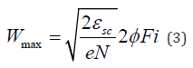
https://biomedres.us/reprints.php
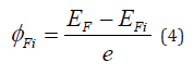
https://biomedres.net/submit-manuscript.php